Embed Size (px)
Citation preview
RESEARCH ARTICLE Open Access
Treatment of aneurysmal bone cysts usingendoscopic curettageHisaki Aiba1, Masaaki Kobayashi1,5*, Yuko Waguri-Nagaya2, Hideyuki Goto1, Jun Mizutani3, Satoshi Yamada1,Hideki Okamoto1, Masahiro Nozaki1, Hiroto Mitsui3, Shinji Miwa1, Makoto Kobayashi1, Kojiro Endo1, Shiro Saito1,Taeko Goto4 and Takanobu Otsuka1
Abstract
Background: Although aneurysmal bone cysts (ABCs) are benign tumours, they have the potential to be locallyaggressive. Various treatment approaches, such as en bloc resection, open curettage, radiotherapy, sclerotherapy,and embolization have been proposed, but the most appropriate treatment should be selected after consideringthe risk of tumour recurrence and treatment complications. Endoscopic curettage (ESC) may be a less invasivealternative to open curettage for ABC treatment. We aimed to describe the use of ESC for the treatment of ABCsand to report our clinical outcomes, including the incidence rate of recurrence, radiological appearance at finalfollow-up, time to solid union, complications, and postoperative function.
Methods: Between 1998 and 2015, 30 patients (18 men and 12 women; mean age, 17.4 years) underwent ESC forthe treatment of primary ABCs at our hospital (mean postoperative follow-up, 55 months). ESC was performedunder arthroscopic guidance for direct visualization, and curettage extended until normal bone was observed inthe medullary cavity. To investigate bone healing after ESC, we evaluated the consolidation of cysts at the finalevaluation (based on the modified Neer classification) and time to solid union after surgery, which was defined assufficient cortical bone thickness to prevent fracture and allow physical activities.
Results: Recurrence was identified in 3 cases (10%). Curative outcomes were obtained after repeated ESC oropen curettage. A log-rank analysis indicated that age < 10 years (p = 0.004) and contact of the tumour withthe physis (p = 0.01) increased the risk of tumour recurrence. Residual tumours were identified in 9 cases(30%); these lesions remained inactive over the extended follow-up period. The average time to solid unionafter endoscopic curettage was 3.2 months. Transient radial nerve palsy was identified in 1 case. Goodpostoperative functional recovery occurred in all cases.
Conclusions: ESC is a minimally invasive technique for the treatment of ABCs, and the tumour recurrencerate is comparable to that of other standard procedures. However, the application of this method should becarefully considered, especially for patients < 10 years and when the tumour comes in contact with thephysis.
Keywords: Endoscopy, Endoscopic curettage, Bone tumour, Aneurysmal bone cyst
* Correspondence: [email protected] of Orthopedic Surgery, Nagoya City University Graduate Schoolof Medical Sciences, 1 Kawasumi, Mizuho-cho, Mizuho-ku, Nagoya 467-8601,Japan5Department of Orthopedic Surgery, Ogaki Municipal Hospital, 4-86Minaminokawa-cho, Ogaki 503-8502, JapanFull list of author information is available at the end of the article
© The Author(s). 2018 Open Access This article is distributed under the terms of the Creative Commons Attribution 4.0International License (http://creativecommons.org/licenses/by/4.0/), which permits unrestricted use, distribution, andreproduction in any medium, provided you give appropriate credit to the original author(s) and the source, provide a link tothe Creative Commons license, and indicate if changes were made. The Creative Commons Public Domain Dedication waiver(http://creativecommons.org/publicdomain/zero/1.0/) applies to the data made available in this article, unless otherwise stated.
Aiba et al. BMC Musculoskeletal Disorders (2018) 19:268 https://doi.org/10.1186/s12891-018-2176-6
BackgroundAneurysmal bone cysts (ABC) are expandable cavitiesthat form within bone and are filled with blood andlined with proliferative fibroblasts, giant cells, and tra-becular bone [1]. An ABC generally arises either as aprimary neoplasm derived from translocation [2] or as asecondary lesion adjacent to osteoblastoma, giant cell tu-mours, or other types of tumours [3]. The goal of treat-ment is to stop the progression of the lesion, relievepain, and prevent pathological fracture of the bone [4].The following various treatment approaches have beenproposed to meet these therapeutic goals: en bloc resec-tion; curettage with or without burring or adjuvant ther-apy [5, 6]; radiotherapy [7]; curopsy, which is a novelbiopsy technique introduced by Reddy et al. [8]; sclero-therapy [9]; and embolization [10]. Selection of the mostappropriate treatment for any case depends on the riskof tumour recurrence and comorbidity associated withthe treatment. In this study, we proposed the use ofendoscopic curettage (ESC) as minimally invasive sur-gery for the treatment of ABCs and reviewed the surgi-cal and clinical outcomes of this treatment approach.
MethodsPatient selection for ESC and relevant characteristics ofthe study groupFrom 1998 to 2014, 37 primary ABCs (37 patients) werediagnosed at our hospital, and histopathological con-firmation was performed by our division of pathology.Among these, ESC was not performed for 7 cases afterthe initial clinical examination because of uncertainty re-garding the diagnosis on imaging; therefore, open biopsy
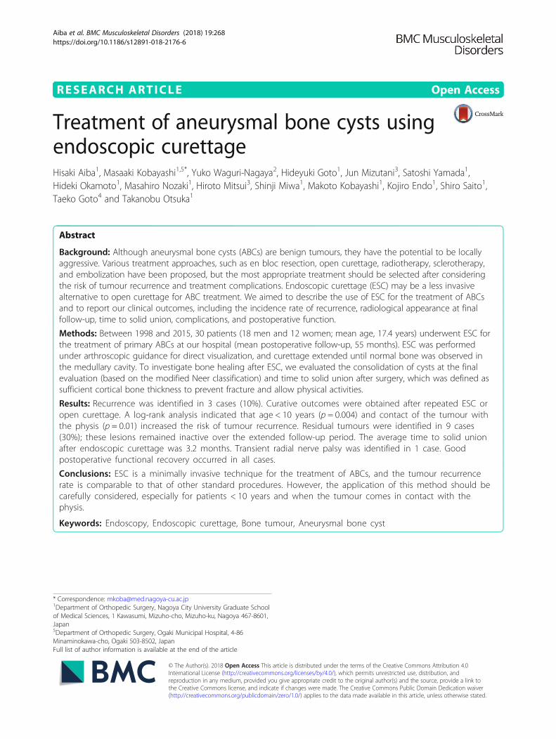
was indicated for exclusion of malignancy (n = 5) or therisk of pathological fracture after ESC (n = 2). Theremaining 30 cases (18 men and 12 women; median age,17.4 years; range, 5.8–40.0 years) were treated with ESC.For cases of pathological fracture before treatment, wewaited 4–5 weeks until the cavities were closed becausecortical continuity was necessary for irrigation of thecyst during surgery (Fig. 1). ABCs were located in tubu-lar bones in 19 cases (12 humeri, 4 tibias, 2 femora, and1 s metatarsal bone), in flat bones in 7 cases (3 ilia, 3 is-chia, and 1 pubis), in short bones in 3 cases (2 calcaneiand 1 os naviculare), and in a sesamoid bone in 1 case(patella). The mean maximum cyst diameter was 66.8(standard deviation [SD], 32.9) mm. Patients werefollowed-up until bone healing; however, when the cystdid not consolidate over time, the patients were moni-tored for at least 2 years postoperatively. The mean over-all follow-up period was 55.0 (SD, 34.3) months.Additional information regarding patient characteristicsis available (Additional file 1).
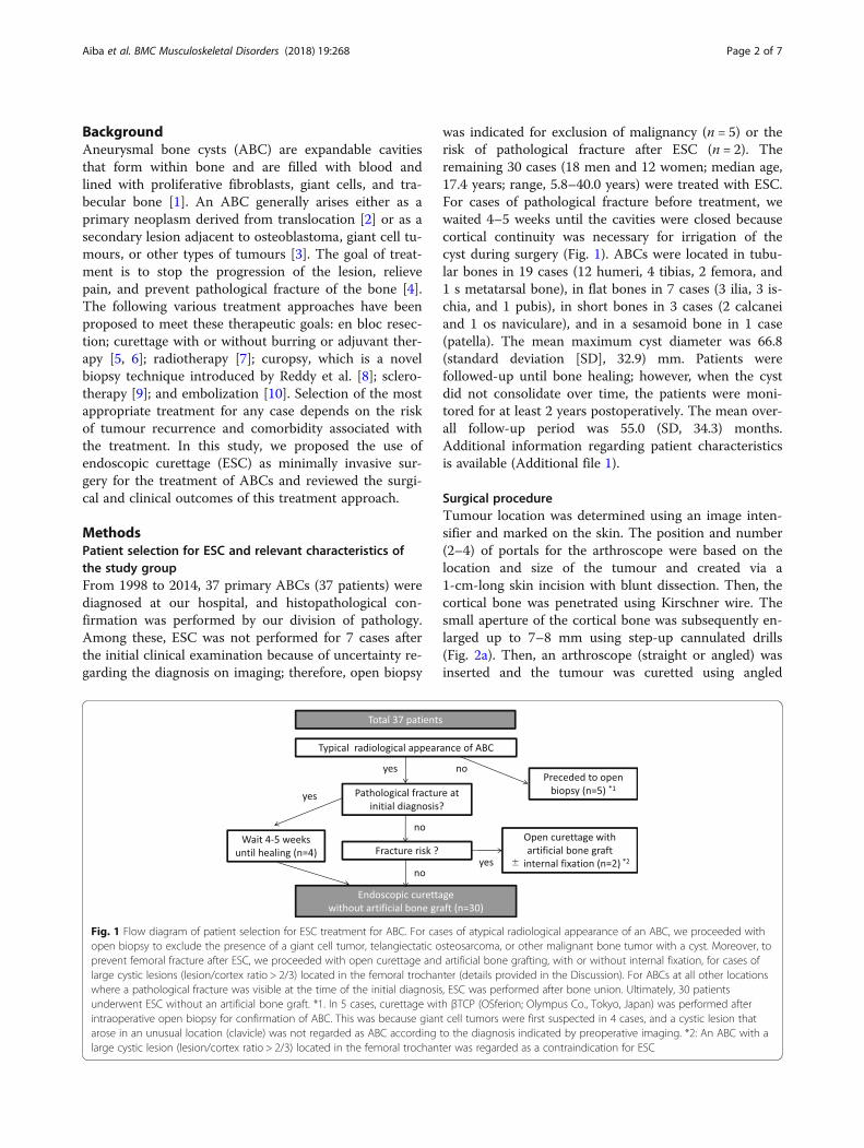
Surgical procedureTumour location was determined using an image inten-sifier and marked on the skin. The position and number(2–4) of portals for the arthroscope were based on thelocation and size of the tumour and created via a1-cm-long skin incision with blunt dissection. Then, thecortical bone was penetrated using Kirschner wire. Thesmall aperture of the cortical bone was subsequently en-larged up to 7–8 mm using step-up cannulated drills(Fig. 2a). Then, an arthroscope (straight or angled) wasinserted and the tumour was curetted using angled
Fig. 1 Flow diagram of patient selection for ESC treatment for ABC. For cases of atypical radiological appearance of an ABC, we proceeded withopen biopsy to exclude the presence of a giant cell tumor, telangiectatic osteosarcoma, or other malignant bone tumor with a cyst. Moreover, toprevent femoral fracture after ESC, we proceeded with open curettage and artificial bone grafting, with or without internal fixation, for cases oflarge cystic lesions (lesion/cortex ratio > 2/3) located in the femoral trochanter (details provided in the Discussion). For ABCs at all other locationswhere a pathological fracture was visible at the time of the initial diagnosis, ESC was performed after bone union. Ultimately, 30 patientsunderwent ESC without an artificial bone graft. *1. In 5 cases, curettage with βTCP (OSferion; Olympus Co., Tokyo, Japan) was performed afterintraoperative open biopsy for confirmation of ABC. This was because giant cell tumors were first suspected in 4 cases, and a cystic lesion thatarose in an unusual location (clavicle) was not regarded as ABC according to the diagnosis indicated by preoperative imaging. *2: An ABC with alarge cystic lesion (lesion/cortex ratio > 2/3) located in the femoral trochanter was regarded as a contraindication for ESC
Aiba et al. BMC Musculoskeletal Disorders (2018) 19:268 Page 2 of 7
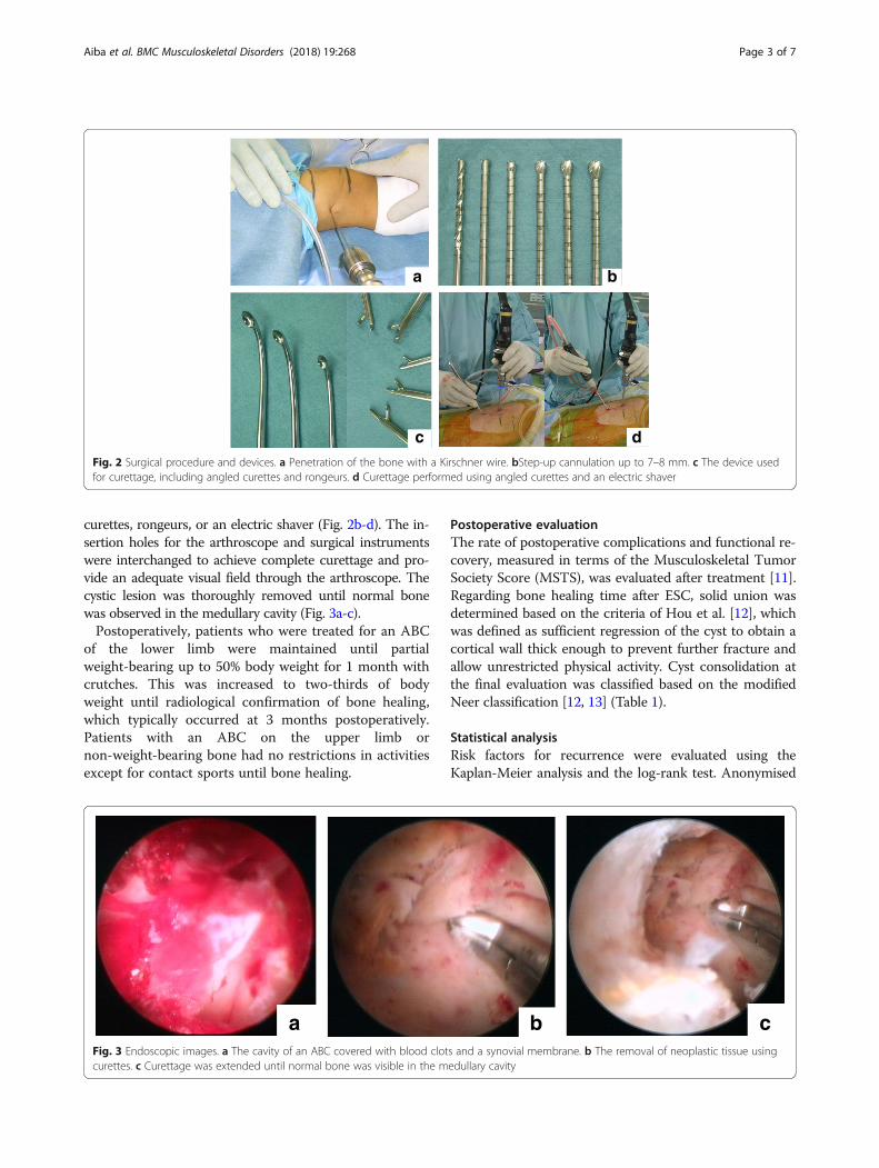
curettes, rongeurs, or an electric shaver (Fig. 2b-d). The in-sertion holes for the arthroscope and surgical instrumentswere interchanged to achieve complete curettage and pro-vide an adequate visual field through the arthroscope. Thecystic lesion was thoroughly removed until normal bonewas observed in the medullary cavity (Fig. 3a-c).Postoperatively, patients who were treated for an ABC
of the lower limb were maintained until partialweight-bearing up to 50% body weight for 1 month withcrutches. This was increased to two-thirds of bodyweight until radiological confirmation of bone healing,which typically occurred at 3 months postoperatively.Patients with an ABC on the upper limb ornon-weight-bearing bone had no restrictions in activitiesexcept for contact sports until bone healing.
Postoperative evaluationThe rate of postoperative complications and functional re-covery, measured in terms of the Musculoskeletal TumorSociety Score (MSTS), was evaluated after treatment [11].Regarding bone healing time after ESC, solid union wasdetermined based on the criteria of Hou et al. [12], whichwas defined as sufficient regression of the cyst to obtain acortical wall thick enough to prevent further fracture andallow unrestricted physical activity. Cyst consolidation atthe final evaluation was classified based on the modifiedNeer classification [12, 13] (Table 1).
Statistical analysisRisk factors for recurrence were evaluated using theKaplan-Meier analysis and the log-rank test. Anonymised
a b
c dFig. 2 Surgical procedure and devices. a Penetration of the bone with a Kirschner wire. bStep-up cannulation up to 7–8 mm. c The device usedfor curettage, including angled curettes and rongeurs. d Curettage performed using angled curettes and an electric shaver
a b cFig. 3 Endoscopic images. a The cavity of an ABC covered with blood clots and a synovial membrane. b The removal of neoplastic tissue usingcurettes. c Curettage was extended until normal bone was visible in the medullary cavity
Aiba et al. BMC Musculoskeletal Disorders (2018) 19:268 Page 3 of 7
radiographical images were evaluated by an orthopaedicsurgeon (H.A.) and a radiologist (T.G.) for double-blindconfirmation of the classification. Then, the kappa valuewas calculated to evaluate inter-observer errors regardingthe morphological appearance of the bone. For statisticalanalysis of the categorical data, a chi-squared analysis wasused. A p-value < 0.05 was considered significant for allanalyses. Measured variables were presented as mean andstandard deviation or median with range, as appropriate,for the data distribution. All statistical analyses were con-ducted using SPSS version 24 (IBM, Chicago, IL).
ResultsAmong the 30 patients included in our study group, ESCwas performed using an average of 2.3 portals (4 portals in
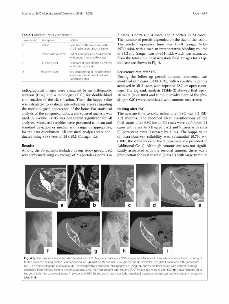
3 cases; 3 portals in 4 cases; and 2 portals in 23 cases).The number of portals depended on the size of the lesion.The median operative time was 107.8 (range, 47.0–197.0 min), with a median intraoperative bleeding volumeof 34.2 mL (range, near 0–354 mL), which was estimatedfrom the total amount of irrigation fluid. Images for a typ-ical case are shown in Fig. 4.
Recurrence rate after ESCDuring the follow-up period, tumour recurrence wasidentified in 3 cases (3/30; 10%), with a curative outcomeachieved in all 3 cases with repeated ESC or open curet-tage. The log-rank analysis (Table 2) showed that age <10 years (p = 0.004) and tumour involvement of the phy-sis (p = 0.01) were associated with tumour recurrence.
Healing after ESCThe average time to solid union after ESC was 3.2 (SD,1.7) months. The modified Neer classifications of thefinal status after ESC for all 30 cases were as follows: 21cases with class A-B (healed cyst) and 9 cases with classC (persistent cyst) (assessed by H.A.). The kappa valueof intra-observer reliability was substantial (0.76; p =0.001; the differences of the 2 observers are provided inAdditional file 1). Although tumour size was not signifi-cantly associated with the residual tumour, there was apredilection for cyst residue (class C) with large tumours
Table 1 Modified Neer classification
Classification Description Details
A Healed Cyst filled with new bone withsmall radiolucent area (< 1 cm)
B Healed with a defect Radiolucent area (< 50% diameter)with enough cortical thickness
C Persistent cyst Radiolucent area (≧50% diameter)with thin cortical rim
D Recurrent cyst Cyst reappearing in the obliteratedarea or in the increased residualradiolucent area
a d
g
b c
f
e
h iFig. 4 Typical case of a suspected ABC treated with ESC. Magnetic resonance (MR) images of a 16-year-old boy who presented with bruising ofhis hip sustained during contact sports participation: (a) axial T2; (b) coronal T2-weighted; and (c) coronal T1-weighted enhanced with gadolinium(Gd). The plain radiograph is shown in (d). The preoperative computed tomography (CT) image (e) shows the bone lesion with cortical thinningextending from the iliac wing to the periacetabular area. Plain radiographs after surgery (f). CT image at 6 months after ESC (g) shows remodeling ofthe cystic lesion by cancellous bone. At 6 years after ESC (h), the pelvic bone was fully remodeled despite a residual cyst and without any symptoms(class B) (i)
Aiba et al. BMC Musculoskeletal Disorders (2018) 19:268 Page 4 of 7
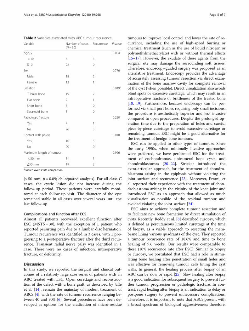
(> 50 mm; p = 0.09; chi-squared analysis). For all class Ccases, the cystic lesion did not increase during thefollow-up period. These patients were carefully moni-tored at each follow-up visit. The diameter of the cystremained stable in all cases over several years until thelast follow-up.
Complications and function after ECSAlmost all patients recovered excellent function afterESC (MSTS = 30), with the exception of 1 patient whoreported persisting pain due to a lumbar disc herniation.Tumour recurrence was identified in 3 cases, with 1 pro-gressing to a postoperative fracture after the third recur-rence. Transient radial nerve palsy was identified in 1case. There were no cases of infection, intraoperativefracture, or deformity.
DiscussionIn this study, we reported the surgical and clinical out-comes of a relatively large case series of patients with anABC treated with ESC. Open curettage and reconstruc-tion of the defect with a bone graft, as described by Jaffeet al. [14], remain the mainstay of modern treatment ofABCs [4], with the rate of tumour recurrence ranging be-tween 40 and 90% [6]. Several procedures have been de-veloped as options for the eradication of micro-residue
tumours to improve local control and lower the rate of re-currence, including the use of high-speed burring orchemical treatment (such as the use of liquid nitrogen orpolymethylmethacrylate) with or without thermal effects[15–17]. However, the exudate of these agents from thesurgical site may damage the surrounding soft tissues.Therefore, endoscopy-guided surgery was proposed as analternative treatment. Endoscopy provides the advantageof accurately assessing tumour resection via direct exam-ination of the bone marrow cavity for complete removalof the cyst (when possible). Direct visualization also avoidsblind spots or excessive curettage, which may result in anintraoperative fracture or brittleness of the treated bone[18, 19]. Furthermore, because endoscopy can be per-formed via small port holes requiring only small incisions,the procedure is aesthetically superior and less invasivecompared to open procedures. Despite the prolonged op-eration time due to the preparation of holes and carefulpiece-by-piece curettage to avoid excessive curettage orremaining tumour, ESC might be a good alternative forthe treatment of benign bone tumours.ESC can be applied to other types of tumours. Since
the early 1990s, when minimally invasive approacheswere preferred, we have performed ESC for the treat-ment of enchondromas, unicameral bone cysts, andchondroblastomas [20–22]. Stricker introduced theextra-articular approach for the treatment of chondro-blastoma arising in the epiphysis without violating thejoint surface and recurrence [23]. Moreover, Errani, etal. reported their experience with the treatment of chon-droblastoma arising in the vicinity of the knee joint andintroduced ESC as an approach that allowed as muchvisualisation as possible of the residual tumour andavoided violating the joint surface [24].ESC aims to achieve complete tumour resection and
to facilitate new bone formation by direct stimulation ofcysts. Recently, Reddy et al. [8] described curopsy, whichis defined as percutaneous limited curettage at the timeof biopsy, as a viable approach to resecting the mem-brane lining various quadrants of the cyst. They reporteda tumour recurrence rate of 18.6% and time to bonehealing of 9.6 weeks. Our results were comparable tothese (10% recurrence rate after ESC). Similar to biopsyor curopsy, we postulated that ESC had a role in stimu-lating bone healing after penetration of small holes andwas effective for removing tumour cells lining the cystwalls. In general, the healing process after biopsy of anABC can be slow or rapid [25]. Slow healing after biopsyis a good indication for subsequent surgery to prevent fur-ther tumour progression or pathologic fracture. In con-trast, rapid healing after biopsy is an indication to delay orpostpone surgery to prevent unnecessary complications.Therefore, it is important to note that ABCs present witha broad spectrum of biological aggressiveness; therefore,
Table 2 Variables associated with ABC tumour recurrence
Variable Number of cases(N = 30)
Recurrence P-value
Age, y 0.004
< 10 8 3
≧10 22 0
Sex 0.776
Male 18 2
Female 12 1
Location 0.949a
Tubular bone 19 2
Flat bone 7 1
Short bone 3 0
Sesamoid bone 1 0
Pathologic fracture 0.220
Yes 4 1
No 26 3
Contact with physis 0.010
Yes 10 3
No 20 0
Maximum length of tumour 0.966
< 50 mm 11 1
≧50 mm 19 2aPooled over strata comparison
Aiba et al. BMC Musculoskeletal Disorders (2018) 19:268 Page 5 of 7
ESC might fail in the case of aggressive lesions [26]. How-ever, in our case series, we did not observe a rapid in-crease in osteolytic lesions after ESC; therefore, insistentcurettage in the cavity by ESC is possibly important toachieve total removal of the resident tumour.We excluded cases in which ESC might have lessened
the strength of the bone during the postoperative periodprior to bone healing. This decision was based on ourprior experience during our early period of performingESC, when we experienced 2 cases of postoperative frac-ture in patients in whom ESC was used for to treat aunicameral bone cyst. Currently, it is not possible to pre-dict pathological fractures with benign bone tumours inpaediatric patients [27]. Kaelin et al. [28] proposed theuse of a cyst index (area of the cyst on a plain radio-graph divided by the square of the diameter of the bonediaphysis at its tubular portion) to predict the risk of apathological fracture, with a cutoff index of > 4 cystsidentified for the humerus and a cutoff index of > 3.4cysts for the femur being predictive of postoperativefractures. However, the positive predictive value of thisindex was limited and had a high false-positive return.Similar to the cyst index, Mirel’s score, which was ori-ginally introduced for the management of metastatic tu-mours and calculated as the sum of each value for thetumour position, pain, and the lesion/cortex ratio (theratio of the maximum width of the lesion to the bone),is a practical method of predicting postoperative frac-tures that provides an indication for preemptive internalfixation [29]. We considered a lesion/cortex ratio of > 2/3 in the trochanteric region as a contraindication toESC. Therefore, we chose to perform preemptive fix-ation in 2 cases with a cystic lesion of the trochanterdue to concerns regarding severe functional restrictionassociated with pathological fractures of the trochantericregion of the femur and the patient anxiety associatedwith such an impairment. The validity of this strategymust be verified in further research.Open growth plates [30] and the proximity of the bone
cyst to the epiphysis [31] have been identified as risk fac-tors for local recurrence. In our study, we identified age< 10 years and tumour involvement of the physis as riskfactors for ABC recurrence after ESC. Based on our re-sults, we suggest that indications for ESC should be con-sidered carefully, and that a watch-and-see strategy isrecommended for patients at high risk for local recur-rence and low risk for pathological fractures.There were some limitations to our study that need to
be acknowledged. First, comparisons with other standardtreatments are necessary to evaluate the outcomes ofESC treatment more precisely. Second, the sample sizewas limited; therefore, the power of our study was notstrong. Third, all diagnoses were not confirmed by agenetic approach but histological appearance. In 2004,
USP6 rearrangement and/or CDH11 rearrangementwere reported in the majority of primary ABC cases(69%) [32]. Because secondary ABC did not involvethese rearrangements, this translocation was consideredto be a useful method of determining the differentialdiagnosis. Therefore, the accuracy of the diagnosis mighthave affected the results of this study. Finally, althoughall procedures were performed by a single surgeon, the ef-fects of the learning curve for the ESC procedure need tobe considered. In fact, 1 case of radial nerve palsy, prob-ably resulting from blunt trauma in the area of the radialgroove during the ESC procedure, was identified duringthe early technical development period. Therefore, pre-operative planning is essential to avoid complications withESC, including identification of the appropriate positionof the holes to avoid the neurovascular structure and de-termination of the appropriate timing for intervention tolower the risk for pathological fractures.
ConclusionTo our knowledge, this is the first report of the out-comes of ESC for ABC treatment involving a relativelylarge case series. Nevertheless, a reliable comparisonwith other methods is difficult. We reported a lower re-currence rate than previous reports, and our patients re-covered without long-lasting complications and achievedgood functional recovery. However, further investiga-tions are necessary to validate our methods.
Additional file
Additional file 1: Additional table. (DOCX 95 kb)
AbbreviationsABC: Aneurysmal bone cyst; ESC: Endoscopic curettage; SD: Standarddeviation
AcknowledgementsWe thank the staff of the Division of Pathology of Nagoya City UniversityHospital for evaluating the histological specimens.
Availability of data and materialsAll data are available in the Additional file.
Authors’ contributionsHA: Software use, validation, visualization, formal analysis, data curation, andoriginal draft preparation. MkK: Conceptualization, main surgeon,methodology, and supervision. YWN: Surgeon and supervision. HG and HO:Surgeon and supervision. JM: Supervision and data curation. SY: Supervisionand data curation. MN: Conceptualization, surgeon, and methodology. HMand MmK: Surgeon. SM: Reviewing and editing. KE and SS: Data curation. TG:Radiological assessment. TO: Reviewing and editing, project administration,and conceptualization. All authors read and approved the final manuscript.
Ethics approval and consent to participateThis retrospective study received the approval of the local committee ofNagoya City University Hospital (no. 60–17-0071, approved on December 6,2017) and was conducted in compliance with the guidelines of the HelsinkiDeclaration of 1975. Informed consent was obtained from the patients andtheir family members.
Aiba et al. BMC Musculoskeletal Disorders (2018) 19:268 Page 6 of 7
Consent for publicationWritten informed consents were received from patients > 16 years or fromthe parent/legal guardian if the patient was < 16 years for publication of thisreport and accompanying images.
Competing interestsThe authors declare that they have no competing interests.
Publisher’s NoteSpringer Nature remains neutral with regard to jurisdictional claims inpublished maps and institutional affiliations.
Author details1Department of Orthopedic Surgery, Nagoya City University Graduate Schoolof Medical Sciences, 1 Kawasumi, Mizuho-cho, Mizuho-ku, Nagoya 467-8601,Japan. 2Department of Joint Surgery for Rheumatic Diseases, Nagoya CityUniversity Graduate School of Medical Sciences, 1 Kawasumi, Mizuho-cho,Mizuho-ku, Nagoya 467-8601, Japan. 3Department of Rehabilitation Medicine,Nagoya City University Graduate School of Medical Sciences, 1 Kawasumi,Mizuho-cho, Mizuho-ku, Nagoya 467-8601, Japan. 4Department of Radiology,Nagoya City University Graduate School of Medical Sciences, 1 Kawasumi,Mizuho-cho, Mizuho-ku, Nagoya 467-8601, Japan. 5Department ofOrthopedic Surgery, Ogaki Municipal Hospital, 4-86 Minaminokawa-cho,Ogaki 503-8502, Japan.
Received: 28 January 2018 Accepted: 10 July 2018
References1. Copley L, Dormans JP. Benign pediatric bone tumors. Evaluation and
treatment. Pediatr Clin North Am. 1996;43:949–66.2. Ye Y, Pringle LM, Lau AW, Riquelme DN, Wang H, Jiang T, Lev D, Welman A,
Blobel GA, Oliveira AM, Chou MM. TRE17/USP6 oncogene translocated inaneurysmal bone cyst induces matrix metalloproteinase production viaactivation of NF-kappaB. Oncogene. 2010;29:3619–29. https://doi.org/10.1038/onc.2010.116.
3. Martinez V, Sissons HA. Cancer. Aneurysmal bone cyst. A review of 123cases including primary lesions and those secondary to other bonepathology 1988;61:2291–2304.
4. Park HY, Yang SK, Sheppard WL, Hegde V, Zoller SD, Nelson SD, FedermanN, Bernthal NM. Current management of aneurysmal bone cysts. Curr RevMusculoskelet Med. 2016;9:435–44.
5. Cummings JE, Smith RA, Heck RK Jr. Argon beam coagulation as adjuvanttreatment after curettage of aneurysmal bone cysts: a preliminary study.Clin Orthop Relat Res. 2010;468:231–7. https://doi.org/10.1007/s11999-009-0914-7.
6. Steffner RJ, Liao C, Stacy G, Atanda A, Attar S, Avedian R, Peabody TD.Factors associated with recurrence of primary aneurysmal bone cysts: isargon beam coagulation an effective adjuvant treatment? J Bone Joint SurgAm. 2011;93:e1221–9. https://doi.org/10.2106/JBJS.J.01067.
7. Feigenberg SJ, Marcus RB Jr, Zlotecki RA, Scarborough MT, Berrey BH,Enneking WF. Megavoltage radiotherapy for aneurysmal bone cysts. Int JRadiat Oncol Biol Phys. 2001;49:1243–7.
8. Reddy KI, Sinnaeve F, Gaston CL, Grimer RJ, Carter SR. Aneurysmal bonecysts: do simple treatments work? Clin Orthop Relat Res. 2014;472:1901–10.https://doi.org/10.1007/s11999-014-3513-1.
9. Varshney MK, Rastogi S, Khan SA, Trikha V. Is sclerotherapy better thanintralesional excision for treating aneurysmal bone cysts? Clin Orthop RelatRes. 2010;468:1649–59. https://doi.org/10.1007/s11999-009-1144-8.
10. Rossi G, Mavrogenis AF, Facchini G, Bartalena T, Rimondi E, Renzulli M,Andreone A, Durante S, Angelini A, Errani C. How effective is embolizationwith N-2-butyl-cyanoacrylate for aneurysmal bone cysts? Int Orthop. 2017;41:1685–92. https://doi.org/10.1007/s00264-016-3364-3. Epub 2016 Dec 8
11. Enneking WF, Dunham W, Gebhardt MC, Malawar M, Pritchard DJ. A systemfor the functional evaluation of reconstructive procedures after surgicaltreatment of tumors of the musculoskeletal system. Clin Orthop Relat Res.1993;(286):241–6.
12. Hou HY, Wu K, Wang CT, Chang SM, Lin WH, Yang RS. Treatment ofunicameral bone cyst: a comparative study of selected techniques. J BoneJoint Surg Am. 2010;92:855–62. https://doi.org/10.2106/JBJS.I.00607.
13. Neer CS 2nd, Francis KC, Marcove RC, Terz J, Carbonara PN. Treatment ofunicameral bone cyst. A follow-up study of one hundred seventy-five cases.J Bone Joint Surg Am. 1966;48:731–45.
14. Jaffe HL, Lichtenstein L. Solitary unicameral bone cyst with emphasis on theroentgen picture, the pathologic appearance and the pathogenesis. ArchSurg. 1942;44:1004–25. https://doi.org/10.1001/archsurg.1942.01210240043003.
15. Ozaki T, Hillmann A, Lindner N, Winkelmann W. Aneurysmal bone cysts inchildren. J Cancer Res Clin Oncol. 1996;122:767–9.
16. Peeters SP, Van der Geest IC, de Rooy JW, Veth RP, Schreuder HW.Aneurysmal bone cyst: the role of cryosurgery as local adjuvant treatment. JSurg Oncol. 2009;100:719–24. https://doi.org/10.1002/jso.21410.
17. Keçeci B, Küçük L, Isayev A, Sabah D. Effect of adjuvant therapies onrecurrence in aneurysmal bone cysts. Acta Orthop Traumatol Turc. 2014;48:500–6. https://doi.org/10.3944/AOTT.2014.14.0020.
18. Choi Y, Kwak JM, Chung SH, Jung GH, Kim JD. Tumor treated by endoscopy.Clin Orthop Surg. 2014;6:72–9. https://doi.org/10.4055/cios.2014.6.1.72.
19. Dietz JF, Kachar SM, Nagle DJ. Endoscopically assisted excision of digitalenchondroma. Arthroscopy. 2007;23:678.e1–4.
20. Sekiya I, Matsui N, Otsuka T, Kobayashi M, Tsuchiya D. The treatment ofenchondromas in the hand by endoscopic curettage without bone grafting.J Hand Surg Br. 1997;22:230–4.
21. Okamoto H, Kobayashi M, Sekiya I, Shinzi M, Endo K, Otsuka T. Surgery forenchondroma on the fingers - endoscopic curettage. Central Jpn J OrthopSurg Traumatol. 2017;60:53–4. [in Japanese]
22. Otsuka T, Kobayashi M, Yonezawa M, Kamiyama F, Matsushita Y, Matsui N.The treatment of enchondromas in the hand by endoscopic curettagewithout bone grafting. Arthroscopy. 2002;18:430–5.
23. Stricker SJ. Extraarticular endoscopic excision of femoral headchondroblastoma. J Pediatr Orthop. 1995;15:578–81.
24. Errani C, Traina F, Chehrassan M, Donati D, Faldini C. Minimally invasivetechnique for curettage of chondroblastoma using endoscopic technique.Eur Rev Med Pharmacol Sci. 2014;18:3394–8.
25. Louahem D, Kouyoumdjian P, Ghanem I, Mazeau P, Perrochia H, L'kaissi M,Cottalorda J. Active aneurysmal bone cysts in children: possible evolutionafter biopsy. J Child Orthop. 2012;6:333–8. https://doi.org/10.1007/s11832-012-0424-0.
26. Tsagozis P, Brosjö O. Current strategies for the treatment of aneurysmalbone cysts. Orthop Rev (Pavia). 2015;7:6182. https://doi.org/10.4081/or.2015.6182.
27. Snyder BD, Hauser-Kara DA, Hipp JA, Zurakowski D, Hecht AC, Gebhardt MC.Predicting fracture through benign skeletal lesions with quantitativecomputed tomography. J Bone Joint Surg Am. 2006;88:55–70.
28. Kaelin AJ, MacEwen GD. Unicameral bone cysts. Natural history and the riskof fracture. Int Orthop. 1989;13:275–82.
29. Mirels H. Metastatic disease in long bones. A proposed scoring system fordiagnosing impending pathologic fractures. Clin Orthop Relat Res. 1989;249:256–64.
30. Kapila R, Sharma R, Sohal YS, Singh D, Singh S. Primary epiphysealaneurysmal bone cyst of distal ulna. J Orthop Case Rep. 2015;5:85–7. https://doi.org/10.13107/jocr.2250-0685.356.
31. Gibbs CP Jr, Hefele MC, Peabody TD, Montag AG, Aithal V, Simon MA.Aneurysmal bone cyst of the extremities. Factors related to localrecurrence after curettage with a high-speed burr. J Bone Joint SurgAm. 1999;81:1671–8.
32. Oliveira AM, Perez-Atayde AR, Inwards CY, Medeiros F, Derr V, Hsi BL,Gebhardt MC, Rosenberg AE, Fletcher JA. USP6 and CDH11 oncogenesidentify the neoplastic cell in primary aneurysmal bone cysts and areabsent in so-called secondary aneurysmal bone cysts. Am J Pathol.2004;165:1773–80.
Aiba et al. BMC Musculoskeletal Disorders (2018) 19:268 Page 7 of 7