Embed Size (px)
Citation preview

Gedikbafl› A, Gül A, Sarg›n A et al., Ultrasound in Trisomy 18 and 13,Gedikbafl› A, Gül A, Sarg›n A et al., Ultrasound in Trisomy 18 and 13,J Turkish-German Gynecol Assoc, Vol. 9(4); 2008:218-223J Turkish-German Gynecol Assoc, Vol. 9(4); 2008:218-223218
IntroductionTrisomy 18 or Edwards’ syndrome, a lethal chromosomalabnormality, after Down syndrome, is the second mostcommon autosomal trisomy and has a reported incidencevarying from one in 3000 to 1 in 8000. It was firstrecognized as a specific entity in 1960 by Edwards et al.
and Jones (1,2). Different severe congenital malformations,intense mental retardation, as well as high rate of infantmortality are typical features of trisomy 18. The conditionis incompatible with long-term survival, and the singularcases that do survive have an extremely low quality of life.The mortality in utero is high and death in those fetuseswhich are live born occurs within the first few weeks oflife, with a median survival period of <1 month (3). Itssonographic features in the second and third trimestershave been well-described (4). These include strawberry-shaped head, ventriculomegaly, choroid plexus cyst,posterior fossa cyst, facial cleft, micrognathia, nuchaledema, diaphragmatic hernia, heart defects, exomphalos,
AbstractObjective: To evaluate the sonographic characteristics of fetuses with trisomy 18 and trisomy 13.Materials and Methods: From March 2002 to December 2006, we reviewed the database and medical records of 25 cases withtrisomy 18 and trisomy 13. The subjects were recruited from pregnant women undergoing prenatal sonographic examinationsat 13-28 weeks of gestation and subsequently proven trisomy 18 or 13. The results of ultrasound findings were reviewed inthese cases with chromosomes confirmed as trisomy 18 and/or 13.Results: All cases had at least one abnormal sonographic finding. The common sonographic findings included choroid ple-xus cysts, abnormal head shape, cardiac anomalies, holoprosencephaly with associated facial anomalies, abnormal feet and/orhands, especially polydactyly, clenched hand, omphalocele. Non-structural abnormal findings such as polyhydroamnios or fe-tal growth restriction were seen in less than one third of the fetuses.Discussion: Nearly all fetuses with trisomy 18 or 13 had characteristic sonographic patterns of abnormalities demonstratedat midpregnancy. Detailed ultrasound at midpregnancy could effectively screen fetuses with trisomy 18 or 13 for furthergenetic testing.Keywords: genetic ultrasound, trisomy 18, trisomy 13
Özet
Trizomi 18 ve 13’te Ultrason
Amaç: Trizomi 18 ve trizomi 13 tan›s› alm›fl fetüslerde sonografik bulgular› de¤erlendirmek.Materyal ve Metot: Mart 2002 ile Aral›k 2006 aras›nda veritaban›m›zda kay›tl› bulunan 25 trizomi 18 ve trizomi 13 olgusude¤erlendirildi. Olgular 13-28. gebelik haftalar›nda prenatal ultrasonografik muayeneleri yap›lm›fl ve trizomi 18 veya 13olarak tan› alm›flt›r. Trizomi 18 ve/veya trizomi 13 tan›s›n› alm›fl bu olgular›n ultrason bulgular› de¤erlendirildi.Sonuçlar: Tüm olgular›n en az iki patolojik ultrason görüntüsü vard›. En s›k gözlenen bulgular koroid pleksus kisti, patolojikkafa flekli, kardiyak patolojiler, holoprozensefali ile iliflkili yüz anomalileri, anormal el ve ayak flekli, polidaktili ve pençe elidi. Yap›sal anatomik bulgular›n d›fl›nda polihidroamnios ve geliflme gerili¤i gibi sonuçlar da saptand›.Tart›flma: ‹kinci trimesterde neredeyse trizomi 18 veya 13 olgular›n›n tümünün karakteristik ultrason bulgular› vard›.Ayr›nt›l› ultrason taramas› ile 2. trimesterde etkin trizomi 18 veya 13 taramas› yapmak olas›d›r.
Anahtar sözcükler: genetik ultrason, trizomi 18, trizomi 13
CLINICAL STUDY
Ultrasound in Trisomy 18 and 13Ali GED‹KBAfiI, Ahmet GÜL, Akif SARGIN, Taner GÜNAY, Yavuz CEYLAN
Bak›rköy Maternity and Children Diseases Hospital, Department of Obstetrics and Gynecology,Perinatology Unit, ‹stanbul, Turkey
Received 14 August 2007; received in revised form 19 December 2007; accepted 15 February 2008;
published online 28 August 2008
Corresponding Author: Dr. Ali Gedikbafl›Bak›rköy Kad›n Do¤um ve Çocuk Hastal›klar› Hastanesi,‹stanbul, TürkiyeGSM : +90 0532 213 51 40E-mail : [email protected]

219
J Turkish-German Gynecol Assoc, Vol. 9(4); 2008J Turkish-German Gynecol Assoc, Vol. 9(4); 2008
esophageal atresia, renal defects, abnormal extremities, andintrauterine growth restriction. However, in the firsttrimester, the sonographic abnormalities are less welldocumented; the features reported include increased nuchaltranslucency, heart defects, and exomphalos (5,6).
Patau first discovered trisomy 13 in 1960 (7). This syndromeoccurs in one of 5000 births, and is the most seen thirdautosomal anomaly (2). Common characteristic features ofthe syndrome consist of central nervous system (CNS)anomalies, especially holoprosencephaly, facial and ocularanomalies, polydactyly, clubbed or rocker-bottom feet, heartdefects, renal anomalies, and a single umbilical artery. Themedian survival is 2.5 days (2). Eighty-two percent ofnewborns with trisomy 13 die in the first month and only 5%survive the first 6 months (2). Survivors have severe mentaldefects, often have seizures, and they fail to thrive. Becausethe prognosis of the syndrome is very poor, early prenataldiagnosis is important. In the past, prenatal diagnosis byamniocentesis was performed only because of maternal age.In the last decade, ultrasonography has been increasinglyused for prenatal diagnosis. The prenatal sonographiccharacteristics of trisomy 13 have been published, but moststudies included fetuses at all stages of pregnancy.
Advances in ultrasound prenatal diagnostics havesignificantly improved the option of early detection ofcongenital anomalies, leading to improvement of perinatalcare and giving the opportunity for pregnancy termination inthe cases of lethal disorders (7-9). Accurate and actualknowledge of survival rate, disease course, and methods oftreatment is therefore very important for clinicians andfamily members who take care of children and fetuses withtrisomy 18, in making correct but also ethically very difficultdecisions. In this article, we describe our ultrasoundexperience from the last 5 years in managing fetuses withEdwards and Patau syndrome. The objective of this studywas to evaluate the efficacy of sonographic screening fortrisomy 18 and 13 at midpregnancy.
Materials and MethodsThis descriptive analysis was undertaken at ‹stanbul Bak›rköyMaternity and Children Diseases Hospital, Department ofObstetrics and Gynecology, Perinatology Unit. Women withabnormal ultrasonographic findings or an abnormal triple testwere offered genetic ultrasonography and a chromosomalstudy. The subjects were recruited from pregnant women whounderwent prenatal sonographic examinations with variousindications at midpregnancy (13-28 weeks of gestation). Theinclusion criteria were fetuses’ proven trisomy 18 byamniocentesis, chordocentesis, or chorion villus biopsy withabnormal ultrasound scan. The gestational age was estimatedby either last menstrual period, crown-rump length in firsttrimester or biparietal diameter with femur length in secondtrimester. Ultrasonographic examinations were performed
transabdominally from March 2002 to November 2006 by oneof four experienced maternal-fetal medicine physiciansonographers using Sonoline-G50 TM, Siemens, Issaquah,WA, USA and Voluson 730 Expert TM, GE Healthcare,Milwaukee, WI, USA; multifrequency convex transducer 2.0-7.0 MHz.
All ultrasound findings were prospectively identified at thetime of first scanning before knowledge of cytogeneticdiagnosis. Reason for referral to our ultrasound unit weresmall-for dates uterus (n=1), positive screening test (n=2),suspected or unexplained abnormal structural findings byultrasound (n=22).
Ultrasonographic imaging included standard biometrymeasurements of BPD (biparietal diameter), HC (headcircumference), AC (abdominal circumference), and FL(femur length) as well as fetal anatomic evaluation of thecerebral ventricles, posterior fossa, spine, four-chamberview of the heart, stomach, kidneys, and bladder inaccordance with ACOG and AIUM criteria (11,12). Inaddition to these anatomic structures, we attempted toevaluate the following structures in the second trimesterscan in all cases; nuchal skin fold area, cerebellum, right andleft ventricular outflow tracts, renal pelvis, and extremities.Color flow imaging of the fetal heart was used when a heartdefect was suspected on the basis of the four-chamber viewand imaging of the outflow tracts. Intrauterine growthretardation (IUGR) was defined as a difference of at least 14days between the mean gestational age by ultrasonographyand last menstrual period (LMP) dating. In cases ofomphalocele, the mean gestational age was calculated fromthe BPD and FL, and in cases of intracranial abnormalities,it was calculated from the AC and FL.
All 25 cases were confirmed as having trisomy 18 or trisomy13 by genetic study – amniocentesis (18), chordocentesis (4),chorionic villus sampling (2) and one cardiocentesis.
The hospital ethics committee made the decision fortermination of pregnancy (TOP), after counseling andapproval of the parents. Autopsy was offered to all patientsafter termination of pregnancy.
ResultsA computerized search identified 21 trisomy 18 fetuses and4 trisomy 13 fetuses from 13-28 weeks’ gestation that hadcomprehensive ultrasound scans and multiple-markerscreening tests before genetic karyotyping from 2002-2006.
The mean maternal age for trisomy 18 groups was 31 (18-42)years. Fourteen of them were under 35 years of age. Sixteenwomen (76.2%) were multiparous, and 5 (23.8%) werenulliparous. Comprehensive ultrasound examinations wereabnormal in 21 of 21 (100%) fetuses. Mean gestational agewhen abnormalities were detected was 19 (13-24) weeks.

220
J Turkish-German Gynecol Assoc, Vol. 9(4); 2008J Turkish-German Gynecol Assoc, Vol. 9(4); 2008Gedikbafl› et al.
Mean gestational age when pregnancy was terminated was23.57 (16-32) weeks. The mean maternal age for trisomy 13groups was 25 (21-32) years. All of them were under 35years of age. Ultrasound examination was abnormal in allfour cases (100%). Mean gestational age at diagnosis was22.25 (16-28) an at termination of pregnancy 24 weeks (18-30 weeks).
All of the cases were sonographically evaluated beforekaryotyping. Therapeutic termination was done in all casesafter proper counseling. All cases had at least two abnormalsonographic finding. The most common findings in trisomy18 were choroid plexus cysts, strawberry shaped head,ventricular septal defects and abnormalities of extremities.The most common findings in trisomy 13 cases were vermishypoplasia, holoprosencephaly, and facial cleft.
The sonographic findings and appropriate illustrations areseen in Table 1, Figure 1 and 2 for trisomy 18 and 13 cases.
Discussion Trisomy 18 (Edwards syndrome) is a chromosomalabnormality that results from the presence of an extra copy ofchromosome 18. The etiologies of the trisomy of 18 areknown as maternal meiotic nondisjunction (over 90%),paternal meiotic nondisjunction (5%), and paternaldislocation. Trisomy 18 is a lethal chromosomal abnormalityleading to fetal or neonatal death. Edwards first described theclinical features of the abnormality in 1960. Every organsystem can be affected by trisomy 18. Fetuses are usuallygrowth-restricted and have facial abnormalities, includingmicrocephaly, prominent occiput, low-set ears, micrognathia,and a small mouth. They also have skeletal anomalies,
Abnormality Trisomy 18; n (%) Trisomy 13; n (%)
Cranium/Brain In 14/21 (66.6%) of cases In 4/4 (100%) of cases
- Choroid plexus cysts 9 (42.8) -
- Strawberry-shaped cranium 8 (38.1) -
- Vermis hypoplasia/agenesis 4 (19.0) 2 (50.0)
- Ventriculomegaly 3 (14.3) -
- Holoprosencephaly 1 (4.8) 2 (50.0)
- Dandy-Walker malformation 1 (4.8) 1 (25.0)
- Corpus callosum agenesis 1 (4.8) -
- Enlarged cisterna magna - 1 (25.0)
Face/Neck In 6/21 (28.6%) of cases In 4/4 (100%) of cases
- Cystic hygroma 4 (19.0) -
- Facial cleft 2 (9.5) 2 (50.0)
- Micrognathia 2 (9.5) -
- Cyclopes - 1 (25.0)
- Nuchal edema - 1 (25.0)
Cardiac In 15/21 (71.4%) of cases In 2/4 (59%) of cases
- Ventricular septal defect (VSD) 9 (42.8) 1 (25.0)
- Atrioventricular septal defect (AVSD) 4 (19.0) 1 (25.0)
- Tetralogy of Fallot (TOF) 3 (14.3) -
- Double outlet right ventricle (DORV) 2 (9.5) -
- Pulmonary stenosis 2 (9.5) -
- Single AV valve 3 (14.3) -
- Hypoplastic left ventricle and arcus aorta 1 (4.8) -
Thorax/Abdomen In 7/21 (33.3%) of cases In none cases
- Hydronephrosis 2 (9.5) -
- Omphalocele 1 (4.8) -
- Absent stomach 1 (4.8) -
- Diaphragmatic hernia 1 (4.8) -
- Hydrothorax 2 (9.5) -
- Ascites 1 (4.8) -
Others
- Abnormal hands/feet 7 (33.3) -
- Single umbilical artery 4 (19.0) 1 (25.0)
- Polyhydroamnios 2 (9.5) -
- Fetal growth restriction 2 (9.5) 1 (25.0)
- Umbilical cord cyst 1 (4.8) -
Table 1. Sonographic abnormalities in 21 fetuses with trisomy 18 and 4 fetuses with trisomy 13
221
J Turkish-German Gynecol Assoc, Vol. 9(4); 2008J Turkish-German Gynecol Assoc, Vol. 9(4); 2008
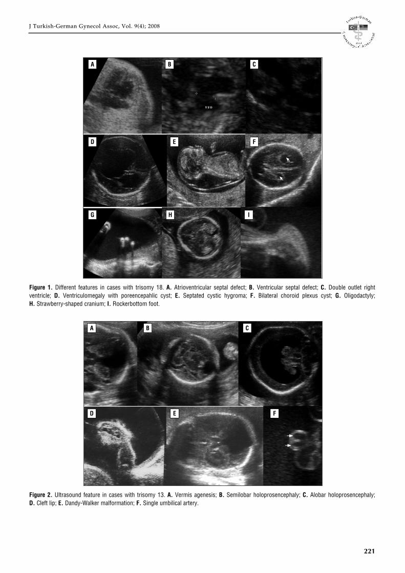
Figure 1. Different features in cases with trisomy 18. A. Atrioventricular septal defect; B. Ventricular septal defect; C. Double outlet rightventricle; D. Ventriculomegaly with poreencepahlic cyst; E. Septated cystic hygroma; F. Bilateral choroid plexus cyst; G. Oligodactyly;H. Strawberry-shaped cranium; I. Rockerbottom foot.
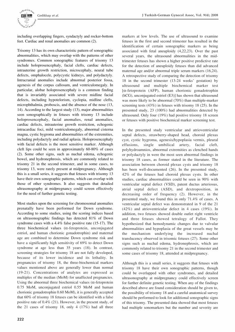
Figure 2. Ultrasound feature in cases with trisomy 13. A. Vermis agenesis; B. Semilobar holoprosencephaly; C. Alobar holoprosencephaly;D. Cleft lip; E. Dandy-Walker malformation; F. Single umbilical artery.
A B C
D E F
G
A
D E F
B C
H I
222
J Turkish-German Gynecol Assoc, Vol. 9(4); 2008J Turkish-German Gynecol Assoc, Vol. 9(4); 2008Gedikbafl› et al.
including overlapping fingers, syndactyly and rocker-bottomfeet. Cardiac and renal anomalies are common (2).
Trisomy 13 has its own characteristic pattern of sonographicabnormalities, which may overlap with the patterns of othersyndromes. Common sonographic features of trisomy 13include holoprosencephaly, facial clefts, cardiac defects,intrauterine growth restriction, microcephaly, neural tubedefects, omphalocele, polycystic kidneys, and polydactyly.Intracranial anomalies include abnormal posterior fossa,agenesis of the corpus callosum, and ventriculomegaly. Inparticular, alobar holoprosencephaly is a common findingthat is invariably associated with severe midline facialdefects, including hypotelorism, cyclopia, midline clefts,microphthalmia, proboscis, and the absence of the nose (13,14). According to the largest study (13), major abnormalitiesseen sonographically in fetuses with trisomy 13 includeholoprosencephaly, facial anomalies, renal anomalies,cardiac defects, intrauterine growth restriction, echogenicintracardiac foci, mild ventriculomegaly, abnormal cisternamagna, cystic hygroma and abnormalities of the extremities,including polydactyly and radial aplasia. Holoprosencephalywith facial defects is the most sensitive marker. Althoughcleft lips could be seen in approximately 60-80% of cases(2). Some other signs, such as nuchal edema, echogenicbowel, and hydronephrosis, which are commonly related totrisomy 21 in the second trimester, and in some cases, totrisomy 13, were rarely present at midpregnancy. Althoughthis is a small series, it suggests that fetuses with trisomy 13have their own sonographic patterns, which can overlap withthose of other syndromes. It also suggests that detailedultrasonography at midpregnancy could screen effectivelyfor the need of further genetic testing.
Most studies upon the screening for chromosomal anomaliesprenatally have been performed for Down syndrome.According to some studies, using the scoring indices basedon ultrasonographic findings has detected 81% of Downsyndrome cases with a 4.4% false positive rate (15-17). Thethree biochemical values (α-fetoprotein, unconjugatedestriol, and human chorionic gonadotrophin) and maternalage are combined to determine Down syndrome risk andhave a significantly high sensitivity of 69% to detect Downsyndrome at age less than 35 years (18). In contrast,screening strategies for trisomy 18 are not fully developed,because of its lower incidence and its lethality. Inpregnancies of trisomy 18, the three-biochemical markersvalues mentioned above are generally lower than normal(19-21). Concentrations of analytes are expressed asmultiples of the median (MoM) for unaffected pregnancies.Using the abnormal three biochemical values (α-fetoprotein0.75 MoM, unconjugated estriol 0.55 MoM and humanchorionic gonadotrophin 0.60 MoM), it is generally acceptedthat 60% of trisomy 18 fetuses can be identified with a falsepositive rate of 0.4% (21). However, in the present study, ofthe 21 cases of trisomy 18, only 4 (17%) had all three
markers at low levels. The use of ultrasound to examinefetuses in the first and second trimester has resulted in theidentification of certain sonographic markers as beingassociated with fetal aneuploidy (4,22,23). Over the pastseveral years, the ultrasound abnormalities in the mid-trimester fetuses has shown a higher positive predictive ratefor the detection of aneuploidy fetuses than did advancedmaternal age and/or abnormal triple serum markers (16,24).A retrospective study of comparing the detection of trisomy18 in the second trimester (13-24 weeks’ gestation) byultrasound and multiple biochemical marker test[α-fetoprotein (AFP), human chorionic gonadotrophin(hCG), unconjugated estriol (E3)] has shown that ultrasoundwas more likely to be abnormal (70%) than multiple-markerscreening tests (43%) in fetuses with trisomy 18 (25). In thepresented study, 23 (100%) had abnormalities detected byultrasound. Only four (19%) had positive trisomy 18 screenor fetuses with positive biochemical marker screening test.
In the presented study ventricular and atrioventricularseptal defects, strawberry-shaped head, choroid plexuscyst, cystic hygroma, agenesis/hypoplasia vermis, pleuraleffusions, single umbilical artery, facial cleft,polyhydroamnios, abnormal extremities as clenched handsor polydactyly in were the most seen abnormal findings intrisomy 18 cases, as former stated in the literature. Theassociation between choroid plexus cysts and trisomy 18has been well-documented (26). In the presented study,42% of the fetuses had choroid plexus cysts. In otherstudies, cardiac abnormalities could be seen in 90% withventricular septal defect (VSD), patent ductus arteriosus,atrial septal defect (ASD), and dextroposition, indecreasing order of frequency (2). However, in thepresented study, we found this in only 71.4% of cases. Aventricular septal defect was demonstrated in 9 of the 21(42%) and atrioventricular defect in 4 cases (19%). Inaddition, two fetuses showed double outlet right ventricleand three fetuses showed tetralogy of Fallot. Theyhypothesized that hemodynamic changes due to valvularabnormalities and hypoplasia of the great vessels may bethe mechanism underlying the increased nuchaltranslucency observed in trisomic fetuses (27). Some othersigns such as nuchal edema, hydronephrosis, which arecommonly related to trisomy 21 in the second trimester andsome cases of trisomy 18, attended at midpregnancy.
Although this is a small series, it suggests that fetuses withtrisomy 18 have their own sonographic patterns, thoughcould be overlapped with other syndromes, and detailedultrasonography at midpregnancy could effectively screenfor further definite genetic testing. When any of the findingsdescribed above are found consideration should be given to,the possibility of trisomy 18 and a careful anatomical surveyshould be performed to look for additional sonographic signsof this trisomy. The presented data showed that most fetuseshad multiple sonomarkers but the number and severity are

223
J Turkish-German Gynecol Assoc, Vol. 9(4); 2008J Turkish-German Gynecol Assoc, Vol. 9(4); 2008
highly variable. It is difficult for this small series to specifywhich abnormality is the best predictor, but the moreabnormal findings, the more likely it is to be the syndrome.Recently, a study about abnormal sonographic features of 38fetuses identified with trisomy 18 and the sensitivity ofsonographic abnormalities was reported. They found that alltrisomy 18 cases had 4 or more prenatally detected sonographicabnormalities, and the sensitivity of sonographicdetection of fetuses with trisomy 18 was 100% (28). Devore(29) reported a detection rate of 99% with the inclusion offetal echocardiogram. In the presented study, all trisomy 18cases had at least two or more anomalous findings.Therefore, our opinion is, that second trimester screening isthe most important way in detecting trisomy 18 cases.
An 11-14 weeks’ gestational nuchal translucency scan,including early cardiac scanning and neurosonography andexcluding cystic hygroma, which is an indication forkaryotyping, can also demonstrative. There are a fewreferences in literature (30-33) which propose first trimesterscreening and report a detection rate of approximately 80%for fetal aneuploidy in this period.
In conclusion, early prenatal diagnosis of this lethalsyndrome can reduce the maternal morbidity and provides anopportunity for the family to decide upon termination. Fetalabnormalities and soft markers must be separated. One softmarker alone is not an indication for karyotyping. Our studyconfirms that no bright test exists for the prenatal screeningtest of this highly lethal congenital anomaly, except forultrasonography, as most cases of trisomy 18 and 13 had atleast two or more sonographically detectable structuralabnormalities. Therefore, prenatal screening should beproposed, first at 11-14 gestational weeks and finally at18-22 gestational weeks for every pregnancy.
References1. Edwards MT, Smith WL, Hanson J et al. A new trisomic syndrome.
Lancet 1960;1:787-9.2. Jones KL. Smith’s Recognizable Patterns of Human Malformation, 5th
edition. Philadelphia: W.B. Saunders; 1997:14-5.3. Rasmussen SA, Wong LY, Yang Q et al. Population-based analyses of
mortality in trisomy 13 and trisomy 18. Pediatrics 2003;111:777-84.4. Nicolaides KH, Snijders RJM, Gosden CM et al. Ultrasonographically
detectable markers of fetal chromosomal abnormalities. Lancet1992;340:704-7.
5. Sherod C, Sebire NJ, Soares W et al. Prenatal diagnosis of trisomy 18at the 10-14 week ultrasound scan. Ultrasound Obstet Gynecol1997;10:387-90.
6. Gembruch U, Baschat AA, Knöpfle G, Hansmann M. Results ofchromosomal analysis in fetuses with cardiac anomalies as diagnosedby first- and early second-trimester echocardiography. UltrasoundObstet Gynecol 1997;10:391-6.
7. Patau K. Multiple congenital anomaly caused by an extra chromosome.Lancet 1960;1:790-5.
8. Nicolaides KH, Azar G, Byrne D et al. Fetal nuchal translucency:Ultrasound screening for chromosomal defects in first trimester of preg-nancy. BMJ 1992;304:967-9.
9. DeVore GR. Second trimester ultrasonography may identify 77-95% offetuses with trisomy 18. J Ultrasound Med 2000;19:565-76.
10. De Vigan C, Baena N, Cariati E, Clementi M, Stoll C, and theEUROSCAN working group. Contribution of ultrasonographic exami-nation to the prenatal detection of chromosomal abnormalities in 19centers across. Europe Ann Genet 2001;44:209-17.
11. American College of Obstetrics and Gynecology: Ultrasonography inpregnancy. ACOG 1994; Technical Bulletin 187:1.
12. American Institute of Ultrasound in Medicine: Antepartum obstetricalguidelines. J Ultrasound Med 1986;5:241.
13. Lehman CD, Nyberg DA, Winter III TC et al. Trisomy 13 syndrome:Prenatal US findings in a review of 33 cases. Radiology 1995;194:217-22.
14. Greene MF, Benacerraf BR, Frigoletto Jr FD. Reliable criteria for theprenatal sonographic diagnosis of alobar holoprosencephaly. Am JObstet Gynecol 1987;56:687-9.
15. Benacerraf BR, Nadel A, Bromley B. Identification of second trimesterfetuses with autosomal trisomy by use of sonographic scoring index.Radiology 1994;193:135-40.
16. Benacerraf BR, Nyberg D, Bromley B, Frigoletto FD. Sonographicscoring index for prenatal detection of chromosomal abnormalities. JUltrasound Med 1992;11:449-58.
17. Bafler ‹. Fetal anöploidi olgular›nda 2. trimester ultrasonografininetkinli¤i ve yeri. T Klin Jinekol Obstet 2002;12:364-70.
18. Wald NJ, Watt HC, Hackshaw AK. Integrated screening for Down’ssyndrome based on tests performed during the first and secondtrimester. N Engl J Med 1999;341:461-7.
19. Merkatz IR, Nitowsky HM, Macri JN, Johnson WE. An associationbetween low maternal serum alpha-fetoprotein and fetal chromosomalabnormalities. Am J Obstet Gynecol 1984;148:886-94.
20. Bogart MH, Pandian MR, Jones OW. 1987. Abnormal maternal serumchorionic gonadotrophin levels in pregnancies with fetal chromosomeabnormalities. Prenat Diagn 1987;7:623-30.
21. Canick JA, Knight GJ, Palomaki GE et al. 1988. Low second trimestermaternal serum unconjugated estriol in pregnancies with Downsyndrome. Br J Obstet Gynecol 1988;195:330-3.
22. Pandya P, Snijders R, Johnson S et al. Screening for fetal trisomies bymaternal age and fetal nuchal translucency thickness at 10-14 weeks ofgestation. Br J Obstet Gynecol 1995;102:957-62.
23. Bahado-Singh RO, Choi SJ, Oz U et al. Early second-trimesterindividualized estimation of trisomy 18 risk by ultrasound. ObstetGynecol 2003;101:463-8.
24. Hobbins JC, Lezotte DC, Persutte WH et al. 2003. An 8-center study toevaluate the utility of midterm genetic sonograms among high-riskpregnancies. J Ultrasound Med 2003;22:33-8.
25. Brumfield CG, Wenstrom KD, Owen J, Davis RO. Ultrasound findingsand multiple marker screening in trisomy 18. Obstet Gynecol 2000;95:51-4.
26. Walkinshaw S, Pilling D, Spriggs A. Isolated choroid plexus cyst –the need for routine offer of karyotyping. Prenat Diagn1994;14:663.
27. Hyett JA, Moscoso G, Nicolaides KH. Cardiac defects in 1st trimesterfetuses with trisomy 18. Fetal Diagn Ther 1995;10:381-6.
28. Yeo Lami, Guzman ER, Day-Salvatore D et al. Prenatal detection offetal trisomy 18 through abnormal sonographic features. J UltrasoundMed 2003;22:581-90.
29. DeVore G. OC125: Genetic ultrasound: 99% detection rate for trisomy13, 18 and 21. Ultrasound Obstet Gynecol 2007;30:405.
30. Geipel A, Daiss T, Katalinic A et al. Changing attitudes towards non-invasive aneuploidy screening at advanced maternal age in a Germantertiary care center. Ultraschall Med 2007;28:67-70.
31. Spencer K, Nicolaides KH. A first trimester trisomy 13 / trisomy 18 riskalgorithm combining fetal nuchal translucency thickness, maternalserum free beta-hCG and PAPP-A. Prenat Diagn 2002;22:877-9.
32. Sen C. The use of first trimester ultrasound in routine practice. J PerinatMed 2001;29:212-21.
33. Hyett JA, Moscoso G, Nicolaides KH. Cardiac defects in 1st trimesterfetuses with trisomy 18. Fetal Diagn Ther 1995 Nov-Dec;10:381-6.