Embed Size (px)
Citation preview
UMTS Base Station-Like Exposure, Well Being andCognitive Performance
Sabine J. Regel, Sonja Negovetic, Martin Röösli, Veronica Berdiñas, Jürgen Schuderer, Anke Huss, Urs Lott,
Niels Kuster, and Peter Achermann
doi:10.1289/ehp.8934 (available at http://dx.doi.org/)Online 6 June 2006
The National Institute of Environmental Health Sciences
National Institutes of Health
U.S. Department of Health and Human Services
ehponline.org
1
UMTS Base Station-Like Exposure, Well Being and Cognitive Performance
Sabine J. Regel1, Sonja Negovetic
1, Martin Röösli
2, Veronica Berdiñas
3, Jürgen Schuderer
3,
Anke Huss2, Urs Lott
3, Niels Kuster
3, and Peter Achermann
1,4
1 University of Zürich, Institute of Pharmacology & Toxicology, Zürich, Switzerland
2 University of Bern, Department of Social and Preventive Medicine, Bern, Switzerland
3 IT'IS Foundation for Research on Information Technologies in Society, Swiss Federal
Institute of Technology (ETH), Zürich, Switzerland
4 University of Zürich, Center for Integrative Human Physiology, Zürich, Switzerland
Corresponding author:
Peter Achermann, PhD
University of Zürich
Institute of Pharmacology & Toxicology
Winterthurerstrasse 190
CH-8057 Zürich
Email: [email protected]
Phone: +41 -44 -63 559 54
Fax: +41 -44 -63 557 07
2
Acknowledgments
The study was supported by the Swiss Research Foundation on Mobile Communication grant
A2004-0. All authors declare no conflict of interest. We thank Nora Burgermeister and
Eveline Honegger for her help with the experiment, Denis Spät for support with exposure
equipment, Dr. Brad Anholt for help with power analysis, Dr. Roland Dürr, Peter Sepan and
Karl Wüthrich for technical support, and Dr. Alexander Borbély for comments on the
manuscript. We acknowledge the support of Dr. Gregor Dürrenberger in fund rising and the
scientific expert panel (Drs. Peter Zwamborn, Heinz-Gregor Wieser, Christian Schindler and
Alexander Grob) in the design of the study.
Article descriptor: Risk assessment or Population health
Running title: UMTS exposure, well being and cognitive performance
Keywords: base station, cognitive function, electromagnetic hypersensitivity, human
exposure, mobile phones, RF EMF
Abbreviations
BMI: Body mass index
BQW: Bern questionnaire on well being
CRT: Two-choice reaction time task
E-field: Electric field
EHS: Electromagnetic hypersensitivity
FDTD: Finite-difference time-domain
GSM: Global System for Mobile Communication; second generation of mobile networks
3
N-back: N-back task
QCD: Short questionnaire on current disposition
QOF: Self designed questionnaire on other well-being related factors
RF EMF: Radio frequency electromagnetic fields (3 kHz to 300 GHz)
SAR: Specific absorption rate
SRT: Simple reaction time task
TNO: Netherlands Organization for Applied Scientific Research
TNO-Q: TNO questionnaire; modified Bulpitt & Fletcher questionnaire on “quality of life”
UMTS: Universal Mobile Telecommunication System; third generation of mobile networks
VSAT: Visual selective attention task
Outline of section headers
- Abstract
- Background
- Objectives
- Methods
- Results
- Conclusions
- Introduction
- Methods
- Study Participants
- Study Design
- Exposure and Dosimetry
- Questionnaires
- Cognitive Tasks

4
- Statistical Analysis
- Results
- Questionnaires
- Cognitive Tasks
- Dosimetry
- Discussion
- References
- Tables
- Table 1
- Table 2
- Table 3
- Table 4
- Table 5
- Figures
- Figure legend 1
- Figure legend 2
- Figure 1
- Figure 2
5
Abstract
Background
Radio-frequency electromagnetic fields (RF EMF) of mobile communication systems are
widespread in the living environment, yet their effects on humans are uncertain despite a
growing body of literature.
Objectives
We investigated the influence of a Universal Mobile Telecommunication System (UMTS)
base station-like signal on well being and cognitive performance in subjects with and without
self-reported sensitivity to RF EMF.
Methods
We performed a controlled exposure experiment (45 min at an electric field strength of 0, 1 or
10 V/m, incident with a polarization of 45º from the left back side of the subject, weekly
intervals) in a randomized, double-blind crossover design. 117 healthy subjects (33 self-
reported sensitive, 84 non-sensitive subjects) participated in the study. We assessed well
being, perceived field strength and cognitive performance with questionnaires and cognitive
tasks and conducted statistical analyses using linear mixed models. Organ and brain tissue
specific dosimetry including uncertainty and variation analysis was performed.
Results
In both groups, well being and perceived field strength were not associated with actual
exposure levels. We observed no consistent condition-induced changes in cognitive
performance except for two marginal effects. At 10 V/m, we observed a slight effect on speed
in one of six tasks in the sensitive subjects and an effect on accuracy in another task in non-
6
sensitive subjects. Both effects disappeared after multiple endpoint adjustment.
Conclusions
In contrast to a recent Dutch study, we could not confirm a short-term effect of UMTS base
station-like exposure on well being. The reported effects on brain functioning were marginal
and may have occurred by chance. Peak spatial absorption in brain tissue was considerably
smaller than during usage of a mobile phone. No conclusions can be drawn regarding short
term-effects of cell phone exposure or the effects of long-term base station-like exposure on
human health.
7
Introduction
In 2003, a Dutch study on the effects of controlled exposure to mobile communication system
radio-frequency electromagnetic fields (RF EMF) at base station intensities on human well
being and cognitive function was published (Zwamborn et al. 2003), hereafter called TNO
study (TNO Netherlands Organization for Applied Scientific Research, Physics and
Electronics Laboratory). Effects of two systems were explored, the second generation Global
System for Mobile Communication (GSM) widely used around the world and its successor
the Universal Mobile Telecommunications System (UMTS), the third generation of mobile
networks. Two groups of subjects were investigated, consisting of individuals with and
without self-reported health complaints attributed to daily life exposures to RF EMF. Whereas
exposure to GSM-like EMF had no effect at the time-averaged incident electric field (E-field)
strength of 0.7 V/m, UMTS-like exposure at an E-field strength of 1 V/m reduced well being
in both groups. No consistent effects on cognitive performance were found. The 3 dB
difference of the averaged incident fields was unlikely to have contributed to the different
outcome of GSM and UMTS exposure on well being. The results were hypothesized to be due
to the different modulation schemes.
The TNO-study was the first study to investigate a base station-like exposure and to indicate a
reduction in well being. With respect to the stronger but much more localized exposure by
mobile phone handsets there is an abundant, yet controversial body of research regarding
potential non-thermal effects on humans. Data on well being are inconclusive (Rubin et al.
2006; for a review see Seitz et al. 2005), yet various studies identified subtle effects regarding
changes in brain activity or influences on cognitive function such as reaction times, working
memory and attention (e.g. Curcio et al. 2005; Freude et al. 2000; Huber et al. 2002; Huber et
al. 2005; Hyland 2000; Koivisto et al. 2000b; Krause et al. 2000a). Some of the reported
changes (e.g., acceleration of response times in certain cognitive tasks, altered oscillatory
8
activity in the EEG as a function of time and task) were however inconsistent and could not
be replicated (Haarala et al. 2003; Krause et al. 2004; Preece et al. 2005).
An ongoing debate in RF EMF research and the general public is concerned with self-reported
electromagnetic hypersensitivity (EHS) relating to persons attributing subjective complaints
of impaired well being (e.g., headache, nausea, sleep disturbances) to EMF exposure
comprising radio frequency, as well as extremely low-frequency fields of domestic power
supplies (e.g., National Institute of Environmental Health Sciences 1998; Röösli et al. 2004).
So far, no causal link was found between exposure to mobile phones and EHS symptoms (for
a review see Rubin et al. 2005) and objective criteria for EHS specification could not be
established.
The persisting uncertainty associated with potential adverse health effects of the new UMTS
technology, together with its rapidly ongoing implementation has lead to widespread public
concern in many countries. We designed the present experiment as a follow-up study to
clarify the reliability of the TNO study that was largely debated in the scientific community.
Meanwhile, additional follow up studies were initiated in Denmark, the U.K. and Japan
(personal communications). We used validated measuring instruments and an improved setup
yielding better uniformity of exposure, as well as an additional E-field strength (10 V/m) to
establish a dose-response relationship. Based on the results reported by Zwamborn et al.
(2003), we hypothesized that exposure to UMTS-like radiation would attenuate subjective
well being in both sensitive and non-sensitive subjects, possibly in a dose-dependent manner,
but would not affect cognitive performance.
9
Methods
Study Participants
We investigated the effects of UMTS-like EMF in subjects with self-reported sensitivity to
RF EMF (N=37) and a reference group without complaints (N=91). Due to non-compliance of
three subjects and eight dropouts, the final study group included N=33 sensitive (14 males, 19
females) and N=84 non-sensitive subjects (41 males, 43 females). Both groups were recruited
from the general public by advertisement in a local newspaper, by flyers and from databases
of two previous studies with sensitive participants willing to participate in future research
projects. Due to a lack of an operational tool for measuring sensitivity to EMF (WHO, 2005),
criteria for recruitment were based on self-reported sensitivity to RF EMF, i.e., purported
sensing of RF EMF or afflictions related to RF EMF as emitted by mobile or cordless phones
and antennas.
Subjects were contacted by telephone and pre-selected by a standardized interview. Exclusion
criteria comprised pacemakers, hearing aids, artificial cochleas, regular consumption of
narcotics or psychoactive drugs in the previous six months, smoking, polymorbidity with
respect to chronic diseases, pregnancy, a medical history of head injuries and or neurologic/
psychiatric diseases, sleep disturbances, an average consumption of alcohol >10 drinks/ week,
and of caffeinated beverages amounting to >450 mg caffeine/ day (e.g., approximately 3 cups
of coffee). We also excluded shift workers and persons undertaking long-haul flights (>3 h
time zone difference) within the last month prior to the experiment.
On their first appointment, all subjects filled in a questionnaire to verify the exclusion and
matching criteria (age (in decades), gender, and residential area). The entire reference group
was frequency matched to the sensitive group and a subgroup was 1:1 matched, also including
body mass index (BMI). Subjects were aged between 20 and 60 years (mean age 37.7±10.9 y
(±SD)), right-handed (Oldfield 1971) and of normal body weight (BMI 19-30 kg/m2). They
10
gave their written informed consent and were reimbursed for participating. The ethical
committee of the Canton Zürich approved of the study protocol.
Study Design
We performed the study at the Institute of Pharmacology and Toxicology, University of
Zürich, between February 1 and May 20, 2005. It consisted of three experimental sessions at
one-week intervals (±1 day) that were preceded by a training session 7±1 days ahead and that
were always scheduled at the same time of day (approximately ±2 h). Subjects were evenly
distributed across experimental period, weekdays and time of day. We asked them to abstain
from any medication 24 h prior to each session and also requested not to use a mobile or
cordless phone for 12 h preceding the sessions.
Exposure was computer controlled providing double blind conditions, which we applied in a
randomized crossover design. Before and after exposure, subjects filled in the questionnaires
in an office room and were then escorted to the exposure chambers. Exposure took place in
two identical and specially adapted, but separate rooms with constant temperature and light
conditions. We randomly assigned pairs of subjects to one of six possible sequences of the
three exposure conditions (0 (sham), 1, 10 V/m), but shifted the subjects in each pair by 20
min to minimize contact between them. Each exposure session lasted 45 min, during which
subjects performed two series of cognitive tasks (session 1 and 2), starting at the beginning
and after 22 min of exposure, respectively. Between sessions, subjects remained in front of
the computer and were allowed to read magazines.
Exposure and Dosimetry
Each experimental room included an exposure area installed as a one side open chamber
shielded with RF radiation absorbers (Figure 1). We placed the antenna (Huber&Suhner type
11
SPA 2000/80/8/0/V) in 1.5 m height and 2 m distance from the subjects, targeting the left side
of the body from behind, with a field incidence angle of 25° with respect to the ear-ear
vertical plane (see Figures 1 and 2). To produce the same polarization as in the TNO study,
we tilted the antenna and thus the E-field 45° from vertical. The antenna possessed a –3 dB
beam width of approximately 75° in horizontal and vertical directions, resulting in a uniform
E-field distribution similar to the far field of a base station. We verified field uniformity
before and after the experimental phase by scanning the exposure area with a field probe. The
UMTS signal format was identical to the one used by Zwamborn et al. (2003), consisting of
four control and synchronization channels (Primary Synchronization Channel at -8.3 dB
below total RF power, Secondary Synchronization Channel, at -8.3 dB, Primary Common
Control Physical Channel, at -5.3 dB, Common Pilot Channel, at -3.3 dB) with a center
frequency of 2140 MHz and chip rate of 3.84 Mchips/s. The signal, generated by a
commercial generator (Agilent E4433B Options 200, 201, UN8, UN9), corresponded to a
UMTS base station frequency division duplex mode downlink configuration with no active
voice calls. Exposure was continuously monitored and regulated (3-axis E-field probe). Each
chamber was equipped with a wooden table and chair, a flat panel monitor with keyboard, a
plastic response box for the cognitive tasks and the UMTS antenna with a field probe (Figure
1). We kept the web cam that recorded the subjects from top left (1 frame/s) and the computer
hardware outside the exposure chamber. The sum of all magnetic fields (frequency range 30
Hz to 400 kHz) was below 0.2 T. We measured background RF radiation levels (80 MHz to
4 GHz) before and after the experiment and they remained below 1 mV/m over the whole
exposure area.
We conducted numerical dosimetry according to Kuster and Schönborn (2000) using the
finite-difference time-domain (FDTD) simulation platform Semcad X (SPEAG, Switzerland)
and three whole-body anatomical phantoms (two male, one female). We treated reflections
12
from furniture as uncertainty, reducing the computational space to 2.6 x 1 x 1.8 m3 (l x w x h).
We modeled the floor as concrete ( = 7.5, = 0.12 S/m), whereas the walls and ceiling were
modeled as perfectly absorbing boundaries. The numerical discretization of the chamber was
5 x 5 x 5 mm3, of the human model 2 x 2 x 2 mm
3, and of parts of the antenna 1 x 0.5 x
1 mm3, resulting in approximately 335 million voxels.
The sources contributing to the absolute uncertainty of the average dosimetry were: 1)
antenna modeling: 0.1 dB (experimentally verified); 2) deviation of incident field exposure
with respect to the target field including transfer calibration, sensor linearity, feedback control
and reflections from furniture: 0.7 dB; and 3) average anatomy, dielectric parameters and
discretizations. The variation as function of weight, gender and position was assessed
separately by 1) scaling the three phantoms in the range of our subjects (47-110 kg; head
tissues were based on non-scaled phantoms), and 2) by rotating the phantoms ±25° around
their axis. Due to good uniformity of the field, we could neglect the effect of movement.
Questionnaires
The short Questionnaire on Current Disposition (QCD) (Müller and Basler 1993) measures
subjective well being within short test-retest intervals using six bipolar items (tense – calm;
apprehensive – unperturbed; worried – unconcerned; anxious – relaxed; skeptical – trusting;
uneasy – comfortable) and was applied before and after each experimental condition.
We used the modified Quality-of-life Questionnaire (Zwamborn et al. 2003), hence referred to
as TNO-Q, as a reference questionnaire for comparison with the TNO study. The validated,
original questionnaire had been developed to estimate “quality of life” during trials of an
antihypertensive drug treatment (Bulpitt and Fletcher 1990) and was modified by Zwamborn
et al. (2003) by using a selection of 23 items separated in five subscales (anxiety, somatic
symptoms, inadequacy, depression and hostility).
13
We applied a self-designed Questionnaire to include Other Factors (QOF) potentially related
to well being (sleep duration, quality of previous night, suffering from a cold, amount of
alcohol and caffeine consumed and medication taken on the day of the experimental session,
(pre-) menstrual complaints and stressful events). Moreover, subjects had to rate the perceived
field strength of the same day’s exposure condition on a visual analogue scale. We applied the
TNO-Q and the QOF after each experimental condition. Completion of all questionnaires
took 5-15 min.
One week prior to the training and one week after the last session, we applied a paper version
of the Bern Questionnaire on Well-being (BQW) (Grob 1995). It measures well being over a
few weeks (39 items separated into two main scales (satisfaction, ill health)) and was used to
assess whether participation per se had an influence on well being, irrespective of exposure.
Cognitive Tasks
We investigated the effects of UMTS-like radiation on brain functioning with the Simple
Reaction Time Task (SRT) and 2-Choice Reaction Time Task (CRT) (Koivisto et al. 2000b;
Preece et al. 1999; Preece et al. 1998), the N-back Task (N-back) (Koivisto et al. 2000a) and
the Visual Selective Attention Task (VSAT) adapted from Zwamborn et al. (2003) and
applied the tasks in fixed order (SRT, CRT, 1-, 2-, 3-back, VSAT). We implemented the tasks
by using software from e-Prime (Psychology Software Tools Inc., USA). We instructed
subjects to respond as quickly and accurately as possible by using their right index (targets)
and middle finger (non-targets). Completion of one series took 15-20 min.
In the SRT, a ”0” appeared on screen until the subjects pressed the corresponding “0” button
on the response box. In the CRT, either "JA" (yes) or "NEIN" (no) was shown and subjects
had to press the “J” (targets) and “N” button (non-targets).
14
In the N-back task, single consonants were randomly presented. Subjects had to compare each
current letter with any letter presented 1-, 2- or 3-trials back and press “J” for same letters and
“N” for different letters.
In the VSAT, a random combination of four letters and/ or crosses in a square was presented.
The targets were “U” and “F” appearing on the diagonal from upper left to lower right. When
one or both appeared, subjects had to press “J” and “N” when no target was presented.
Statistical Analysis
We used linear mixed models for statistical analyses (questionnaires: STATA 9.0 (StataCorp,
USA); cognitive tasks: SAS 8.2 (SAS Institute Inc., USA)). With respect to reaction times, we
excluded individual outliers over all sessions according to a robust rejection-estimation
procedure (4* median deviation) (Hampel 1985). We transformed reaction times (1/ reaction
time), which are referred to as speed [1/s; correct responses only] and checked residuals for
normal distribution.
We performed stratified analyses for the sensitive and non-sensitive group by using a random
intercept model presuming an identical intraclass correlation for all subjects. The base model
included the factor Condition (sham, 1, 10 V/m) and Week (1, 2, 3) to account for possible
order effects. The model for cognitive data also contained the factor Session (S1, S2) and
corresponding interaction effects. We modeled Condition as a continuous variable to test for a
dose response relationship and assessed differences between groups with an overall model
including the factor Sensitivity and a Sensitivity*Condition interaction. We evaluated the
robustness of results by adjusting the model for potential confounding factors (see Table 1
and 2).
We used the percentage of correct answers in the CRT, 1-, 2-, 3-back and VSAT as a measure
of accuracy. Except for the 3-back, residuals were not normally distributed and differences
15
were assessed using non-parametric Wilcoxon-Signed-Rank tests. We performed comparisons
of 1 V/m vs. sham and 10 V/m vs. sham for S1, S2, and the difference between the two
sessions. The resulting p-values were adjusted for multiple testing (six tests) according to
Bonferroni-Holm (Holm 1979).
In order to generally control for multiple testing, a multiple endpoint adjustment was
performed for the cognitive outcomes using the method proposed by Tukey and colleagues
(Tukey et al. 1985).
We analyzed the ability to perceive EMF by calculating Spearman rank correlations between
perceived field intensity and true exposure status for each subject. We tested the number of
positive and negative correlations using Sign test and used the same procedure to evaluate the
association between perceived field intensity and well being (QCD, TNO-Q).
16
Results
Questionnaires
Well being as measured by the QCD and the TNO-Q was not affected by exposure (Table 1).
With respect to the six items in the QCD and the five subscales of the TNO-Q, we found no
significant exposure-response associations in any of the two groups. Irrespective of the actual
condition, sensitive subjects generally reported more health problems, particularly in the
TNO-Q. Neither group showed a relationship between perceived field intensity and true
exposure status (Table 1). Sensitive subjects indicated higher field strengths in all conditions
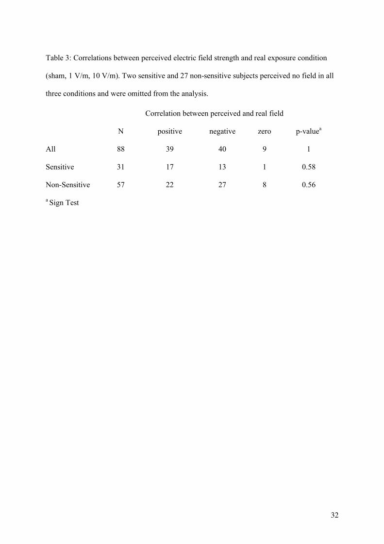
(p<0.001), even though score values were not associated with exposure levels. 17 out of 31
sensitive subjects had a positive correlation between perceived and real field intensity, 13 a
negative correlation (non-sensitive group: 22 and 27 out of 57 subjects, respectively), which
can be expected by chance (Table 3). Irrespective of exposure condition, perceived field
intensity was positively correlated with impaired well being in 68% of sensitive (QCDdiff:
p=0.043) and 64% of non-sensitive subjects (p=0.001). Similar results were found with
respect to the QCDpost and the TNO-Q (data not shown).
In the BQW, comparison of scores one week prior to and after study participation showed no
significant changes for satisfaction and ill health in the sensitive group. In the non-sensitive
group, the score for ill health was lower after the experiment (p=0.004), but satisfaction
remained unchanged.
Cognitive Tasks
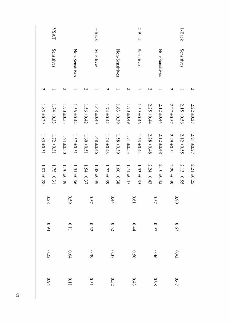
In the course of the entire study, subjects got faster in all tasks (p<0.02) except the SRT. In
both groups and irrespective of condition, speed decreased significantly from S1 to S2 in both
the SRT and CRT, but increased in the 1-, 2-, 3-back and VSAT (p<0.0001). In the following,
only effects including Condition or a Condition*Session interaction are described.
17
In both groups, we observed no condition-induced effects on speed in the SRT, 1-, 2-, 3-back
and VSAT. In the CRT, speed decreased in the sensitive group from S1 to S2 in the sham and
1 V/m condition (~20 ms), but not in the 10 V/m condition (Condition*Session: p=0.007,
Table 2). In contrast, we observed a decrease in speed between sessions irrespective of
exposure condition in the non-sensitive group (p=0.254, Table 2). A mixed model ANOVA
including the factor Sensitivity (sensitive, non-sensitive) corroborated the observed
differences between groups with respect to exposure (Condition*Sensitivity: p=0.005).
Accuracy was not affected by exposure in a dose response manner in any of the cognitive
tasks, except for the 1-back task in the non-sensitive group, where it decreased from 98.2%
(sham) to 97.3% (10 V/m; p=0.046) in session 1.
Adjusting the models for potential confounding factors (see Table 1 and 2) or performing the
analyses with only the 1:1 matched subjects did not alter the results. After multiple endpoint
adjustment (alpha=0.05; number of tests=44, overall correlation among cognitive
outcomes=0.39), however, all reported p-values exceeded the significance level of p=0.0051
(Tukey et al. 1985).
Dosimetry
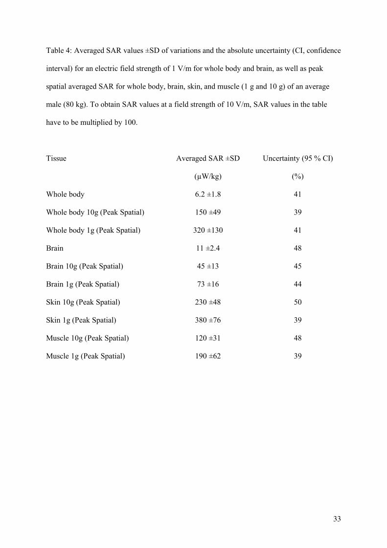
Penetration depth was low and highest specific absorption rate (SAR) values occurred
predominantly at the illuminated side close to the skin (Table 4, Figure 2). Whole-body
average absorption was 6.2 ±1.8 and 620 ±180 W/kg for 1 V/m and 10 V/m, respectively,
with an absolute uncertainty of 41% (Table 4). Peak spatial SAR (averaged over 10 g) was 45
±13 and 4500±1300 W/kg for brain tissue. At 10 V/m, all values were at least 100x below
recommended safety limits (International Commission on Non-Ionizing Radiation Protection
1998). Compared to usage of a mobile phone at the ear or to exposure levels used in other
18
studies, the peak spatial SAR of the brain was more than 100x lower at 10 V/m in our study.
SAR values for head tissues and left/ right differences are provided in Table 5.
The SAR values are strongly dependent on the incidence angle and the polarization of the
field which were fixed in our study. Variation of incidence angle and polarization at the same
field strength will lead to considerable changes of the SAR values in different parts of the
body.
19
Discussion
In contrast to our hypothesis, well being as assessed by the QCD and TNO-Q questionnaires
was not affected by UMTS radiation, neither at the 1 V/m nor at the 10 V/m condition. Even
though sensitive subjects generally reported more health problems, we found no difference
overall between the two groups with respect to the applied field conditions. Similarly,
cognitive performance was not affected, except for two separate and marginal effects in the 10
V/m condition. In the CRT, we could not observe a slight decrease in speed across sessions in
sensitive subjects and in the 1-back task, accuracy was reduced in non-sensitive subjects
compared to the sham condition.
Cognitive tasks with moderate to high workload have frequently been used as a tool to assess
RF EMF effects on brain physiology by measuring simple motor responses requiring selective
attention and higher cognitive functions such as working memory (e.g. Krause et al. 2000b).
Except for the VSAT, which was taken from the TNO battery of cognitive tasks for follow-up
reasons, we chose the SRT, CRT and N-back on the basis of recently published work
attempting to assess EMF-induced changes with respect to brain physiology (Koivisto et al.
2000a; Koivisto et al. 2000b; Preece et al. 1999). However, the described effects showed no
consistent picture and could not be replicated (Haarala et al. 2003; Preece et al. 2005).
In general, exposure in these studies was poorly defined and the inconsistencies in cognitive
outcome may be due to differences in the design, blinding, study population and sample size,
thus preventing a comparison of the results. Alternatively, cognitive tasks used so far may not
be sensitive enough to reliably measure potential RF EMF effects on brain functioning,
leading to a masking of existing effects or resulting in significant effects of tests that
stochastically respond to RF EMF. Moreover, statistical analysis of several tests increases the
risk of false positive findings.
20
In the present study, speed was affected in the sensitive group in one of six cognitive tasks
and accuracy in the non-sensitive group in one of five tasks. Although we cannot exclude an
actual Condition*Session interaction in the CRT in sensitive subjects and, similarly, a
Condition effect in the 1-back task in non-sensitive subjects, the findings seem to be
coincidental because they did not reach significance after multiple endpoint adjustment.
Both the sensitive and the non-sensitive group were unable to identify the applied fields better
than expected by chance. Because we investigated only three conditions per subject, the
likelihood of correct field rating by chance was relatively high. The observed distribution of
39 individuals with a positive correlation between the applied and estimated exposure
condition and 40 individuals with a negative correlation was likely to be expected by chance.
Nevertheless, we cannot exclude that among these subjects a minority was actually able to
perceive the applied exposure. The identification of such individuals has failed in several
provocation studies so far (reviewed in Rubin et al. 2005) and would require a multiple testing
approach in order to reduce the likelihood of a correct rating by chance. Perceived field
strength correlated with an impairment of current well being in both groups irrespective of
exposure condition. Also, sensitive subjects rated perceived field strengths higher than non-
sensitive subjects, yet ratings in both groups were not better than expected by chance and not
associated with exposure levels. This indicates that sensitive subjects overestimate their
ability to better perceive RF EMF than the general public (Leitgeb and Schröttner 2003).
Our results differ with respect to both well being and cognitive performance from the results
reported by Zwamborn et al. (2003). The TNO-Q is an adapted and not validated version of
the original questionnaire (Bulpitt and Fletcher 1990) and was not designed for short retest
intervals. Our findings were corroborated by the results of the QCD, a standardized
questionnaire that more reliably measures changes in well being over short test-retest intervals
(Müller and Basler 1993). Contrary to the TNO study, we found no significant effect on speed

21
in the VSAT. It was however the only task applied in both studies; all other cognitive tasks
were distinct. Zwamborn et al. (2003) found other effects with respect to cognitive tasks and
exposure conditions (GSM and UMTS) and we also report an effect on speed in one out of six
tasks and an effect on accuracy in one out of five tasks used. No clear picture therefore
emerges across the two studies showing reproducible effects of exposure condition or
cognitive task.
A number of other factors may contribute more generally to the discrepancies between the
TNO study and our study. Sample sizes differ substantially (sensitive subjects: 24 versus 33;
non-sensitive subjects: 24 versus 84). Our reference group was frequency matched to the
sensitive group and a subgroup was 1:1 matched with respect to gender, age, residential area
and BMI. In the TNO study, all conditions in a particular subject were carried out on a single
day, whereas we investigated the subjects at the same time of day in weekly intervals to rule
out possible circadian and carry-over effects. We further controlled circadian influences by a
uniform distribution of experimental sessions across the time of day. Carry-over effects may
lead to an accumulation of RF EMF radiation over time, thus falsifying potential effects of
discrete conditions. Furthermore, inclusion of an additional E-field strength of 10 V/m is
likely to have contributed to a more reliable assessment of RF EMF effects.
Technical improvements necessitated the modification of the exposure setup used in the TNO
study to achieve a more uniform and reproducible base station-like exposure. Whereas the
signal (carrier frequency and modulation) and the angle of incidence were identical, the
spatial incident field distribution was less uniform in the TNO study, where a narrow
exposure beam of only 5° width was used resulting in a larger variation due to differences in
height and position of the subjects. In addition, the whole-body exposure conditions applied
in this study correspond better to a base-station exposure scenario. However, exposure of head
tissues was equivalent in both studies, even though we had a smaller inter-subject variability.
22
Further insights regarding the discrepancies between the present and the Dutch study might be
gained from other follow up studies underway in Denmark, the U.K. and Japan, which are
also investigating the effect of UMTS base station-like radiation on well being and cognitive
function (personal communications).
In summary, we found no causal relationship between RF EMF and a decrease in well being
or adverse health effects under the given exposure conditions, but cannot exclude an effect of
UMTS-like EMF on brain functioning. The described effects were weak and not consistent in
the two groups. Regarding the implications for public health due to widespread exposure in
the living environment, no conclusions about long-term effects of UMTS base station-like
EMF can be drawn from the present study, as only a short-term exposure was applied.
23
References
Bulpitt CJ, Fletcher AE. 1990. The measurement of quality of life in hypertensive patients: a
practical approach. Br J Clin Pharmac 30:353-364.
Curcio G, Ferrara M, Moroni F, D'Inzeo G, Bertini M, De Gennaro L. 2005. Is the brain
influenced by a phone call? An EEG study of resting wakefulness. Neurosci Res 53:265-
270.
Freude G, Ullsperger P, Eggert S, Ruppe I. 2000. Microwaves emitted by cellular telephones
affect human slow brain potentials. Eur J Appl Physiol 81:18-27.
Grob A. 1995. Subjective well-being and significant life-events across the life-span. Swiss J
Psychol 54:3-18.
Haarala C, Björnberg L, Ek M, Laine M, Revonsuo A, Koivisto M, et al. 2003. Effect of a
902 MHz electromagnetic field emitted by mobile phones on human cognitive function: a
replication study. Bioelectromagnetics 24:283-288.
Hampel FR. 1985. The breakdown points of the mean combined with some rejection rules.
Technometrics 27:95-107.
Holm S. 1979. A simple sequentially rejective multiple test procedure. Scand J Statist 6:65-
70.
Huber R, Treyer V, Borbély AA, Schuderer J, Gottselig JM, Landolt H-P, et al. 2002.
Electromagnetic fields, such as those from mobile phones, alter regional cerebral blood
flow and sleep and waking EEG. J Sleep Res 11:289-295.
Huber R, Treyer V, Schuderer J, Berthold T, Buck A, Kuster N, et al. 2005. Exposure to
pulse-modulated radio frequency electromagnetic fields affects regional cerebral blood
flow. Eur J Neurosc 21:1000-1006.
Hyland GJ. 2000. Physics and biology of mobile telephony. Lancet 356:1833-1836.
24
International Commission on Non-Ionizing Radiation Protection. 1998. Guidelines for
limiting exposure to time-varying electric, magnetic, and electromagnetic fields (up to
300 GHz). Health Phys 74:494-522.
Koivisto M, Krause CM, Revonsuo A, Laine M, Hämäläinen H. 2000a. The effects of
electromagnetic field emitted by GSM phones on working memory. Neuroreport
11:1641-1643.
Koivisto M, Revonsuo A, Krause C, Haarala C, Sillanmäki L, Laine M, et al. 2000b. Effects
of 902 MHz electromagnetic field emitted by cellular telephones on response times in
humans. Neuroreport 11:413-415.
Krause CM, Haarala C, Sillanmäki L, Koivisto M, Alanko K, Revonsuo A, et al. 2004.
Effects of electromagnetic field emitted by cellular phones on the EEG during an auditory
memory task: a double blind replication study. Bioelectromagnetics 25:33-40.
Krause CM, Sillanmäki L, Koivisto M, Häggqvist A, Saarela C, Revonsuo A, et al. 2000a.
Effects of electromagnetic field emitted by cellular phones on the EEG during a memory
task. Neuroreport 11:761-764.
Krause CM, Sillanmäki L, Koivisto M, Häggqvist A, Saarela C, Revonsuo A, et al. 2000b.
Effects of electromagnetic fields emitted by cellular phones on the electroencephalogram
during a visual working memory task. Int J Radiat Biol 76:1659-1667.
Kuster N, Schönborn F. 2000. Recommended minimal requirements and development
guidelines for exposure setups of bio-experiments addressing the health risk concern of
wireless communications. Bioelectromagnetics 21:508-514.
Leitgeb N, Schröttner J. 2003. Electrosensibility and electromagnetic hypersensitivity.
Bioelectromagnetics 24:387-394.
Müller B, Basler HD. 1993. Kurzfragebogen zur aktuellen Beanspruchung (KAB) [in
German]. Weinheim: Beltz.
25
National Institute of Environmental Health Sciences - NIEHS Working Group Report. 1998.
Assessment of health effects from exposure to power-line frequency electric and
magnetic fields. Minnesota: National Institute of Environmental Health Sciences/National
Institutes of Health. Available:
http://www.niehs.nih.gov/emfrapid/html/WGReport/PDF_Page.html [accessed 19 April
2006]
Oldfield RC. 1971. The assessment and analysis of handedness: the Edinburgh inventory.
Neuropsychologia 9:97-113.
Preece AW, Goodfellow S, Wright MG, Butler SR, Dunn EJ, Johnson Y, et al. 2005. Effect of
902 MHz mobile phone transmission on cognitive function in children.
Bioelectromagnetics 26:S138-S143.
Preece AW, Iwi G, Davies-Smith A, Wesnes K, Butler S, Lim E, et al. 1999. Effect of a 915-
MHz simulated mobile phone signal on cognitive function in man. Int J Radiat Biol
75:447-456.
Preece AW, Wesnes KA, Iwi GR. 1998. The effect of a 50 Hz magnetic field on cognitive
function in humans. Int J Radiat Biol 74:463-470.
Röösli M, Moser M, Baldinini Y, Meier M, Braun-Fahrländer C. 2004. Symptoms of ill
health ascribed to electromagnetic field exposure - a questionnaire survey. Int J Hyg
Environ Health 207:141-150.
Rubin GJ, Das Munshi J, Wessely S. 2005. Electromagnetic hypersensitivity: a systematic
review of provocation studies. Psychosom Med 67:224-232.
Rubin GJ, Das Munshi J, Wessely S. 2006. A systematic review of treatments for
electromagnetic hypersensitivity. Psychother Psychosom 75:12-18.
Seitz H, Stinner D, Eikmann T, Herr C, Röösli M. 2005. Electromagnetic hypersensitivity
(EHS) and subjective health complaints associated with electromagnetic fields of mobile
26
phone communication - a literature review published between 2000 and 2004. Sci Total
Environ 349:45-55.
Tukey JW, Ciminera JL, Heyse JF. 1985. Testing the statistical certainty of a response to
increasing doses of a drug. Biometrics 41:295-301.
WHO (World Health Organization). 2005. Electromagnetic fields and public health. Fact
Sheet No 296, Available: http://www.who.int/mediacentre/factsheets/fs296/en/ [accessed
19 April 2006].
Zwamborn APM, Vossen SHJA, van Leersum BJAM, Ouwens MA, Mäkel WN. 2003.
Effects of global communication system radio-frequency fields on well being and
cognitive functions of human subjects with and without subjective complaints FEL-03-
C148. Netherlands: TNO Physics and Electronics Laboratory. Available:
http://home.tiscali.be/milieugezondheid/dossiers/gsm/TNO_rapport_Nederland_sept_200
3.pdf [accessed 19 April 2006].
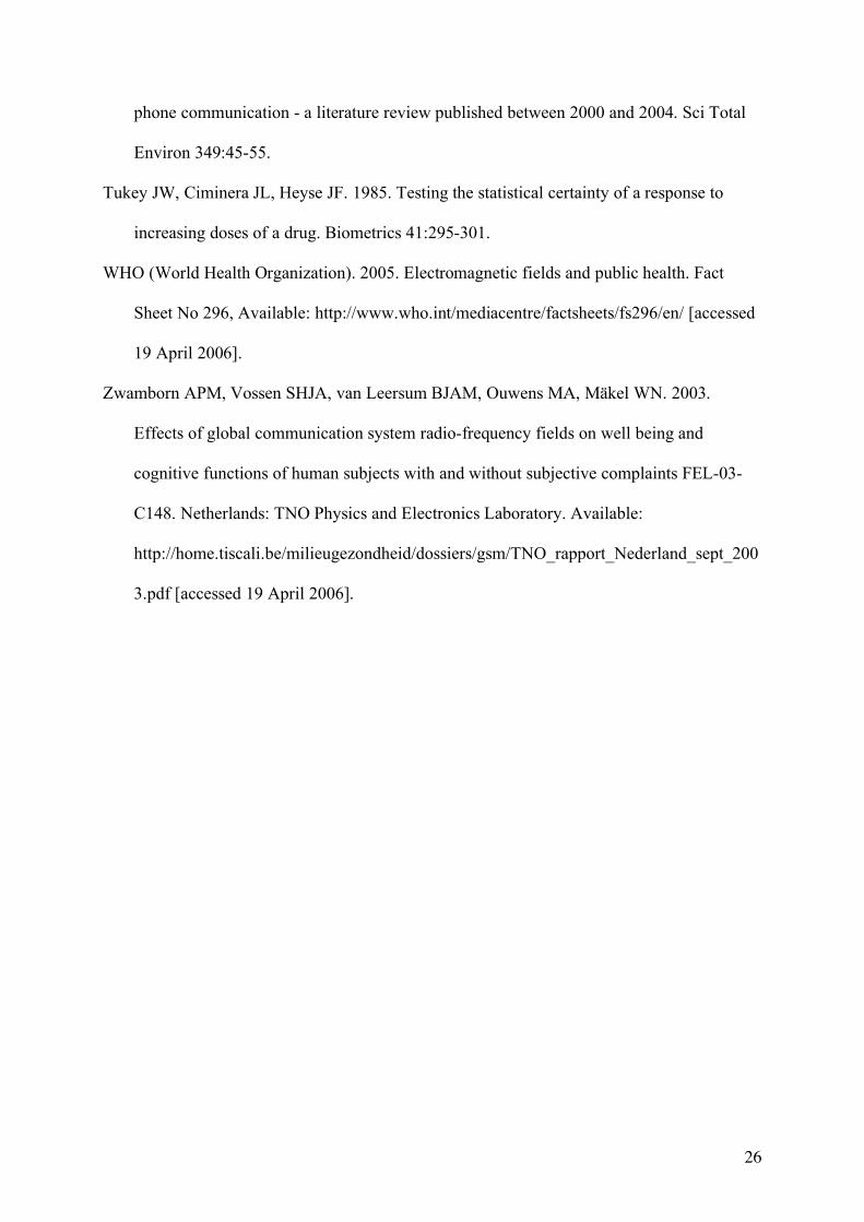
27
Ta
ble
s
Tab
le 1: R
esults o
f applied
questio
nnaires (m
ean sco
res ±S
D; N
=33 sen
sitive an
d N
=84 n
on-sen
sitive su
bjects). O
utco
mes o
f the Q
CD
(Sh
ort
qu
estion
na
ire on
curren
t disp
ositio
n) co
mprise th
e differen
ce betw
een p
re and p
ost ex
perim
ental sco
res (QC
DD
iff ) as well as p
ost ex
perim
ental
scores (Q
CD
po
st ). A d
ifference sco
re >0 co
rresponds to
a deg
radatio
n in
curren
t well b
eing d
urin
g th
e experim
ent. In
the Q
CD
po
st and th
e TN
O-Q
(Qu
ality-o
f-life qu
estion
na
ire) hig
her sco
res refer to a lo
wer w
ell bein
g. W
e measu
red su
bjectiv
e field p
erceptio
n b
y m
eans o
f a 100 m
m v
isual
analo
gue scale ran
gin
g fro
m “n
ot a
t all”
(0) to
“very stro
ng”
(100 m
m). W
e only
report p
-valu
es of C
on
ditio
n (C
ond) (fo
r details see M
ethods).
Outco
me
Gro
up
Sham
1V
/m
10V
/m
Cond
a C
ond
b
Mean
±S
D
Mean
±S
D
Mean
±S
D
p-v
alue
p-v
alue
QC
Dd
iff S
ensitiv
e 0
.30
±0
.83
0
.24
±0
.99
0
.24
±0
.95
0
.88
0
.95
N
on
-Sen
sitive
0.0
5 ±
0.7
3
-0.0
4 ±
0.5
9
0.0
2 ±
0.5
5
0.9
3
0.9
5
QC
Dp
ost
Sen
sitive
2.5
7 ±
1.0
6
2.6
5 ±
1.2
2
2.6
1 ±
0.9
7
0.9
7
0.9
6
N
on
-Sen
sitive
2.1
9 ±
0.7
6
2.0
5 ±
0.8
0
2.1
3 ±
0.7
8
0.9
7
0.8
9
TN
O-Q
S
ensitiv
e 1
0.5
3 ±
9.5
1
9.6
1 ±
8.9
6
9.7
9 ±
8.3
8
0.8
4
0.6
5
N
on
-Sen
sitive
5.2
3 ±
5.0
9
4.4
5 ±
4.9
2
4.9
6 ±
5.0
8
0.7
8
0.9
2
28
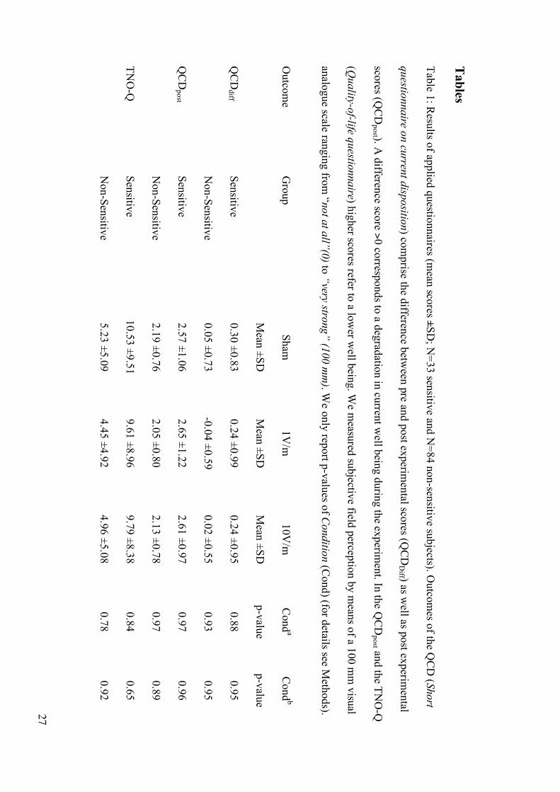
Field
percep
tion
S
ensitiv
e 2
6.0
±3
1.9
3
1.2
±3
3.7
2
9.4
±2
9.7
0
.89
0
.67
N
on
-Sen
sitive
12
.9 ±
22
.8
5.7
±1
3.1
1
2.2
±2
3.2
0
.24
0
.33
a Adju
sted fo
r ord
er; b Ad
justed
for o
rder, ag
e, gen
der, B
MI, caffein
e intak
e, med
ication
, (pre-) m
enstru
al com
plain
ts, sleep q
uality
and
sufferin
g
from
a cold
29
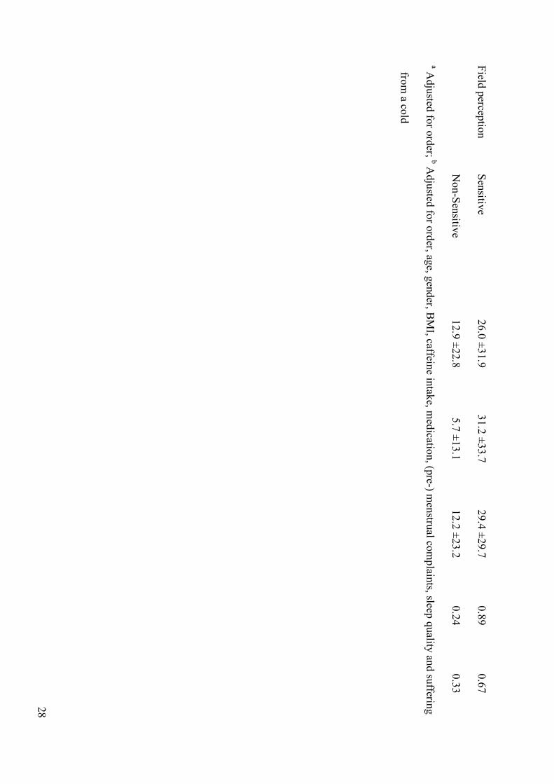
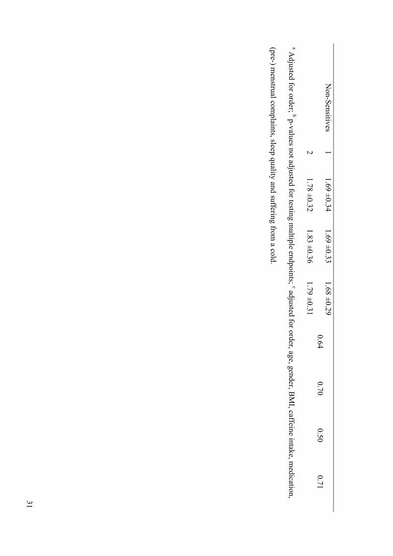
Tab
le 2: R
esults o
f cognitiv
e perfo
rman
ce. Mean
speed
±S
D (1
/Reactio
n tim
e [1/s]; N
=33 sen
sitive an
d N
=84 n
on-sen
sitive su
bjects) in
the tw
o
session
s (1st an
d 2
nd h
alf of ex
po
sure) in
the S
RT
(Sim
ple rea
ction
time ta
sk), CR
T (T
wo ch
oice rea
ction tim
e task), N
-back
task (1
-, 2-, 3
-back),
and V
SA
T (V
isua
l selective atten
tion tim
e task). W
e only
report p
-valu
es of C
on
ditio
n (C
ond) an
d o
f the in
teraction C
on
ditio
n*
Sessio
n (fo
r
details see M
etho
ds). S
tatistical analy
sis is based
on d
ata of all su
bjects. D
ue to
a missin
g sessio
n in
som
e subjects, m
ean v
alues are b
ased o
n
subjects w
ho co
mpleted
both
sessions in
each co
nditio
n (N
= at least 3
2 sen
sitive an
d at N
= at least 7
7 n
on-sen
sitive su
bjects).
Outco
me
Gro
up
Sessio
n
Sham
Mean
±S
D
1V
/m
Mean
±S
D
10
V/m
Mean
±S
D
Cond
a,b
p-v
alue
Cond*S
ession
a,b
p-v
alue
Cond
b,c
p-v
alue
Cond*S
ession
b,c
p-v
alue
SR
T
Sen
sitives
1
3.8
6 ±
0.5
2
3.7
8 ±
0.4
4
3.8
4 ±
0.4
8
2
3.7
3 ±
0.5
6
3.6
5 ±
0.4
3
3.7
8 ±
0.4
7
0.0
9
0.2
7
0.0
7
0.2
7
N
on
-Sen
sitives
1
3.8
5 ±
0.3
7
3.8
5 ±
0.3
8
3.8
4 ±
0.4
3
2
3.7
0 ±
0.4
4
3.7
0 ±
0.4
9
3.6
8 ±
0.4
1
0.5
9
0.5
1
0.3
7
0.5
0
CR
T
Sen
sitives
1
2.3
7 ±
0.2
8
2.3
3 ±
0.2
5
2.3
3 ±
0.2
8
2
2.2
5 ±
0.3
0
2.2
0 ±
0.2
7
2.3
1 ±
0.2
2
0.0
3
0.0
1
0.0
2
0.0
1
N
on
-Sen
sitives
1
2.2
7 ±
0.2
6
2.2
7 ±
0.2
7
2.2
4 ±
0.2
5
0.1
3
0.2
5
0.0
8
0.2
4
30
2
2.2
2 ±
0.2
7
2.2
1 ±
0.2
7
2.2
1 ±
0.2
5
1-B
ack
Sen
sitives
1
2.1
5 ±
0.5
6
2.1
2 ±
0.5
5
2.1
3 ±
0.5
5
2
2.2
7 ±
0.5
7
2.2
9 ±
0.5
4
2.2
9 ±
0.4
9
0.9
0
0.6
7
0.9
3
0.6
7
N
on
-Sen
sitives
1
2.1
2 ±
0.4
4
2.1
2 ±
0.4
8
2.1
0 ±
0.4
2
2
2.2
5 ±
0.4
4
2.2
8 ±
0.4
8
2.2
4 ±
0.4
3
0.5
7
0.9
7
0.4
6
0.9
8
2-B
ack
Sen
sitives
1
1.5
9 ±
0.4
6
1.5
3 ±
0.4
4
1.5
3 ±
0.3
5
2
1.7
0 ±
0.4
9
1.7
1 ±
0.5
3
1.7
1 ±
0.4
7
0.6
1
0.4
4
0.5
0
0.4
3
N
on
-Sen
sitives
1
1.6
3 ±
0.3
9
1.5
8 ±
0.3
9
1.6
0 ±
0.3
8
2
1.7
4 ±
0.4
2
1.7
4 ±
0.4
3
1.7
2 ±
0.3
9
0.4
4
0.5
2
0.3
7
0.5
2
3-B
ack
Sen
sitives
1
1.4
8 ±
0.4
0
1.4
8 ±
0.4
6
1.4
8 ±
0.3
9
2
1.5
6 ±
0.4
2
1.6
0 ±
0.5
1
1.5
4 ±
0.3
7
0.5
7
0.5
2
0.3
9
0.5
1
N
on
-Sen
sitives
1
1.5
6 ±
0.4
4
1.5
7 ±
0.5
1
1.5
1 ±
0.3
6
2
1.7
0 ±
0.5
5
1.6
4 ±
0.5
0
1.7
0 ±
0.4
9
0.5
9
0.1
1
0.6
4
0.1
1
VS
AT
S
ensitiv
es 1
1
.74
±0.3
3
1.7
2 ±
0.3
1
1.7
5 ±
0.3
1
2
1.8
5 ±
0.2
9
1.8
5 ±
0.3
1
1.8
7 ±
0.2
8
0.2
8
0.9
4
0.2
2
0.9
4
31
N
on
-Sen
sitives
1
1.6
9 ±
0.3
4
1.6
9 ±
0.3
3
1.6
8 ±
0.2
9
2
1.7
8 ±
0.3
2
1.8
3 ±
0.3
6
1.7
9 ±
0.3
1
0.6
4
0.7
0
0.5
0
0.7
1
a Adju
sted fo
r ord
er; b p-v
alues n
ot ad
justed
for testin
g m
ultip
le endpoin
ts; c adju
sted fo
r ord
er, age, g
ender, B
MI, caffein
e intak
e, med
ication,
(pre-) m
enstru
al com
plain
ts, sleep q
uality
and
sufferin
g fro
m a co
ld.
32
Table 3: Correlations between perceived electric field strength and real exposure condition
(sham, 1 V/m, 10 V/m). Two sensitive and 27 non-sensitive subjects perceived no field in all
three conditions and were omitted from the analysis.
Correlation between perceived and real field
N positive negative zero p-valuea
All 88 39 40 9 1
Sensitive 31 17 13 1 0.58
Non-Sensitive 57 22 27 8 0.56
a Sign Test
33
Table 4: Averaged SAR values ±SD of variations and the absolute uncertainty (CI, confidence
interval) for an electric field strength of 1 V/m for whole body and brain, as well as peak
spatial averaged SAR for whole body, brain, skin, and muscle (1 g and 10 g) of an average
male (80 kg). To obtain SAR values at a field strength of 10 V/m, SAR values in the table
have to be multiplied by 100.
Tissue
Averaged SAR ±SD
( W/kg)
Uncertainty (95 % CI)
(%)
Whole body 6.2 ±1.8 41
Whole body 10g (Peak Spatial) 150 ±49 39
Whole body 1g (Peak Spatial) 320 ±130 41
Brain 11 ±2.4 48
Brain 10g (Peak Spatial) 45 ±13 45
Brain 1g (Peak Spatial) 73 ±16 44
Skin 10g (Peak Spatial) 230 ±48 50
Skin 1g (Peak Spatial) 380 ±76 39
Muscle 10g (Peak Spatial) 120 ±31 48
Muscle 1g (Peak Spatial) 190 ±62 39
34
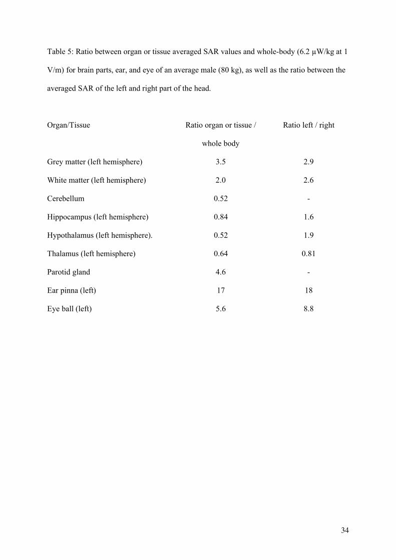
Table 5: Ratio between organ or tissue averaged SAR values and whole-body (6.2 W/kg at 1
V/m) for brain parts, ear, and eye of an average male (80 kg), as well as the ratio between the
averaged SAR of the left and right part of the head.
Organ/Tissue
Ratio organ or tissue /
whole body
Ratio left / right
Grey matter (left hemisphere) 3.5 2.9
White matter (left hemisphere) 2.0 2.6
Cerebellum 0.52 -
Hippocampus (left hemisphere) 0.84 1.6
Hypothalamus (left hemisphere). 0.52 1.9
Thalamus (left hemisphere) 0.64 0.81
Parotid gland 4.6 -
Ear pinna (left) 17 18
Eye ball (left) 5.6 8.8
35
Figure legends
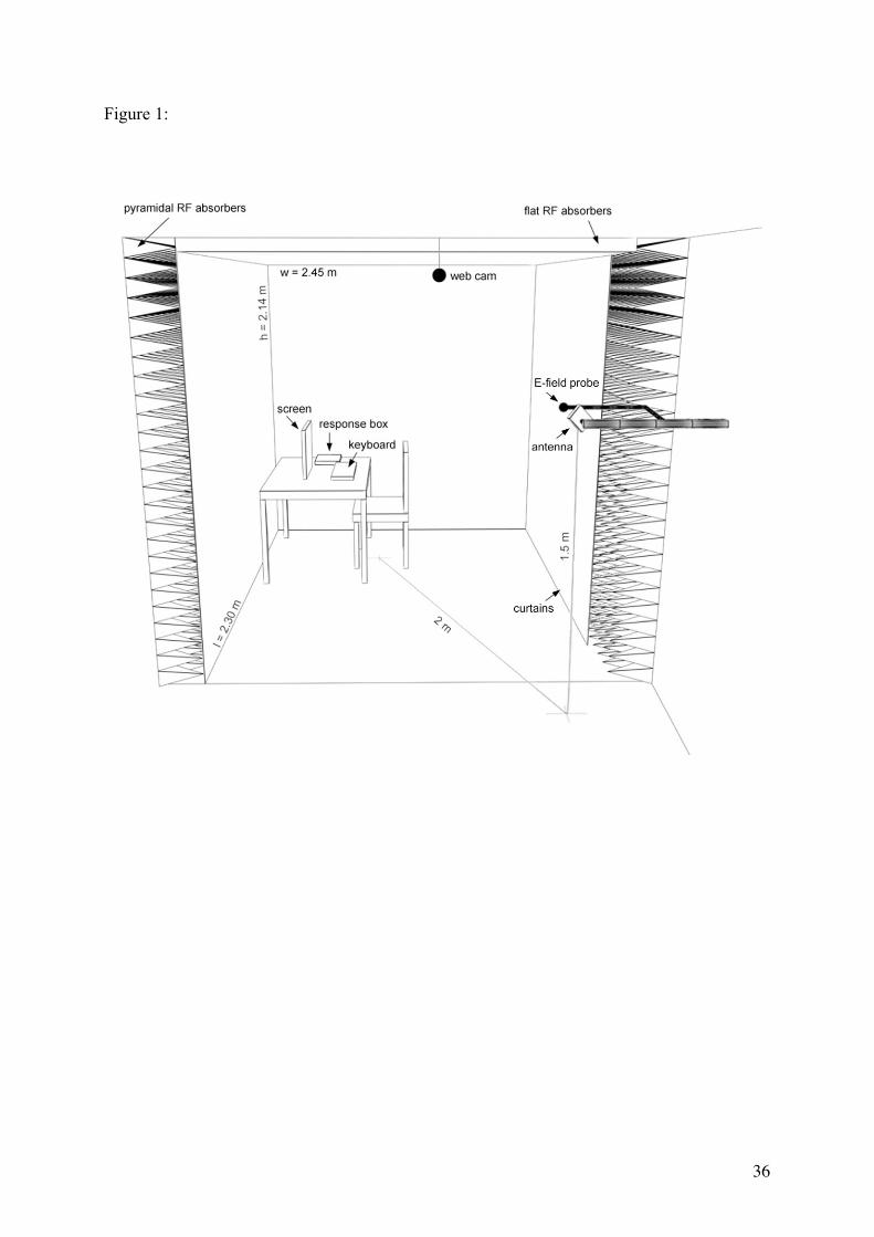
Figure 1: Sketch of the exposure chamber. Walls covered by pyramidal RF absorbers and
non-reflecting curtains. Ceiling covered by flat absorbers. Antenna, electric field probe,
furniture, screen, keyboard, response box, and web cam, inner dimensions (w: width; h:
height; l: length), and position of the antenna are indicated.
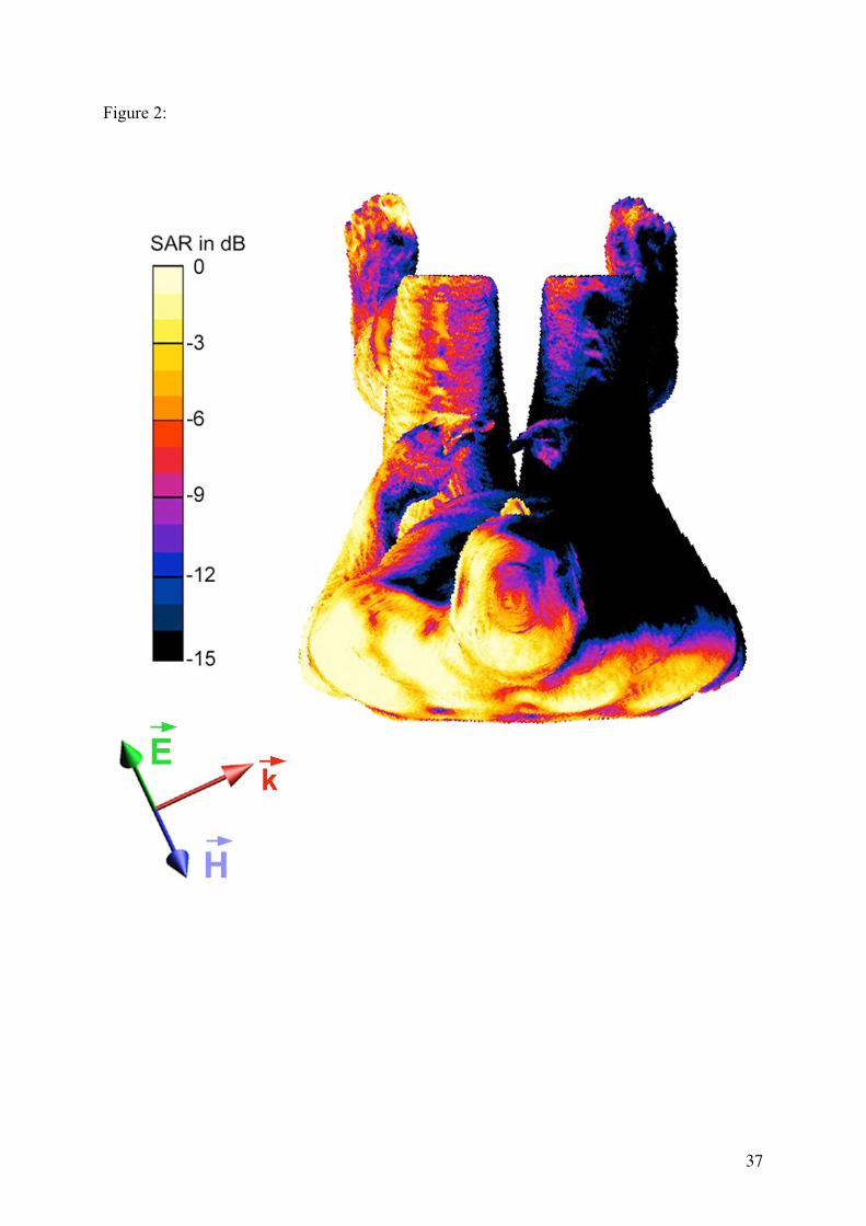
Figure 2: SAR distribution on the surface of a male (80 kg) in a sitting position (top view). 0
dB corresponds to 0.05 W/kg for an electric field strength of 1V/m. The orientation of the
electric field ( r E ), the magnetic field (
r H ), and the propagation direction (
r k ) of the EMF are
indicated.
36
Figure 1:
37
Figure 2:
![T24 Technical Manual - · PDF fileT24-BSi and T24-BSu [Base Station] ... Mantracourt Electronics Limited T24 Technical Manual 4 ... RF Exposure Limits](https://img.pdfslide.net/doc/110x75/5a809fa07f8b9aa24f8c9617/t24-technical-manual-and-t24-bsu-base-station-mantracourt-electronics-limited.jpg)


![2. Feychting ICNIRP principles and practice [互換モード]...Maria Feychting Adèle C. Green Akimasa Hirata Carmela Marino Sharon Miller Gunnhild Oftedal Tsutomu Okuno Martin Röösli](https://img.pdfslide.net/doc/110x75/5fea9076ad511627013dc64f/2-feychting-icnirp-principles-and-practice-fff-maria-feychting.jpg)