Embed Size (px)
Citation preview

1
UNIVERSITÀ DEGLI STUDI DI MILANO
DOTTORATO DI RICERCA IN
SCIENZA DELLO SVILUPPO PRENATALE, DIAGNOSI E TERAPIA FETALE
CICLO 23 (8AR)
DIPARTIMENTO DI OSTETRICIA E GINECOLOGIA
Ospedale dei Bambini V. Buzzi -ICP-
Dipartimento di Scienze Cliniche
NEW SCREENING TESTS IN EARLY PREGNANCY: DIAGNOSIS AND PREVENTION
DOTT.SSA VALENTINA CONSERVA
MATRICOLA
R07635
TUTOR PROF. ENRICO FERRAZZI
COORDINATORE DEL DOTTORATO PROF. ENRICO FERRAZZI
A.A. 2009-2010
2
INDEX
PRESENTATION ………………………………………….………………………………………..3
ABSTRACTS ………………………………………………………………………………………..4
GENERAL INTRODUCTION ……………………………………………………………………. 6
REFERENCES ………………………………………………………………………………………8
PART I ……………………………………….…………………………………………………….. 9
DISTINCTION BETWEEN FETAL GROWTH RESTRICION AND SMALL FOR GESTATIONAL
AGE NEWBORN WEIGHT ENHANCES THE PROGNOSTIC VALUE OF LOW PAPP-A IN THE
FIRST TRIMESTER
INTRODUCTION ………………………………………………………………………………….10
AIM OF THE STUDY ……………………………………………………………………………..13
METHODS AND RESULTS ………………………………………………………………………14
DISCUSSION …………………………………………………………………….………………..16
REFERENCES ……………………………………………………………………………………. 17
FIGURE …………………………………………………………………………………………….20
TABLES ……………………………………………………………………………………………22
PART II …………………………………………………………………………………………… 24
RECURRENCE AND SEVERITY OF ABNORMAL PREGNANCY OUTCOME IN PATIENTS
TREATED BY LOW-MOLECULAR-WEIGHT –HEPARIN: A PROSPECTIVE PILOT STUDY
INTRODUCTION ………………………………………………………………………………… 25
AIM OF THE STUDY ……………………………………………………………………………..26
METHODS ………………....………………………………………………………………………27
RESULTS …………………………………………………………………………………………..30
DISCUSSION ………………………………………………………………………………………32
REFERENCES …………………………………………………………………….……………….36
FIGURE …………………………………………………………………………………………….39
TABLES ……………………………………………………………………………………………41

3
PRESENTATION
“New screening tests in early pregnancy: diagnosis and prevention” is the title chosen to present
two papers I published during these three years.
Conserva V, Signaroldi M, Mastroianni C, Stampalija T, Ghisoni L, Ferrazzi E. Distinction
between fetal growth restriction and small for gestational age newborn weight enhances the
prognostic value of low PAPP-A in the first trimester. Prenat Diagn. 2010 Oct;30(10):1007-9
Conserva V, Muggiasca M, Arrigoni L, Mantegazza V, Edoardo Rossi E, Ferrazzi E. Recurrence
and severity of abnormal pregnancy outcome in patients treated by low-molecular-weight-heparin:
a prospective pilot study. J Matern Fetal Neonatal Med. 2011 Nov 29
These two paper deal with subjects that are apparently different: the first paper studies the
prognostic value of a placental marker measured in the first trimester, PAPP-A, for abnormal
pregnancy outcomes, the second one investigates the possible role of low molecular weight heparin
to improve the development of pregnancy in patients with previous abnormal pregnancy outcomes.
Besides the specific clinical application of both results, the two studies assume the central role of
placentation. A physiologic communication and balance in maternal-fetal interface allows a correct
development of placenta from the earliest days of gestation to twenty four weeks of pregnancy,
although the phenotypic outcome of this process can be clinically evaluated much later in
pregnancy. These two papers tap into this silent and fundamental process in order to evaluate a
potential for early diagnosis or prevention.

4
ABSTRACTS
Distinction between fetal growth restriction and small for gestational age newborn weight
enhances the prognostic value of low PAPP-A in the first trimester.
Conserva V, Signaroldi M, Mastroianni C, Stampalija T, Ghisoni L, Ferrazzi E.
Prenat Diagn. 2010 Oct;30(10):1007-9
Objective Low levels of PAPP-A in maternal blood may become an early marker of obstetrical
complications. The aim of this article was to sort out those outcomes consistently related to an
abnormal placental vascular function and to evaluate their association with low levels of maternal
serum PAPP-A in early pregnancy
Methods We analyzed retrospectively a database of the first trimester combined screening of an
Italian biotech company and investigated the correlation between PAPP-A value < 5th percentile,
0.40 multiples of the median (MoM), and infants with birthweight below the 10th
percentile for
gestational age (small for gestational age SGA), preterm delivery, GH and PE not associated with
intra-uterine growth restriction (IUGR), IUGR isolated with an abnormal umbilical PI or associated
with maternal GH-PE, placental abruption and intra-uterine fetal demise (IUFD) after 22 weeks of
gestation.
Results 1687 patients were analyzed. Overall pregnancy complications were observed in 31.4% of
women with low PAPP-A and in 21.1% women with a PAPP-A value >0.4 MoM (P < 0.0001).
Severe and early fetal growth restriction (<34 weeks) with abnormal umbilical PI or maternal PE,
was significantly associated with low levels of PAPP-A (OR= 10, 95% CI 1.0–97, P = 0.02). No
relationship was observed between SGA newborns and low PAPP-A. Weak association was
observed in with GH and PE not associated with fetal growth restriction (OR = 1.9, 95% CI 1.1–3,
P = 0.01). We also observed a correlation between low PAPP-A and preterm delivery (OR = 1.8,
95% CI 1.1–2,9, P = 0.01). Because of the small number of cases, the OR for placental abruption
and IUFD were not calculated
Conclusions. Low values of PAPP-A are associated with abnormal obstetrical outcome. Evaluating
separately growth-restricted fetuses and small for gestational age fetuses we observe that only
growth-restricted fetuses are significantly associated with low values of PAPP-A, whereas SGA
newborns, simply defined by their percentile rank, are not predicted by this test in the first trimester
of pregnancy.

5
Recurrence and severity of abnormal pregnancy outcome in patients treated by low-
molecular-weight-heparin: a prospective pilot study.
Conserva V, Muggiasca M, Arrigoni L, Mantegazza V, Edoardo Rossi E, Ferrazzi E
J Matern Fetal Neonatal Med. 2011 Nov 29
Objective This prospective pilot study assesses the recurrence rate and severity of abnormal
pregnancy outcome (APO), excluding early pregnancy complications, in pregnant patients, without
acquired thrombophilia, treated by prophylactic doses of LMWH, independently from their
congenital thrombophilic condition.
Methods We recruited a cohort of 128 pregnant patients with previous APO; 100 of whom with
APO and intrauterine growth restriction (IUGR), and 28 with maternal APO only. LMWH
treatment was started at recruitment. Composite cross over recurrence rate IUGR, gestational
hypertension, preeclampsia, HELLP, abruptio was analyzed. The main outcome measure was
severe APOs with iatrogenic delivery ≤ 32 weeks of gestation.
Results Median gestational age at LMWH treatment was 20 weeks. Severe APO decreased in
treated pregnancies from 45% to 4% (R.R.=0.3, CI .95=0.2-0.8). This value was not significantly
different in thrombophilic and non thrombophilic patients. When severe and minor complications
were analyzed altogether the recurrence rate was 28%. In patients with APO and FGR in the index
pregnancy, newborn weights were significantly better in the treated pregnancy: 1090g (1035-1145)
vs. 850g (535-1200), P<0,01)
Conclusions. Prophylactic regimen of LMWH significantly reduced the recurrence rate of severe
composite APO in pregnancies affected in the index pregnancy by APO and fetal growth restriction
or SGA newborns. This result was independent from the patients’ inherited thrombophilic
conditions.
6
GENERAL INTRODUCTION
Prior to implantation, the outer wall of the blastocyst is formed by a single layer of uninucleate
trophectoderm cells. Contact with the maternal uterine interface induces local differentiation of this
layer into an outer multinucleated syncytiotrophoblast, and an underlying population of progenitor
cells, the cytotrophoblast cells (1).
The maternal-fetal interface has certain unique features immunologically. It must exhibit immune
tolerance to the allogenic syncytiotrophoblast, cytotrophoblast and fetus, and at the same time, it
must maintain adequate protection against microbial invasion of the maternal host. The
syncytiotrophoblast forms the terminally differentiated epithelial covering of the placental villous
trees, being in direct contact with maternal blood and behaves, together with decidual cells, as ―non-
classical‖ immune cells at the utero-placental interface expressing toll-like receptors (TLR). These
receptors at maternal-fetal interface not only recognize microorganisms, but also encode for genes
resulting in release of cytokines, prostaglandins and coagulation factors (2).
Cytotrophoblast cells can differentiate along either of two pathways. They may fuse with the
overlying syncytiotrophoblast, thus increasing its mass. Alternatively, where villi make contact with
the decidua they may differentiate along the extravillous pathway and invade the maternal tissues.
In doing so, they start as interstitial trophoblast invading through the stroma of the decidua and then
may remain interstitial or may follow two other routes, towards the lumen of spiral arteries as
endovascular trophoblast (3), or towards the lumen of uterine glands as endoglandular trophoblast
(4). The interstitial trophoblast normally penetrates as far as the inner third of the myometrium.
Trophoblast development and differentiation are crucial processes for a physiologic pregnancy
development. Any insult resulting in an aberrant development and differentiation of the villous
syncytiotrophoblast poses a risk for the integrity of the placental barrier and the release of necrotic
and aponecrotic trophoblast fragments, culminating in a systemic inflammatory response of the
mother (5). This process is shown by the shift of normal Th1/ Th2 balance to Th2/ Th1, resulting in
increased production of inflammatory cytokines (6). Inflammation is known to play a pivotal role
in pathogenesis of Preeclampsia (PE), and other obstetric complications including pretermlabor and
chorioamnionitis (7) by the activation of circulating leukocytes in maternal blood, along with
increased levels of particular subsets of leukocyte-derived microparticles (MP). Other markers of
inflammatory change include Interleukin-1 Receptor Antagonist (IL-1RA) and tumor necrosis
factor (TNF)-R1in women with PE, as compared to non-pregnant cohort (8). Local hemostatic
changes in placental vasculature have been well defined (9). While there is expression of tissue
factor (TF) in the synctiotrophoblast, there is a concomitant decrease in the concentration of tissue
factor inhibitor pathway (TFPI) in the synctiotrophoblast. Furthermore, the balance between TF-

7
TFPI has been found to be altered in women with gestational vascular complications. Select
coagulation factors and global markers of coagulation activation have been partially characterized
in women with placenta mediated complications. For example in women with pre eclampsia,
coagulation abnormalities which have been identified include decreased antithrombin activity,
elevated PAI-1 antigen and elevated Thrombin-Antithrombin (TAT) complexes (10).
Biochemical and immunologic markers reflect the systemic responses of maternal systems to
abnormal pregnancy and may be of an inflammatory or metabolic origin.

8
REFERENCES
1. Cetin I, Huppertz B, Burton G, Cuckle H, Gonen R, Lapaire O, Mandia L, Nicolaides K, Redman
C, Soothill P, Spencer K, Thilaganathan B, Williams D, Meiri H. Pregenesys pre-eclampsia
Markers consensus meeting: What do we require from markers, risk assessment and model Systems
to tailor preventive strategies? Placenta. 2011 Feb;32 Suppl:S4-16.
2. Hossain, Nazli; Schatz, Frederick; Paidas, Michael J. Heparin and maternal fetal interface: Why
should it work to prevent pregnancy complications? Thrombosis Research Volume: 124, Issue: 6,
December, 2009, pp. 653-655
3. P. Kaufmann, S. Black and B. Huppertz, Endovascular trophoblast invasion: implications for the
pathogenesis of intrauterine growth retardation and preeclampsia. Biol Reprod, 69 (2003), pp. 1–7.
4. G. Moser, M. Gauster, K. Orendi, A. Glasner, R. Theuerkauf and B. Huppertz, Endoglandular
trophoblast, an alternative route of trophoblast invasion? Analysis with novel confrontation co-
culture models. Hum Reprod, 25 (2010), pp. 1127–1136.
5. B. Huppertz, Placental origins of preeclampsia: challenging the current hypothesis. Hypertension,
51 (2008), pp. 970–975.
6. Challis J.R., Lockwood C.J., Myatt L., Norman J.E., Strauss J.F., Petraglia F., Inflammation and
pregnancy, Reprod Sci, Volume: 16, Issue: 2 (2009), pp. 206—215
7. Park J.S., Park C.W., Lockwood C.J., Norwitz E.R., Role of cytokines in preterm labor and birth,
Minerva Ginecol, Volume: 57, Issue: 4 (2005), pp. 349—366
8. Lok C.A., Jebbink J., Nieuwland R., Faas M.M., Boer K., Sturk A., et al. Leukocyte activation
and circulating leukocyte-derived microparticles in preeclampsia, Am J Reprod Immunol, Volume:
61, Issue: 5 (2009), pp. 346—359
9. Lockwood C.J., Pregnancy-associated changes in the hemostatic system, Clin Obstet Gynecol,
Volume: 49, Issue: 4 (2006), pp. 836—843
10. Halligan A., Bonnar J., Sheppard B., Darling M., Walshe J., Haemostatic, fibrinolytic and
Endothelial variables in normal pregnancies and pre-eclampsia, Br J Obstet Gynaecol, Volume:
101, Issue: 6 (1994), pp. 488—492

9
PART I
DISTINCTION BETWEEN FETAL GROWTH RESTRICION AND SMALL FOR
GESTATIONAL AGE NEWBORN WEIGHT ENHANCES THE PROGNOSTIC VALUE
OF LOW PAPP-A IN THE FIRST TRIMESTER

10
INTRODUCTION
The placentation, placenta itself and its myriad functions are central to successful reproductive
outcomes. The placenta is the first complex mammalian organ to develop and as development
continues, it becomes a robust endocrine organ, producing a spectrum of hormones that are unique
to the pregnancy, such as human chorionic gonadotropin, estriol, as well as hormones identical to
those produced elsewhere, such as estrogen and progesterone. This hormone production is
intrinsically coupled to the physiologic changes of pregnancy and is critical for pregnancy
maintenance.
Biochemistry and biosynthesis of PAPP-A
Pregnancy-associated plasma protein-A (PAPP-A) was first identified in 1974 (1). Circulating
PAPP-A is principally derived from the syncytiotrophoblast (2,3), and the PAPP-A gene is located
on human chromosome 9q33.1 (4). The protein is secreted as a disulfide-bound homodimer with a
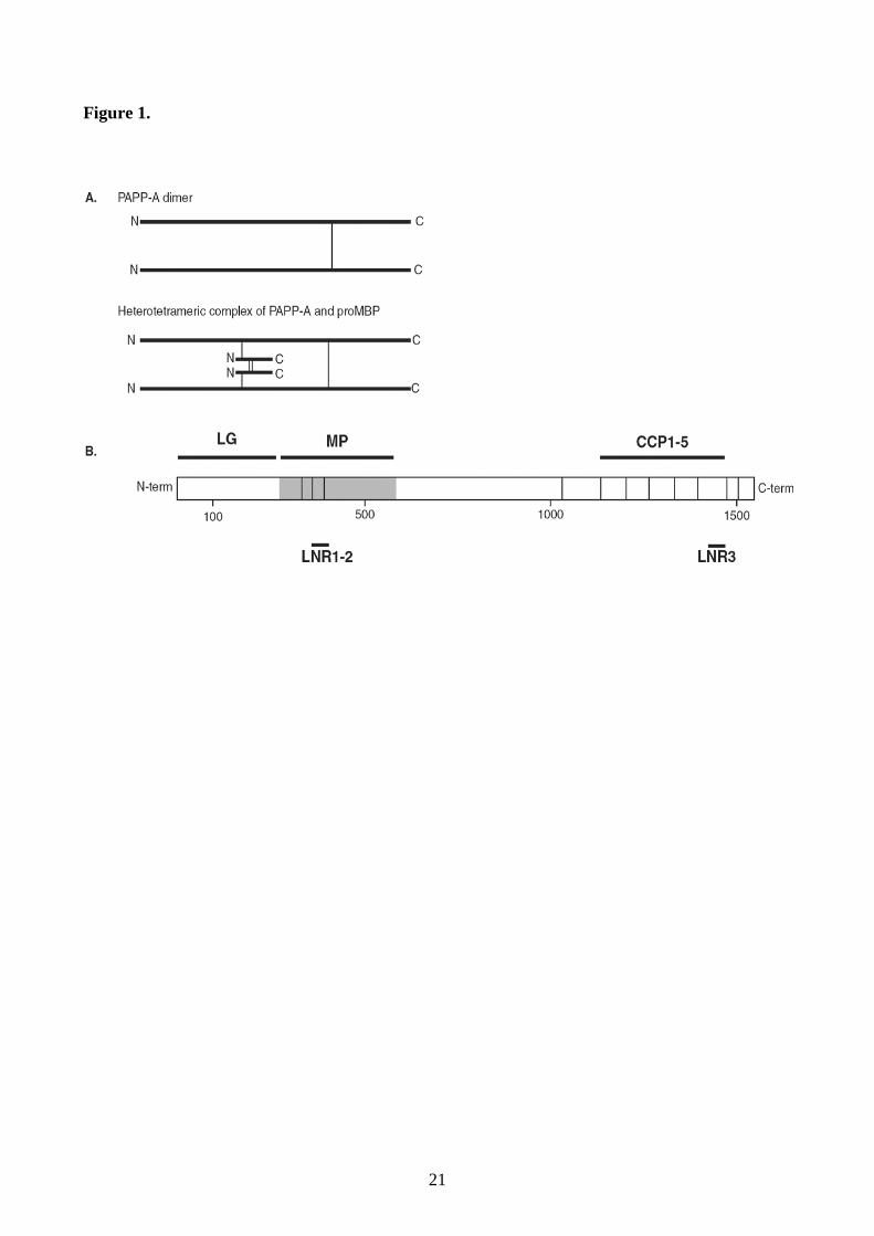
molecular weight of 400,000 g/mol (5). Each subunit consists of 1,547 amino acids (6) and includes
modules known to occur in other proteins (Figure 1), but the degree of identity is generally low. A
distinguishing feature is the metalloproteinase modules (MP), based on which PAPP-A is classified
as a metzincin metalloproteinase, and hence related to the large family of matrix metalloproteinases.
Other modules of PAPP-A include three Lin12-Notch repeat (LNR1-3) modules, a single laminin
G-like module (LG), and five complement control protein modules (CCP1-5) that mediate binding
of PAPP-A to heparan sulfates of cell surfaces.
Almost all of the circulating PAPP-A is covalently bound to proMBP (proform of eosinophil major
basic protein). During pregnancy, both PAPP-A and proMBP are expressed abundantly in the
human placenta (7), but in different cell types. PAPP-A is principally expressed in the
syncytiotrophoblast, whereas proMBP is expressed in extravillous cytotrophoblasts (2), from where
it is secreted without propeptide cleavage. Thus, the process of PAPP-A/proMBP complex
formation occurs in the extracellular environment, probably at the surface of the
syncytiotrophoblast. PAPP-A bound to the cell surface readily forms a complex with proMBP, this
complex is unable to bind to the cell surface and therefore enters the circulation.
PAPP-A and the insulin-like growth factor system

11
Strong evidence suggests that PAPP-A increases the local bioavailability of insulin-like growth
factor (IGF) by cleaving the inhibitors IGFBP-4 and −5 (insulin-like growth factor-binding protein-
4 and −5) (8). IGF is considered mitogenic and anti-apoptotic and is important for the growth of
human cells in most tissues (9). This probably explains why PAPP-A knockout mice are reduced to
60% of normal size at birth, strikingly similar to the IGF-II knockout mouse (10). Only dimeric
PAPP-A, not the PAPP-A/proMBP complex, shows this proteolytic activity (5). At term, about 1%
of the circulating PAPP-A is present as uncomplexed, active PAPP-A. However, the fraction of
active PAPP-A is much higher in the first trimester, although the total level of PAPP-A is lower.
Maternal serum levels of PAPP-A
Maternal PAPP-A blood levels become detectable soon after implantation and increase throughout
pregnancy, with a doubling time of 3–4 days during the first trimester and maximum levels at term
(11,12) The rapid increase in PAPP levels during the first trimester causes the interpretation of a
given value to be very dependent on gestational age. Common practice is therefore to use the unit
MoM (Multiples of Median) as a gestational age-independent expression of PAPP-A concentration.
The average half life of PAPP-A after normal delivery is 53 ± 26 hours (mean plus SD) (13). In
addition to gestational age, a number of maternal and pregnancy-associated characteristics affect the
maternal serum concentration of PAPP-A (Table 1). Some of these (multiple parity and smoking)
are probably related to the mass of trophoblastic tissues, because the PAPP-A concentration
increases with increasing ultrasound-determined placental volume (14). Other factors such as
maternal pre-pregnancy weight have been correlated with the volume of distribution, whereas
associations with parity, ethnicity, and assisted reproduction have so far escaped biological
explanations. Several studies have tested the hypothesis that low maternal serum levels of
pregnancy-associated plasma protein A (PAPP-A) may predict adverse pregnancy outcomes other
than Down syndrome in first trimester (15- 20). Low levels of PAPP-A in maternal blood could
become an early marker of obstetrical complications associated with poor placental function, that is
small babies, gestational hypertension (GH), pre-eclampsia (PE), stillbirth and even premature
delivery. However, contradictory results had been observed in different cohorts (19-21). These
findings could result as a consequence of non-homogeneous criteria in the definition of different
abnormal obstetrical outcomes. Recently, Poon et al. (22) showed that low PAPP-A is significantly
associated with early PE but not with late PE. This data confirmed the need to distinguish among
abnormal obstetrical outcomes those based on similar placental damage. Moreover, when
considering poor obstetrical outcomes, other environmental factors have to be taken into account.

12
Among these, maternal smoking has been largely studied while it is associated with impaired fetal
growth. Therefore, this factor could interfere when evaluating the predictive value of PAPP-A.

13
AIM OF THE STUDY
The aim of this article was to sort out among abnormal obstetrical outcomes those consistently
related to an abnormal placental vascular function and to evaluate their association with low levels
of maternal serum PAPP-A in early pregnancy.

14
METHODS AND RESULTS
The database of the first trimester combined screening of an Italian biotech company (Bi-Tech
Ltd.), certified by the Fetal Medicine Foundation (FMF), was analysed retrospectively by obstetrics
and gynaecology specialists at the University Department of Clinical Sciences, University of Milan,
under the regulations of a research contract. The database was searched for all combined first
trimester screening tests in women with singleton pregnancies performed between January 2007 and
January 2008. All women with high risk for trisomy 21, defined as risk greater than 1/350, were
identified together with the women who presented intermediate risk, defined as risk 1/350 ÷ 1/1000.
A control group of pregnant women at low risk for Down syndrome (<1/1000) was selected by
means of a randomization table. The risk was evaluated by the combination of maternal age, nuchal
translucency (NT) measurement, maternal serum-free β-hCG and PAPP-A. NT had been measured
according to the FMF guidelines (23), while Bi-Tech Ltd. performed the analysis of serum
metabolites and calculated the combined risk. PAPP-A values were corrected for maternal weight
and smoking habit. Pregnancy outcome of all patients was obtained by telephone interviews
conducted by a physician. Women with an aneuploid fetus diagnosed by invasive procedures or at
birth filled an outcome form provided by the Bi-Tech company. In our cohort, the PAPP-A 5th
percentile corresponded to 0.40 multiples of the median (MoM). This was used to define a low
PAPP-A value and it is in accordance with previously observed results by Krantz et al. (16) and
Dugoff et al. (16), respectively, 0.42 and 0.45 MoM. The following pregnancy complications were
evaluated: (1) infants with birthweight below the 10th
percentile for gestational age (small for
gestational age SGA); (2) preterm delivery <37 weeks of gestation; (3) GH and PE not associated
with intra-uterine growth restriction (IUGR), the definition of GH and PE was according to the
criteria proposed by Davey and MacGillivray (1988); (4) IUGR isolated or associated with GH or
PE. IUGR was defined as decrement of the fetal abdominal circumference below the 10th percentile
for local standard intra-uterine growth curves associated with an abnormal umbilical PI or maternal
GH-PE. We also analysed the correlation of low PAPP-A with (5) placental abruption and (6) intra-
uterine fetal demise (IUFD) defined as fetal death after 22 completed weeks of gestation. Patients
with more than one complication were counted only once, taking into account the most severe
complication as shown in increasing order in this list. We performed univariate logistic regression
model with maximum likelihood fitting to evaluate the correlation between PAPP-A and poor
obstetrical outcomes and to estimate odds ratios (OR) and their corresponding 95% confidence
interval (CI). To account simultaneously for the effects of maternal smoking and weight, we also
performed a multiple logistic regression model (25). Statistical significance was assumed for P <
0.05. Over the 12-months period, we identified 401 women with high risk and 867 with

15
intermediate risk for Down syndrome, while 502 subjects were randomly selected out of a larger
sample (13 376) of women at low risk for chromosomal aneuploidies. The final sample size counted
1770 participants. Pregnancy outcomes were available for the whole study population.
Chromosomal abnormalities were diagnosed in 26 subjects and were excluded from the analysis.
Spontaneous miscarriage <18 weeks of gestation occurred in 7 women. Gestational diabetes
developed in 50 patients. These patients were also excluded from the final analysis, and the
remaining 1687 patients were considered for the purpose of this study. Overall, pregnancy
complications were observed in 31.4% (132/421) of women with low PAPP-A and in 21.1%
(267/1266) women with a PAPP-A value >0.4 MoM (P < 0.0001). No significant differences
between low and normal PAPP-A groups were observed for ethnicity (99.1% Caucasians vs 99.5%)
and maternal age (33 years, i.r. 31–35 vs 33 years, i.r. 31–36). The frequency of smoking habit (9.5
vs 6.2%, P value = 0.03) and maternal weight (63 kg, i.r. 56–72 vs 60 kg, i.r. 55–67, P value
<0.001) were significantly different in two groups. Table 2 shows the comparison for the
considered outcomes in the two groups, pregnant patients with low and normal PAPP-A and not
adjusted and adjusted OR. Because of the small number of cases, the OR for placental abruption
and IUFD were not calculated. Similarly, the distinction between the early (<34 weeks) and late
onset (≥34 weeks) of adverse outcome was possible just for the IUGR group. The highest OR for
abnormal outcome was observed for IUGR below 34 weeks of gestation, followed by adjusted OR
for preterm delivery. SGA newborns were observed in about 10% of cases, both in women with low
and normal PAPP-A.

16
DISCUSSION
We observed that PAPP-A values below 0.40 MoM (5th
centile) in first trimester confer a higher
relative risk for complications caused by placental vascular insufficiency and preterm delivery.
Severe fetal growth restriction, in association with vascular placental damage as assessed by an
abnormal umbilical PI or maternal PE, was significantly associated with low levels of PAPP-A in
early pregnancy. When this association was evaluated for severe and early onset IUGR (<34
weeks), the OR predicted by low PAPP-A in this cohort was up to 10 (95% CI 1.0–97, P = 0.02).
By contrast, no relationship was observed between SGA newborns and low PAPP-A in the first
trimester, whereas most studies demonstrated a positive predictive value of low PAPP-A for small
for gestational age fetuses. Our result could be explained by the fact that we separated from the
SGA group the IUGR group, that is those newborns whose growth was restricted due to severe
placental insufficiency, as defined by an abnormal umbilical PI or maternal GH/PE. According to
these criteria, the incidence of SGA that we observed in this cohort was around 10% as expected
when the 10th percentile of a reference population is adopted to sort out SGA newborns. SGA are
considered, by the definition itself, a normal biological variant, not necessarily associated with
growth restriction. Weak associations were observed in pregnant women with GH and PE not
associated with fetal growth restriction (not adjusted OR = 1.9); moreover, the OR adjusted for
smoking habits and maternal weight was not significant. Thus, this ratio may not be clinically
meaningful. We also observed a correlation between low PAPPA and preterm delivery (adjusted
and not OR = 1.8). Preterm delivery is mainly caused by infection, placental vascular damage and
stress (26). Therefore, it is conceivable from a biological point of view that low levels of PAPP-A
in early gestation may result from vascular damage. We acknowledge that the main criticism to our
report could be the small number of cases in the cohort. This is the reason we were not able to
evaluate the correlation between the low PAPP-A and intra-uterine fetal death and placental
abruption, as well as the distinction between early and late onset of adverse outcomes for all groups
As reported (15,16,18), many of the associations between low PAPP-A levels and the adverse
outcomes are statistically highly significant, but the sensitivity and positive predictive values for the
individual outcomes are relatively low: one in ten pregnancies with low PAPP-A concentration
resulted in PE with severe IUGR or severe IUGR with an abnormal umbilical PI. We conclude that
low values of PAPP-A confirm their association with abnormal obstetrical outcome. However, the
distinction between growth-restricted fetuses and small for gestational age fetuses allowed us to
prove that only growth-restricted fetuses are significantly associated with low values of PAPP-A,
whereas SGA newborns, simply defined by their percentile rank, are not predicted by this test in the
first trimester of pregnancy.

17
REFERENCES
1. Lin TM, Galbert SP, Kiefer D, Spellacy WN, Gall S. Characterization of four human pregnancy-
associated plasma proteins. Am J Obstet Gynecol. 1974;118:223–36.
2. Bonno M, Oxvig C, Kephart GM, Wagner JM, Kristensen T, Sottrup-Jensen L, et al. Localization
of pregnancy-associated plasma protein-A and colocalization of pregnancy-associated plasma
protein-A messenger ribonucleic acid and eosinophil granule major basic protein messenger
ribonucleic acid in placenta. Lab Invest. 1994;71:560–6.
3. Tornehave D, Chemnitz J, Teisner B, Folkersen J, Westergaard JG. Immunohistochemical
demonstration of pregnancy-associated plasma protein A (PAPP-A) in the syncytiotrophoblast of
the normal placenta at different gestational ages. Placenta. 1984;5:427–31.
4. Silahtaroglu AN, Tumer Z, Kristensen T, Sottrup-Jensen L, Tommerup N. Assignment of the
human gene for pregnancy-associated plasma protein A (PAPPA) to 9q33.1 by fluorescence in situ
hybridization to mitotic and meiotic chromosomes. Cytogenet Cell Genet. 1993;62:214–16.
5. Overgaard MT, Sorensen ES, Stachowiak D, Boldt HB, Kristensen L, Sottrup-Jensen L, et al.
Complex of pregnancy-associated plasma protein-A and the proform of eosinophil major basic
protein. Disulfide structure and carbohydrate attachment. J Biol Chem. 2003;278:2106–17.
6. Kristensen T, Oxvig C, Sand O, Moller NP, Sottrup-Jensen L. Amino acid sequence of human
pregnancy-associated plasma protein-A derived from cloned cDNA. Biochemistry. 1994;33:1592–
8.
7. Overgaard MT, Oxvig C, Christiansen M, Lawrence JB, Conover CA, Gleich GJ, et al.
Messenger ribonucleic acid levels of pregnancy-associated plasma protein-A and the proform of
eosinophil major basic protein: expression in human reproductive and nonreproductive tissues. Biol
Reprod. 1999;61:1083–9.
8. Laursen LS, Kjaer-Sorensen K, Andersen MH, Oxvig C. Regulation of insulin-like growth factor
(IGF) bioactivity by sequential proteolytic cleavage of IGF binding protein-4 and −5. Mol
Endocrinol. 2007;21:1246–57
9. Clemmons DR. Role of insulin-like growth factor binding proteins in controlling IGF actions.
Mol Cell Endocrinol. 1998;140:19–24.

18
10. Conover CA, Bale LK, Overgaard MT, Johnstone EW, Laursen UH, Fuchtbauer EM, et al.
Metalloproteinase pregnancy-associated plasma protein A is a critical growth regulatory factor
during fetal development. Development. 2004;131:1187–94.
11. Smith R, Bischof P, Hughes G, Klopper A. Studies on pregnancy-associated plasma protein A
in the third trimester of pregnancy. Br J Obstet Gynaecol. 1979;86:882–7.
12. Bischof P, DuBerg S, Herrmann W, Sizonenko PC. Pregnancy-associated plasma protein-A
(PAPP-A) and hCG in early pregnancy. Br J Obstet Gynaecol. 1981;88:973–5.
13. Bischof P, Amandruz M, Weil-Franck C, Baeriswyl JP, Weil A, Hermann WL, et al. The
disappearance rate of pregnancy-associated plasma protein-A (PAPP-A) after the end of normal and
abnormal pregnancies. Arch Gynecol. 1984;236:93–8.
14. Plasencia W, Akolekar R, Dagklis T, Veduta A, Nicolaides KH. Placental volume at 11-13
weeks' gestation in the prediction of birth weight percentile. Fetal Diagn Ther. 2011;30(1):23-8.
15. Barrett SL, Bower C, Hadlow1 NC. 2008. Use of the combined firsttrimester screen result and
low PAPP-A to predict risk of adverse fetal outcomes. Prenat Diagn 28: 28–35.
16. Dugoff L, Hobbins JC, Malone FD, et al. 2004. First-trimester maternal serum PAPP-A and
free-beta subunit human chorionic gonadotropin concentrations and nuchal translucency are
associated with obstetric complications: a population-based screening study (the FASTER Trial).
Am J Obstet Gynecol 191: 1446–1451.
17. Krantz D, Goetzl L, Simpson JL, et al. 2004. Association of extreme first-trimester free human
chorionic gonadotropin-beta, pregnancyassociated plasma protein A, and nuchal translucency with
intrauterine growth restriction and other adverse pregnancy outcomes. Am J Obstet Gynecol 191:
1452–1458.
18. Smith GCS, Shah I, Crossley JA, et al. 2006. Pregnancy-associated plasma protein A and alpha-
fetoprotein and prediction of adverse perinatal outcome. Obstet Gynecol 107: 161–166.
19. Spencer CA, Allen VM, Flowerdew G, et al. 2008a. Low levels of maternal serum PAPP-A in
early pregnancy and the risk of adverse outcomes Prenat Diagn 28: 1029–1036.
20. Spencer K, Cowans Nj, Molina F, et al. 2008b. First-trimester ultrasound and biochemical
markers of aneuploidy and the prediction of preterm or early preterm delivery. Ultrasound Obstet
Gynecol 31: 147–152.
21. Kavak ZN, Basgul A, Koray E, et al. 2006. The efficacy of firsttrimester PAPP-A and free
βhCG levels for predicting adverse pregnancy outcome. J Perinat Med 34: 145–148.

19
22. Poon LCY, Stratieva V, Piras S, Piri S, Nicolaides KH. 2010. Hypertensive disorders in
pregnancy: combined screening by uterine artery Doppler, blood pressure and serum PAPP-A at
11–13 weeks. Prenat Diagn 30: 216–223.
23. Snijders RJ, Noble P, Sebire N, et al. 1998. UK multicentre project on assessment of risk of
trisomy 21 by maternal age and fetal nuchaltranslucency thickness at 10–14 weeks of gestation.
Fetal Medicine Foundation First Trimester Screening Group. Lancet 352(9125): 343–346.
24. Davey DA, MacGillivray I. The classification and definition of the hypertensive disorders of
pregnancy. Am J Obstet Gynecol. 1988 Apr;158(4):892-8.
25. Breslow N, Powers W. Are there two logistic regressions for retrospective studies? Biometrics.
1978 Mar;34(1):100-5
26. Goldenberg RL, Culhane JF, Iams JD, et al. 2008. Epidemiology and causes of preterm birth.
Lancet 371(9606): 75–84.

20
Figure 1 legend
The protein is secreted as a disulfide-bound homodimer with a molecular weight of 400,000 g/mol.
Each subunit consists of 1,547 amino acids and includes modules known to occur in other proteins,
but the degree of identity is generally low (A). Other modules of PAPP-A include three Lin12-
Notch repeat (LNR1-3) modules, a single laminin G-like module (LG), and five complement
control protein modules (CCP1-5) that mediate binding of PAPP-A to heparan sulfates of cell
surfaces.
21
Figure 1.

22
Table 1. Maternal and pregnancy-related factors influencing the level of PAPP-A in maternal serum
during pregnancy.
Twins 1.86–2.12 MoM
Dichorionic 2.25 MoM
Monochorionic 1.76 MoM
Mode of conception
IVF 0.80–0.90 MoM
ICSI 0.66–0.81 MoM
Ethnicity (ratio)
Afro-Carribean vs. Caucasian 1.55–1.57 MoM
South Asian vs. Caucasian 1.03–1.08 MoM
East Asian vs. Caucasian 1.09–1.20 MoM
Maternal pre-pregnancy weight
35–45 kg vs. all 1.55 MoM
115–125 kg vs. all 0.42 MoM
Maternal smoking during pregnancy
Smoking in pregnancy 0.82–0.86 MoM
Nulliparous vs. multiparous 1.01–1.02 MoM
IDDM vs. no IDDM 0.80–1.01 MoM
Maternal HIV-infection vs. HIV-negative 0.84 MoM
ICSI, intracytoplasmic sperm injection; IDDM, insulin dependent diabetes mellitus; IVF, in vitro
fertilization; MoM, multiples of the median; PAPP-A, pregnancy-associated plasma protein-A.

23
Table 2 —Abnormal outcome in pregnant women with PAPP-A ≤0.40 MoM and >0.40 MoM and
not adjusted and adjusted OR for maternal smoking and weight
PAPP-A
≤0.40 MoM
(n = 421)
n (%)
PAPP-A
>0.40 MoM
(n = 1266 )
n (%)
Not adjusted
OR
(95% CI)
P value
Adjusted OR
(95% CI)
P value
Normal
outcome 289 (69)
999 (79)
SGA 48 (11.4) 131 (10.3) 1.1 (0.8–1.0) 0.54 1.3 (0.9–1.9) 0.10
IUGR+
PE/GH or
IUGR
27 (6.4) 36 (2.8) 2.3 (1.4–3.9) 0.001 2.7 (1.6–4.6) 0.0002
<34 weeks 5 1 10 (1.0–97) 0.02
≥34 weeks 22 35 1.9 (1.2–3.4) 0.01
GH-PE* 27 (6.4) 45 (3.6) 1.9 (1.1–3.0) 0.01 1.5 (0.9–2.4) 0.14
PTD 27 (6.4) 46 (3.6) 1.8 (1.1–2.9) 0.01 1.8 (1.1–2.9) 0.02
Abruptio 2 (0.5) 4 (0.3)
IUFD >22
weeks 1 (0.2) 5 (0.4)
* Twenty five patients with PE; 4 <34 weeks of gestation.

24
PART II
RECURRENCE AND SEVERITY OF ABNORMAL PREGNANCY OUTCOME IN
PATIENTS TREATED BY LOW-MOLECULAR-WEIGHT –HEPARIN: A PROSPECTIVE
PILOT STUDY
25
INTRODUCTION
A dynamic balance of inflammatory cellular and molecular mechanisms dominates placental
development and function to its end. An unbalanced inflammatory activation seems to play a
dominant role for placental lesions linked to abnormal pregnancy outcomes (APO) such as IUGR,
preeclampsia and abruption [1-2].
Accordingly, we hypothesize that low molecular weight heparin (LMWH) may be useful in patients
with APO; mainly because of the role LMWH may play in the regulation of inflammation [3-4-5]
rather than on its antithrombotic role [6-7].
Potential benefits of LMWH could be due to enhancement of metalloproteasis, which may cause an
improvement of trophoblastic invasion [8], or by its antiapoptotic role in placental cells [9], or even
by its key role in complement activation inhibition 11 [10].
This background, together with many other recent studies induces to reconsider the original
hypothesis of an antithrombotic usage of LMWH to prevent and treat abnormal obstetrical outcome.
Indeed, many studies, challenging the correlation between congenital thrombophilia with APO,
came almost to a dead end [11-12]. None of the large studies designed to achieve an adequate
power observed but a weak odd ratio between congenital thrombophilia and APO [7].
Actually, Rey and co-workers [13] completely readdressed this issue of LMWH and thrombophilia
simply by including in a randomized trial of LMWH only pregnant patients with previous APO,
without thrombophilic conditions. In this case, patients treated with LMWH showed a significant
reduction of APO versus non-treated patients. More recently Gris found similar significant
reduction of recurrence of ischemic placental disease in pregnant women with previous HELLP
syndrome randomized to LMWH vs. no treatment, independently from congenital thrombophilic
condition and with the exclusion of women affected by antiphospholipid syndrome [14].

26
AIM OF THE STUDY
The objective of this prospective pilot study was to assess the recurrence rate and severity of
abnormal pregnancy outcome, excluding early pregnancy complications, in pregnant patients,
without acquired thrombophilia, treated by prophylactic doses of LMWH, independently from their
congenital thrombophilic condition.

27
MATERIALS AND METHODS
Design of the study
Starting in December 2001 to December 2008, we prospectively recruited a cohort of consecutive
pregnant patients counseled for one or more previous APO at our interdisciplinary Centre for
obstetrical immuno-haematolgy.
All eligible patients were treated by prophylactic doses of LMWH from the time of recruitment.
The standard dose was 4250 U.I. die of enoxaparin. In two cases of coexisting decrease of
fibrinogen and increase of D-dimer it was increased to 8500 UI until delivery.
When the study was commenced the prophylactic dose of LMWH for eligible patients had been a
local standard of care in our interdisciplinary Center since 1998. In 2004, when the EMEA
regulations for human research were accepted and enforced by the Italian law, this prospective
observational study was communicated to the Ethical Committee and it was allowed to continue to
its end with the same informed consent already in use which had been obtained by all patients.
Patients with preeclampsia in their history were given also prophylactic low dose ASA at the time
of recruitment. Each specific complication was treated according to local protocols, i.e. calcium-
antagonist and beta-blockers for hypertension, corticoids for Hellp syndrome. Fetal growth
restriction was monitored according to the TRUFFLE protocol [15].
Inclusion criteria for this study were one or more of the following complications in the previous
index pregnancy: 1) positive history of intrauterine death (IUFD) after 20 weeks of gestation and
before 37 weeks of gestation, associated with weight below the 10th 22 percentile for local
standards (SGA) 2) fetal growth restriction (IUGR) 3) gestational hypertension (GH) based on the
standard pressure measurements above 140/90 and preeclampsia (PE) when GH and 24 hours
proteinuria above 300mg were recorded, 4) Hellp syndrome based on hemolysis, platelet count
below 100.000 per ml and elevated liver enzymes, 5) clinical diagnosis of abruptio placentae. Early
pregnancy loss was not considered an abnormal pregnancy outcome. Fetal growth restriction was
defined as intrauterine assessment of restricted growth of the abdominal circumference according to
local standards [16-17].
Exclusion criteria consisted of any of the following: gestational age at counseling >32 weeks of the
current gestation; multiple gestation; a previous uneventful pregnancy; a previous pregnancy
treated with LMWH or UFH; patients with clinical immune disease and acquired thrombophilia -
Lupus Like Anticoagulant (LAC) or antiphospholipid syndrome (APL); patients with positive
antinuclear, anti mitochondria, anti smooth muscle antibodies; postnatal or post-mortem diagnosis

28
of congenital fetal anomaly or fetal 1 infection; women of non- Caucasian ethnicity; alcohol or
illicit drug use; early pregnancy loss was not consider an abnormal pregnancy outcome.
APO was grouped into two subsets: group 1 characterized by both maternal (GH, PE, HELLP,
Abruptio) and fetal complications (IUGR and SGA with IUD), and group 2 with maternal
complications only.
We could classify previous pregnancy based on the letter of hospital discharge, not on the original
clinical record. This allowed us to define a reliable diagnosis of specific APOs, whereas it was
impossible to go through a detailed classification of severity for each given complication. We
introduced unequivocal clinical criteria to define a severe APO in the index pregnancy: any
complication that led to an iatrogenic delivery ≤ 32 weeks of gestation. These same criteria were
used to assess the overall severity of recurrence in the composite outcome of treated pregnancies.
Women were studied for congenital thrombophilic conditions: Leiden mutation of factor V,
prothrombin G20210A mutation, MTHFR homo-zygotic mutation and homocysteinemia, protein C,
protein S. These examinations were available throughout the whole study.
Additional more recent tests introduced during the study were not considered in this cohort.
For each patient we recorded age, gestational age at the time of recruitment and the main indices of
severity: gestational age at delivery, mode of delivery and neonatal survival.
Follow-up was restricted to the early neonatal period or until discharge from the hospital of the
mother and newborn.
The study was completed after more than one hundred and thirty eligible patients were treated.
Compliance to treatment and follow-up was unexpectedly high given the severe obstetric history.
Statistics
The sample size of 120 patients was calculated by considering a 25% recurrence rate of severe APO
and hypothesizing that LMWH could reduce recurrence by 50%. We calculated that 120 patients
would be enough to detect a significant difference, for α = 0.05, and a power of 98%.
Baseline demographic data were analyzed by parametric and non-parametric descriptive statistics as
appropriate. Incidence and severity of APO in the index pregnancy and in pregnancies treated with
LMWH were analyzed and compared in the whole cohort, by parametric and non-parametric tests.
The primary outcome was the recurrence of any severe APO that led to iatrogenic delivery ≤h32
weeks of gestation. Relative risk (RR) was used to estimate the risk of recurrence of severe outcome
in LMWH treated 1 pregnant patients. Chi Square test with Yates correction and Fisher exact tests
were used as appropriate. In a second step the cohort was subdivided according to the presence or
absence of any congenital thrombophilia, and recurrence rate and severity re-evaluated. A third

29
analysis was performed subdividing the cohort according to the gestational age at recruitment in
order to observe the efficacy of timing of LMWH therapy. No missing data were observed in the
limited items of retrospective information required.

30
RESULTS
One hundred and thirty three Caucasian patients were eligible for this prospective study. One
hundred and twenty eight completed the study. Mean maternal age was 34 years (interquartile range
26-41). Parity was > 1 in 12 (9.3%) women. Median gestational age at initiation of LMWH therapy
was 20 weeks (i.q. 12-27). Low dose ASA had been added to drug regimen in 45 patients with
previous PE.
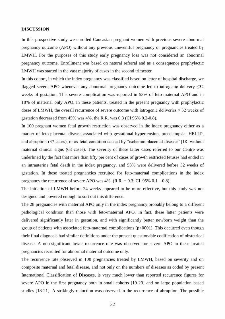
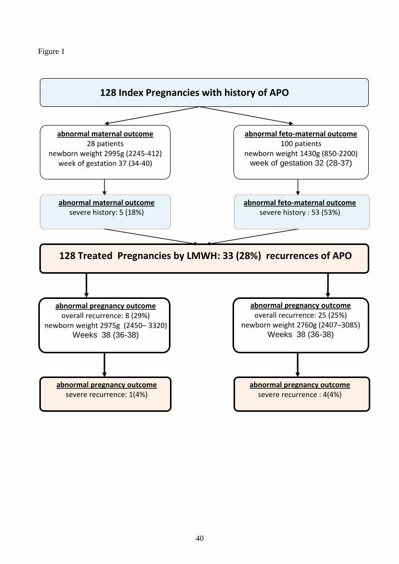
Figure 1 shows the maternal and feto-maternal APO in the index pregnancy and in the treated
pregnancy. The overall recurrence rate in the treated pregnancy was 28%, and not significantly
different in the two groups (p=n.s.). Details on cross over recurrences of APO from feto-maternal to
maternal only and vice versa are reported in tables Ia and Ib. The recurrence of severe APO in all
treated pregnancies, independently from maternal or feto13 maternal APO in the index pregnancy,
was 4% (R.R. = 0.3, CI 95% 0.2-0.8; Chi square test with Yates correction =0.003). In the group
with abnormal feto-maternal outcome, newborn weight was significantly greater even in the treated
pregnancy with severe APOs than in severe APO at index pregnancy: 1090g (1035-1145) vs. 850g
(535-1200), (P<0,01).
Neonatal mortality in the index pregnancy was 10%, no neonatal deaths occurred in the treated
pregnancies.
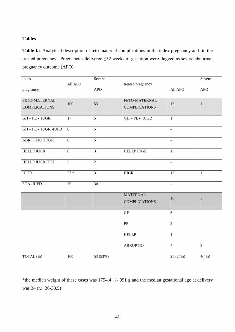
Table Ia shows the details of feto-maternal complications in index pregnancies and their cross over
recurrence in treated pregnancies. In treated pregnancies not a single case of intrauterine fetal death
was observed versus 44 cases of fetal intrauterine death associated with S.G.A. in the index
pregnancy. GH or PE associated with IUGR recurred in only one case out of 17 cases observed in
the index pregnancy.
Severe cases delivered ≤ 32 weeks of gestation are flagged in the index pregnancy and in the treated
pregnancy and reported both as absolute and overall relative figures.
The R.R. for recurrence of severe APO in treated pregnancies was 0.3 (CI .95 = 0.1 – 0.8; Fisher
exact test = 0.001).
In this subset of patients with feto-maternal complications, 70 patients were treated ≤24 weeks of
gestation. Median gestational age at LMWH therapy was 14 weeks (i.q. 10 – 18 completed weeks
of gestation); recurrence rate of severe APO 3 of 70, overall recurrence rate was 20%. In 30 patients
treatment was started ≥ 24 weeks of gestation. Median gestational age at LMWH therapy was 29
week (i.q. 27 - 31 completed weeks of gestation); recurrence rate of severe APO was 1 of 30,
overall recurrence rate was 37%. The Fischer exact test for overall APO recurrence in these two
subsets was <0,08).

31
Table Ib shows the incidence of maternal complication in the index 1 pregnancy and the treated
pregnancy. We found no cases of Hellp syndrome in the treated pregnancy and a decreased
recurrence in abruptio placentae and GH, PE.
Severe cases delivered ≤ 32 weeks of gestation are flagged in the index pregnancy and in the
subsequent complicated pregnancy. The recurrence rate of severe cases was limited to one pregnant
woman who developed a severe fetal growth restriction (980gr) The reduction of severe APO in
treated pregnancies was not significantly different (Fisher exact test = 0.6).
The R.R. was not calculated for this small group that was not large enough to accommodate the
hypothesis of the power calculation.
Fifty patients (39%) screened positive for congenital thrombophilia. Tables 2a and 2b show the
recurrence rate and severity of maternal and feto-maternal complications in the index pregnancy
and the treated pregnancy stratified for the presence or absence of known thrombophilic conditions,
respectively. Severe APO recurred in 4% of treated pregnancies in both groups. The overall
recurrence rate was identical (26%) in the thrombophilic group and non-thrombophilic group.
Significant better outcome were observed for feto-maternal complications both for thrombophilic
and non-thrombophilic patients in the pregnancy treated by LMWH. This different outcome was not
observed for patients with only maternal complications.
32
DISCUSSION
In this prospective study we enrolled Caucasian pregnant women with previous severe abnormal
pregnancy outcome (APO) without any previous uneventful pregnancy or pregnancies treated by
LMWH. For the purposes of this study early pregnancy loss was not considered an abnormal
pregnancy outcome. Enrollment was based on natural referral and as a consequence prophylactic
LMWH was started in the vast majority of cases in the second trimester.
In this cohort, in which the index pregnancy was classified based on letter of hospital discharge, we
flagged severe APO whenever any abnormal pregnancy outcome led to iatrogenic delivery ≤32
weeks of gestation. This severe complication was reported in 53% of feto-maternal APO and in
18% of maternal only APO. In these patients, treated in the present pregnancy with prophylactic
doses of LMWH, the overall recurrence of severe outcome with iatrogenic deliveries ≤ 32 weeks of
gestation decreased from 45% was 4%, the R.R. was 0.3 (CI 95% 0.2-0.8).
In 100 pregnant women fetal growth restriction was observed in the index pregnancy either as a
marker of feto-placental disease associated with gestational hypertension, preeclampsia, HELLP,
and abruption (37 cases), or as fetal condition caused by ―ischemic placental disease‖ [18] without
maternal clinical signs (63 cases). The severity of these latter cases referred to our Centre was
underlined by the fact that more than fifty per cent of cases of growth restricted fetuses had ended in
an intrauterine fetal death in the index pregnancy, and 53% were delivered before 32 weeks of
gestation. In these treated pregnancies recruited for feto-maternal complications in the index
pregnancy the recurrence of severe APO was 4% (R.R. = 0.3; CI .95% 0.1 – 0.8).
The initiation of LMWH before 24 weeks appeared to be more effective, but this study was not
designed and powered enough to sort out this difference.
The 28 pregnancies with maternal APO only in the index pregnancy probably belong to a different
pathological condition than those with feto-maternal APO. In fact, these latter patients were
delivered significantly later in gestation, and with significantly better newborn weight than the
group of patients with associated feto-maternal complications (p<0001). This occurred even though
their final diagnosis had similar definitions under the present questionable codification of obstetrical
disease. A non-significant lower recurrence rate was observed for severe APO in these treated
pregnancies recruited for abnormal maternal outcome only.
The recurrence rate observed in 100 pregnancies treated by LMWH, based on severity and on
composite maternal and fetal disease, and not only on the numbers of diseases as coded by present
International Classification of Diseases, is very much lower than reported recurrence figures for
severe APO in the first pregnancy both in small cohorts [19-20] and on large population based
studies [18-21]. A strikingly reduction was observed in the recurrence of abruption. The possible

33
role of LMWH prophylaxis in reducing this severe APO has already been reported by the NOH-AP
randomized trial 8 [14] in which treatment of pregnancies with previous abruption was started at the
time of early pregnancy and resulted in a recurrence rate of 10 out of 80 cases in the LMWH treated
pregnancies versus 25 out of 80 in non treated cases.
A limitation in our prospective non-randomized study is the possible additional role of ASA in
patients with previous PE. In fact, 45 patients in our study had been treated with LMWH and ASA
because of their history of PE. The appropriateness of this regime had been recently confirmed by a
recent meta-analysis of Bujold [22] observed a significant reduction of the relative risk (RR=0,45)
of recurrence in pregnant women treated by ASA before 16 weeks of gestation. We could only
speculate that this reduction appears to be lower than the one we observed in our cohort in which
PE was not the single most frequent disease both in index and in treated pregnancy. Indeed, only a
prospective randomized trial could solve the relative weight of the two regimens.
Overall, when this cohort was stratified for the presence or absence of thrombophilia the recurrence
rate of severe APO was 4% both in thrombophilic and non-thrombophilic pregnant women. This is
very much in agreement with the figures of the small randomized study on LMWH prophylactic
treatment reported by Rey [13] on non-thrombophilic pregnant women in whom the recurrence rate
of severe APO in the treated arm was 5.5%. To make a complex problem simple we could say that
this is in agreement with the statements of Rodger et al [7] that congenital thrombophilia per se has
not been proven as a cause of placenta mediated pregnancy complications and LMWH might be
beneficial in preventing placental damage based on its potential immune-modulation besides its
known antithrombotic rule 30 [23].
This could be active not only in early gestation by promoting trophoblastic invasion [24] but all
along placental development and ongoing function.
When both severe and all APO were considered altogether in the composite recurrence outcome
analysis, we observed an overall recurrence rate of 25% and 29% for patients with feto-maternal
and maternal complications in the index pregnancy, respectively. These findings lay well within the
huge range reported in literature for observational studies. Even though our small cohort does not fit
the criteria for a proper epidemiological comparison, when we consider our slightly lower
percentage of recurrence versus comparable data reported by Likke 5 [21] (37.8% composite
recurrence rate for preeclampsia occurring between 20 and 36 weeks) we should take into account
the otherwise much higher recurrence expected in our cohort given the high prevalence of
intrauterine fetal death [25] and for HELLP 7 [26], which altogether in the group of 100
pregnancies with feto-maternal complications summed up to 50 cases in the index pregnancy and
was down to two cases only in the LMWH treated pregnancy.
34
A major growing body of evidence is suggesting that under the names of preeclampsia and
gestational hypertension there are at least two diseases with different maternal vascular
abnormalities 13 [27-28-29-30] and quite different placental pathology and fetal growth [31].
Recurrence rate in these two subsets should be investigated by different criteria rather than the
present definition of these syndromes as per the CDI 9.
Gestational age at diagnosis had been frequently adopted to separate these different diseases, and
the name early onset and late onset preeclampsia is now used in clinical settings. However, it is
very likely that this time based criteria are not enough to separate these entities. Placental
pathology, fetal growth and uterine Doppler waveform, or at least the two latter, should be included
into this classification. To assess a proper figure of recurrence, Likke [21], Sibai [26-32] and Van
Rijn [33] corrected the recurrence rate of pregnancies with ―placental ischemic diseases‖ according
to gestational age at diagnosis.
Another confounding factor in assessing the real recurrence of a condition, such as preeclampsia,
IUGR or abruptio placentae, is that in the subsequent pregnancies these abnormal outcomes are
interrelated. Ananth [18], on a large series of 154,810 patients, confirmed a crossover of
preeclampsia, small for gestational age newborns and abruption from one pregnancy to the
subsequent one. This composite outcome is mandatory to assess the true recurrence rate as
confirmed by his larger series of 536,419. Both populations studies by Ananth [18], and Lykke [21]
would have underestimated the recurrence rate of APO from 27.1% to 16.5%, if only preeclampsia
were tracked in the subsequent pregnancy.
The finding that in our present cohort pregnancies recruited for previous maternal APO only,
without clinical evidence of fetal growth restriction, did not show any significant difference
between index and present pregnancy treated by LMWH in terms of gestational age and weight at
birth, is in agreement with this criteria to separate early and late onset preeclampsia.
Beyond speculations, these findings suggest that women with 1 late onset gestational hypertension
and preeclampsia at their first pregnancy should not be included in randomized studies on LMWH
role for this could represent a useless recruitment of patients who are not likely to have any benefit
by LMWH.
The strength of our findings is based on the strict criteria of recruitment, including homogenous
ethnicity. Obviously strict monitoring and possibly better treatment regimen in our referral center
and the strict collaboration between feto-maternal medicine specialists and immune-hematologists
could have contributed to present results, yet given the state of clinical skills in the geographical
area of recruitment where the index pregnancies were monitored and treated, we believe that the
major difference was in fact determined by the usage of LMWH.

35
Major limitations are the non-randomized design of this pilot study and the gestational age at
recruitment and initiation of therapy, based on the timing of referral. Early placentation was already
been completed in most of the cases. However, this limitation together with the striking reduction of
severe APO in recurrence should strongly induce to consider the possible role of LMWH during
second wave of trophoblastic invasion and all along placental function, and not only from the very
early stages of placental developmenT [14-34].
In conclusion, in this prospective study, prophylactic doses of LMWH after the first trimester
reduced the recurrence rate of severe composite APO to 4%. These findings confirm that recurrence
rate of APO in thses studies should consider the overall prevalence as well as severity of recurrence
and the crossover of different APOs caused by placental ischemic disease. This recurrence rate
observed in treated pregnancies was in close agreement with the small but significant randomized
trials [18-19] so far published on LMWH prophylaxis forprevention of APO, and is independent
from inherited thrombophilic conditions.
The less significant role of LMWH in reducing the severity and overall recurrence of APOs
occurring late in pregnancy or without fetal growth restriction addresses the need of overcoming the
bias induced into any clinical study by present definition of these syndromes as per the CDI 9 which
groups under the same heading really different diseases.

36
REFERENCES
[1] Sacks GP, Studena K, Sargent I, et al. Normal pregnancy and preeclampsia both
produceinflammatory changes in peripheral blood leukocytes akin those of sepsis. Am J Obstet
Gynecol 1998; 179, 80-86.
[2] Sargent IL, Borzychowski AM, Redman CW. NK cells and human pregnancy an inflammatory
view. Trends in Immunology 2006; 27 (9): 399-404.
[3] Walker ID, Kujovich JL, Greer IA et al. The use of LMWH in pregnancies at risk: new evidence
or perception? J Thromb Haemost 2005; 3(4): 778-93.
[4] Gori AM, Attanasio M, Gazzini A et al. Cytokine gene expression and production by human
LPS stimulated mononuclear cells are inhibited by sulfated heparin-like semi synthetic derivatives.
J Thromb Haemost 2004; 2(9): 1657-62.
[5] Hochart H, Jenkins PV, Smith OP et al. Low-molecular weight and unfractionated heparins
induce a downregulation of inflammation: decreased levels of proinflammatory cytokines and
nuclear factor-kappaB in LPS-stimulated human monocytes. Br J Haematol 2006; 133(1): 62-7.
[6] Paidas MJ, Ku DH, Langhoff-Roos J et al. Inherited thrombophilias and adverse pregnancy
outcome: screening and management. Semin Perinatol 2005;29(3): 150-63.
[7] Rodger MA, Paidas M, McLintock C et al. Inherited thrombophilia and pregnancy
complications revisited. Obstet Gynecol 2008; 112(2 Pt 1): 320-4.
[8] Di Simone N, Di Nicuolo F, Sanguinetti M et al. Low-molecular weight heparin induces in vitro
trophoblast invasiveness: role of matrix metalloproteinases and tissue inhibitors. Placenta 2007;
28(4):298-304.
[9] Bose P, Black S, Kadyrov M. et al. Heparin and aspirin attenuate placental apoptosis in vitro:
implications for early pregnancy failure. Am J Obstet Gynecol 2005;192(1):23-30.
[10] Oberkersch R, Attorresi AI, Calabrese GC. Low-molecular-weight heparin inhibition in
classical complement activation pathway during pregnancy. Thromb Res 2010;125(5):240-5.
[11] Martinelli I, Taioli E, Cetin I et al. Mutations in coagulation factors in women with
unexplained late fetal loss. N Engl J Med 2000;343(14):1015-8.
[12] Rey E, Kahn SR, David M et al. Thrombophilic disorders and fetal 1 loss: a meta-analysis.
Lancet 2003; 15;361(9361):901-8.
[13] Rey E, Garneau P, David M et al. Dalteparin for the prevention of recurrence of placental-
mediated complications of pregnancy in women without thrombophilia: a pilot randomized
controlled trial. J Thromb Haemost 2009;7(1):58-64.
37
[14] Gris JC, Chauleur C, Faillie JL et al. Enoxaparin for the secondary prevention of placental
vascular complications in women with abruptio placentae. The pilot randomized controlled NOH-
AP trial. Thromb Haemos. 2010; 104(4):771-9.
[15] Protocol : Trial of Umbilical and Fetal Flow in Europe (TRUFFLE). Randomized trial of
timing of delivery in early preterm fetal growth restriction based on early and late fetal Doppler
venous changes versus cardiotocography
[16] Anna Maria Marconi, Stefania Ronzoni, Patrizia Bozzetti, SimonaVailati, Alberto Morabito,
and Frederick C Battaglia. Comparison of Fetal and Neonatal Growth Curves in Detecting Growth
Restriction. Obstet Gynecol. 2008 December ; 112(6): 1227–1234
[17] Todros T, Ferrazzi E, Groli C, Nicolini U, Parodi L, Pavoni M, et al. Fitting growth curves to
head and abdomen measurements of the fetus: a multicentric study. J Clin Ultrasound 1987;15:95–
105.
[18] Ananth CV, Peltier MR, Chavez MR et al. Recurrence of ischemic placental disease. Obstet
Gynecol 2007;110(1):128-33.
[19] Cathelain-Soland S, Coulon C, Subtil D, Houfflin-Debarge V, Subsequent pregnancy outcome
in women with a history of preeclampsia and/or HELLP syndrome] Gynecol Obstet Fertil
2010;38(3):166-72
[20] Chames MC, Haddad B, Barton JR, Subsequent pregnancy outcome in women with a history
of HELLP syndrome at < or = 28 weeks of gestation. Am J Obstet Gynecol 2003;188(6):1504-8.
[21] Lykke JA, Paidas MJ, Langhoff-Roos J. Recurring complications in second pregnancy. Obstet
Gynecol. 2009;113(6):1217-24. Bujold E, Roberge S, Lacasse, Y, Bureau M, A 1 udibert F,
Marcoux S, Forest JC, Giguere Y, Prevention of Preeclampsia and Intrauterine Growth Restriction
With Aspirin Started in Early Pregnancy A Meta-Analysis Obstet Gynecol 2010;116:402–14
[23] Hossain N, Schatz F, Paidas MJ et al.. Heparin and maternal fetal interface: why should lit
work to prevent pregnancy complications? Thromb Res 2009 Dec;124(6):653-5.
[24] Paidas MJ, Thrombosis and regulation of the trophoblast at the maternal interface. Thromb Res
2009;124(4):387-8.
[25] Black M, Shetty A, Bhattacharya S et al. Obstetric outcomes subsequent to intrauterine death
in the first pregnancy. BJOG 2008;115:269–74.
[26] Sibai BM, Ramadan MK, Chari RS et al. Pregnancies complicated by HELLP syndrome
(hemolysis, elevated liver enzymes, and low platelets): subsequent pregnancy outcome and long-
term prognosis Am J Obstet Gynecol 1995 Jan;172(1 Pt 1):125-9.
38
[27] Valensise H, Vasapollo B, Gagliardi G et al. Early and late preeclampsia: two different
maternal hemodynamic states in the latent phase of the disease. Hypertension 2008 Nov;52(5):873-
80.
[28] Masuyama H, Segawa T, Sumida Y et al .Different profiles of circulating angiogenic factors
and adipocytokines between early- and late-onset pre-eclampsia. BJOG 2010 Feb;117(3):314-20.
[29] Wikström AK, Nash P, Eriksson UJ et al. Evidence of increased oxidative stress and a change
in the plasminogen activator inhibitor (PAI)-1 to PAI-2 ratio in early-onset but not late-onset
preeclampsia. Am J Obstet Gynecol 2009 Dec;201(6):597.e1-8.
[30] Cancello R, Clement K. Is obesity an inflammatory illness? Role of low inflammation and
macrophage infiltration in human white adipose tissue. BJOG 2006;113:1141–1147
[31] Cetin I, Huppertz B, Burton G, Cuckle H, Gonen R, Lapaire O, Mandia L, Nicolaides K,
Redman C, Soothill P, Spencer K, Thilaganathan B, Williams D, Meiri H. Pregenesys preeclampsia
markers consensus meeting: what do we require from markers, risk assessment and model to tailor
preventive strategies? Placenta. 2011 Feb;32 Suppl:S4-16
[32] Sibai BM, Mercer B, Sarinoglu Cet al. Severe preeclampsia 1 in the second trimester:
recurrence risk and long term prognosis. Am J Obstet Gynecol 1991; 165:1408-1412.
[33] Van Rijn BB, Hoeks LB, Bots ML, et al: Outcomes of subsequent pregnancy after first
pregnancy with early-onset preeclampsia. Am J Obstet Gynecol 2006,195:723-728.
[34] Multicentre, randomized, controller, open trial on efficacy of low molecular weight eparin in
pregnant women with previous obstetrical complications. HAPPY Study EUDRACT n°2006-
004205.

39
Figure 1
Flow chart of pregnancy outcome of index pregnancies and subsequent pregnancies treated by
prophylactic doses of LMWH
Thin boxes: Index pregnancies. Gestational age and newborn weight (median and i.q.) were
significantly worse in the feto-maternal than in the maternal APO group (p<0001). The prevalence
of severe APO was significantly higher in the feto-maternal APO group (p<0.001)
Thick boxes: treated pregnancies. Feto-maternal APO: gestational age at delivery and newborn
weight (median and i.q.) in the treated pregnancy were highly significantly better than in the index
pregnancy (Mann-Whitney test p<0.001). Maternal APO: gestational age at delivery and newborn
weight in the treated pregnancy were not significantly better than in the index pregnancy (Mann-
Whitney test p<n.s.). The severe and the overall recurrence rate of APO were not significantly
different in the two groups (p=n.s.).
40
Figure 1
abnormal maternal outcome severe history: 5 (18%)
abnormal feto-maternal outcome severe history : 53 (53%)
abnormal maternal outcome 28 patients
newborn weight 2995g (2245-412) week of gestation 37 (34-40)
abnormal feto-maternal outcome 100 patients
newborn weight 1430g (850-2200) week of gestation 32 (28-37)
abnormal pregnancy outcome severe recurrence: 1(4%)
abnormal pregnancy outcome severe recurrence : 4(4%)
128 Index Pregnancies with history of APO
abnormal pregnancy outcome overall recurrence: 25 (25%)
newborn weight 2760g (2407–3085) Weeks 38 (36-38)
abnormal pregnancy outcome overall recurrence: 8 (29%)
newborn weight 2975g (2450– 3320) Weeks 38 (36-38)
128 Treated Pregnancies by LMWH: 33 (28%) recurrences of APO
41
Tables
Table Ia. Analytical description of feto-maternal complications in the index pregnancy and in the
treated pregnancy. Pregnancies delivered ≤32 weeks of gestation were flagged as severe abnormal
pregnancy outcome (APO).
index
pregnancy
All APO
Severe
APO
treated pregnancy
All APO
Severe
APO
FETO-MATERNAL
COMPLICATIONS 100 53
FETO-MATERNAL
COMPLICATIONS 15 1
GH – PE - IUGR 17 5 GH – PE - IUGR 1
GH – PE - IUGR -IUFD 6 5 -
ABRUPTIO IUGR 6 5 -
HELLP IUGR 6 3 HELLP IUGR 1
HELLP IUGR IUFD 2 2 -
IUGR 27 * 3 IUGR 13 1
SGA -IUFD 36 30 -
MATERNAL
COMPLICATIONS 10 3
GH 3
PE 2
HELLP 1
ABRUPTIO 4 3
TOTAL (%) 100 53 (53%) 25 (25%) 4(4%)
*the median weight of these cases was 1754,4 +/- 991 g and the median gestational age at delivery
was 34 (r.i. 36-38.5)

42
Table Ib. Analytical description of maternal complications in the index pregnancy and
complications in the treated pregnancy. Pregnancies delivered ≤32 weeks of gestation were flagged
as severe abnormal pregnancy outcome (APO).
index pregnancy
All APO
Severe APO
treated pregnancy All APO
Severe APO
MATERNAL COMPLICATIONS
28 5 MATERNAL COMPLICATIONS
2 0
GH 5
PREECLAMPSIA 9 2 PREECLAMPSIA 1
HELLP 5 1
ABRUPTIO 9 2 ABRUPTIO 1
FETO-MATERNAL COMPLICATIONS
6 1
IUGR 6 1
TOTAL (%) 28 5 (18%) 8 (28%) 1 (4%)
Legend a) and b) panels.
APO= abnormal pregnancy outcome, excluded first trimester complications; Severe APO =
abnormal pregnancy outcome which led to iatrogenic delivery before 32 completed weeks of
gestation; GH = gestational hypertension; PE= preeclampsia; IUGR= intrauterine growth
restriction; IUFD= intrauterine fetal death; ABRUPTIO= abruption placenta; HELLP = help
syndrome; SGA= small for gestational age.

43
Table IIA. Fifty inherited thrombophilic patients: Analytical description of rate and severity of
recurrence of APO (*). Pregnancies delivered ≤32 weeks of gestation were flagged as severe
abnormal pregnancy outcome (APO). (median and interquartile range in brackets)
Index pregnancy All APO Severe
APO treated pregnancy All APO
Severe
APO P <
FETO-
MATERNAL
COMPLICATION
S
41 16
FETO-
MATERNAL
COMPLICATIONS
8 1
Weeks at delivery 35
(30-38) Weeks at delivery
38
(36,7-38,25) 0,04
Weight (g.) 1600 g
(958-2300) Weight (g.)
2100 g
(1990-2395) 0,08
MATERNAL
COMPLICATION
S
9 0 MATERNAL
COMPLICATIONS 5 1
Weeks at delivery 38
(36-40) Weeks at delivery
37
(35-38) 0,41
Weight (g.) 3000 g
(2440-3200) Weight (g.)
2870 g
(2290-3180) 0,56
Total 50(100%) 16(32%) 13 (26%) 2 (4%)
(*) 19 women (15,6%) were eterozigotic for V Leiden mutation, 19 women (15,6%) were
eterozigotic for G20210A prothrombin mutation, 13 (10,1%) were homozigotic for MTHFR
mutation, two with hyperhomicisteinemia, and 11(8,6%) patients had more than one mutation. Not
a single case of S and C deficiency was recorded in this cohort.

44
Table IIB. Non thrombophilic patients: analytical description of rate and severity of recurrence in .
Pregnancies delivered ≤32 weeks of gestation were flagged as severe abnormal pregnancy outcome
(APO). (median and interquartile range in brackets)
index pregnancy All APO
Severe APO
treated pregnancy All APO
Severe APO
P <
FETO-MATERNAL COMPLICATIONS
59 36 FETO-MATERNAL COMPLICATIONS
13 1
Weeks at delivery 31 (28-36)
Weeks at delivery 36 (35-37)
0,03
Weight (g.) 1295 g (798-2015)
Weight (g.) 2180 g (1890-2450)
0,001
MATERNAL COMPLICATIONS
19 5 MATERNAL COMPLICATIONS
7 2
Weeks at delivery 36 (33-39)
Weeks at delivery 37 (33,5-38)
0,97
Weight (g.) 2990 g (1945-3415)
Weight (g.) 2650 g (2060-3085)
0,72
total 78(100%) 41(53%) 20 (26%) 3 (4%)
Legend a) and b) panels.
APO= abnormal pregnancy outcome, excluded first trimester complications; Severe APO =
abnormal pregnancy outcome which led to iatrogenic delivery before 32 completed weeks of
gestation;