Embed Size (px)
Citation preview
1
Use of a standardized treatment protocol for post-cardiac arrest
treatment
Michael A. Kuiper, MD PhD FCCP FCCM, Neurologist–Intensivist
Department of Intensive Care Medicine,
Medical Center Leeuwarden, PO Box 888
8901 BR Leeuwarden, The Netherlands
Tel +31 58 2866737; e–mail: [email protected]
Academic Medical Center, Amsterdam, Dept. of Intensive Care Medicine
HERMES Critical Care Group, Amsterdam
Member of the scientific council of the Dutch Resuscitation Council
Peter E. Spronk, MD PhD FCCP, Internist–Intensivist
Department of Intensive Care Medicine
Gelre Hospitals (Lukas site), PO Box 9014
7300 DS Apeldoorn, The Netherlands; e–mail: [email protected]
Academic Medical Center, Amsterdam, Dept. of Intensive Care Medicine
HERMES Critical Care Group, Amsterdam
Marcus J Schultz, MD PhD FCCP, Internist–Intensivist
Department of Intensive Care Medicine & Laboratory of Experimental Intensive Care
and Anesthesiology (L·E·I·C·A) e–mail: [email protected]
Mail stop at C3–415, Academic Medical Center at the University of Amsterdam
Meibergdreef 9, 1105 AZ Amsterdam, The Netherlands
HERMES Critical Care Group, Amsterdam
2
Introduction
For a long time outcome of patients after out–of–hospital cardiac arrest (OHCA) has
been extremely poor, with only 5–10% of survivors with good neurological outcome.
In recent years, several studies demonstrated an increase in survival of cardiac
arrest patients admitted to the intensive care unit (ICU), often surpassing 60% with
good neurological outcome [1-6]. Since the introduction of cardiopulmonary
resuscitation (CPR) in the early nineteen–sixties by Safar et al. (mouth–to–mouth
respiration) [7] and Kouwenhoven et al. (closed chest–compression) [8], emphasis in
resuscitation medicine has been on treatment of cardiac arrest until return of
spontaneous circulation (ROSC), with manual CPR and early defibrillation of
convertible cardiac rhythms being the two most important items. The general
consensus was that improvement of outcome of cardiac arrest patients would solely
lie in shortening the period of circulatory standstill, thus minimizing the, mainly
neurological, damage. After having restored circulation, treating physicians ―could
only wait and see what the outcome would be‖. However, alongside the processes of
recovery and compensation, a pathological state may develop with associated organ
failure – the so–called post–resuscitation syndrome. Physicians should be aware of
this condition and actively treat its complications to improve the condition of the
patient and to increase the chance of a good neurological outcome after cardiac
arrest. Induced mild hypothermia is an important factor in approaching this entity and
has become an established treatment for the post–cardiac arrest patient. However,
induced mild hypothermia is not the sole treatment modality that we should use.
Indeed, Sunde et al. [4] reported on the use of a standardized treatment
protocol and showed improved outcomes of patients treated with a bundle of
strategies with historic controls. This chapter discusses the role of several strategies
3
in patients after cardiac arrest and details of post–resuscitation care. It provides a
rationale for the different treatment steps of a standardized treatment protocol for
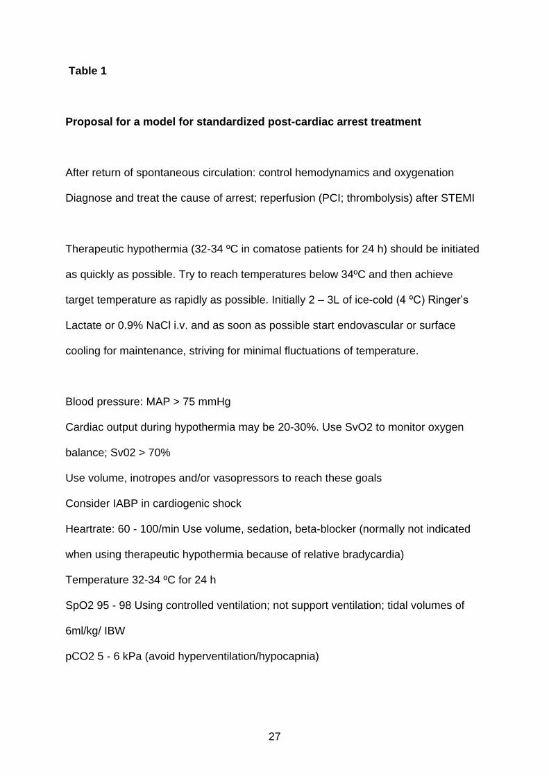
post–cardiac arrest patients (see table 1).
Chain of survival
The chain of survival as advocated by the various societies of resuscitation,
promotes getting help, basic life support (BLS) with manual CPR to buy time, early
defibrillation to restart the circulation and advanced life support (ALS) by
professionals. In the past, emphasis in resuscitation medicine has mainly been on
the first 3 steps of resuscitation: getting help, early and good quality CPR and early
defibrillation. The 4th step of the chain of survival, ALS, was probably never as well
defined as the earlier steps. As early defibrillation is probably the most crucial step in
the early phase of cardiac arrest, the use of automated external defibrillators already
has most probably led to a better neurological outcome in cardiac arrest patients.
Also early coronary interventions have improved outcome.
The 2 landmark studies by the HACA group [9] and by Bernard et al. [10]
showing benefit of induced mild hypothermia after cardiac arrest have shifted the
focus and now there is an increasing interest in post–resuscitation treatment.
However, it was not until 2005 before the 4th ring of the chain of survival was updated
to reflect the importance of post resuscitation care in determining the ultimate
outcome following cardiac arrest [12] (Figure 1).
The post–resuscitation syndrome
The post–resuscitation syndrome is defined as a condition after resuscitation
following (prolonged) cardiac arrest and therefore whole body ischemia and
4
reperfusion with multiple–organ dysfunction, most notably of but not limited to the
brain. Negovsky, one pioneer of ―reanimatology‖ and therapeutic hypothermia, most
probably introduced the concept of the post–resuscitation syndrome [12]. Negovsky
found pathophysiological changes after resuscitation that substantially differed from
those caused by ischemia and hypoxia. In the brain, redistribution of Ca2+, together
with the formation of free radicals was observed, causing damage to DNA and
organelle membranes, followed by development of progressive autoimmune
pathology, reflecting the damage of the blood-brain barrier [12-13]. The post–
resuscitation or post–cardiac arrest syndrome is at present believed to be a form of
systemic inflammatory response syndrome, caused by ischemia–reperfusion. As this
―sepsis–like syndrome‖ [14] may precede the development to multiple–organ
dysfunction syndrome, even after successful and swift restoration of the circulation, a
patient after cardiac arrest may ultimately die of the consequences of this post–
resuscitation syndrome. On the other hand, this syndrome gives physicians the
opportunity to intervene and strive for improvement of outcome by supporting the
function of failing organs by ventilation, hemodynamic support or renal replacement
therapy, thereby improving the outcome of these patients.
Early coronary intervention
Early revascularization after myocardial infarction improves outcome after myocardial
infraction. As compared with thrombolytic therapy, primary coronary intervention
(PCI) results in a higher rate of patency of the infarct–related coronary artery, lower
rates of stroke and re–infarction, and higher long-term survival rates [15]. Induced
mild hypothermia in combination with PCI is feasible and safe in patients resuscitated
after cardiac arrest due to acute myocardial infarction [16]. In patients with cardiac
5
arrest due to ST–elevation myocardial infarction (STEMI), it may be acceptable to
use thrombolytic therapy as the reperfusion strategy of first choice. This applies
especially in hospitals where immediate PCI is not available [17]. The diagnosis of
STEMI can be established in the field immediately after ROSC in most patients. This
may enable an early decision about reperfusion therapy, i.e., immediate out–of–
hospital thrombolytic therapy or targeted transfer for PCI [18]. In view of the existing
evidence, PCI after cardiac arrest due to STEMI should be the preferred mode of
treatment in regions and situations where this is applicable.
Induced mild hypothermia
Since the landmark publications by the HACA group [9] and Bernard et al. [10], many
studies have been published using a non–randomized design or matched historical
controls, all showing a profound improvement of outcome since the implementation
of therapeutic hypothermia (Figure 2). The two original studies provided us with the
evidence for treating patients with mild hypothermia if these fulfilled the criteria for
inclusion (HACA study: witnessed cardiac arrest, ventricular fibrillation or ventricular
tachycardia as the initial cardiac rhythm, presumed cardiac origin of the arrest, age
between 18–75 years, estimated interval of 5–15 minutes from the patient‘s collapse
to the first attempt at resuscitation by emergency medical personnel, and an interval
of no more than 60 minutes from collapse to restoration of spontaneous circulation).
These inclusion criteria must have led to exclusion of many cardiac arrest patients.
Indeed, out of 3551 patients assessed for eligibility in the HACA study, 3246 did not
meet these inclusion criteria. There is accumulating evidence, however, that induced
mild hypothermia is probably also valuable for patients not strictly fulfilling these
criteria, such as patients with asystole or pulse–less electrical activity [19-20] or
6
patients above 75 years of age [21]. It seems reasonable to consider induced mild
hypothermia for all patients admitted to the ICU after cardiac arrest, which is
supported by the ILCOR statement on therapeutic hypothermia [22].
Why should all patients after circulatory arrest and ROSC receive induced
hypothermia? Therapeutic hypothermia affects brain metabolism, lowering the
metabolism with 40–50% when temperature decreases from 380C to 320C, thereby
decreasing oxygen demand. Moreover, the level of inflammation is attenuated and
intracranial pressure decreases [19]. However, we might view mild hypothermia not
so much as a neuro–protective strategy alone, but as an active treatment strategy to
improve the function of the brain as well as other organs. Apart from improving brain
function, therapeutic hypothermia may (or can) improve function of other organs:
studies suggest positive effects in cardiogenic shock, seizures, acute respiratory
distress syndrome, nephropathy, hepatic failure, or arrhythmias [20]. These
strategies remain to be proven however. A non–significant reduction of infarct size in
myocardial infarction has also been reported [23].
Data from surveys in Europe and the United States suggest that rates of use
among physicians of induced mild hypothermia in post-cardiac arrest patients may be
as low as 30–40% [24]. A recent e–mail–invitation guided anonymous web–based
survey was held amongst ICUs in the Netherlands. Thirty–seven ICUs (50%)
mentioned always to treat these patients with therapeutic hypothermia, 42% only
treat patients when CPR fulfilled several criteria, such as ventricular fibrillation as
presenting cardiac rhythm at arrival of ambulance (82%) and duration of time to
return of spontaneous circulation (55%). Six ICUs (8%) never induced hypothermia.
The most important reason for not inducing hypothermia was lack of equipment.
Surface cooling (86%) and cold intravenous fluids (71%) were most frequently used
7
to reach the target temperature. Hemodynamic instability was most cited as a reason
to discontinue treatment with therapeutic hypothermia. Thus, in the Netherlands,
therapeutic hypothermia after CPR is implemented in almost all ICUs, which is
compared to previous reports of other countries, exceedingly high [unpublished data].
Nevertheless, despite varying acceptance and implementation across the world, lack
of scientific evidence for the use of induced mild hypothermia after cardiac arrest
may no longer be used as an argument for not implementing this treatment.
Cerebral blood flow and mean arterial blood pressure
Initially, shortly after ROSC, there is a change in cerebral blood flow, which in the
later stage, after 72 hours, normalizes [25]. The mean flow velocities, as assessed
using trans–cranial Doppler are lowered, while oxygen extraction was initially normal
but decreased after time. This latter effect was significantly more pronounced in non–
survivors.
After cardiac arrest, cerebral autoregulation is disturbed, but not completely
absent [26]. As this mechanism regulates the cerebral blood flow to maintain a
steady oxygen delivery to the brain during changes in blood pressure, this means
that cerebral blood flow is highly pressure dependent during impairment of the
autoregulation and therefore a high mean arterial blood pressure (MAP) is most
probably needed. There is no clinical evidence for a specific limit for the mean arterial
blood pressure, but as the intracranial pressure following CPR is not necessarily
elevated, and in general remains below 15 mm Hg [27], maintaining the MAP > 75
mm Hg and thus a cerebral perfusion pressure of > 60 mm Hg, is justifiable. The
optimal MAP for post–cardiac arrest patient may even be higher [28]. Of note, the
MAP in the Bernard study [10] was approximately 90 mmHg in both the hypothermia
8
as well as in the normothermia group. In patients with severe myocardial dysfunction
after cardiac arrest, these values may be difficult to obtain, and a balance needs to
be found between the optimal blood pressure for cerebral blood flow and the burden
for the myocardium to meet this demand.
Hemodynamic support and monitoring
Hemodynamic monitoring is warranted to optimize circulation and balance the
oxygen supply with the demand. Heart rate, blood pressure, cardiac output, SvO2 or
ScvO2, lactate, and arterial blood gasses need to be monitored. While all of these
methods of monitoring the circulation have their own obvious limitations, monitoring
may prevent severe misbalances between oxygen delivery and oxygen consumption.
Cardiac output during hypothermia may be 20–30% lower and therefore be accepted
below normal limits. The use of ScvO2 or SvO2 measurements to monitor oxygen
balance and maintaining Scv02 > 70% is probably a reasonable approach. Volume,
inotropics and/or vasopressors will often be necessary to reach these goals. We may
need to consider the use of an intra-–aortic balloon pump (IABP) in patients with
(refractory) cardiogenic shock due to myocardial infarction or myocardial stunning.
In line with guidelines for pharmacological prevention of peri-operative cardiac
complications in high risk patients undergoing non-cardiac surgery aiming to maintain
adequate oxygen delivery, while preventing myocardial ischemia, the heart rate
needs to be maintained between 60–100/minute, using volume, sedation, beta–
blockers or a pacemaker to obtain this. As hypothermia often leads to relative
bradycardia, beta–blockers are rarely needed, however. Anti–arrhythmic drugs may
be needed to maintain sinus rhythm. In patients who come to survive but who are
prone to arrhythmias, implantable cardiac defibrillators can be considered. Close
9
monitoring of the circulation is warranted in order to balance oxygen demand and
supply, while at the same time taking care of relieving the myocardial burden and
preventing secondary myocardial ischemia.
Ventilation
In post–resuscitation patients, the use of controlled ventilation as opposed to a
support mode of ventilation is rational, as this in most cases decreases oxygen
consumption [29]. The use of tidal volumes of 6 ml/kg predicted or ideal bodyweight
(IBW) is advocated to diminish the occurrence of acute lung injury (ALI) as there is
accumulating and convincing clinical and preclinical evidence that ventilation with
tidal volumes of 6 ml/kg predicted or ideal bodyweight prevents the development of
ALI [30].
As the metabolic rate drops 5–8% per each degree °C, there is a concomitant
drop in oxygen consumption and carbon dioxide production. Ventilator settings need
to be adjusted accordingly to prevent hyperventilation and thus hypocapnia.
Hypocapnia leads to cerebral vasoconstriction and diminished cerebral blood flow
and oxygen delivery. Blood gasses need to be repeatedly checked to be able to
maintain normoventilation.
While there is no discussion about the need to treat and prevent hypoxia, this
invariably leads to hyperoxia, which may be detrimental in it self, especially after a
period of hypoxia. While there is preclinical evidence to promote the use of a limited
fraction of inspired oxygen (FiO2), there is no evidence for limiting FiO2 in humans.
Striving for a an peripheral oxygen saturation (SpO2) of 95–98% would seem
reasonable to prevent hypoxemia and hyperoxemia, but while hyperoxemia is
10
potentially detrimental after ischemia, especially out beyond 5 minutes, hypoxemia is
obviously worse.
Blood glucose and electrolyte monitoring
As hypothermia can decrease insulin sensitivity as well as reduce insulin secretion by
pancreatic islet cells, patients who are cooled have a high risk of developing
hyperglycemia. Control of blood glucose using insulin therapy is therefore necessary.
While there is no clinical outcome study showing benefit in a strictly defined subgroup
of post cardiac arrest patients, there is sufficient evidence for controlling blood
glucose using insulin from the van den Berghe studies [31-32] to include this strategy
in the post–resuscitation treatment.
Electrolyte disturbances are to be expected in patients treated with
hypothermia because of changes in renal function, combined with electrolyte shifts to
the intracellular compartment. Especially hypomagnesemia can easily occur and is
associated with increased risks for adverse outcome. As magnesium has a pivotal
role in many central nervous and cardiovascular processes, it is advisable to early
start magnesium supplementation. Administering magnesium helps prevention of
hypokalemia, hypophosfatemia, hypocalcemia, and hyponatremia, so controlling
magnesium facilitates the control of other electrolytes. During hypothermia
hypokalemia may also occur, especially in patients with increased urine production. It
is beyond the scope of this paper to discuss all possible effects of electrolyte
disturbances during induced mild hypothermia. It is generally advised to keep
magnesium, potassium and phosphate in the normal to high-normal range, and to
keep sodium in the normal range. [33]
11
Patients after cardiac arrest will have a lowered pH, due to increases in pCO2
and lactate. As the patient is mechanically ventilated, the pCO2 will often not pose a
problem and normalize, and restoration of the circulation will most often lead to
clearance of lactate, reducing these levels and normalizing the pH. As a low pH
induces a pro-inflammatory state, and because many enzyme mediated processes
are compromised in case of a pH lower than 7.20–7.25, measures need to be taken
to maintain a pH > 7.20. Sunde et al. showed that using a goal directed, standardized
approach in the treatment of post resuscitation patients leads to a significantly less
negative base excess, and therefore a higher pH, without changes in pCO2 [4].
In patients with electrolyte disturbances, low pH and/or acute kidney failure,
one might consider using a form of continuous renal replacement therapy (CRRT), as
continuous veno-venous hemofiltration (CVVH). CVVH may aid in maintaining a
normal acid–base and electrolyte balance.
Thrombolytic therapy aiming at improvement of brain perfusion
Fischer et al. have shown in a cat model that thrombolytic therapy with recombinant
tissue type plasminogen activator and heparin after cardiac arrest and successful
cardiopulmonary resuscitation reduces the non-perfused brain areas and improves
microcirculatory reperfusion [34]. In a retrospective study, Richling et al. found a
trend towards better neurological outcome using thrombolytic therapy compared to
PCI In patients with cardiac arrest due to STEMI [17]. At the moment however, there
are insufficient clinical data to support this strategy.
12
Brain monitoring
We may consider monitoring the brain using an (continuous) EEG. A study
evaluating the use of continuous EEG monitoring after cardiac arrest showed in 26
out of 94 evaluated cardiac arrest patients development of electrographic status
epilepticus during therapeutic hypothermia, and this status epilepticus correlated with
poor outcome [35]. The continuous EEG monitoring provides the opportunity to treat
electrographic status epilepticus in absence of clinical signs (non–convulsive status
epilepticus) in patients treated with hypothermia, sedation and paralyzing agents,
which prevent the clinical signs and symptoms to become overt. As electrographic
status epilepticus is known to increase oxygen consumption of the brain, it seems
reasonable to try to terminate the electrographic status epilepticus. Anti-convulsant
drugs as well as sedatives can be used to prevent and/or treat this status epilepticus.
While reasonable, there are yet no convincing data on outcome to support this
strategy.
Prevention of infection
Therapeutic hypothermia increases the risk on infections due to immune
suppression. A retrospective cohort study of patients after cardiac arrest treated with
mild hypothermia showed an increased incidence of lower respiratory tract infections
of 88% [36]. Sunde et al. compared a prospective cohort treated with hypothermia
with a retrospective cohort before the use of therapeutic hypothermia [4]. They found
no increase in the rate of pneumonia as a result of hypothermia, but their reported
percentage of pneumonia was also high: 57 % in the cardiac arrest patient cohort
before implementing therapeutic hypothermia compared with 48% of in the cohort
treated with therapeutic hypothermia. This is most probably a detrimental factor as
13
ventilator–associated pneumonia (VAP) is known to increase mortality and also
because infections may cause fever, which is harmful for the compromised nervous
system. There are various options to decrease the possibility of an infection to occur,
one of these being the use of selective decontamination of the digestive tract, which
is a well–proven strategy to prevent VAP and mortality in ICU patients [37-38].
Use of sedation, analgesia and paralyzing agents
Use of sedation in comatose post cardiac arrest patients is rational, as it not only
facilitates the use of hypothermia and controlled ventilation, but also reduces oxygen
consumption. The use of analgesics is rational as well: a state of post-anoxic coma
does not eliminate the need for anesthesia and/or analgesia [13]. Patients with an
acute myocardial infarction may experience pain, which may even be aggravated by
prolonged periods of chest compressions, necessitating the use of analgesic agents,
which on their own can reduce oxygen consumption. Sedation and analgesia are
also used to avoid shivering during hypothermia. Shivering greatly increases oxygen
demand and needs to be diagnosed and treated. If sedation and analgesia are not
sufficient to abolish shivering, magnesium may be used, as well as meperidine
(pethidine®). As the patient still shows signs of shivering, a paralyzing agent needs to
be added to the treatment.
The choice of sedatives is not always considered crucial. It needs mentioning
that the protocol of the Bernard study [10] demanded the use of midazolam, while in
the HACA study [9] the combination of midazolam and fentanyl was used. If we
prefer the use of other sedatives or analgesics, we need to consider the potential
drawbacks of these drugs. Propofol for instance, often used as a sedative for patients
with neurological critical care disorders, has a more profound negative inotropic
14
effect than midazolam, thereby possibly compromising the circulation and thus
negatively affecting the prognosis. Shivering was treated with paralyzing agents as
vecuronium in the Bernard study [10] and pancuronium in the HACA trial [9].
Slow, passive or active re–warming and the prevention of fever.
Re–warming after therapeutic hypothermia needs to be slow and controlled (0.2–0-
5ºC/h). Rapid re–warming in patients with traumatic brain injury and in the peri–
operative setting has resulted in worse outcome than slow re–warming [19]. Animal
studies have shown that rapid re–warming can adversely affect outcome and that
slow re–warming preserves the benefits of cooling [39]. Rapid re–warming might
cause regional or general imbalances between cerebral blood flow and oxygen
consumption and thus cause hypoxia, leading to additional ischemic neuronal
damage [19]. In clinical studies, rapid re–warming also increases the risk of
electrolyte shifts and especially of hyperkalemia. Re–warming also affects the
sensitivity of the cell to insulin; so during re–warming the glucose should closely be
monitored.
Increasing evidence suggests that fever is harmful to the injured brain, and it
seems reasonable to maintain normothermia in most patients with neurological
injuries who have decreased consciousness (especially in those previously treated
with hypothermia) for at least 72 h after injury [19].
Passive re–warming cannot be strictly controlled; as slow re–warming and
prevention of fever are of the utmost importance, it is reasonable to choose for slow,
active and controlled re–warming.
15
Prognostication
Unpublished data of the PROPAC study [40] by Zandbergen et al. on differences in
outcome of patients after cardiac arrest related to early do–not–resuscitate (DNR)
orders after admittance suggest that installing treatment limitations within the first 24
hours leads to an decreased chance of survival.
Pupillary light response, corneal reflexes, motor responses to pain, and
somatosensory evoked potential studies can reliably assist in accurately predicting
poor outcome in comatose patients after cardiopulmonary resuscitation for cardiac
arrest [41]. Myoclonus status epilepticus, which also is regarded as a reliable
predictor of unfavorable outcome, is most probably not as useful as previously
thought. Myoclonus status epilepticus is rare and should not be confused with the
more frequent myoclonus that can be elicited by touch or noise. As mild hypothermia
mandates the use of analgesics, sedatives and sometimes of paralytic agents, and
as the pharmacokinetics of these drugs are changed due to hypothermia, resulting in
a reduced clearance, the utmost care needs to be taken when performing a
neurological assessment of these patients. Often a longer period is needed before
prognostications can be made.
While an absent cortical response (N20) of the SEP has been demonstrated to
have a positive predictive value of 100%, the presence of a N20 does not in any way
predict good outcome.
Serum neuron–specific enolase (NSE) has also been suggested as a
predictable parameter in establishing prognosis after cardiac arrest, but recent
studies in patients treated with hypothermia show higher serum NSE values than
previously reported in patients surviving with good neurological outcome. [42] This
probably limits the usefulness of NSE in establishing a reliable prognosis.
16
Prognosis cannot be based on circumstances of CPR itself. Witnessed or not
witnessed, BLS or no BLS, and time to ROSC do not reliably predict outcome in an
individual patient. For prognostication we should only use established predictors.
Poor outcome in post anoxic coma can be reliably predicted with good neurological
assessment after three days and with somatosensory evoked potentials in a
substantial number of patients. Installing DNR orders within the first 24 hours after
cardiac arrest leads to a decreased chance of survival of the post resuscitation
patient.
Future perspectives
While we now witness a great leap forward in the treatment of post cardiac arrest
patients, many possible treatment modes are under investigation to further improve
outcome.
1) During ALS, a device for automated chest compressions is potentially
useful in assisting and improving CPR. We have yet to await convincing clinical data
before widespread use can be advocated. Impedance threshold devices to produce
negative intra thoracic pressure during ventilation in CPR can be used to improve
preload and thereby hemodynamics and cerebral blood flow during CPR [43]. On–
site cooling after OHCA has been shown to be feasible [44]. No clinical data are yet
available to show survival benefit.
2) Cardio-cerebral or chest compression-only resuscitation has been
advocated by Ewy et al., claiming substantial outcome benefit after cardiac arrest
[45]. On March 31 of 2008, the American Heart Association issued a statement that
recommended to perform chest-compression-only CPR if the rescuer is a bystander
without CPR training or "previously trained in CPR but not confident in his or her
17
ability to provide conventional CPR, including high-quality chest compressions (ie,
compressions of adequate rate and depth with minimal interruptions) with rescue
breath." [www.americanheart.org]. This recommendation has not been adopted by
the European Resuscitation Council.
3) Coenzyme Q10 (CoQ10) is an essential mitochondrial cofactor that has
been shown to possess neuroprotective qualities in neurodegenerative disorders and
may also have a cardioprotective effect in cardiosurgery. Combining CoQ10 with mild
hypothermia immediately after CPR may improve survival and may improve
neurological outcome in survivors. [46].
4) Erythropoietin (Epo) is suggested to have neuroprotective properties as
well. A small clinical study using Epo in OHCA patients failed however to show
significant survival benefit [47].
5) In a different way will applying stopping rules for pre–hospital termination of
resuscitation in OHCA will affect outcome of patients surviving to the ED or the ICU.
In a recent retrospective validation study, Sasson et al. found that BLS and ALS
termination-of-resuscitation rules performed well in identifying OHCA patients who
have little or no chance of survival [48]. Strict BLS and ALS stopping rules were
defined. For BLS: Event not witnessed by emergency medical services personnel; No
automated external defibrillator used or manual shock applied in out of-hospital
setting; No return of spontaneous circulation in out–of–hospital setting. Additional for
ALS: Arrest not witnessed by bystander; No bystander–administered
cardiopulmonary resuscitation. A patient must meet all of the criteria included in
either rule to warrant termination of resuscitation in the out–of–hospital setting. Pre-
hospital selection of post cardiac arrest patients who will invariably die will increase
the likelihood of survival for the remaining cohort, and probably change the attitude of
18
the treating physicians towards these patients, theoretically resulting in more
aggressive treatment and better outcome [48].
Conclusion
After cardiac arrest, immediate restoration of the circulation is of the utmost
importance. Good quality basic life support (BLS) and early defibrillation are crucial
steps in this phase of CPR. After resumption of spontaneous circulation (ROSC),
considerable improvement of outcome of the post–cardiac arrest patient can be
achieved by actively treating many complications of the ischemia–reperfusion
phenomena known as the post–resuscitation syndrome. The most important
treatment modality of these is induced mild hypothermia. Other important treatment
modalities include early coronary reperfusion, controlled ventilation to achieve normal
arterial blood pO2 and pCO2, hemodynamic optimization, judicious use of sedatives
and analgesics and prevention of shivering to reduce oxygen consumption, control of
electrolytes and glucose, prevention and treatment of seizures, prevention of
complications as infections and the use of validated predictors for prognosis.
Presently, there is accumulating evidence to support the view that a standardized
protocol should be used to optimize the treatment of the post-cardiac arrest patient
admitted to the ICU.
19
References
1. Oddo M, Schaller MD, Feihl F, Ribordy V, Liaudet L. (2006) From evidence to
clinical practice: effective implementation of therapeutic hypothermia to improve
patient outcome after cardiac arrest. Crit Care Med 34:1865–73
2. Al-Senani FM, Graff agnino C, Grotta JC, et al. (2004) A prospective,
multicenter pilot study to evaluate the feasibility and safety of using the CoolGard
System and Icy catheter following cardiac arrest. Resuscitation 62:143–50
3. Busch M, Soreide E, Lossius HM, Lexow K, Dickstein K. (2006) Rapid
implementation of therapeutic hypothermia in comatose out-of-hospital cardiac arrest
survivors. Acta Anaesthesiol Scand 50:1277–83
4. Sunde K, Pytte M, Jacobsen D, et al. (2007) Implementation of a standardised
treatment protocol for post resuscitation care after out-of-hospital cardiac arrest.
Resuscitation 73:29–39
5. Kim F, Olsufka M, Longstreth WT Jr, et al. (2007) Pilot randomized clinical trial
of prehospital induction of mild hypothermia in out-of-hospital cardiac arrest patients
with a rapid infusion of 4 degrees C normal saline. Circulation 115:3064–70
6. Belliard G, Catez E, Charron C, et al. (2007) Efficacy of therapeutic
hypothermia after out-of-hospital cardiac arrest due to ventricular fibrillation.
Resuscitation 75: 252–59
20
7. Safar P, McMahon M (1958) Mouth-to-airway emergency artificial respiration.
J Am Med Assoc 166:1459-60
8. Kouwenhoven WB, Jude JR, Knickerbocker GG (1960) Closed chest cardiac
massage. J Am Med Assoc 173:1064–1067
9. The HACA Study Group (2002) Mild therapeutic hypothermia to improve the
neurologic outcome after cardiac arrest. N Engl J Med 346:549-56
10. Bernard S, Gray TW, Buist MD, et al (2002) Treatment of comatose survivors
of out-of-hospital cardiac arrest with induced hypothermia. N Engl J Med 346:557-63
11. Nolan J, Soar J, Eikeland H. (2006) The chain of survival. Resuscitation
71:270-1
12. Negovsky VA (1972) The second step in resuscitation--the treatment of the
'post-resuscitation disease'. Resuscitation 1:1-7
13. Negovsky VA, Gurvitch AM (1995) Post-resuscitation disease--a new
nosological entity. Its reality and significance. Resuscitation 30:23-7
14. Adrie C, Adib-Conquy M, Laurent I, et al (2002) Successful cardiopulmonary
resuscitation after cardiac arrest as a ‗‗sepsis-like‘‘syndrome. Circulation 106:562-8
21
15. Zijlstra F, Hoorntje JC, de Boer MJ et al (1999) Long-term benefit of primary
angioplasty as compared with thrombolytic therapy for acute myocardial infarction. N
Engl J Med 341:1413-9
16. Wolfrum S, Pierau C, Radke PW, Schunkert H, Kurowski V (2008) Mild
therapeutic hypothermia in patients after out-of-hospital cardiac arrest due to acute
ST-segment elevation myocardial infarction undergoing immediate percutaneous
coronary intervention. Critical Care Medicine 36:1780-6
17. Richling N, Herkner H, Holzer M, Riedmueller E, Sterz F, Schreiber W. (2007)
Thrombolytic therapy vs primary percutaneous intervention after ventricular fibrillation
cardiac arrest due to acute ST-segment elevation myocardial infarction and its effect
on outcome. Am J Emerg Med 25:545-50
18. Müller D, Schnitzer L, Brandt J. Arntz HR. The Accuracy of an Out-of-Hospital
12-Lead ECG for the Detection of ST-Elevation Myocardial Infarction Immediately
After Resuscitation. Ann Emerg Med. 2008 Aug 21. [Epub ahead of print]
19. Polderman KH (2008) Induced hypothermia and fever control for prevention
and treatment of neurological injuries. Lancet 371:1955-69
20. Polderman KH, Sterz F, van Zanten ARH, et al (2003) Induced hypothermia
improves neurological outcome in asystolic patients with out-of hospital cardiac
arrest. Circulation 108:IV–581 (abst)
22
21. Van Lelyveld LE, Tjan DH, van Zanten AR (2008) Mild therapeutic
hypothermia after cardio-pulmonary resuscitation; patients over the age of 75.
Intensive Care Medicine 34:S212
22. Nolan JP, Morley PT, Hoek TL, Hickey RW (2003) Therapeutic hypothermia
after cardiac arrest. An advisory statement by the Advancement Life support Task
Force of the International Liaison committee on Resuscitation. Resuscitation 57:231-
5
23. Dixon SR, Whitbourn RJ, Dae MW, et al (2002) Induction of mild systemic
hypothermia with endovascular cooling during primary percutaneous coronary
intervention for acute myocardial infarction. J Am Coll Cardiol 40:1928–34
24. Brooks SC, Morrison LJ (2008) Implementation of therapeutic hypothermia
guidelines for post-cardiac arrest syndrome at a glacial pace: Seeking guidance from
the knowledge translation literature. Resuscitation 77:286-92
25. Lemiale V, Huet O, Vigué B, et al (2008) in cerebral blood flow and oxygen
extraction during post-resuscitation syndrome. Resuscitation 76:17-24
26. Nishizawa H, Kudoh I (1996) Cerebral autoregulation is impaired in patients
resuscitated form cardiac arrest. Acta Anaesthesiol Scand 40:1149-53
27. Sakabe T, Tateishi A, Miyauchi Y, et al (1987) Intracranial pressure following
cardiopulmonary resuscitation. Intensive Care Medicine 13:256-9
23
28. Leonov Y, Sterz F, Safar P, Johnson DW, Tisherman SA, Oku K (1992)
Hypertension with hemodilution prevents multifocal cerebral hypoperfusion after
cardiac arrest in dogs. Stroke 23:45-53
29. Lewis WD, Chwals W, Benotti PN et al (1988) Bedside assessment of the
work of breathing. Critical Care Medicine 16:117-22
30. Schultz MJ, Determann RM, Wolthuis EK (2008)Ventilation with lower tidal
volumes as compared with normal tidal volumes for patients without acute lung injury
– a preventive randomized controlled trial. Intensive Care Medicine 34:S10 (abst)
31. van den Berghe G, Wouters P, Weekers F et al (2001) Intensive insulin
therapy in the critically ill patients. N Engl J Med 345:1359-67
32. Van den Berghe G, Wilmer A, Hermans G et al (2006) Intensive insulin
therapy in the medical ICU. N Engl J Med 354:449-61
33. Behringer W, Bernard S, Holzer M, Polderman K, Tiainen M, Roine RO (2007)
Prevention of postresusctiation neurologic dysfunction and injury by the use of
therapeutic hypothermia. In: Paradis NA, Halperin HR, Kern KB, Wenzel V,
Chamberlain DA (eds) Cardiac Arrest. The science and practice of resuscitation
medicine. 2nd edn. Cambrigde University Press, Cambridge, pp 848-884
24
34. Fischer M, Böttiger BW, Popov-Cenic S, Hossmann K-A. (1996) Thrombolysis
using plasminogen activator and heparin reduces cerebral no-reflow after
resuscitation from cardiac arrest: an experimental study in the cat. Intensive Care
Med 1996;22:1214–23
35. Rundgren M, Westhall E, Cronberg T, Rosén I, H. Friberg H (2008) Amplitude
integrated EEG (AEEG) predicts outcome in hypothermia treated cardiac arrest
patients. Intensive Care Medicine 34:S102 (abst)
36. Nieuwendijk R, Struys AA, Gommers D, Simoons ML, Bakker J (2008)
Treatment with induced hypothermia after out-of-hospital cardiac arrest has a high
incidence of lower respiratory infections. Int Care Medicine 34:S211 (abst)
37. Stoutenbeek CP, van Saene HFK, Miranda DR, Zandstra DF (1984) The
effect of selective decontamination of the digestive tract on colonisation and infection
rate in multiple trauma patients. Int Care Medicine 10:185-192
38. de Jonge E, Schultz MJ, Spanjaard L, et al (2003) Effects of selective
decontamination of digestive tract on mortality and acquisition of resistant bacteria in
intensive care: a randomised controlled trial. Lancet 362:1011-6
39. Alam HB, Rhee P, Honma K, et al. (2006) Does the rate of rewarming from
profound hypothermic arrest influence the outcome in a swine model of lethal
hemorrhage? J Trauma 60:134–46
25
40. Zandbergen EG, Hijdra A, Koelman JH, et al (2006) Prediction of poor
outcome within the first 3 days of postanoxic coma. Neurology 66:62-8
41. Wijdicks EF, Hijdra A, Young GB, Bassetti CL, Wiebe S, Quality Standards
Subcommittee of the American Academy of Neurology (2006) Practice parameter:
prediction of outcome in comatose survivors after cardiopulmonary resuscitation (an
evidence-based review): report of the Quality Standards Subcommittee of the
American Academy of Neurology. Neurology 67:203-10
42. Reisinger J, Höllinger K, Lang W (2007) Prediction of neurological outcome
after cardiopulmonary resuscitation by serial determination of serum neuron-specific
enolase. Eur Heart J 28:52-8
43. Aufderheide TP, Lurie KG (2006) Vital organ blood flow with the impedance
threshold device. Crit Care Med 34:S466-73
44. Busch H, Brunner M, Schwab H, Inderbitzen B, Barbut D, Schwab T (2008)
Pre-treatment with trans-nasal cooling for the induction of therapeutic hypothermia in
patients with cardiac arrest leads to a significant faster achievement of target
temperature during systemic cooling. Intensive Care Med 34:S250 (abst)
45. Ewy GA (2007) Cardiac arrest--guideline changes urgently needed. Lancet
369:882-4
26
46. Damian MS, Ellenberg D, Gildemeister R, et al (2004) Coenzyme Q10
combined with mild hypothermia after cardiac arrest: a preliminary study. Circulation
110:3011-6
47. Cariou A, Claessens YE, Pène F, et al (2008) Early high-dose erythropoietin
therapy and hypothermia after out-of-hospital cardiac arrest: a matched control study.
Resuscitation 76:397-404
48. Sasson C, Hegg AJ, Macy M, Park A, Kellermann A, McNally B, CARES
Surveillance Group (2008) Prehospital termination of resuscitation in cases of
refractory out-of-hospital cardiac arrest. JAMA 300:1432-8
49. Arawwawala D, Bret SJ (2007) Clinical review: beyond immediate survival
from resuscitation-long-term outcome considerations after cardiac arrest. Crit Care
11:235
27
Table 1
Proposal for a model for standardized post-cardiac arrest treatment
After return of spontaneous circulation: control hemodynamics and oxygenation
Diagnose and treat the cause of arrest; reperfusion (PCI; thrombolysis) after STEMI
Therapeutic hypothermia (32-34 ºC in comatose patients for 24 h) should be initiated
as quickly as possible. Try to reach temperatures below 34ºC and then achieve
target temperature as rapidly as possible. Initially 2 – 3L of ice-cold (4 ºC) Ringer‘s
Lactate or 0.9% NaCl i.v. and as soon as possible start endovascular or surface
cooling for maintenance, striving for minimal fluctuations of temperature.
Blood pressure: MAP > 75 mmHg
Cardiac output during hypothermia may be 20-30%. Use SvO2 to monitor oxygen
balance; Sv02 > 70%
Use volume, inotropes and/or vasopressors to reach these goals
Consider IABP in cardiogenic shock
Heartrate: 60 - 100/min Use volume, sedation, beta-blocker (normally not indicated
when using therapeutic hypothermia because of relative bradycardia)
Temperature 32-34 ºC for 24 h
SpO2 95 - 98 Using controlled ventilation; not support ventilation; tidal volumes of
6ml/kg/ IBW
pCO2 5 - 6 kPa (avoid hyperventilation/hypocapnia)
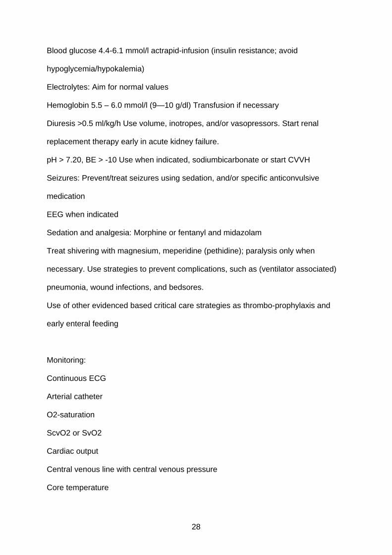
28
Blood glucose 4.4-6.1 mmol/l actrapid-infusion (insulin resistance; avoid
hypoglycemia/hypokalemia)
Electrolytes: Aim for normal values
Hemoglobin 5.5 – 6.0 mmol/l (9—10 g/dl) Transfusion if necessary
Diuresis >0.5 ml/kg/h Use volume, inotropes, and/or vasopressors. Start renal
replacement therapy early in acute kidney failure.
pH > 7.20, BE > -10 Use when indicated, sodiumbicarbonate or start CVVH
Seizures: Prevent/treat seizures using sedation, and/or specific anticonvulsive
medication
EEG when indicated
Sedation and analgesia: Morphine or fentanyl and midazolam
Treat shivering with magnesium, meperidine (pethidine); paralysis only when
necessary. Use strategies to prevent complications, such as (ventilator associated)
pneumonia, wound infections, and bedsores.
Use of other evidenced based critical care strategies as thrombo-prophylaxis and
early enteral feeding
Monitoring:
Continuous ECG
Arterial catheter
O2-saturation
ScvO2 or SvO2
Cardiac output
Central venous line with central venous pressure
Core temperature
29
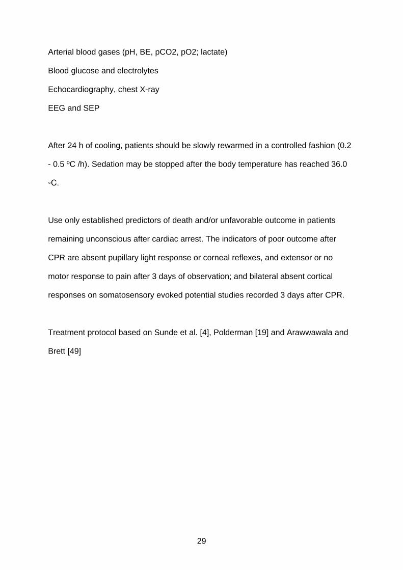
Arterial blood gases (pH, BE, pCO2, pO2; lactate)
Blood glucose and electrolytes
Echocardiography, chest X-ray
EEG and SEP
After 24 h of cooling, patients should be slowly rewarmed in a controlled fashion (0.2
- 0.5 ºC /h). Sedation may be stopped after the body temperature has reached 36.0
◦C.
Use only established predictors of death and/or unfavorable outcome in patients
remaining unconscious after cardiac arrest. The indicators of poor outcome after
CPR are absent pupillary light response or corneal reflexes, and extensor or no
motor response to pain after 3 days of observation; and bilateral absent cortical
responses on somatosensory evoked potential studies recorded 3 days after CPR.
Treatment protocol based on Sunde et al. [4], Polderman [19] and Arawwawala and
Brett [49]