Embed Size (px)
Citation preview

905
INTRODUCTION
THE AORTIC VALVE THRIVES in a complex and dynamicmechanical environment while maintaining unidi-
rectional circulation and preventing left ventricular over-load. Many pathologies and congenital heart defects ad-
versely affects the aortic valve, compromising its func-tion and ability to repair itself,1,2 Homograft or prosthetic(mechanical or biological) valve replacements can pro-vide approximately 10 to 20 years of function before fail-ing in a number of ways, mainly associated with the factthat they are nonliving substitutes.3–5 Tissue engineering
TISSUE ENGINEERINGVolume 12, Number 4, 2006© Mary Ann Liebert, Inc.
Valvular Endothelial Cells Regulate the Phenotype ofInterstitial Cells in Co-culture: Effects of Steady Shear Stress
JONATHAN T. BUTCHER and ROBERT M. NEREM
ABSTRACT
Valvular endothelial cells interact with interstitial cells in a complex hemodynamic and mechanicalenvironment to maintain leaflet tissue integrity. The precise roles of each cell type are difficult toascertain in a controlled manner in vivo. The objective of this study was to develop a three-dimen-sional aortic valve leaflet model, comprised of valvular endothelium and interstitial cells, and de-termine the cellular responses to imposed lumenal fluid flow. Two leaflet models were created us-ing type I collagen hydrogels. Model 1 contained 1 million/mL porcine aortic valve interstitial cells(PAVICs). Model 2 added a seeding of the lumenal surface of Model 1 with approximately 50,000/cm2
porcine aortic valve endothelial cells (PAVECs). Both leaflet models were exposed to 20 dynes/cm2
steady shear for up to 96 h, with static constructs serving as controls. Endothelial cell alignment,matrix production, and cell phenotype were monitored. The results indicate that PAVECs align per-pendicularly to flow similar to 2D culture. We report that PAVICs in model 1 express vimentinstrongly and �-smooth-muscle actin (SMA) to a lesser extent, but SMA expression is increased byshear stress, particularly near the lumenal surface. Model 1 constructs increase in cell number,maintain protein levels, but lose glycosaminoglycans in response to shear. Co-culture with PAVECs(Model 2) modulates these responses in both static and flow environments, resulting in PAVIC phe-notype that is more similar to the native condition. PAVECs stimulated a decrease in PAVIC pro-liferation, an increase in protein synthesis with shear stress, and reduced the loss of glycosamino-glycans with flow. Additionally, PAVECs stimulated PAVIC differentiation to a more quiescentphenotype, defined by reduced expression of SMA. These results suggest that valvular endothelialcells are necessary to properly regulate interstitial cell phenotype and matrix synthesis. Addition-ally, we show that tissue-engineered models can be used to discover and understand complex bio-mechanical relationships between cells that interact in vivo.
Petit Institute for Bioengineering and Bioscience, Woodruff School of Mechanical Engineering, Georgia Institute of Technol-ogy, Atlanta, Georgia.
shows great potential to develop a valve substitute thatcan grow and remodel similar to the success reported withpulmonary autografts in the Ross procedure.6 Attemptsto engineer heart valve tissue have focused on the main-tenance of mechanical integrity using a variety of matrixscaffolds.7–10 One of the most successful animal trials us-ing engineered valvular substitutes, reported by Hoer-strup and colleagues, used biodegradable polymer scaf-folds seeded with vascular cells. They reported that after20 weeks’ implantation in a sheep model, the engineeredvalves exhibited similar tissue structure, cytoskeletalmarkers, and mechanical properties as those normalvalves.11 Unfortunately, these valves maintained persis-tent nontrivial regurgitation that may preclude their usein the clinical setting. Although the reasons for theseshortcomings are unclear, they may be due to the factthat nonvalvular cells were used to initially populate thematrices, which perhaps miss key phenotypic behaviors.The future success of engineered heart valves may there-fore depend ultimately on the ability of the cells popu-lating the scaffolds to thrive within the dynamic and com-plex mechanical environment, remodel the tissue matrix,and resemble native cell phenotypes.
Surprisingly, very little is known about native valvu-lar cells and their interactions in vivo. Advances in theunderstanding of valvular cell biology have not kept upwith the understanding of valve tissue mechanics andscaffold design. Indeed, most tissue-engineering applica-tions have used cells isolated from blood vessels8,12,13 orstem cells14 to populate their matrices. The aortic valveleaflet is populated with interstitial cells, while the sur-faces are lined with endothelial cells. Valvular endothe-lial dysfunction has been implicated as the initiator ofmany clinical sequelae, including inflammatory reac-tions, calcification, and blood clots.1,15,16 We have pre-viously demonstrated that aortic valve endothelial cellsrespond differently to shear stress than aortic endothelialcells do. Aortic valve endothelial cells align perpendicu-larly to the direction of flow, in contrast to aortic endo-thelial cells, which align parallel, and this morphologychange may be associated with unique mechanotrans-duction pathways.17 Transcriptional profiling also indi-cates that valvular endothelial cells may be different fromaortic endothelial cells, most notably that aortic valve en-dothelial cells are more proliferative than aortic endo-thelial cells.18 In vivo evidence suggests that valvular in-terstitial cells are highly dynamic producers of proteinand glycosaminoglycans, unlike smooth-muscle cells,19
and express markers that suggest a hybrid myofibroblast-like phenotype.20 Cultured interstitial cells express �-smooth-muscle actin to a variable degree, as well as fi-broblast surface antigen, and this was not mimicked bycells from other sources.21 Taylor and colleagues dem-onstrated that interstitial cells in three-dimensional cul-ture in vitro expressed markers similar to interstitial cells
BUTCHER AND NEREM
in vivo, and the relative proportions of specific markerexpressing cells was maintained.22 We have previouslyshown that interstitial cells expressed �-smooth-muscleactin in vitro to a similar degree as smooth-muscle cells,desmin to a lesser degree, and that three-dimensional cul-ture also influences interstitial cell phenotype.23
What has not been addressed by any of the previouslymentioned studies is the influence that native valvular en-dothelium may exert on the underlying interstitial cells.It is well known that proper interaction between vascu-lar endothelial and smooth-muscle cells is paramount tonormal vessel function. Vascular endothelial stimulationof underlying smooth-muscle cells is critical for the main-tenance of vessel tone and inhibition of pathologicalsmooth-muscle cell differentiation. In vitro co-culturemodels have shown that endothelial cells inhibit smooth-muscle proliferation under flow.24,25 Gene expression ofendothelial cells is also altered when soluble factors areallowed to exchange with smooth-muscle cells.26 It islikely that interactions between valvular endothelial cellsand interstitial cells are similarly important.
The objective of this study therefore was to use tissue-engineering techniques to develop a three-dimensionalco-culture model of an aortic valve leaflet. By entomb-ing native aortic valve cells in a known amount of rele-vant biological matrix protein, much more experimentalcontrol can be gained over biological variations, and per-haps a more clear understanding of the cell behaviors.Valvular endothelial-interstitial cell co-cultures were alsoexposed to steady laminar shear for 48 or 96 h to inves-tigate the role of hemodynamics in the interaction be-tween the cells as determined by morphological, bio-chemical, and immunohistochemical assays.
MATERIALS AND METHODS
Cell culture and model creation
Porcine aortic valve endothelial cells (PAVECs) andaortic valve interstitial cells (PAVICs) were isolated us-ing collagenase digestion as previously described,17,23
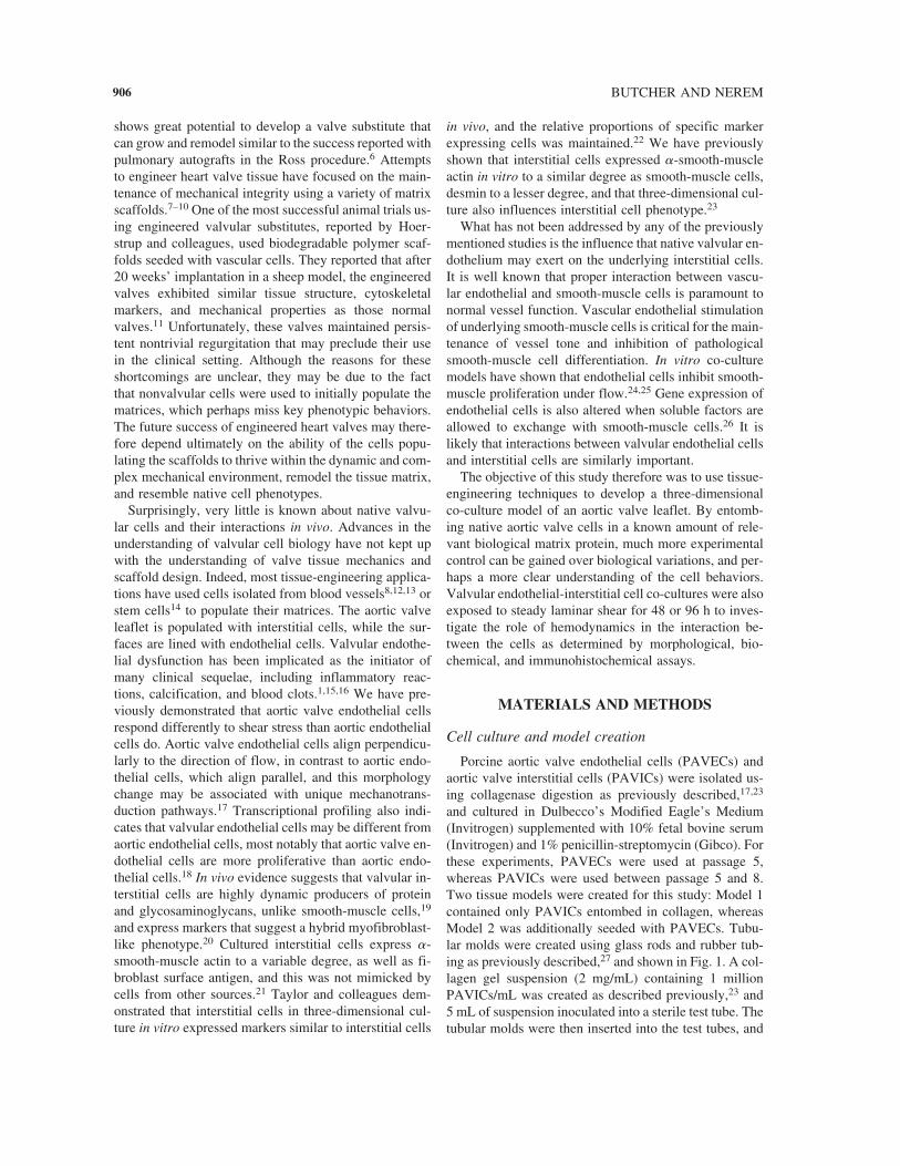
and cultured in Dulbecco’s Modified Eagle’s Medium(Invitrogen) supplemented with 10% fetal bovine serum(Invitrogen) and 1% penicillin-streptomycin (Gibco). Forthese experiments, PAVECs were used at passage 5,whereas PAVICs were used between passage 5 and 8.Two tissue models were created for this study: Model 1contained only PAVICs entombed in collagen, whereasModel 2 was additionally seeded with PAVECs. Tubu-lar molds were created using glass rods and rubber tub-ing as previously described,27 and shown in Fig. 1. A col-lagen gel suspension (2 mg/mL) containing 1 millionPAVICs/mL was created as described previously,23 and5 mL of suspension inoculated into a sterile test tube. Thetubular molds were then inserted into the test tubes, and
906

the constructs were allowed to gel for 1 h. The moldswere then removed from the tubes, placed in a sterile dishcontaining 150 mL of culture media, and incubated at37°C, 5% CO2. The constructs were then allowed to com-pact circumferentially around the mandrel for 6 days, in-creasing the density and strength of the tissue so that itcould be manipulated. Each construct was then gently re-moved from the mold and sectioned longitudinally to cre-ate a rectangular tissue structure. The surface contactingthe glass mandrel was placed face down on a glass mi-croscope slide, and any media were aspirated from un-der the tissue to ensure complete surface contact. A rec-tangular polycarbonate mold was placed around theconstruct, and 6 mL of a 3.5% agar solution at 47°C wasinoculated over the construct and immediately sealedwith another glass slide. The agar solidified after 1 minat room temperature. The sandwich was flipped over andthe slide covering the flat surface was removed to exposeit to media. The resulting embedded construct was thenplaced in 35 mL of culture media. To create the co-cul-ture model (Model 2), Model 1 constructs were addi-tionally seeded with 50,000 PAVECs/cm2 for 1 h. Bothmodels were then cultured for an additional 48 h to ac-commodate PAVEC monolayer formation in Model 2constructs.
Shear stress experiments
The embedded constructs were placed into a modifiedversion of the parallel plate flow chamber previously de-scribed.17 A smaller rubber spacer was used to accom-modate the glass coverslip and rectangular mold con-taining the embedded construct. A larger spacer was usedto create the flow channel and to accommodate the in-creased variation in channel height due to any undula-tions in the construct surface. The lumenal surfaces werethen exposed to 20 dynes/cm2 steady laminar shear stressfor 48 or 96 h. The hemodynamics of the native valveare very complex and difficult to mimic in vitro, but pre-vious studies have shown that 20 dynes/cm2 approxi-mates the mean wall shear stress averaged over a normalcardiac cycle.28 This value was used both as a first ap-
SHEER STRESS AND 3D VALVE LEAFLET MODEL
proximation of the hemodynamic environment and tocompare with our previous results using valvular cells.17
Nonsheared Model 1 and Model 2 constructs at 96 h, aswell as immediately before the application of flow (0 h),served as controls.
Endothelial morphology
Upon completion of the experimental time points, theconstructs (Model 2) were removed, and the agar moldwas cut away and then fixed in 3.7% paraformaldehydefor 24 h. The constructs were then gently peeled awayfrom the agar and placed in phosphate-buffered saline so-lution (Gibco). The constructs were then permeabilizedwith 0.1% Triton X-100 for 5 min, followed by blockingin 1% neonatal goat serum for 1 h. Endothelial cell phe-notype was then labeled by incubation with anti-humanvon Willebrand factor (vWF) (Sigma F-3520, 1:100, inrabbit) for 1 h, secondary antibody incubation (goat anti-rabbit, FITC, Molecular Probes A-11008, 1:100, 1 h) andcounterstained for f-actin (rhodamine phalloidin, SigmaR-418, 1:400) and cell nuclei (Hoechst, Sigma F-32258,1:1000) for 30 min, followed by additional rinsing andcoverslipping. Constructs were then imaged using laserscanning confocal microscopy (Zeiss LSM 510). The lu-menal surface of the constructs was placed face down(closest to the objective) on a glass coverslip and fiverepresentative pictures were taken of each construct.Morphological parameters (shape index and orientationangle) were assessed as previously described using im-age analysis software (LSM Image, Zeiss).17 Measure-ments were then compared with the morphology ofPAVEC monolayers on glass slides. Eight constructs percondition were used for statistical analyses. Two-factoranalysis of variance was used to determine significancebetween cell type and condition, with p � 0.05 consid-ered significant.
Biochemical assays and histology
Constructs were sectioned in half widthwise: half ofthe construct was fixed for histology, and half was im-
907
FIG. 1. Schematic of valvular co-culture model creation. (Color images are available online at �www.liebertpub.com/ten�.)

mediately frozen at -80°C until analyzed biochemically.PAVIC only constructs (Model 1) and co-cultures (Model2) were analyzed for cell number, total protein content,and sulfated glycosaminoglycan content as previously de-scribed.23 Cell number was normalized to dry weight toaccount for differences in size of the samples, while ma-trix production was normalized to DNA content to ap-proximate per cell values. Additionally, all construct val-ues were normalized by day 0 averages to account forvariations between batches of constructs. Samples from sixconstructs in each condition were used to determine sig-nificant differences. Statistical significance was deter-mined using analysis of variance for time duration, and Ttests for differences between flow condition and cell type.p � 0.05 was considered significant for these tests. PAVICphenotype was qualitatively assessed using immunohisto-chemistry. Construct samples were fixed overnight in 3.7%paraformaldehyde and placed in 70% ethanol. Constructswere then paraffin embedded and sectioned at 5 �m. Slideswere deparaffinized, washed twice in phosphate-bufferedsaline, and blocked in 1% bovine serum albumin for 30min. Antibodies to vWF (Sigma F-3520, 1:600), vimentin(Cy3 conjugate, Sigma V2228, 1:200), �-smooth-muscleactin (Cy3 conjugate, Sigma 0-6198, 1:400), or myosinheavy chain (Sigma, M-7786, 1:100) were incubatedsingly for 1 h. Slides were then washed, followed by in-cubation in secondary antibody for vWF (MolecularProbes, A-11008, 1:100) for 40 min. Slides were thenwashed twice and coverslipped with a DAPI counterstainfor cell nuclei. Slides were viewed using a fluorescent mi-croscope (Nikon E600), and several representative pictureswere taken from each construct stained.
RESULTS
Co-culture model characterization
PAVICs compacted the collagen hydrogels over the 6-day period (Fig. 2A), with a majority of the compactionoccurring by day 3, similar to our previous data.23 Imag-ing by polarized light showed the collagen fibril align-ment through the thickness of the construct (Fig. 2E).Highly aligned fibers were concentrated near the surfaceof the tissue closest to the glass mandrel, and more ran-domly oriented near the opposite surface. It is likely thatthe developed tissue anisotropy is due to the physical con-straint placed at this surface.
The surface with highly aligned underlying matrix be-came the “lumenal” surface upon longitudinal sectioning,which was then seeded with PAVECs, creating the Model2 co-culture. Immunofluorescent staining for vWF indi-cated that the entire surface of the leaflet was not com-pletely covered with endothelial cells, but large patchesas shown in Fig. 2C existed throughout the surface, lead-
BUTCHER AND NEREM
ing us to estimate the surface to be approximately 70%covered with endothelial cells, with some variation be-tween individual constructs. Z-stacked confocal mi-croscopy showed a contact-inhibited endothelial mono-layer on top of a closely apposed interstitial cellpopulation (Fig. 2D). Some PAVICs were closely ap-posed to surface PAVECs in the model, suggesting thatthese cells could readily interact with each other in theco-culture model, but it was unclear whether these rep-resented junctional contacts.
Endothelial cell alignment under flow
Fig. 3 shows that on an aligned three-dimensional ma-trix in co-culture with PAVICs, the alignment tendenciesof PAVECs were not significantly different overall fromculture on coated slides. Valvular endothelial cells in bothconditions decreased their Shape Indexes from approxi-mately 0.8 to 0.6 under flow, and changed orientationfrom 45° (random) to 75° (perpendicular to flow). Thelarger standard deviation in orientation angle forPAVECs in co-culture was due to slight variations in un-derlying matrix and interstitial cell orientations, whichsomewhat influenced endothelial cell alignments.
Cell organization and proliferation
Model 1 and Model 2 constructs were exposed to 20dynes/cm2 steady shear with static cultures of both con-ditions serving as controls. Fig. 4 shows the histologi-cally determined cellular arrangement in seeded and non-seeded constructs over time. Interstitial cells arerandomly oriented within the Model 1 constructs, withslight circumferential alignment near the lumenal surface.The application of flow enhances this alignment some-what, but only right at the surface. Model 2 constructshad cells throughout the construct, but there were pref-erentially more cells near the lumenal surface and oppo-site edge of the constructs. This may be due to diffusionconstraints imposed by the addition of PAVECs, limit-ing perfusion through the collagen matrix. Biochemicalanalysis of cell number (DNA content) across experi-mental conditions is shown in Fig. 5A. Model 1 con-structs increase cell number over time, with shear stresshaving no effect. This was in contrast to the Model 2 con-structs, which maintained cell number during static cul-ture, and decreased cell number under flow. By day 4,cell content in sheared Model 1 constructs was signifi-cantly higher than sheared Model 2 constructs.
Matrix production
Fig. 5B and 5C show the matrix production of theModel 1 and Model 2 constructs under static and fluidflow conditions. Protein content was regulated in a man-ner opposite to that of cell content. Model 1 constructs
908

SHEER STRESS AND 3D VALVE LEAFLET MODEL 909
FIG. 3. Endothelial cell alignment on valve leaflet co-culture models. (A) Static culture. (B) Forty-eight-hour flow (20dynes/cm2). (C) Shape index comparison. (D) Cell orientation angle comparison. Green � von Willebrand factor, red � f-actin,blue � cell nuclei. Scale bar � 50 �m. N � 8 for each condition. (Color images are available online at �www.liebertpub.com/ten�.)
FIG. 2. Co-culture model characterization. (A) Tubular mold. (B) Embedded construct. (C) Confocal microscope image indi-cating endothelial monolayer. (D) Three-dimensional confocal image indicating cellular arrangement. (E) Polarized light imageindicating matrix alignment (bright field). Confocal staining: green (von Willebrand factor), red (f-actin), blue (cell nuclei). Scalebar � 1 cm (A,B), 100 �m (C–E). (Color images are available online at �www.liebertpub.com/ten�.)

tended to have less protein over time, with no effects ofshear. Model 2 constructs, on the other hand, had nochange in protein content in static culture, but increasedprotein content under flow. At day 4, sheared Model 2constructs had significantly more protein than Model 1constructs. Sulfated glycosaminoglycan (sGAG) contentregulation was much different, however. sGAG contentwas decreased in Model 1 constructs over time and withshear. Both day 2 and day 4 sheared Model 1 constructshad significantly less sGAGs than day 0, but no signifi-cant difference between static sheared constructs at day4. In Model 2 constructs, no significant difference insGAG content was detected in static culture or under flow(p � 0.055). There was significantly more sGAG contentin day 4 sheared Model 2 constructs compared to shearedModel 1 constructs, but no significant difference betweenstatically cultured models (p � 0.18).
Changes in interstitial cell phenotype
Interstitial cell phenotype was determined through theexpression of vimentin, �-smooth-muscle actin (�-SMA), and smooth-muscle myosin. Fig. 6 shows the vi-mentin expression in the Model 1 and Model 2 constructs.Vimentin expression is maintained throughout the timepoints investigated in both models, with little differencesbetween flow and static culture. The cells appear morealigned after 4 days of flow, with vimentin expressionthroughout the cytoplasm. Fig. 7 shows the expression of�-SMA in both models and flow conditions. In VIC con-structs, only a basal level of expression is observed instatic culture throughout the time period investigated. Theapplication of flow, however, increases expression of �-SMA, especially near the lumenal surface. The expres-
BUTCHER AND NEREM
sion of �-SMA is also regulated with the addition ofPAVECs. At day 0, a basal level of expression is noted,but this expression drops off during the culture period.By day 4, virtually no expression is detectable in staticculture, and only slightly more with the application offlow, again in the cells closest to the lumenal surface.These results suggest that the PAVECs decrease the ex-pression of �-SMA in PAVICs in static and fluid flowconditions. No expression of smooth-muscle myosin wasobserved in either model under any culture condition(data not shown). PAVEC phenotype on the lumenal sur-face of the Model 2 constructs was confirmed throughvWF staining, as shown in Fig. 7. A confluent monolayerof endothelial cells was more difficult to demonstratethrough immunohistochemistry, because these constructswere extremely fragile, and the handling necessary forparaffin processing likely detached some of the cells. Itwas interesting to note that on rare occasions, a few cellsexpressing vWF were detected in the subendothelial lay-ers of the constructs, suggesting that some migration mayhave occurred (arrow), but not to any significant degree.
DISCUSSION
This study is the first expose an engineered co-culturemodel of a valvular leaflet to a defined hemodynamic en-vironment. Native aortic valve endothelial cells wereplaced in close proximity to valvular interstitial cells,which were homogeneously entombed in an anisotropicbiological collagen matrix. This model was then incor-porated into a steady shear flow system to investigate in-teractions between fluid flow, endothelial cells, and in-terstitial cells.
910
FIG. 4. Hematoxylin and eosin staining of constructs depicting matrix and cellular architecture. Left insets depict cell align-ment. Right insets depict endothelial layer. Scale bar � 200 �m. (Color images are available online at �www.liebertpub.com/ten�.)

Numerous studies have demonstrated the importanceof appropriate vascular endothelial–smooth-muscle in-teractions for maintenance of blood vessel tone, and theimportance of mechanical forces in regulating cell phe-notype.29–32 Surprisingly little is known about how na-tive valvular endothelial and interstitial cells interact or
SHEER STRESS AND 3D VALVE LEAFLET MODEL
how mechanical forces influence these interactions.Rothenburger and colleagues created valvular leaflet–liketissue with valvular cells co-cultured in a collagen ma-trix and found that these cells synthesized collagen andproteoglycans, but did not expose these to any hemody-namic forces or identify any particular aspects of endo-thelial regulation of interstitial cell function.33 Westonand Yoganathan developed an organ culture model ofaortic valve leaflets and exposed them to shear stress forup to 48 h.34 They found a dramatic increase in cell num-ber and protein and glycosaminoglycan synthesis withstatic incubation, but no differences with the applicationof any flow regimes. Our results indicated no significantchanges in cell or protein content by 48 h, but a loss insGAG content for Model 1 constructs. Weston and Yo-ganathan reported that their leaflet isolation protocol mayhave removed all viable endothelium from the leaflet sur-faces, creating a model more like the VIC, and these re-sults seem to support that notion. The native aortic valveleaflet has other matrix components besides type I colla-gen,35 so some of the differences may also be a result ofadditional matrix signals not present in these models.
The results of our in vitro studies do compare well withpreviously reported in vivo and ex vivo observations. Thestatic culture-induced cell proliferation in Model 1 con-structs may mimic a mitogenic wound healing responseas has been reported by Lester et al., who showed an in-crease in interstitial cell proliferation with endothelial de-nudation in an organ culture model.36 This proliferationwas not seen in organ cultures with intact endothelium,nor was it reported here in Model 2 co-culture constructs.Vascular endothelium inhibits medial smooth-muscle cellproliferation in in vivo37 and in vitro co-culture models,25
and this inhibition is more apparent with shear stress, sim-ilar to our results with valvular cells. In our studies, thepresence of valvular endothelial cells also increased pro-tein synthesis, and reduced in the loss of sGAGs inducedby shear in comparison to controls. Short-term studies inrat aortic valves indicated more active production of pro-teins and GAGs in comparison to other tissues such asaortic wall,19 suggesting that native valvular interstitialcells secrete more matrix than smooth-muscle cells. Ac-tivated interstitial cells, as determined by �-SMA ex-pression,38 persisted throughout the culture periods inModel 1 constructs, but was reduced in co-cultures withvalvular endothelial cells (Model 2 constructs). The pres-ence of positive �-SMA expression at day 0 may be dueto interstitial cell activation resulting from in vitro cul-ture,39 which may persist through the embedding process.We previously showed high expression of �-SMA in two-dimensional culture, which was dramatically reduced inthree-dimensional culture, but still positive.23 Only in-terstitial cells near the lumenal surface expressed �-SMAafter 4 days of steady shear stress in Model 2 constructs,which was similar to the pattern of immunohistochemi-
911
FIG. 5. Cell content (A), protein content (B), and sulfated gly-cosaminoglycan content (C) of EC and non-EC seeded con-structs. Data normalized to Day 0 values and presented asmean � SEM. Asterisk denotes significant with respect to dif-ferent condition (p � 0.05), double asterisk denotes significantwith respect to Day 0 value (p � 0.05). N � 6 for each condi-tion.

cal staining of the ventricularis side of the native leafletshown in Fig. 7. Vascular endothelial cells, in contrast,enhance �-SMA expression in medial cells while in co-culture and in the presence of fluid shear stress.40
The differences in interstitial cell phenotype observedbetween the two culture models and reports of vascularcell interactions may suggest some important differencesbetween valvular and vascular cells. Both interstitial cellsand vascular smooth-muscle cells proliferate when not incommunication with their respective endothelial cells.Valvular interstitial cells secreted matrix in a valvular en-dothelial– and flow-dependent manner, but smooth-mus-cle cell matrix synthesis is inhibited by vascular endo-thelial factors and flow. Valvular endothelium decreases�-SMA expression in interstitial cells, whereas vascularendothelial cells increase expression in medial smooth-muscle cells. These results suggest that the quiescentstates of interstitial cells and smooth-muscle cells are dif-
BUTCHER AND NEREM
ferent in potentially significant ways. The demanding he-modynamic and mechanical environment of the aorticvalve may require a nonproliferative, nonactivated celltype capable of secreting matrix to remodel damaged ma-trix before it progresses to gross tissue failure. Quiescentsmooth-muscle cells in this environment may remaincontractile (activated) and not adequately replace matrixproteins. Synthetic smooth-muscle cells are characteris-tically mitogenic41 and may cause excess tissue growthor disrupt the complex nutrient balance requirements,perhaps leading to a pathological angiogenic response asseen in diseased valves.42 The implantation of tissue-en-gineered valvular conduits using vascular cells by theMayer group43 resulted in apparent valvular-like cell phe-notypes at 20 weeks, but it was unclear whether the cellsthat remained were donor or host cells. The persistenttransvalvular gradients and increased leaflet stiffness re-ported by this group11 suggests that some phenotypic dif-
912
FIG. 6. Vimentin expression in PAVIC constructs and PAVEC-seeded co-cultures (TEVL). Counterstained for cell nuclei (blue).Bottom left panel indicates native leaflet endothelium (von Willebrand factor). Scale bar � 100 �m. (Color images are availableonline at �www.liebertpub.com/ten�.)
FIG. 7. �-Smooth-muscle actin expression in PAVIC constructs and PAVEC-seeded co-cultures (TEVL). Counterstained forcell nuclei (blue). Bottom left panel indicates TEVL endothelium (von Willebrand factor). Scale bar � 100 �m. (Color imagesare available online at �www.liebertpub.com/ten�.)

ferences between the cells in these tissues and nativevalves may remain.
From these studies it is apparent that the valvular en-dothelial cell is the key regulator of interstitial cell phe-notype and shear flow enhances this regulation, but theexact mechanisms are as yet unclear. Gotlieb et al. haveshown that valvular interstitial cell wound repair is me-diated by fibroblast growth factor 2 (FGF-2),44 and sev-eral researchers have shown that transforming growthfactor b1 (TGF�1) expression is associated with valvu-lar pathology.45,46 FGF2 and TGF�1 signaling pathwaysmay therefore be likely candidates for the investigationof valvular endothelial cell regulation of interstitial cellphenotype. It is not currently known whether valvular en-dothelial cells would stimulate vascular smooth-musclecells toward a more valvular interstitial cell-like pheno-type or how vascular endothelial cells would regulatevalvular interstitial cells. We previously reported thatvalvular endothelial cells responded to shear stress dif-ferently than vascular endothelial cells,17 which suggeststhat the aforementioned pairings would interact hetero-geneously.
The long-term success of a tissue-engineered valvu-lar substitute may pivot more on the maintenance ofappropriate cell phenotypes rather than adequate tissuestrength, and therefore the interactions between the na-tive cell types should serve as the “gold standard” forappropriate valvular cell biology. Given the fact thatvalvular cells are generally not available for isolationand repopulation of a tissue-engineered autograft47 andthat allogeneic cells elicit an immune response,48 othercell sources may need to be explored. Recent work cre-ating valvular conduits populated with stem cells mayhave great potential in that these cells might be stimu-lated more easily to differentiate into valvular pheno-types.49 Much more work, however, is needed tobroaden our understanding of the phenotypes of thesecells and their interactions to progress further with thispursuit, and tissue engineering may serve as a usefultool to accomplish this. Valvular mechanics and he-modynamics are truly complex, as is the three-dimen-sional tissue structure. Attempts to expose the entireaortic root to approximate native tissue forces andflows in vitro may not only cause premature tissue de-struction due to nutrient transport limitations, but alsoimpairs the determination of causal parameters. Engi-neered tissue models provide a well-defined three-di-mensional matrix environment where the interactionsbetween relevant cell types can be investigated underwell-controlled stimuli. These models can complementmore involved and expensive animal trials in a posi-tive feedback loop both as a pre-animal feasibility as-sessment and as a post-animal model of mechanisticunderstanding. The field may therefore be able toprogress more quickly without sacrificing biological
SHEER STRESS AND 3D VALVE LEAFLET MODEL
understanding that will become critical as these engi-neered conduits progress to preclinical phases.
ACKNOWLEDGMENTS
We gratefully acknowledge the assistance of TraceyCouse in the processing of the histology. This work wasfunded by the Georgia Tech/Emory Center for the Engi-neering of Living Tissues, an NSF-ERC (RMN), and bya predoctoral fellowship from the American Heart Asso-ciation (JTB).
REFERENCES
1. Cooper, M.D., Jeffery, C., Gall, D.L., and Anderson, A.S.Scanning electron microscopy studies of staphylococcal ad-herence to heart valve endothelial cells in organ culture: anin vitro model of acute endocarditis. Scan. Electron Mi-crosc. 1231, 1985;(pt.3).
2. Poggianti, E., Venneri, L., Chubuchny, V., Jambrik, Z.,Baroncini, L.A., and Picano, E. Aortic valve sclerosis is as-sociated with systemic endothelial dysfunction. J. Am.Coll. Cardiol. 41, 136, 2003.
3. Hammermeister, K., Sethi, G.K., Henderson, W.G.,Grover, F.L., Oprian, C., and Rahimtoola, S.H. Outcomes15 years after valve replacement with a mechanical versusa bioprosthetic valve: final report of the Veterans Affairsrandomized trial. J. Am. Coll. Cardiol. 36, 1152, 2000.
4. Staab, M.E., Nishimura, R.A., Dearani, J.A., and Orszulak,T.A. Aortic valve homografts in adults: a clinical perspec-tive. Mayo Clin. Proc. 73, 231, 1998.
5. North, R.A., Sadler, L., Stewart, A.W., McCowan, L.M.,Kerr, A.R., and White, H.D. Long-term survival and valve-related complications in young women with cardiac valvereplacements. Circulation 99, 2669, 1999.
6. Turrentine, M.W., Ruzmetov, M., Vijay, P., Bills, R.G.,and Brown, J.W. Biological versus mechanical aortic valvereplacement in children. Ann. Thorac. Surg. 71, S356,2001.
7. Shinoka, T., Ma, P.X., Shum-Tim, D., Breuer, C.K., Cu-sick, R.A., Zund, G., Langer, R., Vacanti, J.P., and Mayer,J.E., Jr. Tissue-engineered heart valves. Autologous valveleaflet replacement study in a lamb model. Circulation 94,II164, 1996.
8. Numata, S., Niwaya, K., Fujisato, T., Funamoto, S.,Nakatani, T., Yagihara, T., and Kitamura, S. Decellular-ized allograft valve for tissue engineering: experimentalstudy of heart valves using decellularized cryopreserved al-lografts. Heart Surg. Forum 6, 2, 2002.
9. Dohmen, P.M., Ozaki, S., Verbeken, E., Yperman, J., Fla-meng, W., and Konertz, W.F. Tissue engineering of anauto-xenograft pulmonary heart valve. Asian Cardiovasc.Thorac. Ann. 10, 25, 2002.
10. Shi, Y., Ramamurthi, A., and Vesely, I. Towards tissue en-gineering of a composite aortic valve. Biomed. Sci. In-strum. 38, 35, 2002.
913

11. Hoerstrup, S.P., Sodian, R., Daebritz, S., Wang, J., Bacha,E.A., Martin, D.P., Moran, A.M., Guleserian, K.J., Sper-ling, J.S., Kaushal, S., Vacanti, J.P., Schoen, F.J., andMayer, J.E., Jr. Functional living trileaflet heart valvesgrown in vitro. Circulation 102, III44, 2000.
12. Kim, W.G., Park, J.K., Park, Y.N., Hwang, C.M., Jo, Y.H.,Min, B.G., Yoon, C.J., and Lee, T.Y. Tissue-engineeredheart valve leaflets: an effective method for seeding autol-ogous cells on scaffolds. Int. J. Artif. Organs 23, 624, 2000.
13. Stock, U.A., Nagashima, M., Khalil, P.N., Nollert, G.D.,Herden, T., Sperling, J.S., Moran, A., Lien, J., Martin, D.P.,Schoen, F.J., Vacanti, J.P., and Mayer, J.E., Jr. Tissue-en-gineered valved conduits in the pulmonary circulation. J.Thorac. Cardiovasc. Surg. 119, 732, 2000.
14. Perry, T.E., Kaushal, S., Sutherland, F.W., Guleserian, K.J.,Bischoff, J., Sacks, M., and Mayer, J.E. Thoracic SurgeryDirectors Association Award. Bone marrow as a cell sourcefor tissue engineering heart valves. Ann. Thorac. Surg. 75,761, 2003.
15. Drake, T.A., and Pang, M. Effects of interleukin-1,lipopolysaccharide, and streptococci on procoagulant ac-tivity of cultured human cardiac valve endothelial and stro-mal cells. Infect. Immun. 57, 507, 1989.
16. Campbell, K.M., and Johnson, C.M. Identification ofStaphylococcus aureus binding proteins on isolated porcinecardiac valve cells. J. Lab. Clin. Med. 115, 217, 1990.
17. Butcher, J.T., Penrod, A.M., Garcia, A.J., and Nerem, R.M.Unique morphology and focal adhesion development ofvalvular endothelial cells in static and fluid flow environ-ments. Arterioscler. Thromb. Vasc. Biol. 24, 1429, 2004.
18. Farivar, R.S., Cohn, L.H., Soltesz, E.G., Mihaljevic, T.,Rawn, J.D., and Byrne, J.G. Transcriptional profiling andgrowth kinetics of endothelium reveals differences betweencells derived from porcine aorta versus aortic valve. Eur.J. Cardiothorac. Surg. 24, 527, 2003.
19. Schneider, P.J., and Deck, J.D. Tissue and cell renewal inthe natural aortic valve of rats: an autoradiographic study.Cardiovasc. Res. 15, 181, 1981.
20. Filip, D.A., Radu, A., and Simionescu, M. Interstitial cellsof the heart valves possess characteristics similar to smoothmuscle cells. Circ. Res. 59, 310, 1986.
21. Taylor, P.M., Allen, S.P., and Yacoub, M.H. Phenotypicand functional characterization of interstitial cells from hu-man heart valves, pericardium and skin. J. Heart Valve Dis.9, 150, 2000.
22. Taylor, P.M., Allen, S.P., Dreger, S.A., and Yacoub, M.H.Human cardiac valve interstitial cells in collagen sponge:a biological three-dimensional matrix for tissue engineer-ing. J. Heart Valve Dis. 11, 298, 2002.
23. Butcher, J.T., and Nerem, R.M. Porcine aortic valve inter-stitial cells in three-dimensional culture: comparison ofphenotype with aortic smooth muscle cells. J. Heart ValveDis. 13, 478, 2004.
24. Nackman, G.B., Fillinger, M.F., Shafritz, R., Wei, T., andGraham, A.M. Flow modulates endothelial regulation ofsmooth muscle cell proliferation: a new model. Surgery124, 353, 1998.
25. Ziegler, T., Alexander, R.W., and Nerem, R.M. An endo-thelial cell-smooth muscle cell co-culture model for use in
BUTCHER AND NEREM
the investigation of flow effects on vascular biology. Ann.Biomed. Eng. 23, 216, 1995.
26. Chiu, J.J., Chen, L.J., Lee, P.L., Lee, C.I., Lo, L.W., Us-ami, S., and Chien, S. Shear stress inhibits adhesion mol-ecule expression in vascular endothelial cells induced bycoculture with smooth muscle cells. Blood 101, 2667, 2003.
27. Seliktar, D., Black, R.A., Vito, R.P., and Nerem, R.M. Dy-namic mechanical conditioning of collagen-gel blood ves-sel constructs induces remodeling in vitro. Ann. Biomed.Eng. 28, 351, 2000.
28. Weston, M.W., LaBorde, D.V., and Yoganathan, A.P. Es-timation of the shear stress on the surface of an aortic valveleaflet. Ann. Biomed. Eng. 27, 572, 1999.
29. Harrison, D.G., Sayegh, H., Ohara, Y., Inoue, N., and Ven-ema, R.C. Regulation of expression of the endothelial cellnitric oxide synthase. Clin. Exp. Pharmacol. Physiol. 23,251, 1996.
30. Brown, G.C. Nitric oxide as a competitive inhibitor of oxy-gen consumption in the mitochondrial respiratory chain.Acta. Physiol. Scand. 168, 667, 2000.
31. Ignarro, L.J. Endothelium-derived nitric oxide: actions andproperties. FASEB J 3, 31, 1989.
32. Bochaton-Piallat, M.L., Gabbiani, F., Redard, M.,Desmouliere, A., and Gabbiani, G. Apoptosis participatesin cellularity regulation during rat aortic intimal thicken-ing. Am. J. Pathol. 146, 1059, 1995.
33. Rothenburger, M., Volker, W., Vischer, J.P., Berendes, E.,Glasmacher, B., Scheld, H.H., and Deiwick, M. Tissue en-gineering of heart valves: formation of a three-dimensionaltissue using porcine heart valve cells. ASAIO J. 48, 586,2002.
34. Weston, M.W., and Yoganathan, A.P. Biosynthetic activ-ity in heart valve leaflets in response to in vitro flow en-vironments. Ann. Biomed. Eng. 29, 752, 2001.
35. Christie, G.W. Anatomy of aortic heart valve leaflets: theinfluence of glutaraldehyde fixation on function. Eur. J.Cardiothorac. Surg. 6 Suppl 1, S25, 1992.
36. Lester, W.M., Damji, A.A., Tanaka, M., and Gedeon, I.Bovine mitral valve organ culture: role of interstitial cellsin repair of valvular injury. J. Mol. Cell. Cardiol. 24, 43,1992.
37. Mason, R.A., Hui, J.C., Campbell, R., and Giron, F. Theeffects of endothelial injury on smooth muscle cell prolif-eration. J. Vasc. Surg. 5, 389, 1987.
38. Rabkin, E., Aikawa, M., Stone, J.R., Fukumoto, Y., Libby,P., and Schoen, F.J. Activated interstitial myofibroblastsexpress catabolic enzymes and mediate matrix remodelingin myxomatous heart valves. Circulation 104, 2525, 2001.
39. Yperman, J., De Visscher, G., Holvoet, P., and Flameng,W. Molecular and functional characterization of ovine car-diac valve-derived interstitial cells in primary isolates andcultures. Tissue Eng. 10, 1368, 2004.
40. Villaschi, S., and Nicosia, R.F. Paracrine interactions be-tween fibroblasts and endothelial cells in a serum-free co-culture model. Modulation of angiogenesis and collagengel contraction. Lab Invest. 71, 291, 1994.
41. Worth, N.F., Rolfe, B.E., Song, J., and Campbell, G.R.Vascular smooth muscle cell phenotypic modulation inculture is associated with reorganisation of contractile
914

and cytoskeletal proteins. Cell Motil. Cytoskeleton 49,130, 2001.
42. Soini, Y., Salo, T., and Satta, J. Angiogenesis is involvedin the pathogenesis of nonrheumatic aortic valve stenosis.Hum. Pathol. 34, 756, 2003.
43. Rabkin, E., Hoerstrup, S.P., Aikawa, M., Mayer, J.E., Jr.,and Schoen, F.J. Evolution of cell phenotype and extracel-lular matrix in tissue-engineered heart valves during in-vitro maturation and in-vivo remodeling. J. Heart ValveDis. 11, 308, 2002.
44. Gotlieb, A.I., Rosenthal, A., and Kazemian, P. Fibroblastgrowth factor 2 regulation of mitral valve interstitial cellrepair in vitro. J. Thorac. Cardiovasc. Surg. 124, 591, 2002.
45. Jian, B., Narula, N., Li, Q.Y., Mohler, E.R. 3rd, and Levy,R.J. Progression of aortic valve stenosis: TGF-beta1 is pres-ent in calcified aortic valve cusps and promotes aortic valveinterstitial cell calcification via apoptosis. Ann. Thorac.Surg. 75, 457, 2003.
46. Ng, C.M., Cheng, A., Myers, L.A., Martinez-Murillo, F.,Jie, C., Bedja, D., Gabrielson, K.L., Hausladen, J.M.,Mecham, R.P., Judge, D.P., and Dietz, H.C. TGF-beta-de-pendent pathogenesis of mitral valve prolapse in a mousemodel of Marfan syndrome. J. Clin. Invest. 114, 1586,2004.
SHEER STRESS AND 3D VALVE LEAFLET MODEL
47. Hoffman-Kim, D., Maish, M.S., Krueger, P.M., Lukoff, H.,Bert, A., Hong, T., and Hopkins, R.A. Comparison of threemyofibroblast cell sources for the tissue engineering of car-diac valves. Tissue Eng. 11, 288, 2005.
48. Shinoka, T., Breuer, C.K., Tanel, R.E., Zund, G., Miura,T., Ma, P.X., Langer, R., Vacanti, J.P., and Mayer, J.E. Jr.Tissue engineering heart valves: valve leaflet replacementstudy in a lamb model. Ann. Thorac. Surg. 60, S513, 1995.
49. Sutherland, F.W., Perry, T.E., Yu, Y., Sherwood, M.C.,Rabkin, E., Masuda, Y., Garcia, G.A., McLellan, D.L., En-gelmayr, G.C., Jr., Sacks, M.S., Schoen, F.J., and Mayer,J.E., Jr. From stem cells to viable autologous semilunarheart valve. Circulation 111, 2783, 2005.
Address reprint requests to:Jonathan T. Butcher
Petit Institute for Bioengineering and BioscienceWoodruff School of Mechanical Engineering
Georgia Institute of Technology315 Ferst Drive
Atlanta, GA 30332
E-mail: [email protected]
915