Embed Size (px)
Citation preview
IRAP November 17, 2014
Case 1Presenter: Ian Hughes MD, PGY IIIAttendings: Xiuzhen Duan MD, Stefan E Pambuccian MD
Clinical History: A 78-year-old woman presented with post-menopausal bleeding for the past few months. She reported a past medical history of diabetes and anxiety but denies any other current complaints. Workup included pelvic exam (normal, no current signs of bleeding) and pelvic ultrasound which reveals a thick endometrium (18 mm). Both adnexa were normal. Her last Pap smear was negative. For further evaluation the patient underwent hysteroscopy with endometrial curettage. A representative section is submitted for your review.
Final Diagnosis: Endometrial adenocarcinoma, endometrioid type, FIGO grade 1 with involvement by metastatic carcinoma consistent with patient's colonic primary.
Differential Diagnosis:
Endometrial adenocarcinoma-Endometrioid-Serous-Clear cell
Dedifferentiated/undifferentiated endometrial carcinomaEndometrial carcinoma with neuroectodermal differentiationCombined endometrioid-small cell neuroendocrine carcinomaMetastatic carcinoidMetastatic adenocarcinoma
-Breast-Colon-Upper GI-Lung
Key Features:Histology:At low power the curettage specimen consists of portions of tissue which appear very blue. At higher power the tissue can be seen to contain numerous crowded glands lined by ovoid, somewhat monotonous cells with mild to moderate atypia and little or no intervening stroma. Focal squamous differentiation is noted. Additionally there are groups of higher-grade cells in sheets and poorly formed glands with high nuclear-cytoplasmic ratio, and dark, irregular nuclei. The polypectomy specimen, taken at the same time as the curettage, consists almost entirely of these higher-grade cells.
Immunohistochemistry:
Low-grade areas:Positive: ER, CK7Negative: CK20, CDX2
High-grade areas:Positive: CK20, CDX2Negative: ER, CK7
Both:Negative: Synaptophysin, chromogranin, p53
Discussion: The low-grade, glandular areas are morphologically consistent with endometrial adenocarcinoma, endometrioid type, FIGO grade 1. For the higher grade areas, a number of diagnoses were considered including dedifferentiated/undifferentiated endometrial carcinoma, endometrial carcinoma with neuroendocrine features, metastatic carcinoid, and various metastatic adenocarcinomas (breast, upper GI, colon, lung).
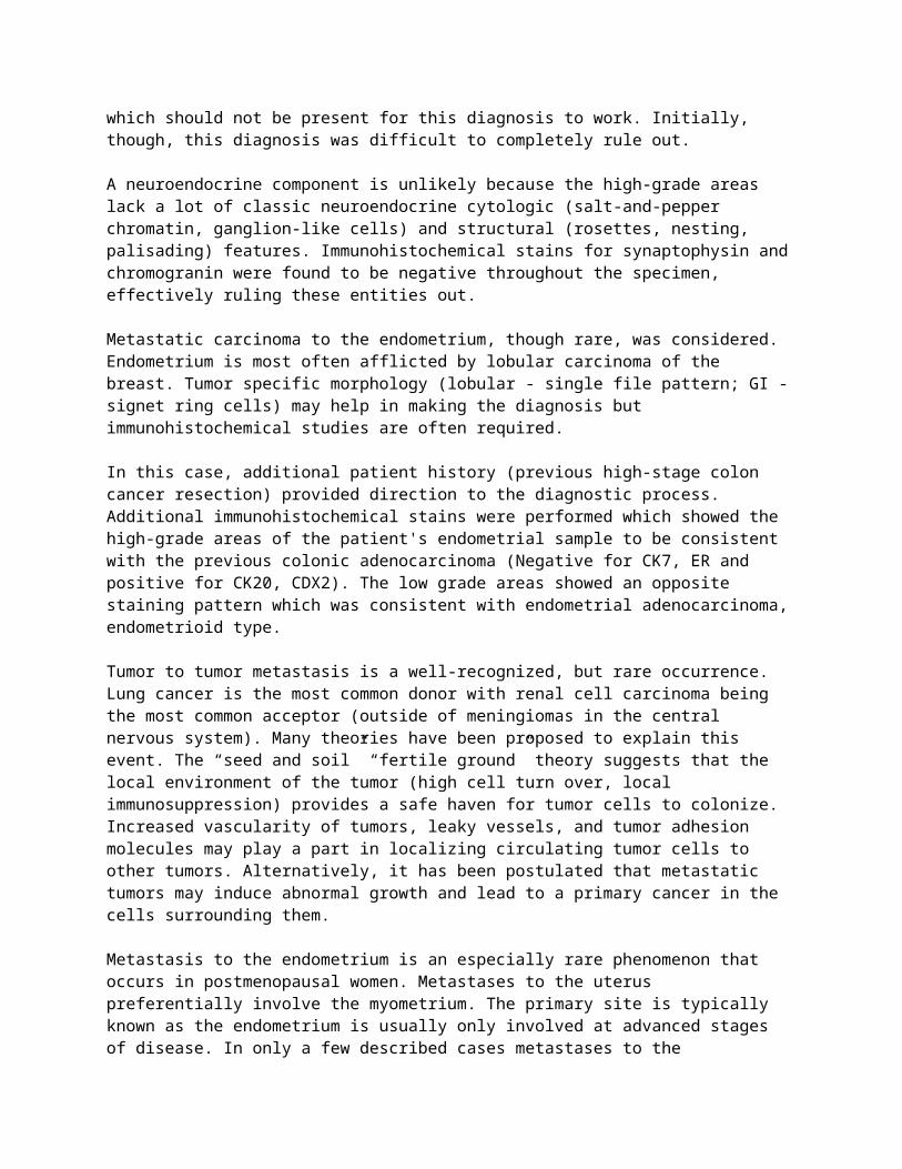
The nuclei of the high-grade areas fit well with undifferentiated endometrial carcinoma, however there were some poorly formed glands which should not be present for this diagnosis to work. Initially, though, this diagnosis was difficult to completely rule out.
A neuroendocrine component is unlikely because the high-grade areas lack a lot of classic neuroendocrine cytologic (salt-and-pepper chromatin, ganglion-like cells) and structural (rosettes, nesting, palisading) features. Immunohistochemical stains for synaptophysin and chromogranin were found to be negative throughout the specimen, effectively ruling these entities out.
Metastatic carcinoma to the endometrium, though rare, was considered. Endometrium is most often afflicted by lobular carcinoma of the breast. Tumor specific morphology (lobular - single file pattern; GI - signet ring cells) may help in making the diagnosis but immunohistochemical studies are often required.
In this case, additional patient history (previous high-stage colon cancer resection) provided direction to the diagnostic process. Additional immunohistochemical stains were performed which showed the high-grade areas of the patient's endometrial sample to be consistent with the previous colonic adenocarcinoma (Negative for CK7, ER and positive for CK20, CDX2). The low grade areas showed an opposite staining pattern which was consistent with endometrial adenocarcinoma, endometrioid type.
Tumor to tumor metastasis is a well-recognized, but rare occurrence. Lung cancer is the most common donor with renal cell carcinoma being the most common acceptor (outside of meningiomas in the central nervous system). Many theories have been proposed to explain this event. The “seed and soil” “fertile ground” theory suggests that the local environment of the tumor (high cell turn over, local immunosuppression) provides a safe haven for tumor cells to colonize. Increased vascularity of tumors, leaky vessels, and tumor adhesion molecules may play a part in localizing circulating tumor cells to other tumors. Alternatively, it has been postulated that metastatic tumors may induce abnormal growth and lead to a primary cancer in the cells surrounding them.
Metastasis to the endometrium is an especially rare phenomenon that occurs in postmenopausal women. Metastases to the uterus preferentially involve the myometrium. The primary site is typically known as the endometrium is usually only involved at advanced stages of disease. In only a few described cases metastases to the endometrium have been the initial presentation of a malignancy. As demonstrated by this case, “pseudoendometrioid” metastatic colon carcinoma and endometrial cancer may be difficult to differentiate morphologically due to a number of similar features. Milder nuclear grade, monomorphism, surrounding premalignant changes, and squamous differentiation suggest a diagnosis of primary endometrial carcinoma.
Patients with concurrent colon and endometrial cancer should be investigated for Lynch Syndrome. Immunohistochemical stains for MLH1, PMS2, MSH2, MSH6 performed on our patient showed no loss of expression, ruling out Lynch Syndrome.
Take-Home Points:Metastasis to the endometrium is rare, but should be considered when an unusual bimorphic pattern is present.
Patient history is important in directing the diagnostic process as most patients with endometrial metastases have known primaries.
Each patient presents unique challenges and should be handled on a case-to-case basis.
References1. Imachi M, Tsukamoto N, Amagase H, Shigematsu T, Amada S, Nakano H. Metastatic adenocarcinoma to the uterine cervix from gastric cancer. A clinicopathologic analysis of 16 cases. Cancer. 1993;71: 3472-3477.2. Scopa CD, Aletra C, Lifschitz-Mercer B, Czernobilsky B. Metastases of breast carcinoma to the uterus. Report of two cases, one harboring a primary endometrioid carcinoma, with review of the literature. Gynecol Oncol. 2005;96: 543-547.3. Metastases to the Uterus. http://www.pathologyoutlines.com/topic/uterusmet.html 4. Gordon, M, Ireland, K. Pathology of Endometrial Carcinoma. Global Library of Women’s Health. Glob. libr. women's med. (ISSN: 1756-2228) 2008; DOI 10.3843/GLOWM.10238 http://www.glowm.com/section_view/heading/Pathology%20of%20Endometrial%20Carcinoma/item/238#16575. Kennebeck CH, Alagoz T. Signet ring breast carcinoma metastases limited to the endometrium and cervix. Gynecol Oncol. 1998;71: 461-464.6. van Leeuwen FE, Benraadt J, Coebergh JW, et al. Risk of endometrial cancer after tamoxifen treatment of breast cancer. Lancet. 1994;343: 448-452.7. Pasini A, Mandelli P, Belloni C. Endometrial metastases from gastric adenocarcinoma: a case report. Tumori. 1995;81: 383-386.8. Tiseo M, Bersanelli M, Corradi D, et al. Endometrial metastasis of lung adenocarcinoma: a case report. Tumori. 2011;97: 411-414.9. Hart WR. Diagnostic challenge of secondary (metastatic) ovarian tumors simulating primary endometrioid and mucinous neoplasms. Pathol Int. 2005;55: 231-243.10. Tafe LJ, Garg K, Chew I, Tornos C, Soslow RA. Endometrial and ovarian carcinomas with undifferentiated components: clinically aggressive and frequently underrecognized neoplasms. Mod Pathol. 2010;23: 781-789.11. Silva EG, Deavers MT, Malpica A. Undifferentiated carcinoma of the endometrium: a review. Pathology. 2007;39: 134-138.
12. Euscher ED, Deavers MT, Lopez-Terrada D, Lazar AJ, Silva EG, Malpica A. Uterine tumors with neuroectodermal differentiation: a series of 17 cases and review of the literature. Am J Surg Pathol. 2008;32: 219-228.13. Momeni M, Kolev V, Costin D, et al. Primary pulmonary carcinoid tumor with metastasis to endometrial polyp. Int J Surg Case Rep. 2013;4: 91-93.14. Atienza-Amores M, Guerini-Rocco E, Soslow RA, Park KJ, Weigelt B. Small cell carcinoma of the gynecologic tract: a multifaceted spectrum of lesions. Gynecol Oncol. 2014;134: 410-418.15. Colling R, Lopes T, Das N, Mathew J. Endometrial metastasis of colorectal cancer with coincident endometrial adenocarcinoma. BMJ Case Rep. 2010;2010.16. Zannoni GF, Vellone VG, Fadda G, Petrillo M, Scambia G. Colonic carcinoma metastatic to the endometrium: the importance of clinical history in averting misdiagnosis as a primary endometrial carcinoma. Int J Surg Pathol. 2011;19: 787-790.
Case 2Presenter: Haiyan Chen MD, PhD, PGY IIIAttendings: Yi Zhou MD, Stefan E. Pambuccian MD and Güliz A. Barkan MD
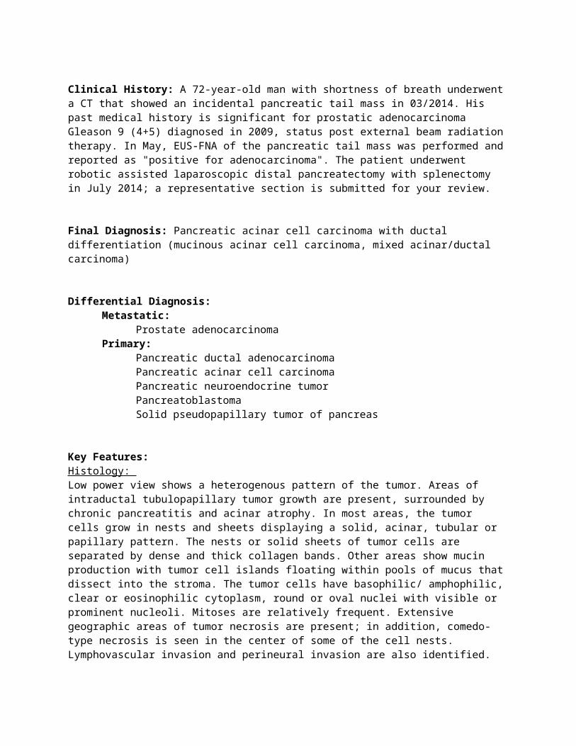
Clinical History: A 72-year-old man with shortness of breath underwent a CT that showed an incidental pancreatic tail mass in 03/2014. His past medical history is significant for prostatic adenocarcinoma Gleason 9 (4+5) diagnosed in 2009, status post external beam radiation therapy. In May, EUS-FNA of the pancreatic tail mass was performed and reported as "positive for adenocarcinoma". The patient underwent robotic assisted laparoscopic distal pancreatectomy with splenectomy in July 2014; a representative section is submitted for your review.
Final Diagnosis: Pancreatic acinar cell carcinoma with ductal differentiation (mucinous acinar cell carcinoma, mixed acinar/ductal carcinoma)
Differential Diagnosis:Metastatic:
Prostate adenocarcinomaPrimary:
Pancreatic ductal adenocarcinomaPancreatic acinar cell carcinomaPancreatic neuroendocrine tumorPancreatoblastomaSolid pseudopapillary tumor of pancreas
Key Features:Histology: Low power view shows a heterogenous pattern of the tumor. Areas of intraductal tubulopapillary tumor growth are present, surrounded by chronic pancreatitis and acinar atrophy. In most areas,
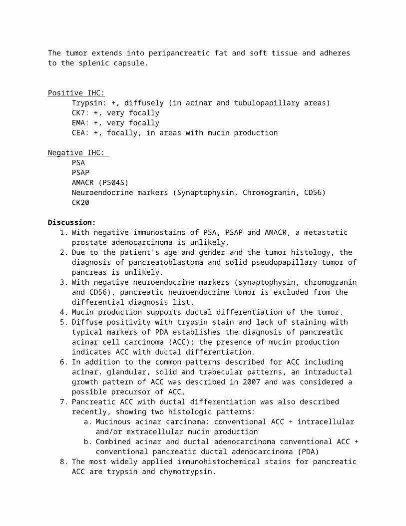
the tumor cells grow in nests and sheets displaying a solid, acinar, tubular or papillary pattern. The nests or solid sheets of tumor cells are separated by dense and thick collagen bands. Other areas show mucin production with tumor cell islands floating within pools of mucus that dissect into the stroma. The tumor cells have basophilic/ amphophilic, clear or eosinophilic cytoplasm, round or oval nuclei with visible or prominent nucleoli. Mitoses are relatively frequent. Extensive geographic areas of tumor necrosis are present; in addition, comedo-type necrosis is seen in the center of some of the cell nests. Lymphovascular invasion and perineural invasion are also identified. The tumor extends into peripancreatic fat and soft tissue and adheres to the splenic capsule. Positive IHC:
Trypsin: +, diffusely (in acinar and tubulopapillary areas)CK7: +, very focallyEMA: +, very focallyCEA: +, focally, in areas with mucin production
Negative IHC: PSAPSAPAMACR (P504S)Neuroendocrine markers (Synaptophysin, Chromogranin, CD56)CK20
Discussion:1. With negative immunostains of PSA, PSAP and AMACR, a metastatic prostate
adenocarcinoma is unlikely.2. Due to the patient's age and gender and the tumor histology, the diagnosis of
pancreatoblastoma and solid pseudopapillary tumor of pancreas is unlikely.3. With negative neuroendocrine markers (synaptophysin, chromogranin and CD56),
pancreatic neuroendocrine tumor is excluded from the differential diagnosis list.4. Mucin production supports ductal differentiation of the tumor.5. Diffuse positivity with trypsin stain and lack of staining with typical markers of PDA
establishes the diagnosis of pancreatic acinar cell carcinoma (ACC); the presence of mucin production indicates ACC with ductal differentiation.
6. In addition to the common patterns described for ACC including acinar, glandular, solid and trabecular patterns, an intraductal growth pattern of ACC was described in 2007 and was considered a possible precursor of ACC.
7. Pancreatic ACC with ductal differentiation was also described recently, showing two histologic patterns:
a. Mucinous acinar carcinoma: conventional ACC + intracellular and/or extracellular mucin production
b. Combined acinar and ductal adenocarcinoma conventional ACC + conventional pancreatic ductal adenocarcinoma (PDA)
8. The most widely applied immunohistochemical stains for pancreatic ACC are trypsin and chymotrypsin.
9. Pancreatic ductal adenocarcinoma can arise from ductal cells, with well known precursor lesions: PanIN, IPMN, MCN, or from acinar cells with less well described precursor lesions, referred to as atypical flat lesions, possibly through a process of acinar-ductal
metaplasia. The origin of ductal adenocarcinoma from acinar cells would potentially explain the ductal differentiation in acinar carcinomas.
10. Pancreatic tumors are defined based on their differentiation (ductal, endocrine or acinar), NOT based on their cell of origin.
11. For pancreatic ACC:
A distinctive pancreatic carcinoma with a somewhat better prognosis (especially at lower stages) than PDA
Combined acinar endocrine carcinomas are relatively common Combined acinar-ductal carcinomas are very rare, and can show two histological patterns: mucinous acinar carcinoma and combined acinar and ductal adenocarcinoma
The few cases of combined acinar-ductal carcinomas reported to date have had a very aggressive course and poor prognosis
Treated as typical ACCs (surgery +/- adjuvant therapies)
12. Take home messages:
Not all adenocarcinomas with an acinar growth pattern in a patient with history of prostate adenocarcinoma is a metastasis
Mucin production and intraductal growth pattern can both be occasionally seen in pancreatic acinar cell carcinoma
References:1. Basturk O, Klimstra DS. 2011. Acinar cell carcinoma of the pancreas and related
neoplasms: a review. Diagnostic Histopathology 18(1): 8-162. Basturk O, Zamboni G, Klimstra DS, Capelli P, Andea A, Kamel NS, Adsay NV. 2007.
Intraductal and papillary variants of acinar cell carcinoma: a new addition o the challenging differential diagnosis of intraductal neoplasms. Am J Surg Pathol 31(3): 363-370
3. Toll AD, Mitchell D, Yeo CJ, Hruban RH, Witkiewicz AK. 2011. Acinar cell carcinoma with prominent intraductal growth pattern: case report and review of the literature. Int J Surg Pathol 19(6): 795-799
4. Stelow EB, Shaco-Levy R, Bao F, Garcia J, Klimstra DS. 2010. Pancreatic acinar cell carcinomas with prominent ductal differentiation: mixed acinar ductal carcinoma and mixed acinar endocrine differentiation carcinoma. Am J Surg Pathol 34(4):510-580
5. Schmidt CM, Matos JM, Bentrem DJ, Talamonti MS, Lillemoe KD, Bilimoria KY. 2008. Acinar cell carcinoma of the pancreas in the United States: prognostic factors and comparison to ductal adenocarcinoma. J Gastrointest Surg 12(12): 2078-2086
6. Liu W, Shia J, Gönen M, Lowery MA, O'Reilly EM, Klimstra DS. 2014. DNA mismatch repair abnormalities in acinar cell carcinoma of the pancreas, frequency and clinical significance. Pancreas 43(8):1264-1270
7. Volmar KE1, Jones CK, Xie HB. 2004. Metastases in the pancreas from nonhematologic neoplasms: report of 20 cases evaluated by fine-needle aspiration. Diagn. Cytopathol 3(4)1:216 –220.
8. Chetty R & Serra S. 2009. Intraductal tubular adenoma (pyloric gland-type) of the pancreas: a reappraisal and possible relationship with gastric-type intraductal papillary mucinous neoplasm. Histopathology 55(3), 270–276.
9. Yamaguchi H, Shimizu M, Ban S, Koyama I, Hatori T, Fujita I, Yamamoto M, Kawamura S, Kobayashi M, Ishida K, Morikawa T, Motoi F, Unno M, Kanno A,Satoh K, Shimosegawa T, Orikasa H, Watanabe T, Nishimura K, Ebihara Y, Koike N, Furukawa T. 2009. Intraductal tubulopapillary neoplasms of the pancreas distinct from pancreatic intraepithelial neoplasia and intraductal papillary mucinous neoplasms. Am J Surg Pathol 33(8):1164-1172
10. Esposito I, Konukiewitz B, Schlitter AM, Klöppel G. 2014. Pathology of pancreatic ductal adenocarcinoma: Facts, challenges and future developments. World J Gastroenterol 20(38): 13833-13841
Case 3 Presenter: Monica Aldulescu DO, PGY-IAttendings: Stefan Pambuccian MD, Ewa Borys MD, Amit Kini MD
Clinical History: A 67y male with morbid obesity (BMI 70), atrial fibrillation, and type 2 diabetes was transferred to our institution for workup of anemia. The patient had a prior diagnosis of a monoclonal paraproteinemia, but had declined a bone marrow biopsy at that time. He was scheduled for a colonoscopy for workup of a possible iron deficiency anemia. His procedure was complicated by atrial fibrillation and hypotension, and thus postponed. However, before evaluation a repeat esophagogastroscopy/colonoscopy could be performed, the patient became bradycardic and unresponsive and expired. An autopsy was performed; a representative section of a grossly normal pituitary gland is submitted for your review.
Final Diagnosis: Intravascular Large B-cell Lymphoma
Differential Diagnosis:Pituitary lesion incidentally identified at autopsy: -Mass-forming lesions of the pituitary gland-Non-mass-forming lesions of the pituitary gland
Pituitary HyperplasiaLymphocytic Hypophysitis and other inflammatory conditionsLangerhans Cell HistiocytosisMetastasesLymphoma
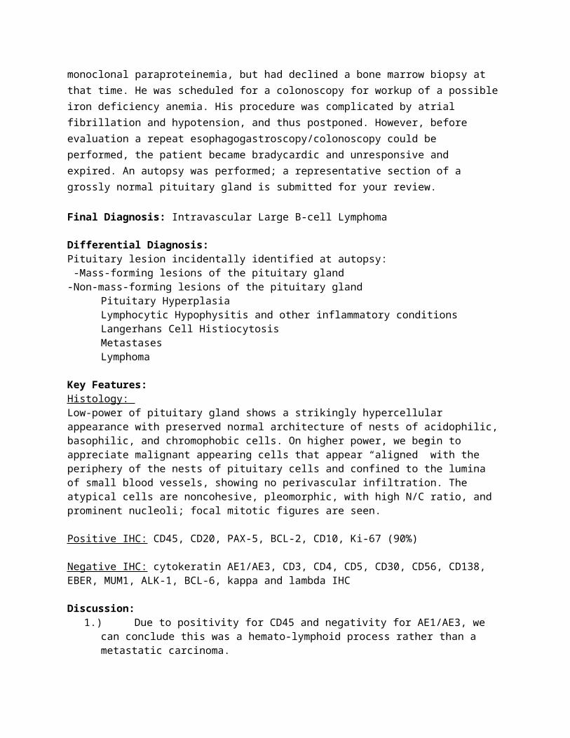
Key Features:Histology: Low-power of pituitary gland shows a strikingly hypercellular appearance with preserved normal architecture of nests of acidophilic, basophilic, and chromophobic cells. On higher power, we begin to appreciate malignant appearing cells that appear “aligned” with the periphery of the nests of pituitary cells and confined to the lumina of small blood vessels, showing no perivascular infiltration. The atypical cells are noncohesive, pleomorphic, with high N/C ratio, and prominent nucleoli; focal mitotic figures are seen.
Positive IHC: CD45, CD20, PAX-5, BCL-2, CD10, Ki-67 (90%)
Negative IHC: cytokeratin AE1/AE3, CD3, CD4, CD5, CD30, CD56, CD138, EBER, MUM1, ALK-1, BCL-6, kappa and lambda IHC
Discussion:1.) Due to positivity for CD45 and negativity for AE1/AE3, we can conclude this was a
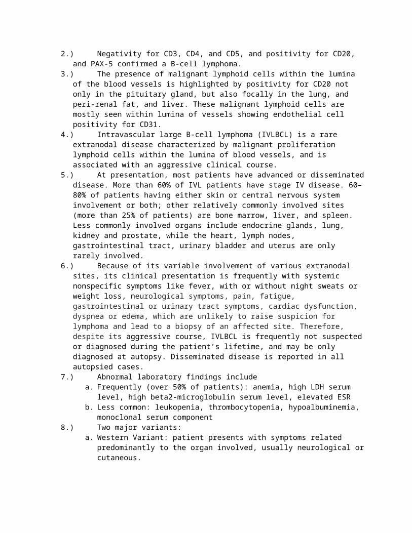
hemato-lymphoid process rather than a metastatic carcinoma.2.) Negativity for CD3, CD4, and CD5, and positivity for CD20, and PAX-5 confirmed a B-
cell lymphoma.3.) The presence of malignant lymphoid cells within the lumina of the blood vessels is
highlighted by positivity for CD20 not only in the pituitary gland, but also focally in the lung, and peri-renal fat, and liver. These malignant lymphoid cells are mostly seen within lumina of vessels showing endothelial cell positivity for CD31.
4.) Intravascular large B-cell lymphoma (IVLBCL) is a rare extranodal disease characterized by malignant proliferation lymphoid cells within the lumina of blood vessels, and is associated with an aggressive clinical course.
5.) At presentation, most patients have advanced or disseminated disease. More than 60% of IVL patients have stage IV disease. 60–80% of patients having either skin or central nervous system involvement or both; other relatively commonly involved sites (more than 25% of patients) are bone marrow, liver, and spleen. Less commonly involved organs include endocrine glands, lung, kidney and prostate, while the heart, lymph nodes, gastrointestinal tract, urinary bladder and uterus are only rarely involved.
6.) Because of its variable involvement of various extranodal sites, its clinical presentation is frequently with systemic nonspecific symptoms like fever, with or without night sweats or weight loss, neurological symptoms, pain, fatigue, gastrointestinal or urinary tract symptoms, cardiac dysfunction, dyspnea or edema, which are unlikely to raise suspicion for lymphoma and lead to a biopsy of an affected site. Therefore, despite its aggressive course, IVLBCL is frequently not suspected or diagnosed during the patient’s lifetime, and may be only diagnosed at autopsy. Disseminated disease is reported in all autopsied cases.
7.) Abnormal laboratory findings include a. Frequently (over 50% of patients): anemia, high LDH serum level, high beta2-
microglobulin serum level, elevated ESRb. Less common: leukopenia, thrombocytopenia, hypoalbuminemia, monoclonal
serum component8.) Two major variants:
a. Western Variant: patient presents with symptoms related predominantly to the organ involved, usually neurological or cutaneous.
b. Asian Variant (hemophagocytosis-associated): patient presents with multi-organ failure, hepatosplenomegaly, thrombocytopenia, and hemophagocytic syndrome.
c. A less common exclusively cutaneous variant, seen in women, has also been described, and is associated with a better prognosis.
A neoplasm of “homeless lymphocytes”. Although the mechanism is not well-defined, it has been proposed that the pathogenesis may be related to lack of homing receptors, adhesion molecules, namely CD29 (beta-1 integrin) and CD54 (intracellular adhesion molecule-1), which are needed for successful tumor invasion from the vasculature into the extravascular space.
References:1. Anila, K.R., et. al. Primary Intravascular large B-cell Lymphoma of Pituitary. Indian Journal of Pathology and Microbiology. (2012). 55(4): 549-551. 2. Asa, Sylvia L. Practical Pituitary Pathology, What Does the Pathologist Need to Know? Arch Pathol Lab Med. (2008) 132:1231-1240.
3. Carrasco, Carmen A., et. al. Primary Pituitary Pituitary Lymphoma in Immunocompetent Patient: Diagnostic Problems and Prolonged Follow-up. Pituitary (2012) 15:93-96. 4. Kurtulmus, Neslihan, et. al. The Pituitary Gland in Patients with Langerhans Cell Histiocytosis: A Clinical and Radiological Evaluation. Endocrine (2014) 5. Lury, Kenneth M. Inflammatory and Infectious Processes Involving the Pituitary Gland. Top Magn Reson Imaging (2005) 16:301-306. 6. Manabu, Kinoshita, et. al. Immunohistochemical analysis of adhesion molecules and matrix metalloproteinases in malignant CNS lymphomas: a study comparing primary CNS malignant and CNS intravascular lymphomas. Brain Tumor Pathology (2008) 25:73-78.7. Moshkin, Olga et. al. Primary Pituitary Lymphoma: A histological, Immunohistochemical, and Ultrastructural Study with Literature Review. Endocrinology Pathology (2009) 20:46-49. 8. Ogilvie, Megan C. et. al. Lymphoma Metastasizing to the Pituitary: An Unusual Presentation of a Treatable Disease. Pituitary (2005) 8:139-146. 9. Orwat, Dennis E, Batalis, Nicholas I: Intravascular Large B-Cell Lymphoma. Arch Pathol Lab Med. (2012) 136:333-338. 10. Pitfalls in the Diagnosis of Intravascular Large B-Cell Lymphoma. European Journal of Hematology. (2013) 91:563-564. 11. Ponzoni M, et. al. Lack of CD29 (beta 1 integrin) and CD54 (ICAM-1) adhesion molecules in intravascular lymphomatosis. Human Pathology (2000) 31:220-226.12. Ponzoni, M, Ferreri, JM. Intravascular lymphoma: a neoplasm of ‘homeless’ lymphocytes?Hematology Oncology (2006) 24: 105–112
11. Scangas, A. George, Laws R. Edward Jr. Pitu
Case 4Presenter: Christina Kwong MD, PhDAttendings: Xianzhong Ding MD, Stefan Pambuccian MD, Ameet Kini MD, PhD
Clinical History: The patient is a 48 year old male who presents with a 4 month history of malaise, fatigue, fevers, and abdominal pain. His past medical history is significant for cystic fibrosis status post bilateral lung transplants in 2003. His medications include prednisone and tacrolimus. Physical examination shows hepatosplenomegaly. Routine labs show elevated liver function enzymes (AST:129 IU/L, ALT: 85 IU/L), increased total bilirubin 5.3 mg/dL, elevated alkaline phosphatase 1588 IU/L, elevated lactate dehydrogenase 1204 IU/L, and elevated GGT 579 IU/L. Ferritin is markedly elevated at 35496 ng/ml. A liver biopsy was performed to elucidate the etiology of the liver function test abnormalities. The patient developed acute kidney and lung failure requiring hemodialysis and intubation respectively and expired 4 days after admission. An autopsy was performed and a representative section of his liver is submitted for your review.
Final Diagnosis 1. Liver biopsy: Liver sinusoids involved by hemophagocytic cells and extensive macro/micro
vesicular steatosis2. Liver (autopsy): Liver sinusoids involved by hemophagocytic cells and classic Hodgkin
lymphoma type post transplant lymphoproliferative disorder
Differential DiagnosisBiliary obstructionCholestatic hepatitisPrimary biliary cirrhosis
Primary sclerosing cholangitisAutoimmune hepatitisViral hepatitisNonalcoholic steatohepatitis (NASH)Drug-induced hepatitisSinusoidal obstruction: Erythrocytic vs Lymphocytic vs Hematopoietic vs Histiocytic
Key FeaturesAdditional tests:
1. Serology/DNA PCR: Positive for EBV DNA (650009 copies/ml); Negative for Hepatitis A, Hepatitis B, and Hepatitis C; Negative for CMV, BK virus, HIV, HSV 1/2, and Parvovirus.
2. Abdominal ultrasound: Hepatosplenomegaly. No gallstones or dilatation of the common bile duct.
Histology:1. Liver biopsy: Low magnification shows overall preserved liver architecture. There is mild
lobular disarray with slightly dilated sinusoids. Portal triads show minimal mononuclear infiltrates with rare eosinophils, and no increase in plasma cells. Central veins show no significant pathology. There is minimal focal necrosis; rare acidophilic bodies are present. Overall the liver biopsy is slightly hypercellular. Trichrome stain does not highlight increased fibrosis. Reticulin stain highlights the lobular disarray and slightly hypercellular sinusoids. Additional special stains: GMS, PAS, copper and iron are non-contributory. Immunostains for viral antigens (Adenovirus, Hepatitis B, CMV, HSV and EBV-LMP) are negative. The cells within the sinusoids contributing to the hypercellular appearance have abundant cytoplasm with what appears to be red blood cells within the cytoplasmic space. These cells are CD68 positive. There are also rare large atypical binucleate cells with prominent nuclei within the sinusoids. These cells have the following immunostain profile:Positive IHC: CD30Negative IHC: CD3, CD4, CD5, CD8, CD20, CD138, ALK, EBV-LMPAdditional stains could not be performed because the core biopsy tissue was exhausted.
2. Liver (autopsy): There is extensive centrilobular zone 3 necrosis and apoptosis. Slightly hypercellular sinusoids are identified similar to that on the liver core biopsy. The cells within the sinusoids are of similar morphology with cytoplasmic erythrocytes and are CD68 positive. The atypical binucleate cells are also seen with identical immunostaining profile as the cells seen on liver core biopsy, and are additionally CD15 positive. In addition EBER ISH is positive.
Discussion1. Abdominal ultrasound did not identify a biliary obstruction therefore this can be ruled
out. 2. The labs show a cholestatic elevated liver enzyme pattern which can be found in
cholestatic hepatitis, primary biliary cirrhosis, and primary sclerosing cholangitis however these diagnoses are unlikely due to the lack of intrahepatic cholestasis and morphologic features found in each entity.
3. Autoimmune hepatitis is eliminated due to the lack of periportal plasma cells and appropriate clinical and serologic context.
4. The only viral hepatitis that is considered on the differential is EBV hepatitis due to the high EBV titers. However the minimal mononuclear infiltrate does not display the
atypical plasmacytoid morphology seen in EBV hepatitis making this differential less likely.
5. The extensive steatosis seen is most likely due to the patient's long history of corticosteroid use. Combined with the minimal inflammatory infiltrate, the patient may have mild steatohepatitis however this diagnosis does not explain the markedly elevated liver enzymes.
6. Drug-induced hepatitis is also unlikely as the patient has not had a change in his medication regimen.
7. The hypercellularity of the sinusoids is evaluated and based on morphology and the patient's medical history, erythrocytic, lymphocytic and hematopoietic infiltrate is ruled out leaving histiocytes as a possibility. Immunostain for CD68 confirms the presence of abundant histiocytes within the sinusoidal space. Based on the presence of erythrocytes within the histiocyte cytoplasm, the type of histiocyte (not pigmented, not ceroid-laden, not foamy) is determined to be a hemophagocytic histiocyte.
8. Hemophagocytic lymphohistiocytosis (HLH)a. Primary HLH is due to genetic mutations. Secondary HLH (more common) is due
to infection, autoimmunity, drugs, immunosuppression, or malignancy.b. Criteria set by the Histiocyte Society: ≥ 5 out of 8 criteria (Low/absent NK cell
function, Prolonged fever, Blood cell abnormalities, Splenomegaly, Increased Triglycerides/Decreased Fibrinogen, Increased Ferritin, Increased CD25 (sIL2Ra), Hemophagocytic cells in the bone marrow). Our case fulfilled 7/8 criteria (in Bold).
c. Involvement of the liver by HLH manifests clinically as elevated liver enzymes. This can be due to the underlying cause of the HLH affecting the liver or due to obstruction of the sinusoids by hemophagocytic histiocytes.
d. The most common histologic features of secondary HLH involving the liver include preserved liver architecture, Kupffer cell hyperplasia, hemophagocytic cells with erythrophagocytosis, sinusoidal dilatation, and portal lymphocytic infiltrates.
e. The most common underlying cause of the HLH with liver involvement is a lymphoproliferative disorder (lymphoma, CLL, Hodgkin's).
9. The atypical binucleate cells were morphologically identified as Hodgkin’s lymphoma Reed-Sternberg cells. The immunostains listed above confirm their identity. Taking the patient's transplant history and elevated EBV viremia, a diagnosis of classic Hodgkin’s lymphoma type post transplant lymphoproliferative disorder is made.
10. Take home pointsa. Hemophagocytosis is difficult to identify in liver biopsies.b. If the clinical presentation does not match the minimal findings on the liver
biopsy, a closer examination is in order.c. Negative EBV-LMP IHC does not preclude EBV involvement.
References
1. Adams DH, Eksteen B. Aberrant homing of mucosal T cells and extra-intestinal manifestations of inflammatory bowel disease. Nat Rev Immunol. 2006;6: 244-251.2. Brunt EM, Gouw AS, Hubscher SG, et al. Pathology of the liver sinusoids. Histopathology. 2014;64: 907-920.3. Chen JH, Fleming MD, Pinkus GS, et al. Pathology of the liver in familial hemophagocytic lymphohistiocytosis. Am J Surg Pathol. 2010;34: 852-867.
4. de Kerguenec C, Hillaire S, Molinie V, et al. Hepatic manifestations of hemophagocytic syndrome: a study of 30 cases. Am J Gastroenterol. 2001;96: 852-857.5. Fischer HP, Flucke U, Zhou H. [Pathology along the liver sinusoids: endothelial and perisinusoidal findings]. Pathologe. 2008;29: 37-46.6. Prendki V, Stirnemann J, Lemoine M, et al. Prevalence and clinical significance of Kupffer cell hyperplasia with hemophagocytosis in liver biopsies. Am J Surg Pathol. 2011;35: 337-345.7. Rubbia-Brandt L. Sinusoidal obstruction syndrome. Clin Liver Dis. 2010;14: 651-668.8. Terada T, Maeta H, Endo K, Horie S, Matsunaga Y, Ohta T. Marked histiocytosis in the portal tract in a patient with reactive hemophagocytic syndrome: An autopsy case. Pathol Int. 1999;49: 672-6759. Approach to the patient with abnormal liver biochemical and function tests. http://www.uptodate.com/contents/approach-to-the-patient-with-abnormal-liver-biochemical-and-function-tests?source=search_result&search=nash&selectedTitle=8%7E64#H19405903. Last accessed 11/1/2014.10. Cholestatic hepatitis. http://livertox.nih.gov/Phenotypes_chol.html. Last accessed 11/4/2014. 11. Interpretation of liver biopsy specimens. http://www.uptodate.com/contents/interpretation-of-liver-biopsy-specimens?source=machineLearning&search=nash&selectedTitle=6%7E64§ionRank=1&anchor=H4622205#H1763618223. Last accessed 11/1/2014.12. Transplant pathology Internet Services. http://tpis.upmc.com/changebody.cfm?url=/tpis/liver/LEBV.jsp. Last accessed 11/3/2014.13. Necrotic, inflammatory and vascular lesions of the liver. http://www.niehs.nih.gov/research/resources/liverpath/necrotic/. Last accessed 11/4/2014.
Case 5:Presenter: John Biemer MDAttending: Dariusz Borys, MD
Clinical History:
A 28-year-old male immigrant from Serbia with no significant past medical history presented with an indurated, swollen, growing mass on his left medial thigh for about one year. The mass was painless and did not affect his knee or leg movement. The patient did not report recent trauma to his left thigh but stated that he did sustain an injury to the left medial thigh as a child more than 10 years ago.
Physical exam demonstrates a well-appearing male with a large 20-cm firm, non-tender, nom-moveable mass of his left anterior thigh. The patient was referred to medical oncology for possible neoadjuvant therapy. A biopsy was obtained and an MRI performed
Final Diagnosis: Cystic Echinococcus (Hydatid Cyst)Differential Diagnosis:
Sarcoma (poor sampling, outside capsule)/Granulomatous response to tumorLow-grade fibromyxoid sarcomaSynovial sarcomaMalignant fibrous histiocytoma (MFH)
Non-infective granulomatous response:
Granuloma annulare, rheumatic nodule, necrobiosis lipoidica Foreign body reaction; Injection reaction (steroids)
Infective granulomatous inflammation: TB/mycobacteria Cat-scratch disease: Bartonella henselaeFungal: Candida, Cryptococcus, Aspergillus, Histoplasma, Sporotrichosis, etc.Parasitic: Cysticercosis, Trichinosis
Bacterial myositis:S. aureus, Group A and Group B Strep, Clostridial gas gangrene
Key Features:Radiology:Large intramuscular cystic mass within the quadriceps muscle demonstrates multiple intralesional cystic lesions. Variation in signal within the internal cystic lesions may represent differences in protein content. This architecture would be highly unusual for a soft tissue sarcoma.
Histology:Biopsy showed a cystic lesion with fragments of fibromuscular tissue with granulation tissue, lymphohistiocytic infiltrates and detached lamellar material, suggestive of cystic echinococcus. Excision of the mass showed cystic echinococcosis with extensive necrosis and inflammatory response, chronic myositis with foreign body giant cells and increased eosinophils. Hooklets were identified in protoscolices.
Immunohistochemical stains: Positive
PAS GMS
Discussion:
1. Hydatid Cyst has been recognized since ancient times; Hippocrates was the first to illustrate a liver hydatid cyst and propose techniques of treatment.
2. Adult E. granulosis tapeworms are found in nature in the intestines of canines (dogs, foxes, wolves, coyotes, jackals, dingoes). The larval cyst stage is present in the viscera of herbivores (sheep, cattle, pigs, deer). Humans are a dead-end intermediate host.
3. When ingested by humans, a six-hooked larval stage called an oncosphere hatches in the duodenum. The oncosphere penetrates the intestinal wall and enters the circulation. Blood carries it to various tissue sites.
4. The disease can involve any part of the body except the hair, teeth and nails, but the vast majority is found in the liver and lungs (more than 80-90% in liver or lungs or both).
5. The unilocular cyst grows slowly, so five to 20 years may pass until symptoms appear. Pressure of the expanding cysts on adjacent organs or pain is usually the first signs of infection. The cyst can act as a locally invasive tumor -- cause brain damage, bone destruction.
6. Diagnosis is difficult and depends on clinical, radiological, serologic and pathologic findings. Aspiration may demonstrate presence of protoscolices and hooklets.
7. On histology, a hydatid cyst is bilayered and surrounded by fibroblasts, mononuclear cells, eosinophils, multinucleated giant cells. It consists of an opaque, elastic, acellular laminated layer of varying thickness and an inner nucleated germinal layer of epithelium. Hooklets also may be seen on histology.
References:- Algorithms in the Diagnosis and Management of Exotic Diseases. X. Echinococcosis
Journal of Infectious Diseases. (1976) 133 (3): 354-358 - Aggressive hydatid cysts: characteristics of six cases, Surgery Today, Official Journal of
the Japan Surgical Society Gürhan Oz , Mehmet Eroglu, Ersin Gunay, Ahmet Bal, Emre Kacar, Olcay Eser and Okan Solak Department of Thoracic Surgery, Afyon Kocatepe University School of Medicine, 03200 Afyonkarahisar, Turkey, 10.1007/s00595-014-1019-9
- Atypical localizations of hydatid disease: experience from a single institute. Nigerian Journal of Surgery Mushtaque M, Mir MF, Malik AA, Arif SH, Khanday SA, Dar RA. 2012 Jan;18(1):2-7. doi: 10.4103/1117-6806.95466.
- Uncommon locations and presentations of hydatid cyst. Annals of Medical and Health Sciences Research, Sachar S, Goyal S, Goyal S, Sangway S. 2014 May;4(3):447-52. doi: 10.4103/2141-9248.133476.
- University of South Carolina School of Medicine, Department of Microbiology and Immunology: http://pathmicro.med.sc.edu/parasitology/cestodes.htm
- Medical Microbiology, P. Murray, K. Rosenthal & M. Pfaller, Sixth Edition, Mosby Elsevier, 2009
- Atlas of Human Parasitology, 4th Edition, Lawrence Ash and Thomas Oribel, American Society of Clinical Pathologists, 1997
Case 6Derek Bumgarner MD, Dariusz Borys MD
Clinical History: A 49 year-old male, with no significant past medical history, presented with a mass on his left pinna. The mass was present for the last 30 years, but increased in size over the last year. Physical exam revealed a firm 2.0 cm mass which was painless to palpation. The patient denied any associated symptoms such as hearing loss, tinnitus, or otorrhea. Representative sections from the resection specimen are submitted for your review.
Final Diagnosis: Solitary Fibrous Tumor, Giant Cell Angiofibroma Variant
Differential Diagnosis:
Tenosynovial giant cell tumorSpindle cell/pleomorphic lipomaSynovial sarcomaAngiofibroma of soft tissueGiant cell fibroblastomaSolitary fibrous tumor
Giant cell angiofibroma
Key Features:
Histology: Histologically our case demonstrates a well-circumscribed, subdermal lesion with a "patternless" proliferation of ovoid and spindle shaped cells. There are alternating hypercellular and hypocellular areas. A background of dense, wiry collagen is present. There are interspersed, floret-like giant cells within the lesion. The vasculature displays a staghorn architecture and perivascular hyalinization.
Positive IHC:CD34Vimentin BCL-2STAT-6
Negative IHC: S100CD31SMA
Discussion:1. Our lesion is subdermal and well-circumscribed. There is a background of bland, fibroblastic spindled cells with a biphasic pattern of hyper/hypocellularity. The collagen is dense and wiry and there are interspersed floret-like giant cells. The vasculature demonstrates staghorn architecture with perivascular hyalinization. The tumor cells stain for CD34, vimentin, BCL-2, and STAT6, but are negative for S100, CD31, and SMA.
2. Giant cell tenosynovial tumor (GCTT) can be excluded from the differential as it presents with plump, epithelioid spindle cells, osteoclast like giant cells, and pigmented macrophages. The immunohistochemical profile also does not match as GCTT does not stain for CD34 and bcl-2.
3. Spindle cell/pleomorphic lipoma demonstrates similarity to our lesion in that it presents with floret-like giant cells, but differs in that it does not have bimorphic areas of hyper/hypocellularity, the background collagen is more ropey, and it contains fat while our lesion does not. The immunohistochemical profile also does not match as spindle cell lipoma stains positive for CD34 but negative for BCL-2.
4. Synovial sarcoma (monophasic) may have a “hemangiopericytoid” appearance, with staghorn shaped vessels, but demonstrates intensely hyperchromatic, hypercellular spindle cells and lacks giant cells or alternating areas of hyper- and hypo-cellularity. It is CD34 negative and BCL-2 positive.
5. Angiofibroma of soft tissue is a newly described lesion (2012). It presents with bland spindle cells and consistent cellularity. It also demonstrates thin walled, staghorn like vasculature. Its immunohistochemical profile does not match as it stains negative for both CD34 and BCL-2.
6. Giant cell fibroblastoma presents with floret-like giant cells and a similar (CD34+/bcl2) immunohistochemical profile, but presents as an infiltrative lesion which invades the subcutis. It also has a less dense collagen background and lacks biphasic alternating cellularity.
7. Solitary fibrous tumor presents with bland spindle cells with alternating areas of hyper/hypocellularity. It demonstrates staghorn vasculature with perivascular hyalinization. These histologic features are similar to those of giant cell angiofibroma with the exception of floret-like giant cells, which are characteristic for giant cell angiofibroma. Both lesions stain positive for CD34 and BCL-2.
8. STAT6 immunohistochemistry with a nuclear staining pattern is positive in nearly 100% of solitary fibrous tumors as well as in the few giant cell angiofibromas that were stained. Given the similarity in clinical presentation, morphology, and immunohistochemistry, giant cell angiofibroma likely represents a variant of solitary fibrous tumor, rather than a distinct entity.
References
1. Cheah AL, Billings SD, Goldblum JR, Carver P, Tanas MZ, Rubin BP. STAT6 rabbit monoclonal antibody is a robust diagnostic tool for the distinction of solitary fibrous tumour from its mimics. Pathology. 2014;46: 389-395.
2. Folpe A, Inwards C. Bone and Soft Tissue Pathology. Saunders-Elsevier, 2010.
3. Gengler C, Guillou L. Solitary fibrous tumour and haemangiopericytoma: evolution of a concept. Histopathology. 2006;48: 63-74.
4. Goldblum JR, Folpe AL, Weiss SW, Enzinger FM, Weiss SW. Enzinger and Weiss's soft tissue tumors. 6th ed. Philadelphia, PA: Saunders/Elsevier, 2014.
5. Guillou L, Gebhard S, Coindre JM. Orbital and extraorbital giant cell angiofibroma: a giant cell-rich variant of solitary fibrous tumor? Clinicopathologic and immunohistochemical analysis of a series in favor of a unifying concept. Am J Surg Pathol. 2000;24: 971-979.
6. Hanau CA, Miettinen M. Solitary fibrous tumor: histological and immunohistochemical spectrum of benign and malignant variants presenting at different sites. Hum Pathol. 1995;26: 440-449.
7. Marino-Enriquez A, Fletcher CD. Angiofibroma of soft tissue: clinicopathologic characterization of a distinctive benign fibrovascular neoplasm in a series of 37 cases. Am J Surg Pathol. 2012;36: 500-508.
8. Salih K, Singhi A, Montgomery E. Selected lesions featuring giant cells. Surgical Pathology. 2011;4: 887-913.
9. Thomas R, Banerjee SS, Eyden BP, et al. A study of four cases of extra-orbital giant cell angiofibroma with documentation of some unusual features. Histopathology. 2001;39: 390-396.
10. Yoshida A, Tsuta K, Ohno M, et al. STAT6 immunohistochemistry is helpful in the diagnosis of solitary fibrous tumors. Am J Surg Pathol. 2014;38: 552-559.