Embed Size (px)
Citation preview
WGTRBceiefpaewdppPpwataspiiT
WESYSEbcscesuev�(p(wisomhtsspsapifop
WRGSMTIfwpo
Abstracts
w
1560astric Adenocarcinoma and Helicobacter Pylori Infection inhailandatha-Korn Vilaichone, Thawee Ratanachu-Ek, Varocha Mahachaiackground and aims: Gastric adenocarcinoma is the second leading cause ofancer death worldwide. This study was designed to evaluate the clinical,ndoscopic findings, pathological features and molecular study of H. pylorinfection in Thai gastric adenocarcinoma patients.Methods: Clinical information,ndoscopic findings, histological features and H. pylori status were collectedrom all gastric adenocarcinoma patients during June 2000-October 2009. H.ylori isolates were genotyped by PCR based on cagA, cag right end junctionnd vacA genotypes.Results: Total of 116 gastric adenocarcinoma patients werenrolled in this study. Common presenting symptoms were dyspepsia (70%),eight loss (64%) and anorexia (44%). Mean duration of symptoms was 107ays. All gastric adenocarcinoma patients were in advanced stage. Overallrevalence of H. pylori infection was 83.6%. There was no difference of H.ylori infection in diffuse type and intestinal type gastric cancer (80% vs. 84%;� 0.05). East Asian genotype (cagA 1a, vacA s1c and/or vacA m1b) was highlyrevalent in Thai gastric adenocarcinoma patients (65%). East Asian genotypeas more common in gastric adenocarcinoma age � 50 years than those patientsge � 50 (74% vs 43%: P�0.01) and more common in patients from city thanhose from rural area (79% vs 50%; P�0.05).Conclusion: Patients with gastricdenocarcinoma in Thailand usually delay diagnosis and present at advancedtage with 5-year survival less than 15%. H. pylori infection was highlyrevalence in Thai gastric adenocarcinoma patients and associated with both
ntestinal type and diffuse type. East Asian genotype may be regarded anmportant factor for pathogenesis and prediction gastric adenocarcinoma inhailand.
1561xpansion of Curative Condition After Endoscopic Resection forubmucosal Invasive Gastric Canceroji Sanomura, Shiro Oka, Shinji Tanaka, Makoto Higashiyama,higeto Yoshida, Koji Arihiro, Kazuaki Chayama, Fumio Shimamotondoscopic resection (ER) is a therapeutic but simulataneously a total excisionaliopsy technique. The AIM of this study is to evaluate the significance of severallinicopathological features in prediction of lymph node (LN) metastasis ofubmucosal gastric cancer and to examine the feasibility of expansion curativeondition after ER for submucosal gastric cancer. PATIENTS and METHODS: Wexamined 220 submucosal gastric cancers that were resected endoscopically orurgically at Hiroshima University Hospital or affiliated hospitals. Patients with ERnderwent additional surgical resection and the presence of LN metastasis wasvaluated. We evaluated sex (male vs. female), age, location (upper vs. middles. lower), gross type (depressed vs. non-depressed), tumor size (30mm vs.30mm), ulceration (positive vs. negative), dominant histlogical type
differentiated vs. undifferentiated), histological type at the deepest invasiveortion (differentiated vs. undifferentiated), size of undifferentiated type20mm vs. �20mm), depth of submucosal invasion (1000�m vs. �1000�m),idth of submucosal invasion (6000�m vs. �6000�m), INF (� vs. �/�), vessel
nvolvement (positive vs. negative) using resected specimen, which were notatisfied with curative condition for ER alone in Japanese guideline for treatmentf gastric cancer. Then relation between cliniccopathological findings and LNetastasis was evaluated. Pathological examination was performed byematoxylin-eosin staining. Data were evaluated by Chi-square or Fisher’s exactest, and P values of less than 0.05 were considered significant. RESULTS: Tumorize�30mm, undifferentiated type at the deepest invasive portion, depth ofubmucosal invasion�1000�m, width of submucosal invasion�6000�m, INF�/�,ositive lymphatic involvement, and positive vessel involvement showed aignificantly higher incidence of LN metastasis by univariate analysis. Multivariatenalysis showed tumor size�30mm, undifferentiated type at the deepest invasiveortion, depth of submucosal invasion�1000�m, width of submucosalnvasion�6000�m, and positive lymphatic involvement are independent riskactors for LN metastasis. All lesions that were negative for all or positive for onlyne feature were showing no LN metastasis.CONCLUSION: Our results can allowrediction of exact LN metastasis and can expand curative condition after ER.
1562etrospective Study of Complication Associated Withastrostomy Tube Replacement and Their Effective Treatmenthinji Nishiwaki, Hiroshi Araki, Jun Takada, Takahiko Asano,asahide Iwashita, Atsushi Tagami, Hiroo Hatakeyama,akao Hayashi, Teruo Maeda, Koshiro Saito
ntroduction: A gastrostomy tube replacement is required during prolonged tubeeeding or on accidental tube dysfunction. Although complications associatedith tube replacement are rare, they may cause life-threatening events, such as
eritonitis. We investigated complications after gastrostomy tube replacement ofver 9 years.Methods: We retrospectively studied 992 consecutive cases ofww.giejournal.org Volu
gastrostomy tube replacement carried out from Mar. 2000 to Sept. 2009 at ourinstitution. PEG was mainly conducted employing the pull-through method after2 point-fixation of the gastric wall against the abdominal wall using a double-lumen gastropexy device. Tube replacement was conducted periodically every 6months after PEG, or on accidental tube dysfunction or dislodgement. A newgastrostomy tube was inserted from the skin incision using an obturator aftertraction removal of the previous one from the skin. Replacement of thegastrostomy tube was confirmed by fistulography with Gastrografin.Results: The992 tube-replacement cases comprised 281 patients (94 males and 187 females,mean age: 80.7 � 8.6 years old); among them, 11 cases had complications,including hemorrhage in 4 and fistula disruption in 7, Hemorrhage occurred 6 to24 hours after tube replacement, originating from vascular injury or mucosal teararound the stoma. The 4 hemorrhage cases underwent anti-coagulation therapyby the administration of warfarin or aspirin, in which hemostasis was achievedusing an endoscopic hemoclip in 2 cases and by compression between the outerand inner bolster in the other 2 cases. The 7 fistula disruption cases wereidentified by extravasation on fistulograms; among them, gastrocutaneous tractsof 3 cases were secured by inserting a guide wire from the skin incision afterintraperitoneal malplaced tube removal. The remaining 4 cases were probedregarding the stomach route by ultrathin endoscope insertion (GIF XP-260 orXP-260N, Olympus Optical Co., Ltd. Tokyo, Japan) from the skin incision. All 7fistula disruption cases successfully received a new gastrostomy tube using thepull-thorough technique. There were no lethal events associated with tubereplacement complications.Conclusion: Hemorrhage and fistula disruption werethe most common complications associated with tube replacement. In casesundergoing anti-coagulation therapy, caution in necessary to avoid hemorrhageafter replacement. When fistula disruption occurs, ultrathin endoscope insertionfrom the skin incision is considered effective for recanalization of thegastrocutaneous tract.
W1563Therapeutic Outcomes of Early Gastric Cancer in ExpandedCriteria - Single Center ExperienceJoo Young Cho, Won Young Cho, Taehee Lee, Hyun Gun Kim,Jin-Oh Kim, Joon Seong Lee, So Young JinBackgrounds and ObjectivesIntroduction of endoscopic submucosal dissectionextends endoscopic therapeutic criteria of early gastric cancer. The aim of thisstudy was to investigate therapeutic outcomes of EGC included in expandedindications.Patients and MethodsFrom March 2003 to October 2009, 228 cases of222 patients who were include in expanded criteria of early gastric cancer wereevaluated of 490 cases of EGC underwent endoscopic submucosaldissection.ResultsMale was 164 and female was 58. Average age was 62(33-86).Complete resection and incomplete resection rate were 78.5%(179 cases) and19.7%(45 cases). 4 cases(1.8%) were undetermined due to autolysis orcoagulation effects. In incomplete group, lateral margin positive was 35cases(77.8%), vertical margin positive was 3 cases(6.7%), lymphatic invasion was4 cases(8.9%), and venous invasion was 4 cases(8.9%). In 219 cases ofdifferentiated cancer(94.7%), complete resection was 79%(173 cases) and 6 out of9 undifferentiated cancer was completely resected. According to tumor size morethan 3cm(58 cases, 25.4%), complete resection was 37 cases(63.8%), incompleteresection was 19 cases(32.8%). Lateral margin positive was 18 case(94.7%) andno vertical margin positive was noted. Average tumor size was bigger inincomplete resection group(3.2cm(incomplete) vs2.4cm(complete)(p�0.01)ConclusionsOur results of ESD in expanded indicationof gastric resection for early gastric cancer were not related in size, location anddifferentiated types, but the depth of invasion was related with completeresection.
W1564Concordance for the Adequate Direction of Lateral MarginBetween Gross, Pathologic, and Stereoscopic Finding ofSpecimen After Endoscopic Submucosal Dissection in GastricEpithelial NeoplasmWon Young Cho, Joo Young Cho, Taehee Lee, Hyun Gun Kim,Jin-Oh Kim, Joon Seong Lee, So Young JinBackground and Aim: For evaluation of correct lateral margin, the sectiondirection of specimen is important. The section direction should be a right angleof the closest area between the lesion and the lateral margin. We aimed toevaluate the correspondence of the gross finding and stereoscopic findingcompared to the patholgic finding in the setting of the correct lateral margindirectionMethod: The 48 specimen after ESD from December 2008 to June 2009was enrolled retrospectively. We analyzed the specimen using the stereoscopyand the image analysis program. To evaluate the direction of the closest lateralmargin, we compared the gross finding, the stereoscopic finding, the pathologicfinding deviding eight direction per 45 angle based on oral side.Result: The
correspondence rate of the gross finding compared with the pathlogic finding inthe closest lateral margin direction is 68% (33/48) and Kappa value is 0.626. Theme 71, No. 5 : 2010 GASTROINTESTINAL ENDOSCOPY AB359
cifin
WHBTNDBadBpiaakTwctMesewhptdccs1pPgp9bmelk
WTITMBiarrpabbiaMampwtqtFt(ub
Abstracts
A
orrespondence rate of the stereoscopic finding is 83% (42/48) and Kappa values 0.773.Conclusion: In the setting of correct lateral margin direction, Grossnding was inferior to the stereoscopic finding. The endoscopist should beeeded the cautious inspection in the setting of lateral margin direction.
1565ow Can Endoscopic Submucosal Dissection for Gastric Tumorse Improved Better Using the Dual Knife?oshifumi Mitani, Shu Hoteya, Toshiro Iizuka, Yuichiro Kuroki,oriko Nishida, Akira Matsui, Ai Fujimoto, Masanori Nakamura,aisuke Kikuchi, Satoshi Yamashita, Naohisa YahagiACKGROUND: Endoscopic submucosal dissection (ESD) has been establisheds a standard endoscopic method for the gastric tumors in Japan. Large andifficult lesions and lesions with ulcer scar can be resected in an en bloc fashion.ut all disadvantages are not yet improved, that is a complexity of therocedure, risk of complication, long procedure time and high cost. Many
nstruments and techniques have been developed to improve those problemsnd the dual knife is one of new devices.AIM: To evaluate the potential benefitsnd efficacies of gastric ESD using the dual knife compared to othernives.METHODS: Among consecutive 384 gastric ESD procedures conducted inoranomon Hospital from June 2008 to September 2009, each 192 proceduresere located in both group D using the dual knife and group O using the otheronventional knife. Two groups by devices were retrospectively compared withheir clinical background, tumor characteristics and clinicopathological results.ore, the area of the resected specimen was considered as the approximativellipse and the average of submucosal dissection speed was calculated as indexcore to evaluate the performance speed (PS) of ESD. The same trials werexamined in each groups by operators, three operators belonged to expert grouphich had performed more than 200 cases, two belonged to intermediate whichad performed 50 to 200 cases and nine belonged to beginner which haderformed less than 50 cases. Statistical analysis was performed by chi-squaredests and student’s t-tests comparing two groups (statistical significance wasefined as p�0.05).RESULTS: There was no significant difference for theirlinical background, tumor characteristics and rate of en bloc resection andomplete resection in two groups. But the procedure time was significantlyhorter and PS was higher in group D compared to group O (66.5: 96.9min, 31.1:8.7mm2/min, p�0.01, respectively). Moreover, the rate of requirement forlural knives in one procedure was significantly lower in group D (p�0.01).ostoperative hospitalization period and complication rate were similar in tworoups.In spite of experience of ESD, the same results were obtained inrocedure time, PS and rate of requirement for plural knives (expert; 67.6:4.0min, 42.8: 21.8 mm2/min, intermediate; 69.0: 91.3min, 25.9: 18.4 mm2/min,eginner; 64.3: 107.6min, 22.3: 14.7 mm2/min). In particular, expert group wasore improved than other two groups.CONCLUSIONS: The dual knife improvedndoscopic submucosal dissection for gastric tumors faster and smoother in eachevel of operator. Also it could save the cost by reducing the number of usingnife and operation time.
1566he Investigation of Upper Gastrointestinal Bleeding Aftermplantation of Drug-Eluting Stents; Prospective Cohort Studyatsuya Toyokawa, Tomoki Inaba, Shigenao Ishikawa,orihito Nakatsu, Masaharu Ando, Jun Tomodaackground: Recently, the use of drug-eluting stents (DES) for treatment of
schemic coronary disease has increased; following implantation, two or morenti-thrombotic agents such as low dose aspirin and ticlopidine are needed. Theisk of upper GI bleeding due to low dose aspirin has been identified;ecommendations regarding its use vary between countries. In Japan, therevalence of Helicobacter pylori (H. pylori) infection, which can lead totrophic chronic gastritis and low intra-gastric acidity, is very high; upper GIleeding following DES implantation is thought to have a more serious impact,ut incidence and suitable methods for its prevention have not yet beennvestigated. The aims of this study are to clarify incidence of upper GI bleedingfter DES implantation and awareness among cardiologists about the condition.ethods: Subjects were 397 consecutive patients with DES implantations (mediange 69, range 36-94 years, 296 male/101 female, mean observation period 22.4onths). After implantation, endoscopic examinations were performed foratients with hematemesis and/or tarry stools. The concomitant use of antacidsith anti-thrombotic agents for prevention of gastro-duodenal injury was left to
he cardiologists’ discretion. In addition, 37 cardiologists answered auestionnaire about post-implantation upper GI bleeding. Results: All patientsook low dose aspirin and ticlopidine; 36 patients (9.1%) also received warfarin.orty-six patients (12%) had past history of peptic ulcers. Antacids/anti-horombotic agents were concomitantly administered to 224 patients [56%; PPI 6730%), H2RA 157 (70%)], especially to the 32 patients with past history of peptic
lcer [70%; PPI 16 (50%), H2RA 16 (50%)]. Under these conditions, upper GIleeding developed in 5 cases (1.3%), all cases were gastric ulcer. One case hadB360 GASTROINTESTINAL ENDOSCOPY Volume 71, No. 5 : 2010
past history of duodenal ulcer. One of the five had been administered half doseH2RA and all 5 patients were H. pylori-positive. In the patients with concomitantuse of PPI, upper GI bleeding did not occur. The incidence of upper GIbleeding was 4.0 per 1,000 patient-years. The survey of cardiologists indicatedrecognition of the risk of upper GI bleeding after DES implantation and thenecessity of the prevention, but the adequacy of its management in Japan,particularly H. pylori eradication therapy, has not been established. Conclusions:This study determined the frequency of upper GI bleeding after DESimplantation was 4.0 per 1,000 patient-years. Further study by gastroenterologistsregarding the prophylaxis of upper GI bleeding after DES implantation in theJapanese populations is necessary.
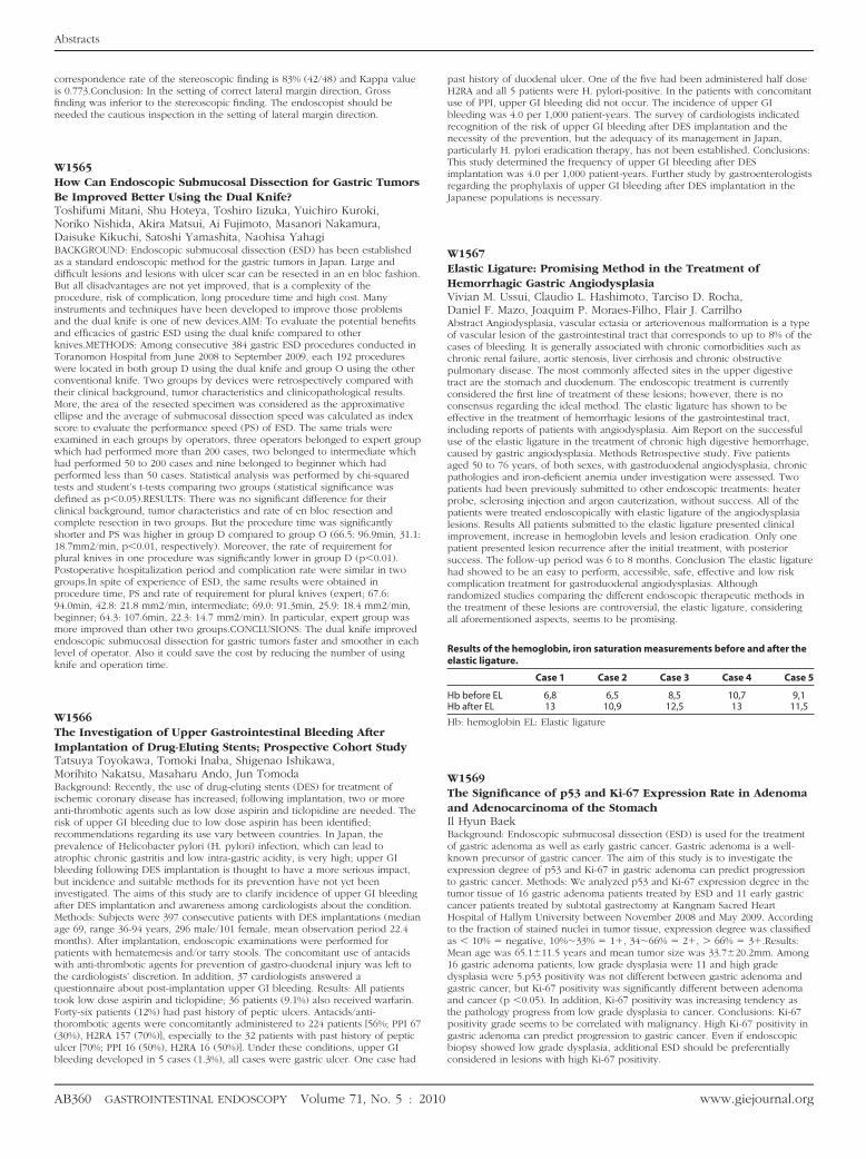
W1567Elastic Ligature: Promising Method in the Treatment ofHemorrhagic Gastric AngiodysplasiaVivian M. Ussui, Claudio L. Hashimoto, Tarciso D. Rocha,Daniel F. Mazo, Joaquim P. Moraes-Filho, Flair J. CarrilhoAbstract Angiodysplasia, vascular ectasia or arteriovenous malformation is a typeof vascular lesion of the gastrointestinal tract that corresponds to up to 8% of thecases of bleeding. It is generally associated with chronic comorbidities such aschronic renal failure, aortic stenosis, liver cirrhosis and chronic obstructivepulmonary disease. The most commonly affected sites in the upper digestivetract are the stomach and duodenum. The endoscopic treatment is currentlyconsidered the first line of treatment of these lesions; however, there is noconsensus regarding the ideal method. The elastic ligature has shown to beeffective in the treatment of hemorrhagic lesions of the gastrointestinal tract,including reports of patients with angiodysplasia. Aim Report on the successfuluse of the elastic ligature in the treatment of chronic high digestive hemorrhage,caused by gastric angiodysplasia. Methods Retrospective study. Five patientsaged 50 to 76 years, of both sexes, with gastroduodenal angiodysplasia, chronicpathologies and iron-deficient anemia under investigation were assessed. Twopatients had been previously submitted to other endoscopic treatments: heaterprobe, sclerosing injection and argon cauterization, without success. All of thepatients were treated endoscopically with elastic ligature of the angiodysplasialesions. Results All patients submitted to the elastic ligature presented clinicalimprovement, increase in hemoglobin levels and lesion eradication. Only onepatient presented lesion recurrence after the initial treatment, with posteriorsuccess. The follow-up period was 6 to 8 months. Conclusion The elastic ligaturehad showed to be an easy to perform, accessible, safe, effective and low riskcomplication treatment for gastroduodenal angiodysplasias. Althoughrandomized studies comparing the different endoscopic therapeutic methods inthe treatment of these lesions are controversial, the elastic ligature, consideringall aforementioned aspects, seems to be promising.
Results of the hemoglobin, iron saturation measurements before and after theelastic ligature.
Case 1 Case 2 Case 3 Case 4 Case 5
Hb before EL 6,8 6,5 8,5 10,7 9,1Hb after EL 13 10,9 12,5 13 11,5
Hb: hemoglobin EL: Elastic ligature
W1569The Significance of p53 and Ki-67 Expression Rate in Adenomaand Adenocarcinoma of the StomachIl Hyun BaekBackground: Endoscopic submucosal dissection (ESD) is used for the treatmentof gastric adenoma as well as early gastric cancer. Gastric adenoma is a well-known precursor of gastric cancer. The aim of this study is to investigate theexpression degree of p53 and Ki-67 in gastric adenoma can predict progressionto gastric cancer. Methods: We analyzed p53 and Ki-67 expression degree in thetumor tissue of 16 gastric adenoma patients treated by ESD and 11 early gastriccancer patients treated by subtotal gastrectomy at Kangnam Sacred HeartHospital of Hallym University between November 2008 and May 2009. Accordingto the fraction of stained nuclei in tumor tissue, expression degree was classifiedas � 10% � negative, 10%�33% � 1�, 34�66% � 2�, � 66% � 3�.Results:Mean age was 65.1�11.5 years and mean tumor size was 33.7�20.2mm. Among16 gastric adenoma patients, low grade dysplasia were 11 and high gradedysplasia were 5.p53 positivity was not different between gastric adenoma andgastric cancer, but Ki-67 positivity was significantly different between adenomaand cancer (p �0.05). In addition, Ki-67 positivity was increasing tendency asthe pathology progress from low grade dysplasia to cancer. Conclusions: Ki-67positivity grade seems to be correlated with malignancy. High Ki-67 positivity ingastric adenoma can predict progression to gastric cancer. Even if endoscopic
biopsy showed low grade dysplasia, additional ESD should be preferentiallyconsidered in lesions with high Ki-67 positivity.www.giejournal.org