Embed Size (px)
Citation preview

Radiology, CMU
Juntima Euathrongchit, MD.Department of RadiologyFaculty of Medicine, CMU
June 17, [email protected]
Saranair Vorapitirangsi
Welcome to Radiology world 2014
Radiology, CMU
Objective
• Introduction to Investigation Methods for Chest
• Limitation vs Precaution on chest film
Radiology, CMU Radiology, CMU
Radiology, CMU
Anterior junctional lineoblique course crossing the upper two-thirds of the sternum from the upper right to lower left and does not extend above the manubriosternaljoint
Posterior junctional linethin, vertical line projecting through
the trachea that extends to the pleural dome above the clavicles to the level of the aortic arch
Azygoesophagealal recess straight stripe running from the azygos
arch to the level of the right hemidiaphram
Radiology, CMU
On Chest Films
Apical lordotic
PA upright Lateral AP supinePA upright Lateral

Radiology, CMU
Dual Energy Subtraction
• Find out calcification.• Find bone, rib lesion.
Standard soft tissue bone
Radiology, CMU
Digital tomosynthesis
Radiology, CMU
Special Imaging Tools
VQ scanUltrasoundAngiography
MRA MRI
Radiology, CMU
MRIAdvantage: No Iodine contrastDisadvantage: Time consuming, Expensive, not good for lung detection
Radiology, CMU
MRI: Oxygen-Enhanced MR Ventilation
http://www.ajronline.org/content/177/1/185.full Radiology, CMU
CT scans -
HRCT
CT chest

Radiology, CMU
Special CT scans
CT pulmonary angiogram MIP –Av IP
3DVR Virtual bronchoscopyRadiologyadadRadRadRadRadRaR ioliolioliolioliologyogyogyogggogog
MnIPRadiology, CMU
New CT technique: Perfusion• An iodine map from dual-energy CT can showed
the distribution of pulmonary perfusion• Photoelectric effect of Iodine
*R Kaewlai http://radiologyinthai.blogspot.com/2010/12/dual-energy-ct-2.html M Riedel, An introduction to dual energy CTKang et al RadioGraphics 2010; 30:685–698
Radiology, CMU
New CT technique: Ventilation
Air trapping
Chae et al Radiology: Volume 248: Number 2—August 2008 Radiology, CMU
PET – CT (FDG – glucose)
http://www.ajronline.org/content/194/1/W91.full
Radiology, CMU
CT CHEST
Radiology, CMU
MDCT
• CT scans– Incremental
– Spiral single
– MDCT
Volume images
images

Radiology, CMU
MDCT
• 4-slice to 16-slice 64-slice multidetector CT
• Progressively No of detectors
scan acquisition times. • In clinical use now, 64-slice CT systems
– gantry rotation times = 0.33 sec. – a spatial resolution = 0.4 mm.
Radiology, CMU
Indication of CT chest• ACR: American College of Radiology• SCBT-MR: Society of Computed Body
Tomography and Magnetic Resonance• SPR: Society for Pediatric Radiology
PRACTICE GUIDELINE FOR THE PERFORMANCE OF THORACIC COMPUTED TOMOGRAPHY (CT)
Radiology, CMU
Indications1. Evaluation of abnormalities discovered on chest images [1].2. Evaluation of clinically suspected cardiothoracic pathology.3. Staging and follow-up of lung cancer and other primary thoracic malignancies, and
detection and evaluation of metastatic disease [2-5].4. Evaluation of cardiothoracic manifestations of known extrathoracic diseases [6-9].5. Evaluation of known or suspected thoracic cardiovascular abnormalities (congenital or
acquired), including aortic stenosis, aortic aneurysms, and dissection [10-12].6. Evaluation of suspected acute or chronic pulmonary emboli [13-22].7. Evaluation of suspected pulmonary arterial hypertension [23].8. Evaluation of known or suspected congenital cardiothoracic anomalies [24,25].9. Evaluation and follow-up of pulmonary parenchymal and airway disease [26-33].10. Evaluation of blunt and penetrating trauma [34,35].11. Evaluation of postoperative patients and surgical complications [36,37].12. Performance of CT-guided interventional procedures [38-41].13. Evaluation of the chest wall [42-44].14. Evaluation of pleural disease [45,46].15. Treatment planning for radiation therapy [47,48].16. Evaluation of medical complications in the intensive care unit or other settings [49,50].
Radiology, CMU
HRCT:- Indication vs contraindication
Indications 1. Evaluation of diffuse pulmonary
disease discovered on chest radiographs, conventional CT of the chest or other CT examinations that include portions of the chest, including selection of the appropriate site for biopsy of diffuse lung disease.
2. Evaluation of the lungs in patients with clinically suspected pulmonary disorders with normal or equivocal chest radiographs.
3. Evaluation of suspected small and/or large airway disease.
4. Quantification of the extent of diffuse lung disease for evaluating effectiveness of treatment.
Contraindications1. There are no absolute
contraindications to HRCT of the lungs.
2. Precaution in Pregnancy
Unable hold breathing
Radiology, CMU
Lobe
RLL: superior, RLL: superior, anterior~,posterioror~, lateral~, anterior ,postemedial basal
RUL: apical, anterior, posterior
RML: lateral, medial
LUL: apicoposterior, anterior,lingula(superior&inferior)
LLL: superior, anteromedial~, posterior~, lateral basal
Radiology, CMU
Chest lobe, segment

Radiology, CMU
Radiology Anatomy: CT chest
Radiology, CMU
CT images: Mediastinum window
http://www.med.wayne.edu/diagradiology/Anatomy_Modules/Mediastinum/Mediastinum.html
Radiology, CMU
CT anatomy
Radiology, CMU
Chest Xray, PA upright Normal, after mastectomy
Radiology, CMU
Abnormality on film• Increased density, Opacity
– Infiltration– Mass– Pleural effusion– Atelectasis
Radiology, CMU
Interesting case• 17 – year-old man • Chronic cough and dyspnea on exertion for 3 months,
– Clear sputum, no pus, no hemoptysis, no fever• Physical examination:-
– T36.7 C , PR 100/min , RR 18/min , BP 110/60 mmHg , O2 sat 98% (Room air)
– Chest: Decrease chest wall movement Lt. ,no accessory muscle use
– Lung: BS decrease entire Lt. lung, Dullness on percussion, Decrease tactile fremitus and Vocal resonance, no egophony, no adventitious sound (wheezing, Rhonchi)

Radiology, CMU
Chest PA upright
Radiology, CMU
Chest PA upright, what is this lesion?
A. Large pleural effusionB. Total left lung atelectasisC. Combined effusion and
atelectasisD. Huge lung massE. Huge mediastinal mass
Radiology, CMU
Chest PA & Left lateral
Radiology, CMU
Chest PA upright, what is this lesion?
A. Large pleural effusionB. Total left lung
atelectasisC. Combined effusion
and atelectasisD. Huge lung massE. Huge mediastinal mass
Radiology, CMU
Answer
Radiology, CMU
Unilateral hemithorax opacityDifferential Diagnosis• Positioning: rotation or scoliosis• Large pleural effusion, pleural thickening,
mesothelioma• Lung: consolidation, mass, collapse, fibrosis, agenesis• Pneumonectomy, thoracoplasty• Chest wall: mass (breast, chest wall musculatures)• Extrathoracic: external structures
Radiology, CMU
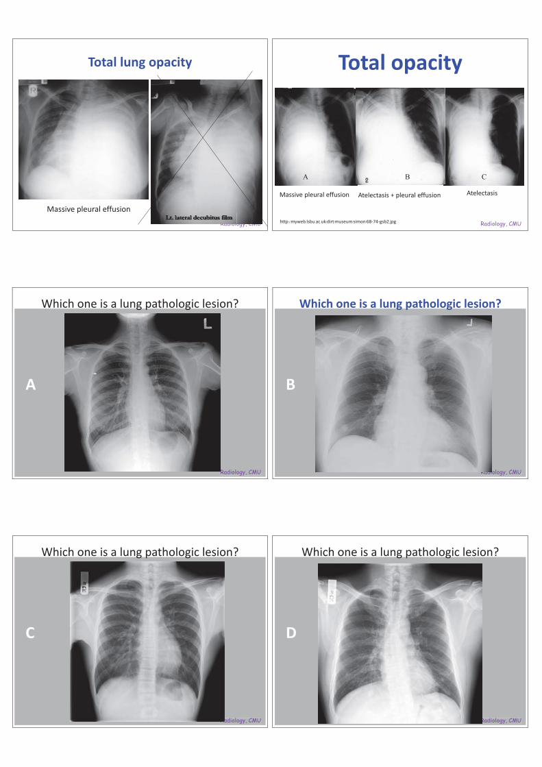
Total lung opacity
RadioloLt. lateral decubitus film
Massive pleural effusionRadiology, CMU
Total opacity
http://myweb.lsbu.ac.uk/dirt/museum/simon/68-74-gsb2.jpg
Massive pleural effusion Atelectasis + pleural effusion Atelectasis
A B C
Radiology, CMURadiology, CMU
A
Which one is a lung pathologic lesion?
Radiology, CMURadiology, CMU
Which one is a lung pathologic lesion?
B
Radiology, CMURadiology, CMU
C
Which one is a lung pathologic lesion?
Radiology, CMURadiology, CMU
D
Which one is a lung pathologic lesion?

Radiology, CMUogy, CMU
A B
C D
Which one is a lung pathologic lesion?
Radiology, CMU
Adequate quality image - A• Remove object
Radiology, CMU
Adequate quality image
• Removable vs non removableobject
Radiology, CMU
Mimiced nodule - B
• Chest wall lesion • Bone island
Radiology, CMU
Additional techniques• Repeat film with changed position• Dual energy subtraction• Digital tomosynthesis• CT scan
Radiology, CMU
Chest wall abnormality - C
• Pectus excavatum– depression of the sternum– Incidence - 0.13 – 0.4% of general population (Fraser et al. 1999)– PA chest: left-sided heart deviation & rotation a mitral
configuration. Parasternal opacity liked RML infiltration or mediastinal mass

Radiology, CMU
Blind areas – D
As complexity of thoracic organs overlying each other in the same plane on each view, they could obscure the lung pathology , these areas called blind areas.
Radiology, CMU
Blind areas
• On PA view – Central airway– Apical lung– Mediastinum– Hila– Retrocardiac field– Inferior lung base– Thoracic cage– Upper abdomen
Radiology, CMU
Tracheal lesion
Min IP 3DVRRadiology, CMU
Sub- diaphragmatic lesion
Radiology, CMU
CT
Radiology, CMU
Steak artifact from Dense CM, CTA - PE
http://radiology.casereports.net/index.php/rcr/article/viewArticle/233/563
Right sided venous approach

Radiology, CMU
Conventional – routine CT• Cover range – thoracic
inlet to whole lung• Scan type – Helical• KV, mA, rotation time
Machine, • Respiratory – inspiration• Image reconstruction: 5
mm thickness, axial mediastinum and lung / coronal
• IV contrast + delayed 40 sec (arterial phase liver)
Radiology, CMU
CTA – systemic circulation
Hemoptysis, Aortic disease
Radiology, CMU
CTA A –– pulmonary circulation
Radiology, CMU
HRCT
Radiology, CMU
HRCT
Radiology, CMURadRadRadRadRadRadRadioliolioliolioliolologyogyogyygygyogyogyogyogygygy, , , ,,,,,,,,,,,,,,,

Radiology, CMU
Plain arterial liver portovenous lung image
Radiology, CMU
CT Chest for SPN protocol - CMU
Cover range – Inlet upper abdomenRepeat nodules at delayed phase
Frist study: Plain nodule + Post contrast scan thorax + delayed 30 sec, 1, 2, 3, 4 min
Reconstruction -* CMUSlice thickness: 5 mm, at leastInterval 5 mm no skip Axial imagesSoft tissue W 1 set - 5 mm slice thickness Lung W 1 set – 5 mmSoft tissue W 1 set – 1 mm slice thickness and intervalCoronal vs sagittal – up to PACS systemReconstruction nodule, 1 – 2 mm thickness for each series
Radiology, CMU
CT for solitary pulmonary nodule
Plain 1-2 mm slice thickness 30 sec D
1 min 2 min 3 min 4 min
Total Iodine contrast 420 mgI / kg, 2 ml/sec injection rate
Radiology, CMU
Low dose CT• The National lung screening trial (NLST)• LDCT screening could reduce lung cancer
mortality to 20% when compared with chest X-ray screening
Radiology, CMU
Risk groupsRisk criteria Screening
RecommendationHigh risk• yr-old, and• yrs yrs)
Get baseline LDCT
High risk• yr-old, and• yrs of smoking, and • One other risk factor (except for second-hand smoke)
Get baseline LDCT
Moderate risk• yr-old, and• yrs of smoking or second-hand smoke, and • No other risk factors
No screening at this time
Low risk• < 50 yr-old, and/or• < 20 pack yrs smoking
No screening at this time
Radiology, CMU
Low dose CT scan• Hypothesis: Generic factor and/or indirect receiving carcinogen or second
smoker could be cause of lung cancer• Cost – effectiveness analysis
Parameter Hs) SLST (care dose)Somatom definition CT
Voltage (kVp) 120 – 140 120
Tube current time product (mAs) 40 – 80 25
Slice thickness (mm) 1.0 – 3.2 1.0 – 5.0
Reconstruction interval (mm) 1 – 2.5 0.8 – 1.0
Number of studies 26,722 60
RiskFamily Hx (closed relative)
20 – 65 YrsNo other cancer

Radiology, CMU
Compare, normal dose vs low dose
Low dose - noise
Radiology, CMU
Tumor growth
Radiology, CMU
Hemoptysis – bronchial a. systemic a.In over 90% of cases of hemoptysis requiring intervention with arterial embolization or surgery,the bronchial arteries are responsible for the bleeding
Radiology, CMU
Protocol
Radiology, CMUR ddR dRadRaddR dRRRaR i li li llolioliiio
Bronchial a enlargement
Radiology, CMU
Bronchiectasis, HRCT

Radiology, CMU
Airway
Radiology, CMU
AirwayTracheal Stenosis •• Bronchial Stenosis • Tracheal-Esophageal Fistula
• Suspected Tracheal or Bronchial Injury or Fracture
• Tracheomalacia• Tracheobronchomalacia• Mounier-Kuhn Syndrome
Radiology, CMU
Protocol CT bronchi - CTISUS
Radiology, CMU
Tracheomalacia: Full inspired vs expired
Radiology, CMU
D VR
Radiology, CMU
Reconstruction

Radiology, CMURadRadRadRaddRadRadRadRaddRadRadRadRadadR ddRadRadRadddR dRadRadR dR ddR dRadRaaRRRaR ioliolioliolioliollioliololioliollolioioiioioioooiooii ogyogyyogyyogyooooo CCCCCCCCCCCCCCCCCCCCCCCCCCCCCCCCCCCCCCCCCCCCCMUMUMUMUMUMMMMMMMMMMMMMMMMMMMMM
Tracheal bronchus
Radiology, CMU
Abnormal chest wall
Radiology, CMU
Pectus excavatum
Radiology, CMU
Early filled aorta – CTA PE
ASD
Radiology, CMU
Cardiac arrest
• During exam – not uncommon• Awareness• Risk factors
– Unstable patient : previous shock, poor station– Multiple trauma– To much monitors– Drug allergy *
Radiology, CMU
Normal not cardiac arrest
• Motion normal artifact

Radiology, CMU
Normal respiration – can control
Radiology, CMU
Except cardiac gating
Radiology, CMU
• At cardiac arrest
• CT features characterized by a
in the dependent parts of
the right side of the body, including the
venous system and the right lobe of the liver.
Radiology, CMU
• --yr man, cardiac arrest st –– polytrauma•
r man, cardiac aryryMarked enhanced
rresstac ard d azygos
polytraumaptss v., pooling CM in dependent part of IVC, hepatic v., right Marked enMMMarked enMarkMar
renal v.; •
e a ;renal v.; no CM in Lt heart, aorta, kidney
Ref: Indian J Radiol Imaging, May 2010.
Radiology, CMU
26- yr man, accident.• Dense contrast opacified azygos v, SVC, great cardiac v, hemiazygos v, right
lumbar v, back venues, Rt atrium, Rt hepatic v, Rt kidney, splenic v, SMV, • No CM in aorta, left heart.• Ref: Indian J Radiol Imaging, May 2010.
Great cardiac v
Radiology, CMU
Near arrest –hypotension.
• Contrast layering in IVC, in cardiogenic shock.
• Note bilateral pleural effusions and pericardial effusion without tamponade.

Radiology, CMU
Cardiac tamponade• Gross pericardial effusion with pressure effect to the heart.• Mod. bilateral pleural effusions.
aaaRaRaaaRaRaRaRaRaRaRRRRRRRRRRRRRRRRRRRRRRRRR Radiology, CMU
Contrast layering in abdomen:- IVC & Rt renal vein• 30 –year-old male with disseminated Tb,
hypotension and worsening of breathlessness.• Pt developed cardiogenic shock within a few hours
and died.
RadRadRadRadRadRadRadRRRRR ioliolioliollollolllioioioo ogyogyogyogyyy CCCCCCCCCCCCCCCCCCCCCCCCMUUMUMUMMMMMMMMMMMMMMMMMMMMMMMMMMMMMMMM
Radiology, CMU
What happen ?
• Injection: - right side• Regurgitation of CM
to the left system: jugular, subclavian, back venules, and hemiazygos v.
A
B
SVC obstruction
AJR:178, May 2002
Radiology, CMU
Rt brachiocephalic venous obstruction with collateral vessels
Radiology, CMULayering in SVC, reflux CM into the azygos v, hemiazygos v. AJR:178, May 2002 Radiology, CMU
Opacification of right ventricle, right atrium, right hepatic veins, and vena cava. Note regurgitation of contrast agent into coronary sinus (arrowhead)
AJR:178, May 2002