Embed Size (px)
Citation preview


What is radiation therapy (RT)?
• Cancer treatment
• Tumor versus normal tissues
• External photon beam RT
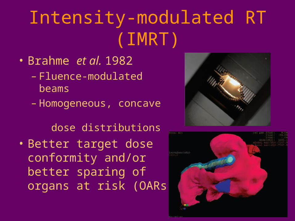
Intensity-modulated RT (IMRT)
• Brahme et al. 1982– Fluence-modulated beams– Homogeneous, concave
dose distributions
• Better target dose conformity and/or better sparing of organs at risk (OARs)
Imaging for RT
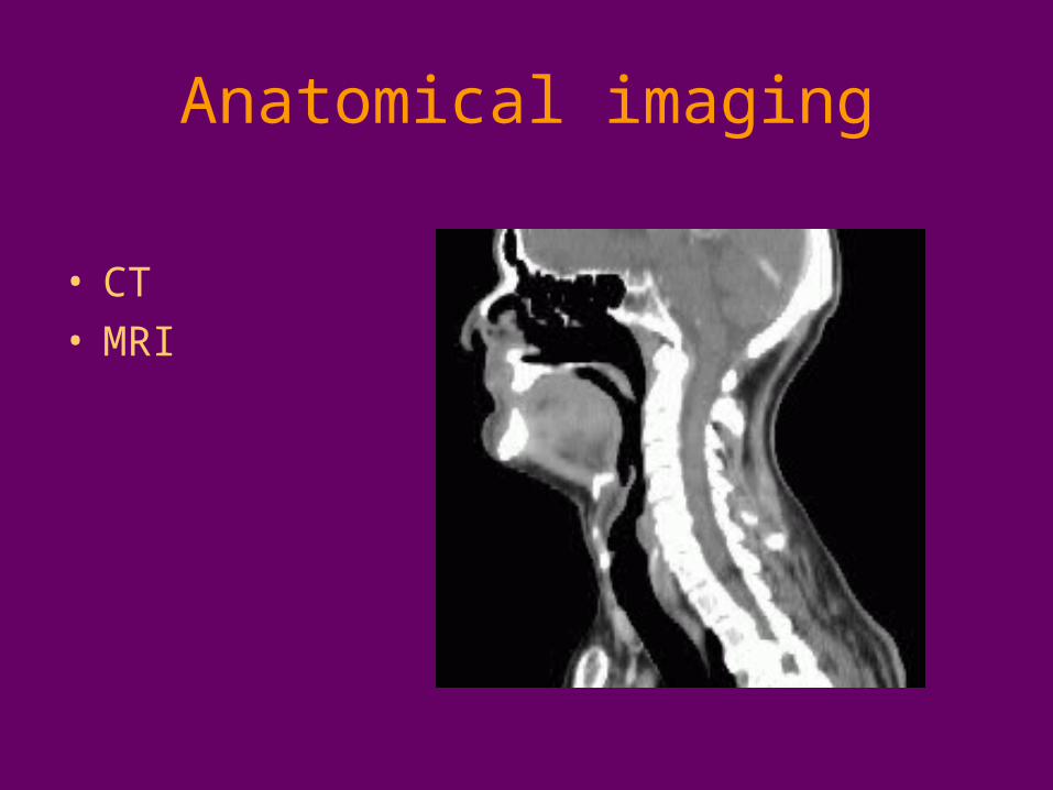
Anatomical imaging
• CT• MRI
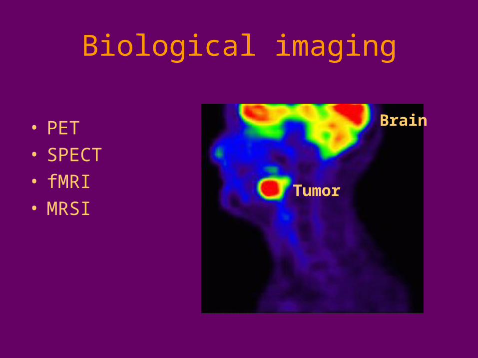
Biological imaging
• PET• SPECT• fMRI• MRSI
Brain
Tumor
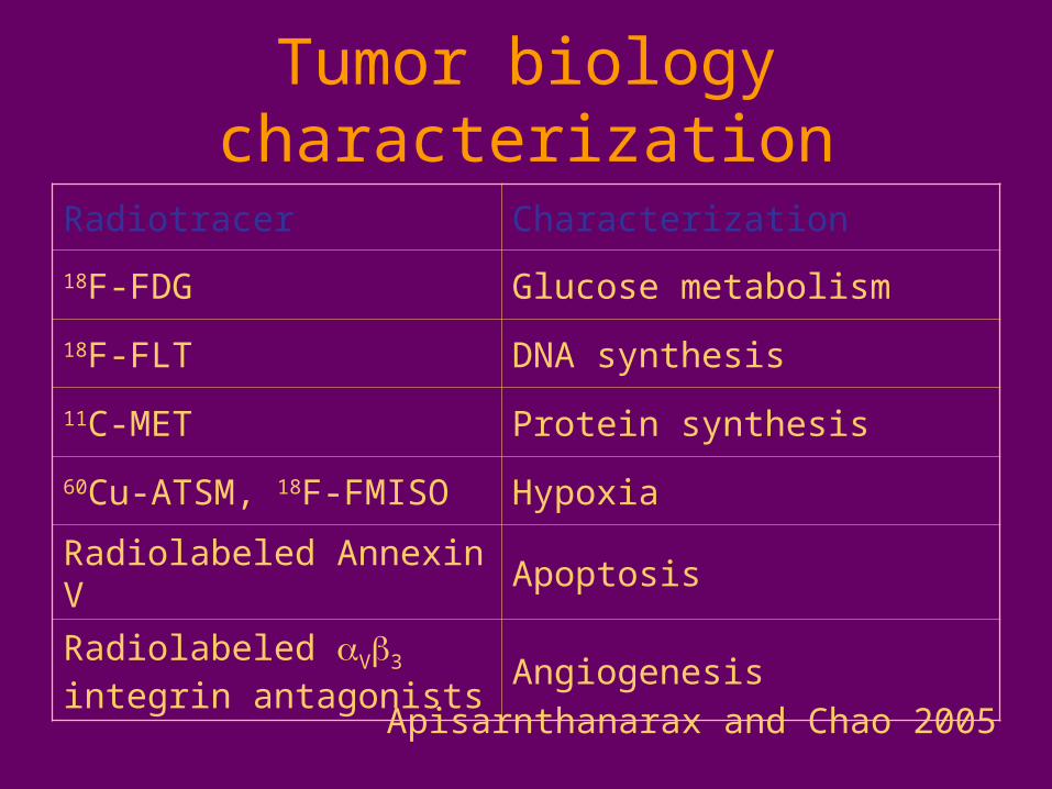
Tumor biology characterization
Radiotracer Characterization
18F-FDG Glucose metabolism
18F-FLT DNA synthesis
11C-MET Protein synthesis
60Cu-ATSM, 18F-FMISO Hypoxia
Radiolabeled Annexin V Apoptosis
Radiolabeled V3 integrin antagonists
Angiogenesis
Apisarnthanarax and Chao 2005
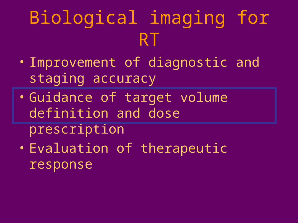
Biological imaging for RT
• Improvement of diagnostic and staging accuracy
• Guidance of target volume definition and dose prescription
• Evaluation of therapeutic response
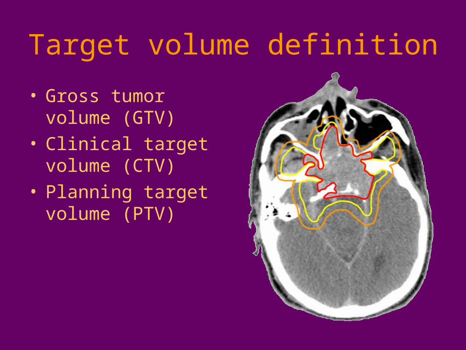
Target volume definition
• Gross tumor volume (GTV)
• Clinical target volume (CTV)
• Planning target volume (PTV)
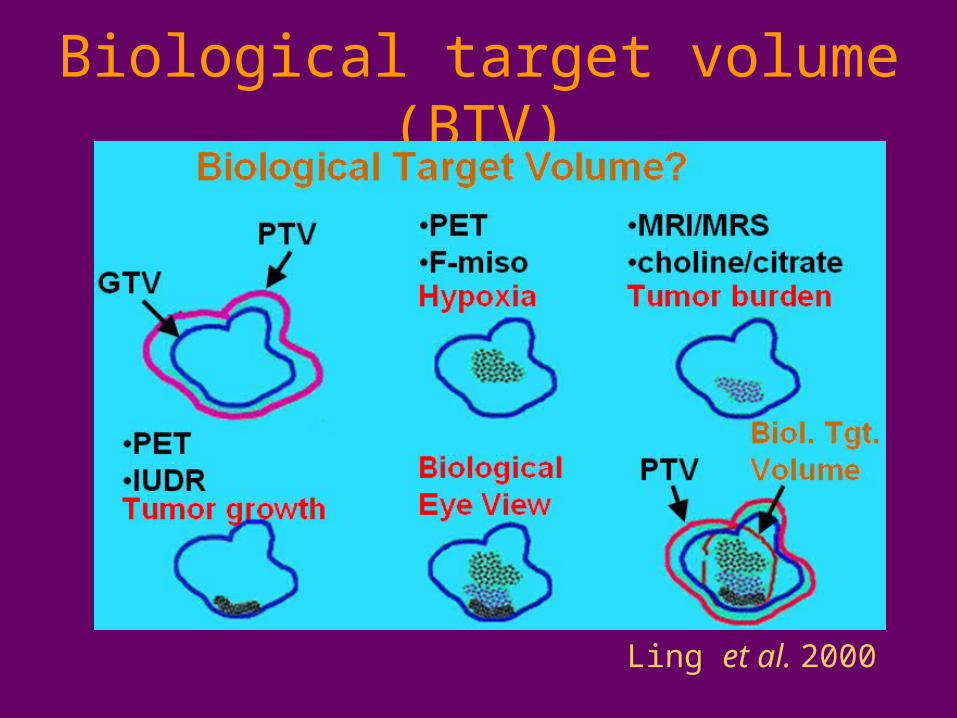
Biological target volume (BTV)
Ling et al. 2000
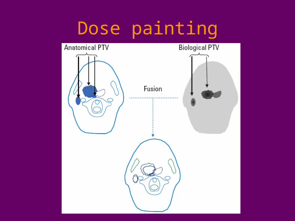
Dose painting
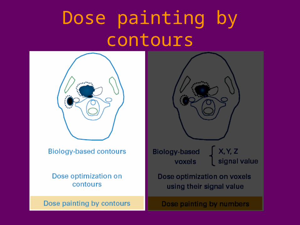
Dose painting by contours
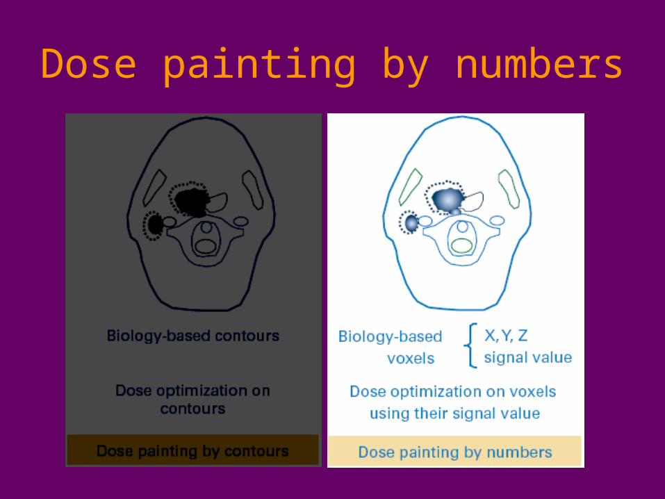
Dose painting by numbers
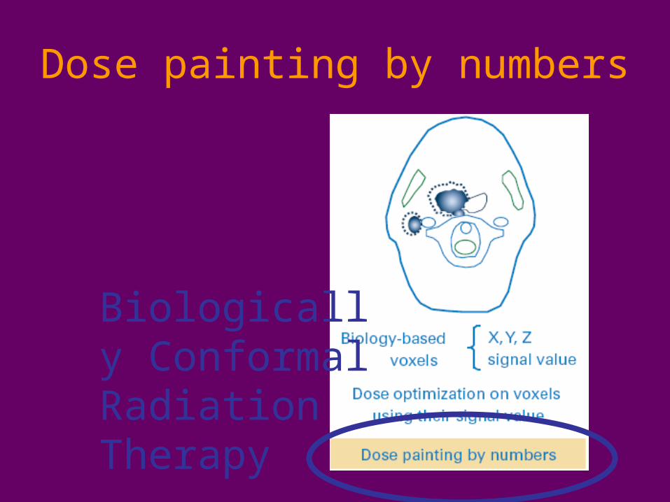
Dose painting by numbers
Biologically Conformal Radiation Therapy
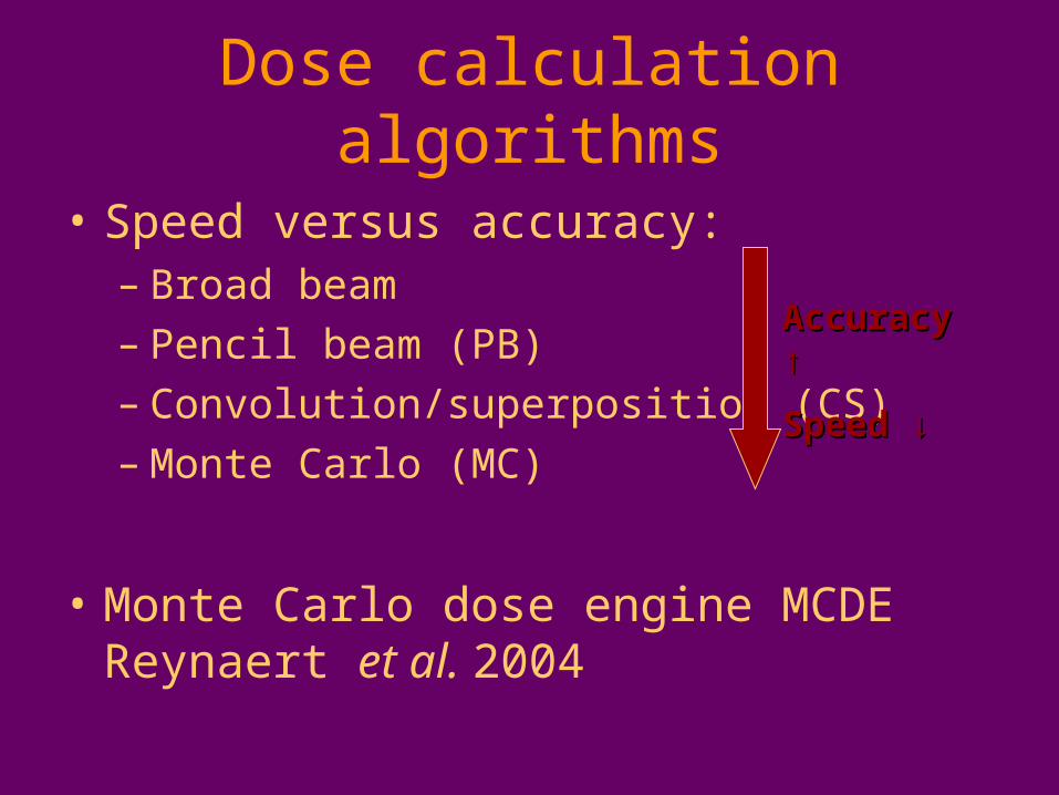
Dose calculation algorithms
• Speed versus accuracy:– Broad beam– Pencil beam (PB)– Convolution/superposition (CS)– Monte Carlo (MC)
• Monte Carlo dose engine MCDE Reynaert et al. 2004
Accuracy Accuracy ↑↑
Speed ↓Speed ↓
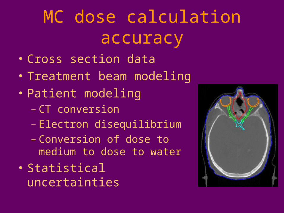
MC dose calculation accuracy
• Cross section data
• Treatment beam modeling
• Patient modeling– CT conversion – Electron disequilibrium– Conversion of dose to medium
to dose to water
• Statistical uncertainties

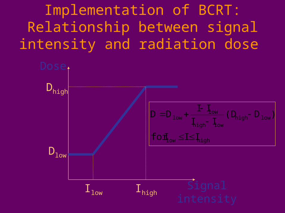
Implementation of BCRT:Relationship between signal intensity
and radiation dose
Dose
Ilow Ihigh
Dlow
Dhigh
Signal intensity
highlow
lowhighlowhigh
lowlow
IIIfor
)D(DII
IIDD

Implementation of BCRT: Treatment planning strategy
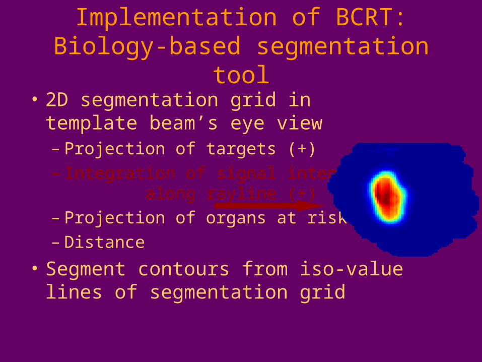
Implementation of BCRT:Biology-based segmentation tool
• 2D segmentation grid in template beam’s eye view– Projection of targets (+)– Integration of signal intensities
along rayline (+)– Projection of organs at risk (-)– Distance
• Segment contours from iso-value lines of segmentation grid
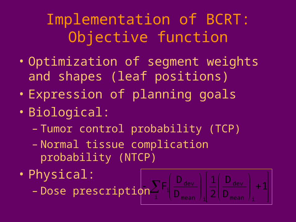
Implementation of BCRT:Objective function
• Optimization of segment weights and shapes (leaf positions)
• Expression of planning goals
• Biological:– Tumor control probability (TCP)– Normal tissue complication probability (NTCP)
• Physical:– Dose prescription
1
D
D
2
1
D
DF
imean
dev
imean
dev
ii
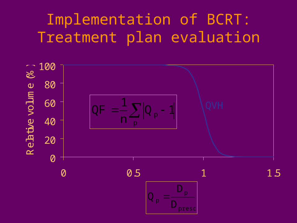
Implementation of BCRT:Treatment plan evaluation
0
20
40
60
80
100
0 0.5 1 1.5
Q
Re
lativ
e v
olu
me
(%
)
p
p 1Qn
1QF
presc
pp D
DQ
QVH
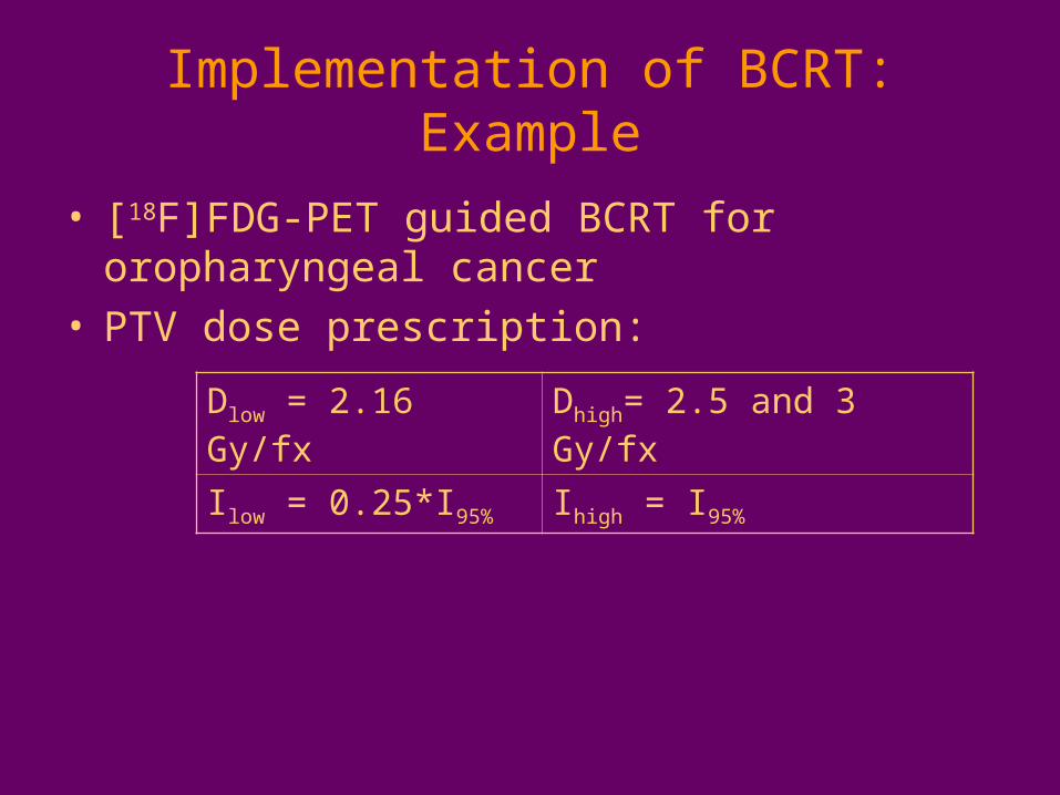
Implementation of BCRT:Example
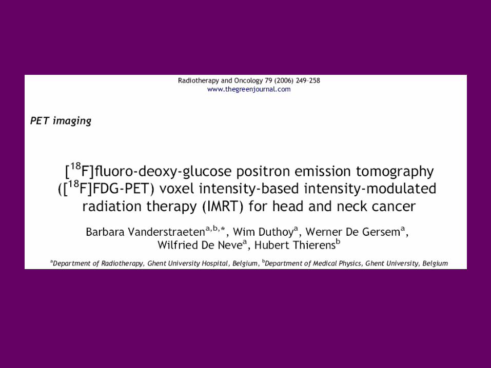
• [18F]FDG-PET guided BCRT for oropharyngeal cancer
• PTV dose prescription:
Dlow = 2.16 Gy/fx Dhigh= 2.5 and 3 Gy/fx
Ilow = 0.25*I95% Ihigh = I95%
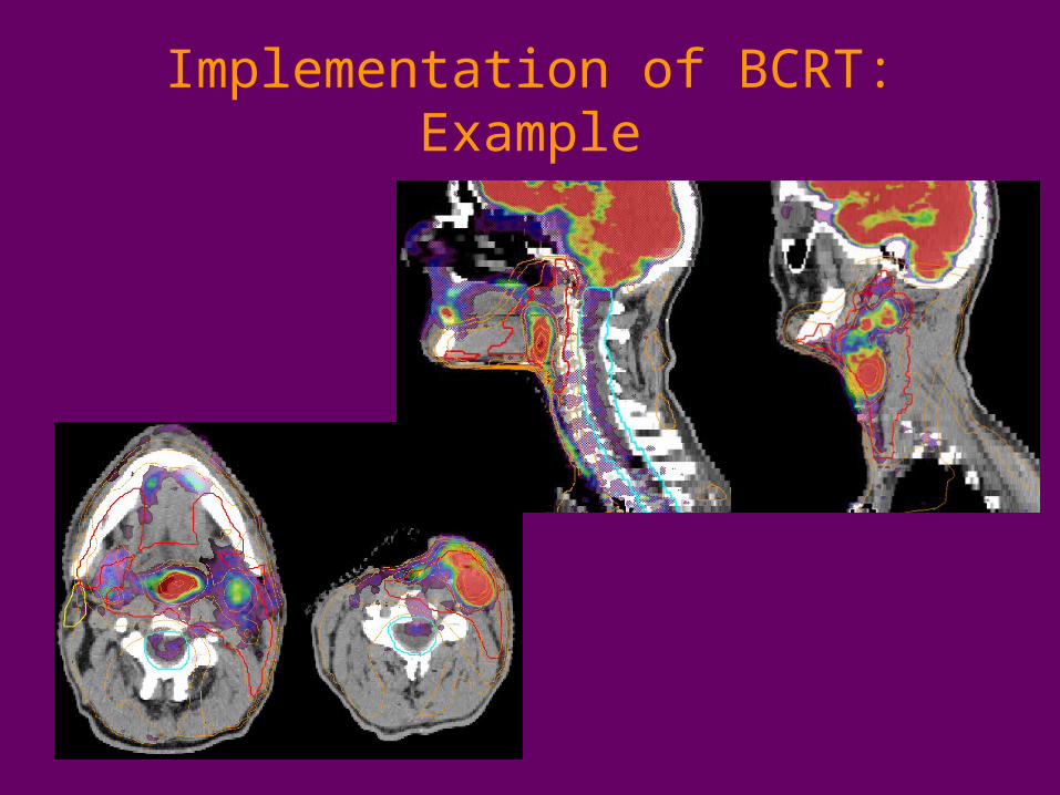
Implementation of BCRT:Example
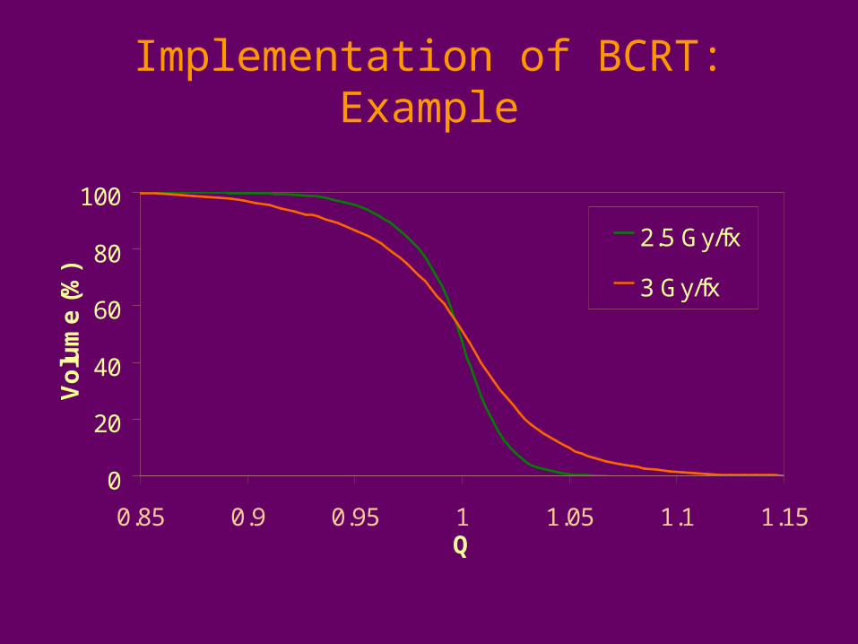
Implementation of BCRT:Example
0
20
40
60
80
100
0.85 0.9 0.95 1 1.05 1.1 1.15Q
Vo
lum
e (
%)
2.5 Gy/fx
3 Gy/fx
Implementation of BCRT:Conclusions
• Technical solution– Biology-based segmentation tool– Objective function
• Feasibility– Planning constraints OK– Best biological conformity for the lowest level
of dose escalation
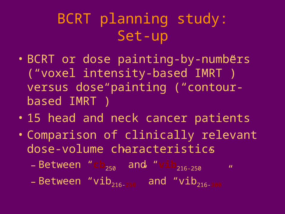
BCRT planning study:Set-up
• BCRT or dose painting-by-numbers (“voxel intensity-based IMRT”) versus dose painting (“contour-based IMRT”)
• 15 head and neck cancer patients
• Comparison of clinically relevant dose-volume characteristics– Between “cb250” and “vib216-250”
– Between “vib216-250” and “vib216-300”
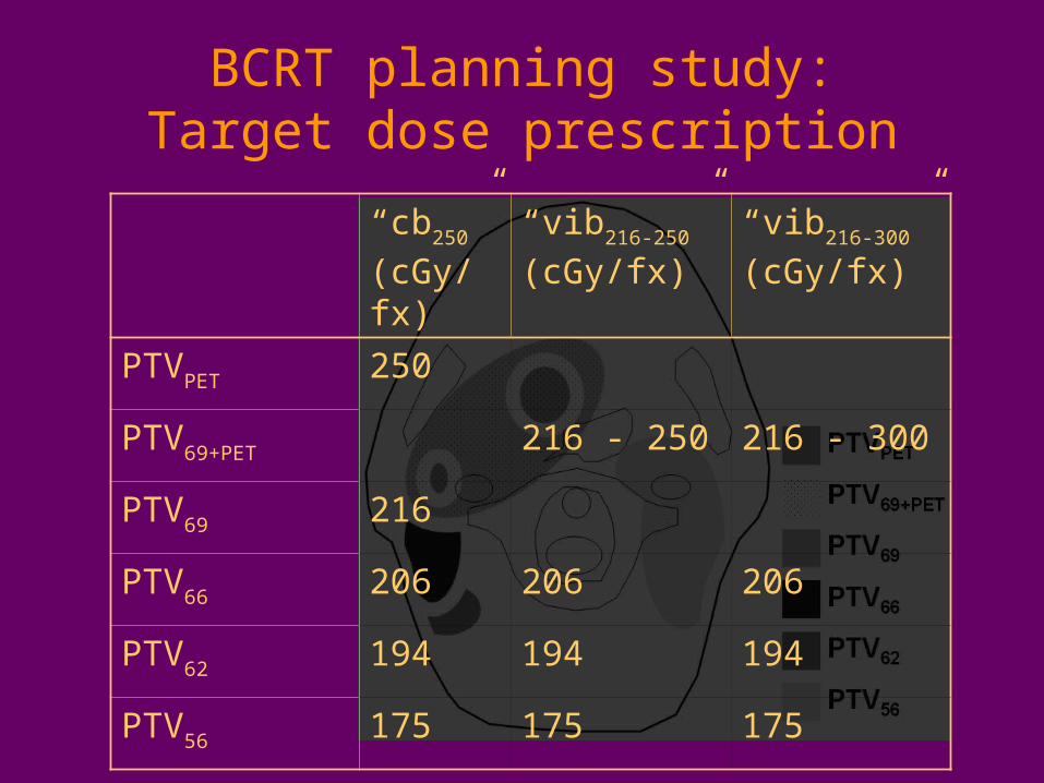
BCRT planning study:Target dose prescription
“cb250”
(cGy/fx)
“vib216-250”
(cGy/fx)
“vib216-300”
(cGy/fx)
PTVPET 250
PTV69+PET 216 - 250 216 - 300
PTV69 216
PTV66 206 206 206
PTV62 194 194 194
PTV56 175 175 175
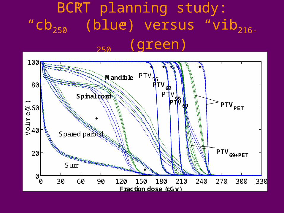
BCRT planning study:“cb250” (blue) versus “vib216-250” (green)
0 30 60 90 120 150 180 210 240 270 300 3300
20
40
60
80
100
Fraction dose (cGy)
Vo
lum
e(%
)
Surr
PTV56
Spared parotid
PTV69+PET
Spinal cordPTV
PET
PTV66
PTV69
MandiblePTV
62
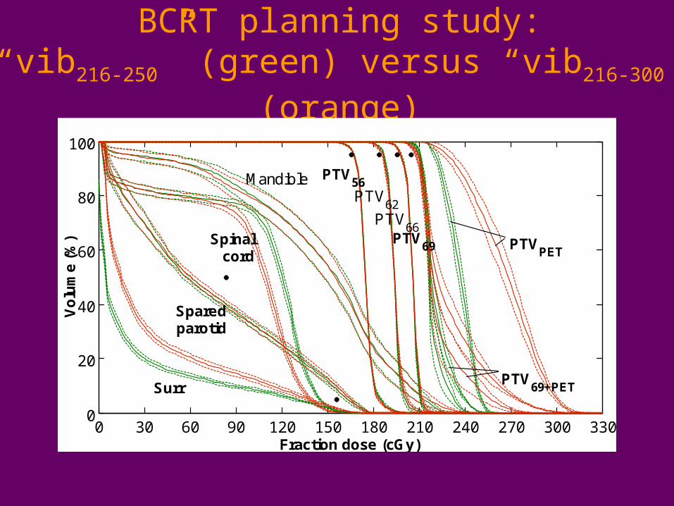
BCRT planning study:“vib216-250” (green) versus “vib216-300” (orange)
0 30 60 90 120 150 180 210 240 270 300 3300
20
40
60
80
100
Fraction dose (cGy)
Vo
lum
e (%
)
MandiblePTV
62
PTVPET
PTV69+PET
PTV66
PTV69
PTV56
Spinalcord
Sparedparotid
Surr
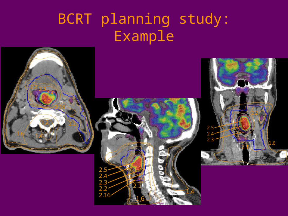
BCRT planning study:Example
2.11.2
2.5
2.16
2.22.4
1.6 1.4
2.3
2.11.2
2.5
2.16
2.22.4
1.6 1.4
2.3
2.1
2.5
2.162.2
2.4
1.61.4
2.32.1
2.5
2.162.2
2.4
1.61.4
2.3
2.1
2.5
2.2
2.4
1.62.3
2.1
2.5
2.2
2.4
1.62.3
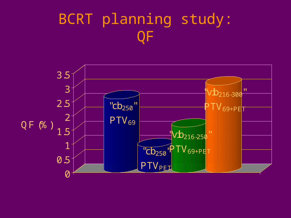
BCRT planning study:QF
"cb250"
PTV69
"cb250"
PTVPET
"vib216-250"
PTV69+PET
"vib216-300"
PTV69+PET
0
0.5
1
1.5
2
2.5
3
3.5
QF (%)
BCRT planning study:Conclusions
• BCRT did not compromise the planning constraints for the OARs
• Best biological conformity was obtained for the lowest level of dose escalation
• Compared to dose painting by contours, improved target dose coverage was achieved using BCRT
MC dose calculations in the clinic
• Comparison of PB, CS and MCDE for lung IMRT
• Comparison of 6 MV and 18 MV photons for lung IMRT
• Conversion of CT numbers into tissue parameters: a multi-centre study
• Evaluation of uncertainty-based stopping criteria
• Feasibility of MC-based IMRT optimization
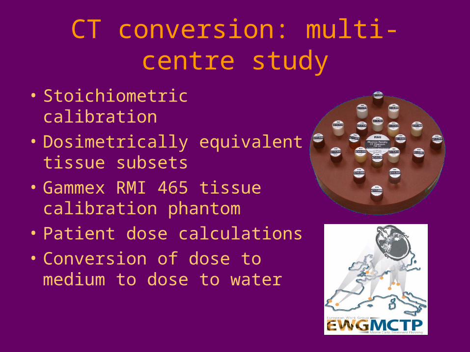
CT conversion: multi-centre study
• Stoichiometric calibration
• Dosimetrically equivalent tissue subsets
• Gammex RMI 465 tissue calibration phantom
• Patient dose calculations
• Conversion of dose to medium to dose to water
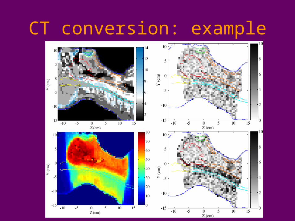
CT conversion: example
CT conversion: conclusions
• Accuracy of MC patient dose calculations
• Proposed CT conversion scheme:
Air, lung, adipose, muscle, 10 bone bins
• Validated on phantoms
• Patient study:
Multiple bone bins necessary if dose is converted to dose to water

Biologically conformal RT
• Technical solution– Bound-constrained linear model– Treatment plan optimization
• Biology-based segmentation tool• Objective function
– Treatment plan evaluation
• Feasibility of FDG-PET guided BCRT for head and neck cancer
MC dose calculations
• Individual patients may benefit from highly accurate MC dose calculations
• Improvement of MCDE– CT conversion– Uncertainty-based stopping criteria
• Feasibility of MC-based IMRT optimization
• MCDE is unsuitable for routine clinical use, but represents an excellent benchmarking tool

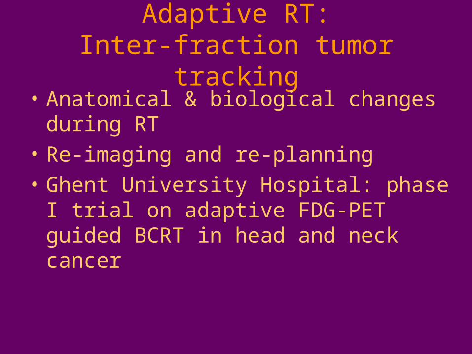
Adaptive RT:Inter-fraction tumor tracking
• Anatomical & biological changes during RT
• Re-imaging and re-planning
• Ghent University Hospital: phase I trial on adaptive FDG-PET guided BCRT in head and neck cancer
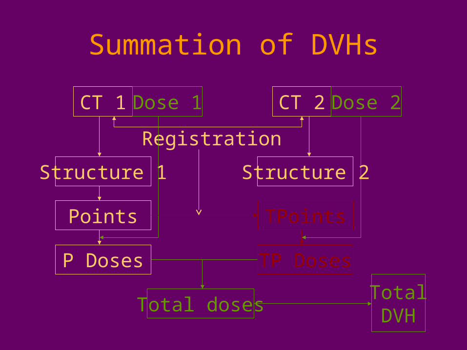
Summation of DVHs
CT 1 Dose 1 CT 2 Dose 2
Registration
Structure 1
Points
P Doses
TPoints
TP Doses
Total dosesTotalDVH
Structure 2
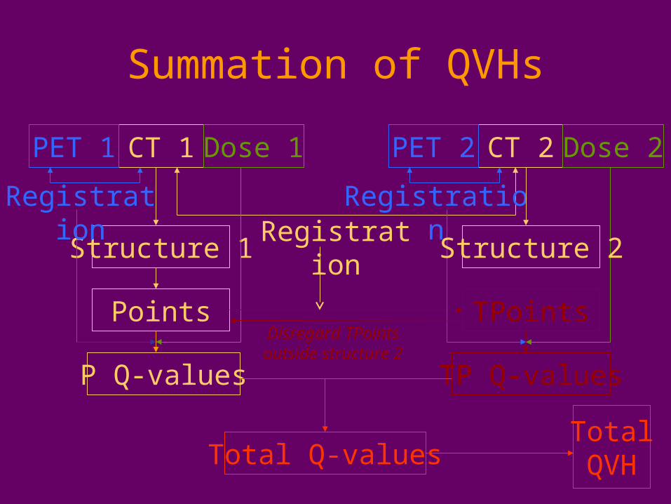
Summation of QVHs
CT 1 Dose 1 CT 2 Dose 2
RegistrationStructure 1
Points
P Q-values
TPoints
TP Q-values
Total Q-valuesTotalQVH
PET 1 PET 2
Registration Registration
Disregard TPointsoutside structure 2
Structure 2
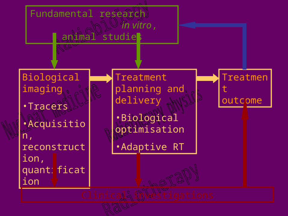
Treatment planning and delivery
•Biological optimisation
•Adaptive RT
Biological imaging
•Tracers
•Acquisition, reconstruction, quantification
Clinical investigations
Fundamental research in vitro, animal studies
Treatment outcome