Embed Size (px)
Citation preview
X-ray Structure of Active Site-inhibited Clotting Factor XaIMPLICATIONS FOR DRUG DESIGN AND SUBSTRATE RECOGNITION*
(Received for publication, July 22, 1996, and in revised form, August 26, 1996)
Hans Brandstetter‡§, Anja Kuhne‡, Wolfram Bode‡, Robert Huber‡, Wolfgang von der Saal¶,Klaus Wirthensohni, and Richard A. Engh‡**
From the ‡Max-Planck-Institut fur Biochemie, Strukturforschung, D-82125 Martinsried, ¶Boehringer Mannheim GmbH,Sandhofer Strasse 116, D-68305 Mannheim, and iBoehringer Mannheim GmbH, Nonnenwald 2,D-82372 Penzberg, Federal Republic of Germany
The 3.0-Å resolution x-ray structure of human des-Gla-coagulation factor Xa (fXa) has been determined in com-plex with the synthetic inhibitor DX-9065a. The bindinggeometry is characterized primarily by two interactionsites: the naphthamidine group is fixed in the S1 pocketby a typical salt bridge to Asp-189, while the pyrrolidinering binds in the unique aryl-binding site (S4) of fXa.Unlike the large majority of inhibitor complexes withserine proteinases, Gly-216 (S3) does not contribute tohydrogen bond formation. In contrast to typical throm-bin binding modes, the S2 site of fXa cannot be used byDX-9065a since it is blocked by Tyr-99, and the aryl-binding site (S4) of fXa is lined by carbonyl oxygen at-oms that can accommodate positive charges. This hasimplications for natural substrate recognition as well asfor drug design.
Hemostasis is the blood clotting process that, when function-ing properly, occurs when an injury to the vasculature leads toa series of vasculomotor and cellular reactions and the activa-tion of the blood coagulation cascade. The latter process isinitiated via the extrinsic pathway, leading first to thrombinactivation and then massively amplified thrombin activationdue to the positive feedback of the intrinsic pathway (1). Bothextrinsic and intrinsic pathways merge at the factor X activa-tion step. An imbalance between these clotting processes, clot-ting inactivation processes (protein C inactivation of hemosta-sis cofactors), and thrombolytic processes (tissue plasminogenactivator, plasminogen) can lead to thrombotic or bleeding dis-orders. Antithrombotics include inhibitors of thrombin, factorXa and factor IXa, factors involved in both the extrinsic andintrinsic pathways (2). As evidence accumulates that thrombinhas other important functions in cellular (3, 4) and neurological(5–10) processes, new synthetic anticoagulants increasinglytarget factor Xa. Daiichi published the first tight binding (Ki 541 nM), specific inhibitor of fXa,1 DX-9065a (11–13).We present here the crystal structure of the factor XazDX-
9065a complex. The inhibitor binds in the active site in anextended conformation, which was expected from earlier stud-
ies (14, 15). Both hydrophobic and electrostatic interactionscharacterize the complex formation, which is also accompaniedby local rearrangements in the active site of fXa. Consideringthese subtle interactions and the unpredictable ligand-inducedmotions involved in binding, there is clearly a need for a seriesof fXa-ligand complex structures to provide adequate informa-tion for structure-based drug design and an understanding ofhow fXa recognizes physiological substrates.
EXPERIMENTAL PROCEDURES
fX was isolated from human plasma; des-Gla-fX was produced viachymotryptic cleavage (removing amino acids L1–L44; chymotrypsino-gen numbering is used for the catalytic domain; the sequential fXnumbering, which is used for the light chain, will be indicated by theprefix “L”). Subsequently, fX was activated with the purified factor Xactivator from Russell’s viper venom, resulting in des-Gla-fXa.The inhibitor DX-9065a (Fig. 1) was prepared as described previously
(12). Initial crystals were obtained by a fine pH screen (0.1 M MES/OH,pH 5.8, 10 mM CaCl, 18% polyethylene glycol 6000) after 2 weeks. Thesefirst rod-like crystals were orthorhombic, with cell constants a 5 56.93Å, b 5 73.17 Å, and c 5 79.08 Å and a 5 b 5 g 5 90.0, but diffractedweakly. Seeding techniques improved both size (0.1 3 0.1 3 0.4 mm3)and intrinsic order. Data to 2.8 Å were collected on a Siemens multiwirearea detector and processed with SAINT data reduction software (16).The final data set used in refinement included reflections to 3.0-Åresolution with completeness of 85% and Rmerge of 31% in the outershell. The crystals belong to the space group P212121 with one moleculein the asymmetric unit. Patterson search techniques (17–19), modelbuilding, electron density map calculation, and model refinement wasdone within MAIN (20) and X-PLOR (21). The model (without additionof water molecules) has been refined to a crystallographic R-value of19.7% (Rfree 5 26.1%) with good stereochemistry (root mean squaredeviation of bonds 5 0.009 Å and angles 5 2.2° from ideality (22)).
RESULTS
Overall fXa Structure—We describe here the structure of thecomplex of Daiichi inhibitor DX-9065a in des-Gla-fXa, i.e. thecatalytic serine protease domain and both the EGF2 (proxi-mate to the catalytic domain) and the EGF1 domains. Theprotein is thus identical to that of the tetragonal fXa crystalstructure previously described (23, 24). Differences betweenthe structures thus arise from the different crystal packinginteractions or different ligand interactions (calcium and inhib-itor) and can usually be attributed to the most local cause; theextent to which apparent ligand-induced structural changesdetermine crystal packing interactions and vice versa remainsambiguous. In the case of the tetragonal fXa crystal structure,the crystal packing interaction between the S1 specificitypocket and the EGF2 C-terminal Arg residue of a symmetry-related fXa molecule is simultaneously an enzyme-ligand in-teraction. This arginine binding interaction is eliminated in thefXazDX-9065a structure. Similarly, the presence of calcium inthe orthorhombic fXazDX-9065a structure is associated simul-taneously with differences in structure and with different crys-
* The costs of publication of this article were defrayed in part by thepayment of page charges. This article must therefore be hereby marked“advertisement” in accordance with 18 U.S.C. Section 1734 solely toindicate this fact.§ Present address: Dept. of Chemistry, MIT, 77 Massachusetts Ave.,
Cambridge, MA 02139.** To whom correspondence should be addressed. Tel.: 49-89-8578-
2630; Fax: 49-89-8578–3516; E-mail: [email protected] The abbreviations used are: fXa, factor Xa; MES, 4-morpho-
lineethanesulfonic acid; EGF, epidermal growth factor; NAPAP, Na-(2-naphthylsulfonylglycyl)-DL-p-amidinophenylalanyl-piperidine; TAPAP,N-(4-toluenesulfonyl)-DL-p-amidinophenylalanyl-piperidine.
THE JOURNAL OF BIOLOGICAL CHEMISTRY Vol. 271, No. 47, Issue of November 22, pp. 29988–29992, 1996© 1996 by The American Society for Biochemistry and Molecular Biology, Inc. Printed in U.S.A.
This paper is available on line at http://www-jbc.stanford.edu/jbc/29988
by guest on March 24, 2018
http://ww
w.jbc.org/
Dow
nloaded from
tal packing interactions.The EGF1 domain, disordered in the tetragonal, arginine-
bound crystal form, was suggested to become ordered in thepresence of calcium (24). However, the orthorhombic crystals offXazDX-9065a grown at 10 mM calcium also show apparentdisorder of the EGF1 domain. Furthermore, the crystal packingof the two crystal forms is not compatible with a unique EGF1orientation (relative to EGF2). The tetragonal crystal packingof arginine-bound fXa suggested an extended EGF2-EGF1 ar-rangement similar to the one observed in fIXa (25). In contrast,the orthorhombic crystal structure of fXazDX-9065a allows onlytwo alternate orientations, both of which result in a compact,globular quarternary fold with EGF1 in contact with the cata-lytic domain. This arrangement can be excluded for the tetrag-onal crystal. This is evidence against a rigid factor X modulearrangement in solution or physiologically at the membranesurface. Rather, several conformations seem possible that maybe physiologically important (possibly distinguishing, for ex-ample, fX as substrate and fXa as an enzyme). This is consist-ent with fluorescence energy transfer measurements that indi-cate that the distance of the active site to the membranechanges significantly upon cofactor (fVa) binding (26). Such achange is not observed in the analogous experiment with fIXaand cofactor VIIIa (27).The calcium-binding site in the catalytic domain is conserved
in factor X, and studies based on synthetic factor X peptidessuggest that the calcium-binding site participates in the pro-thrombinase complex (28). This site is well characterized intrypsin and other serine proteinases (29): calcium binding in-duces an ordering transition in the ligating residues, i.e. theside chains of Asp-70 and Glu-80 as well as the carbonyl groupsof Asn-72 and Ala-75. In the fXazDX-9065a structure, all theseresidues are ordered, and there is density at the expectedcalcium position, indicating at least partial occupation of thecalcium-binding site. The tetragonal, arginine-bound fXa crys-tal structure differs somewhat in the calcium loop, but thedifferences are confined largely to side chains at the bindingsite and a 1–2-Å rigid shift of the loop (residues 72–79). Ahydrogen bond (Thr-73NH–Thr-153CO) links the calcium-bind-ing loop with the autolysis loop, and local differences in thestructures might be correlated with calcium binding. There isno obvious correlation extending to the inhibitor-binding site orelsewhere, however; further crystallographic studies may clar-ify this.In the tetragonal crystal form, the C-terminal Arg-138 of
EGF2 binds into the active site of a neighboring, symmetry-equivalent molecule. Although this seems likely to affect theconformation of this segment, we observe only minor differ-ences in the last three residues, Leu-L136, Glu-L137, and Arg-L138. Also in the orthorhombic crystal structure, the latter hassymmetry contacts with Arg-222 of the catalytic domain.Inhibitor Binding—The binding of DX-9065a to fXa is char-
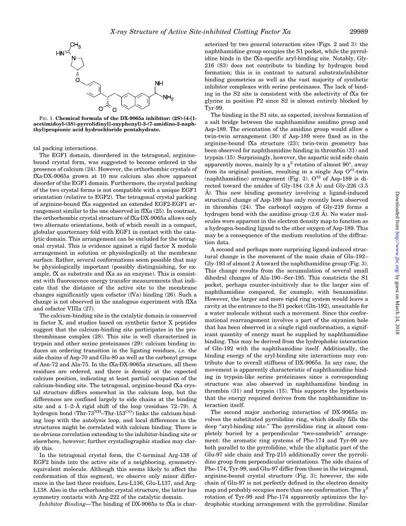
acterized by two general interaction sites (Figs. 2 and 3): thenaphthamidine group occupies the S1 pocket, while the pyrrol-idine binds in the fXa-specific aryl-binding site. Notably, Gly-216 (S3) does not contribute to binding by hydrogen bondformation; this is in contrast to natural substrate/inhibitorbinding geometries as well as the vast majority of syntheticinhibitor complexes with serine proteinases. The lack of bind-ing in the S2 site is consistent with the selectivity of fXa forglycine in position P2 since S2 is almost entirely blocked byTyr-99.The binding in the S1 site, as expected, involves formation of
a salt bridge between the naphthamidine amidino group andAsp-189. The orientation of the amidino group would allow atwin-twin arrangement (30) if Asp-189 were fixed as in thearginine-bound fXa structure (23); twin-twin geometry hasbeen observed for naphthamidine binding in thrombin (31) andtrypsin (15). Surprisingly, however, the aspartic acid side chainapparently moves, mainly by a x2 rotation of almost 90°, awayfrom its original position, resulting in a single Asp Od1-twin(naphthamidine) arrangement (Fig. 3). Od2 of Asp-189 is di-rected toward the amides of Gly-184 (3.8 Å) and Gly-226 (3.5Å). This new binding geometry involving a ligand-inducedstructural change of Asp-189 has only recently been observedin thrombin (24). The carbonyl oxygen of Gly-219 forms ahydrogen bond with the amidino group (2.6 Å). No water mol-ecules were apparent in the electron density map to function asa hydrogen-bonding ligand to the other oxygen of Asp-189. Thismay be a consequence of the medium resolution of the diffrac-tion data.A second and perhaps more surprising ligand-induced struc-
tural change is the movement of the main chain of Gln-192–Gly-193 of almost 2 Å toward the naphthamidine group (Fig. 3).This change results from the accumulation of several smalldihedral changes of Ala-190–Ser-195. This constricts the S1pocket, perhaps counter-intuitively due to the larger size ofnaphthamidine compared, for example, with benzamidine.However, the larger and more rigid ring system would leave acavity at the entrance to the S1 pocket (Gln-192), unsuitable fora water molecule without such a movement. Since this confor-mational rearrangement involves a part of the oxyanion holethat has been observed in a single rigid conformation, a signif-icant quantity of energy must be supplied by naphthamidinebinding. This may be derived from the hydrophobic interactionof Gln-192 with the naphthamidine itself. Additionally, thebinding energy of the aryl-binding site interactions may con-tribute due to overall stiffness of DX-9065a. In any case, themovement is apparently characteristic of naphthamidine bind-ing in trypsin-like serine proteinases since a correspondingstructure was also observed in naphthamidine binding inthrombin (31) and trypsin (15). This supports the hypothesisthat the energy required derives from the naphthamidine in-teraction itself.The second major anchoring interaction of DX-9065a in-
volves the substituted pyrrolidine ring, which ideally fills thedeep “aryl-binding site.” The pyrrolidine ring is almost com-pletely buried by a perpendicular “two-sandwich” arrange-ment: the aromatic ring systems of Phe-174 and Tyr-99 areboth parallel to the pyrrolidine, while the aliphatic part of theGlu-97 side chain and Trp-215 additionally cover the pyrroli-dine group from perpendicular orientations. The side chains ofPhe-174, Tyr-99, and Glu-97 differ from those in the tetragonal,arginine-bound crystal structure (Fig. 3); however, the sidechain of Glu-97 is not perfectly defined in the electron densitymap and probably occupies more than one conformation. The x2
rotation of Tyr-99 and Phe-174 apparently optimizes the hy-drophobic stacking arrangement with the pyrrolidine. Similar
FIG. 1. Chemical formula of the DX-9065a inhibitor: (2S)-{4-[1-acetimidoyl-(3S)-pyrrolidinyl]-oxyphenyl}-3-(7-amidino-2-naph-thyl)propionic acid hydrochloride pentahydrate.
X-ray Structure of Active Site-inhibited Clotting Factor Xa 29989
by guest on March 24, 2018
http://ww
w.jbc.org/
Dow
nloaded from

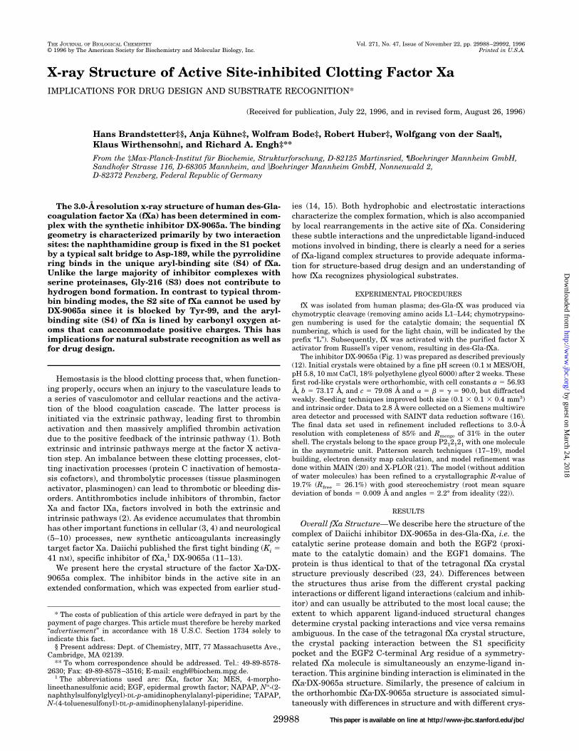
to naphthamidine binding in the S1 pocket, the interaction inthe aryl-binding site is not solely hydrophobic in character. Thecarbonyl oxygen atoms of Lys-96 and Glu-97 together with theGlu-97 side chain form a “nitrogen-cation hole” or simply “cat-ion hole,” analogous to the oxyanion hole of the catalytic site.The cation hole interacts with the delocalized positive charge ofthe pyrrolidine ring and the charged acetimidoyl group point-ing toward Glu-97. The electron density map indicates that thechiral center of the pyrrolidine is in the S-configuration (Fig. 2),as expected from the synthesis of the enantiomerically purecompound (Fig. 1).It is clear from the electron density map that the enantiomer
with an S-configuration of the propionic acid binds the enzyme.The charge of the propionic acid is not involved in a direct ionicinteraction, but is exposed to solvent, reminiscent of the sulfo-nyl group of the NAPAP/TAPAP inhibitor series (34). Ne2 ofGln-192 forms a weak hydrogen bond with propionic acid (4.1Å), while Oe1 interacts with the guanidinium group of Arg-143,i.e. the side chain of Gln-192 forms an ionic bridge between thepropionic acid and Arg-143.The center of the phenyl ring almost coincides with Ca of the
D-Phe residue of a superimposed D-Phe-Pro-Arg chloromethyl
ketone-thrombin complex structure. An aromatic ring systemis advantageous entropically as a spacer between S1 and thearyl-binding site (S4) since it restricts the conformational de-grees of freedom of the compound. This structure demonstratesthe feasibility of ignoring hydrogen bonding at Gly-216. Thus,the geometric restraints of binding in the S1 and aryl-bindingpockets should allow considerable variation of this spacer ininhibitor design.
DISCUSSION
Implications for Natural Substrate Interactions—The physi-ological role of fXa within the blood coagulation cascade con-sists of activation of prothrombin to thrombin and, as a form ofpositive feedback within the extrinsic pathway, activation offactor VII. There is, however, an ongoing debate about thephysiologically relevant fVII activator since fVIIa, fIXa, andthrombin can also activate fVII to fVIIa in vitro (1). Prothrom-bin activation occurs by two cleavages. The more rapid cleavagetakes place at Arg-323–Ile-324 (designated as prothrombin spe-cies 2 in Table I), so meizothrombin accumulates as the prin-cipal intermediate of the activation reaction under physiologi-cal conditions (35). As the resulting two chains remain
FIG. 2. The DX-9065a inhibitor is unambiguously determined by the electron density map. The 2uFou 2 uFcu density map shown iscontoured at 1s over the average. The figure is shown with MAIN (20).
FIG. 3. Binding interactions of DX-9065a with fXa. The Ca plot and side chains involved in inhibitor binding of DX-9065a-bound fXa (yellow)are superimposed with the corresponding atoms of arginine-bound fXa (turquoise). The ligand-induced structural changes at the S1-binding sitemay be seen at the side chain of Asp-189 and along the main chain at Gln-192. The hydrophobic sleeve at the aryl-binding site (S4) is also apparent,with the cation hole formed by Glu-97 and the carbonyl oxygens of Glu-97 and Lys-96 at the back.
X-ray Structure of Active Site-inhibited Clotting Factor Xa29990
by guest on March 24, 2018
http://ww
w.jbc.org/
Dow
nloaded from
disulfide-linked, the proteolytically fully active meizothrombinremains membrane-bound (analogous to fVIIa, fIXa, fXa, oractivated protein C) and is released only with the second cleav-age (Arg-274–Thr-275).The two ligand-induced motions of the S1 site, namely the
aspartic acid 189 rotamer transition and a rigid body motion ofresidues of the oxyanion hole, have also been observed in recentthrombin-inhibitor complexes (24, 31). The oxyanion hole re-mains intact, so motions of the main chain including residues190–195 might also be induced by attractive polar interactionswith the carbonyl group of natural substrates. Alternatively,ligand or cofactor binding might induce motions of this segmentand alter substrate recognition. Thus, this motion may havephysiological relevance as well.The S2 site is hardly accessible to bulky substrate residues.
The 60-loop is in direct contact with the 99-loop, partly via thepolar interaction of the system of Tyr-60 with Lys-96 (3 Å) andpartly via a hydrogen bond between the hydroxyl group ofTyr-60 and the amine of Lys-90. In its low energy conformation,Tyr-99 then blocks access to the S2 site for any side chain,which is consistent with the natural substrate specificity of fXa(Table I). The S2 site of fXa, and in particular Tyr-99, shouldnot, however, be considered absolutely rigid; a strong bindingligand may provide the energy necessary to move Tyr-99 andallow access in an expanded S2 site. This is reflected by the factthat fXa can cleave fVIII at thrombin-specific activation sites(36). The S2 site of fXa is in stark contrast to that of fIXa, wherethe 60-loop (with identical length) opens a large channel for P2and possibly also other substrate residues. The structural basisfor the observed difference of the 60-loop in both enzymes isprobably related to the missing Tyr-60 (and its stabilizinginteractions) in fIXa.The clear preference for glycine at P2 for fXa substrates, the
clear preferences for other residues at P2 for thrombin and fIXasubstrates, and the conservation of Gly at this position in thelimited number of fVII sequences available are evidence for aphysiological role of fXa as activator of fVII. While the identi-ties of the primed site residues at the activation site of serineproteases are restricted by structural requirements, the con-servation of unprimed site residues, if real, most likely reflectsrequirements for substrate recognition. Alternatively, conser-vation of a sequence less optimal for thrombin or fIXa cleavagemight arise from a need to limit cleavage rates for these en-zymes if required by the dynamics of the coagulation reactions.
The P3 residue typically forms a two-rung antiparallel b-lad-der with Gly-216 and by this constraint leaves the side chain ofan L-amino acid exposed to solvent. Therefore, we expect apreference for hydrophilic residues at P3. The propionic acid inDX-9065a mimics P3 in this respect. It should be emphasizedthat conservation observed in cleavage sites may not necessar-ily imply a special binding interaction, but may reflect otherrequirements of the substrate; this is certainly the case withthe P19, P29, and P49 residues of prothrombin species 2 and fVIIcleavage sites as they are critical for the activation mechanism(37, 38).The deep aryl-binding site (S4) is also distinctive with its
surrounding carbonyl groups (from Lys-96 and Glu-97) to-gether with the side chain of Glu-97. The importance of thecation hole for the recognition of physiological substrates hasnot been established. This structure suggests that besides hy-drophobic residues (such as Ile), histidine in particular shouldbe an ideal candidate for binding at the S4 site, although thesequential position on a substrate might not correspond to P4,considering the structure of thrombin-fibrinopeptide A (39):this site can also be occupied by substrate residue Pn, where nmay be different from 4 (n $ 4).Autolysis Loop: A Model for Substrate Recognition?—The
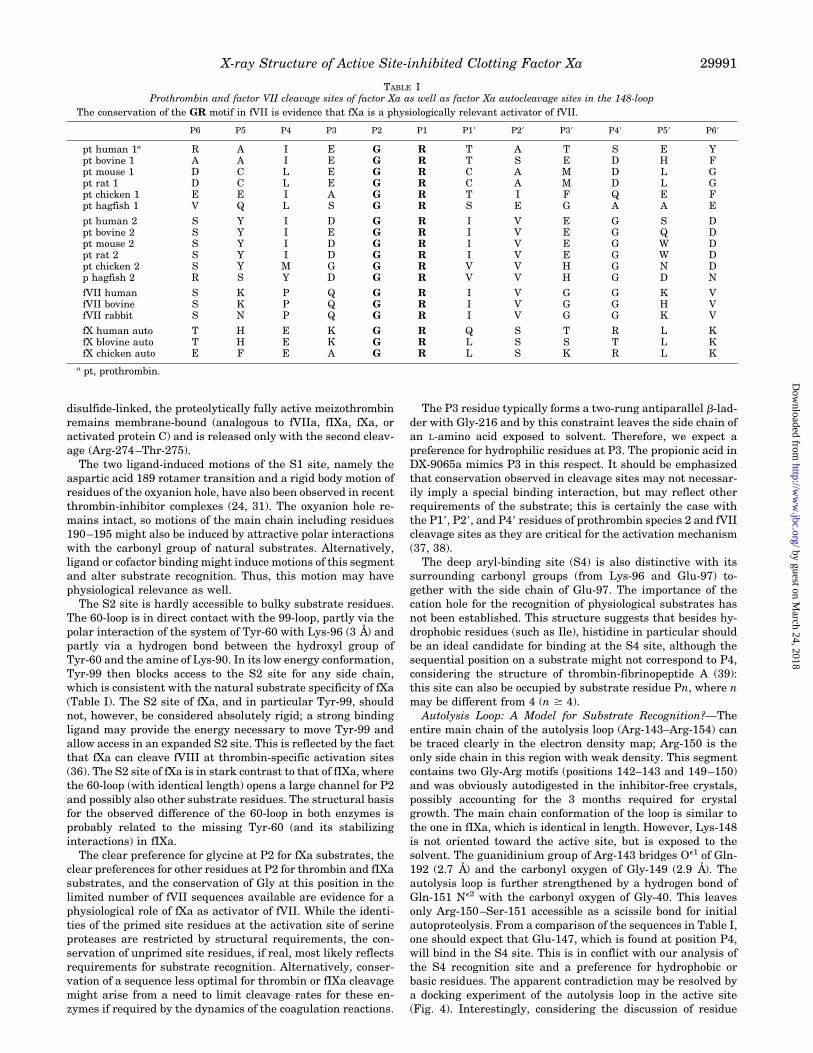
entire main chain of the autolysis loop (Arg-143–Arg-154) canbe traced clearly in the electron density map; Arg-150 is theonly side chain in this region with weak density. This segmentcontains two Gly-Arg motifs (positions 142–143 and 149–150)and was obviously autodigested in the inhibitor-free crystals,possibly accounting for the 3 months required for crystalgrowth. The main chain conformation of the loop is similar tothe one in fIXa, which is identical in length. However, Lys-148is not oriented toward the active site, but is exposed to thesolvent. The guanidinium group of Arg-143 bridges Oe1 of Gln-192 (2.7 Å) and the carbonyl oxygen of Gly-149 (2.9 Å). Theautolysis loop is further strengthened by a hydrogen bond ofGln-151 Ne2 with the carbonyl oxygen of Gly-40. This leavesonly Arg-150–Ser-151 accessible as a scissile bond for initialautoproteolysis. From a comparison of the sequences in Table I,one should expect that Glu-147, which is found at position P4,will bind in the S4 site. This is in conflict with our analysis ofthe S4 recognition site and a preference for hydrophobic orbasic residues. The apparent contradiction may be resolved bya docking experiment of the autolysis loop in the active site(Fig. 4). Interestingly, considering the discussion of residue
TABLE IProthrombin and factor VII cleavage sites of factor Xa as well as factor Xa autocleavage sites in the 148-loop
The conservation of the GR motif in fVII is evidence that fXa is a physiologically relevant activator of fVII.
P6 P5 P4 P3 P2 P1 P19 P29 P39 P49 P59 P69
pt human 1a R A I E G R T A T S E Ypt bovine 1 A A I E G R T S E D H Fpt mouse 1 D C L E G R C A M D L Gpt rat 1 D C L E G R C A M D L Gpt chicken 1 E E I A G R T I F Q E Fpt hagfish 1 V Q L S G R S E G A A E
pt human 2 S Y I D G R I V E G S Dpt bovine 2 S Y I E G R I V E G Q Dpt mouse 2 S Y I D G R I V E G W Dpt rat 2 S Y I D G R I V E G W Dpt chicken 2 S Y M G G R V V H G N Dp hagfish 2 R S Y D G R V V H G D N
fVII human S K P Q G R I V G G K VfVII bovine S K P Q G R I V G G H VfVII rabbit S N P Q G R I V G G K V
fX human auto T H E K G R Q S T R L KfX blovine auto T H E K G R L S S T L KfX chicken auto E F E A G R L S K R L K
a pt, prothrombin.
X-ray Structure of Active Site-inhibited Clotting Factor Xa 29991
by guest on March 24, 2018
http://ww
w.jbc.org/
Dow
nloaded from
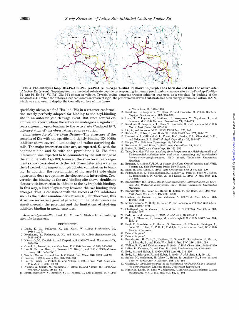
specificity above, we find His-145 (P5) in a rotamer conforma-tion nearly perfectly adapted for binding to the aryl-bindingsite in an autocatalytic cleavage event. But since several ex-amples are known where the substrate undergoes a significantrearrangement upon binding to the active site (“induced fit”),interpretation of this observation requires caution.Implication for Future Drug Design—The structure of the
complex of fXa with the specific and tightly binding DX-9065ainhibitor shows several illuminating and rather surprising de-tails. The major interaction sites are, as expected, S1 with thenaphthamidine and S4 with the pyrrolidine (15). The firstinteraction was expected to be dominated by the salt bridge ofthe amidine with Asp-189; however, the structural rearrange-ments show (consistent with the lack of any detectable water inthe S1 pocket) the important hydrophobic contribution to bind-ing. In addition, the reorientation of the Asp-189 side chainapparently does not optimize the electrostatic interaction. Con-versely, the binding at the aryl-binding site shows significantelectrostatic interaction in addition to the hydrophobic binding.In this way, a kind of symmetry between the two binding sitesemerges. This is consistent with the success of fXa inhibitorssuch as the bisbenzamidine derivatives (40). Furthermore, thisstructure serves as a general paradigm in that it demonstratessimultaneously the potential and the limitations of studyinginhibitor binding in model enzymes.
Acknowledgment—We thank Dr. Milton T. Stubbs for stimulatingscientific discussions.
REFERENCES
1. Davie, E. W., Fujikawa, K., and Kisiel, W. (1991) Biochemistry 30,10363–10370
2. Komiyama, Y., Pedersen, A. H., and Kisiel, W. (1990) Biochemistry 29,9418–9425
3. Nierodzik, M., Klepfish, A., and Karpatkin, S. (1995) Thromb. Haemostasis 74,282–290
4. Grand, R., Turnell, A., and Grabham, P. (1996) Biochem. J. 313, 353–3685. Lee, K., Betz, A., Keep, R., Chenevert, T., Kim, S., and Hoff, J. (1995) J. Neu-
rosurg. 83, 1045–10506. Teo, M., Manser, E., and Lim, L. (1995) J. Biol. Chem. 270, 26690–266977. Brewer, G. (1995) Brain Res. 683, 258–2638. Liu, Y., Fields, R., Festoff, B., and Nelson, P. (1994) Proc. Natl. Acad. Sci.
U. S. A. 91, 10300–103049. Nishino, A., Suzuki, M., Yoshimoto, T., Otani, H., and Nagura, H. (1994) Acta
Neurochir. Suppl. 60, 86–8810. Smith-Swintosky, V., Zimmer, S., II, Fenton, J., and Mattson, M. (1995)
J. Neurochem. 65, 1415–141811. Katakura, S., Nagahara, T., Hara, T., and Iwamoto, M. (1993) Biochem.
Biophys. Res. Commun. 197, 965–97212. Hara, T., Yokoyama, A., Ishihara, H., Yokoyama, Y., Nagahara, T., and
Iwamoto, M. (1994) Thromb. Haemostasis 71, 314–31913. Katakura, S., Nagahara, T., Hara, T., Kunitada, S., and Iwamoto, M. (1995)
Eur. J. Med. Chem. 30, 387–39414. Lin, Z., and Johnson, M. E. (1995) FEBS Lett. 370, 1–515. Stubbs, M., Huber, R., and Bode, W. (1995) FEBS Lett. 375, 103–10716. Howard, A. J., Gilliland, G. L., Finzel, B. C., Poulos, T. L., Ohlendorf, D. H.,
and Salemme, F. R. (1987) J. Appl. Crystallogr. 20, 383–38717. Hoppe, W. (1957) Acta Crystallogr. 10, 750–75118. Rossmann, M., and Blow, D. (1962) Acta Crystallogr. 15, 24–3119. Huber, R. (1965) Acta Crystallogr. 19, 353–35620. Turk, D. (1992) Weiterentwicklung eines Programms fur Molekulgraphik und
Elektronendichte-Manipulation und seine Anwendung auf verschiedeneProtein-Strukturaufklarungen. Ph.D. thesis, Technische UniversitatMunchen
21. Brunger, A. (1993) X-PLOR: A System for X-ray Crystallography and NMR,Version 3.1, Yale University Press, New Haven, CT
22. Engh, R., and Huber, R. (1991) Acta Crystallogr. Sect. A 47, 392–40023. Padmanabhan, K., Padmanabhan, K., Tulinsky, A., Park, C., Bode, W., Huber,
R., Blankenship, D., Cardin, A., and Kisiel, W. (1993) J. Mol. Biol. 232,947–966
24. Brandstetter, H. (1994) Rontgenkristallographische Untersuchungen an Enzy-men des Blutgerinnungssystems. Ph.D. thesis, Technische UniversitatMunchen
25. Brandstetter, H., Bauer, M., Huber, R., Lollar, P., and Bode, W. (1995) Proc.Natl. Acad. Sci. U. S. A. 92, 9796–9800
26. Husten, E., Esmon, C., and Johnson, A. (1987) J. Biol. Chem. 262,12953–12961
27. Mutucumarana, V., Duffy, E., Lollar, P., and Johnson, A. (1992) J. Biol. Chem.267, 17012–17021
28. Chattopadhyay, A., James, H. L., and Fair, D. S. (1992) J. Biol. Chem. 267,12323–12329
29. Bode, W., and Schwager, P. (1975) J. Mol. Biol. 98, 693–71730. Singh, J., Thornton, J., Snarey, M., and Campbell, S. (1987) FEBS Lett. 224,
161–17131. Engh, R., Brandstetter, H., Sucher, G., Baumann, U., Kuhne, A., Eichinger, A.,
Bode, W., Huber, R., Poll, T., Rudolph, R., and von der Saal, W. (1996)Structure, in press
32. Deleted in proof33. Deleted in proof34. Brandstetter, H., Turk, D., Hoeffken, H., Grosse, D., Sturzebecher, J., Martin,
P., Edwards, B., and Bode, W. (1992) J. Mol. Biol. 226, 1085–109935. Walker, R. K., and Krishnaswamy, S. (1994) J. Biol. Chem. 269, 27441–2745036. Lollar, P., Knutson, G., and Fass, D. (1985) Biochemistry 24, 8056–806437. Bode, W., and Huber, R. (1976) FEBS Lett. 68, 231–23638. Bode, W., Schwager, P., and Huber, R. (1978) J. Mol. Biol. 118, 99–11239. Stubbs, M., Oschkinat, H., Mayr, I., Huber, R., Angliker, H., Stone, S., and
Bode, W. (1992) Eur. J. Biochem. 206, 187–19540. Gabriel, B. (1996) Bisbenzamidine als Inhibitoren von Faktor Xa und verwand-
ten Serinproteasen. Diploma thesis, Universitat Regensburg41. Huber, R., Kukla, D., Bode, W., Schwager, P., Bartels, K., Deisenhofer, J., and
Steigemann, W. (1974) J. Mol. Biol. 89, 73–101
FIG. 4. The autolysis loop (His-P5–Glu-P4–Lys-P3–Gly-P2–Arg-P1–Gln-P1*; shown in purple) has been docked into the active siteof factor Xa (green). Superimposed is a modeled substrate peptide corresponding to human prothrombin cleavage site 2 (Ile-P4–Asp-P3–Gly-P2–Arg-P1–Ile-P19–Val-P29–Glu-P39; shown in yellow). Trypsin-bovine pancreas trypsin inhibitor was used as a template for docking of thesubstrates (41). While the autolysis loop conformation was kept rigid, the prothrombin-derived substrate has been energy-minimized within MAIN,which was also used to display the Connolly surface of this figure.
X-ray Structure of Active Site-inhibited Clotting Factor Xa29992
by guest on March 24, 2018
http://ww
w.jbc.org/
Dow
nloaded from
Klaus Wirthensohn and Richard A. EnghHans Brandstetter, Anja Kühne, Wolfram Bode, Robert Huber, Wolfgang von der Saal,
DRUG DESIGN AND SUBSTRATE RECOGNITIONX-ray Structure of Active Site-inhibited Clotting Factor Xa: IMPLICATIONS FOR
doi: 10.1074/jbc.271.47.299881996, 271:29988-29992.J. Biol. Chem.
http://www.jbc.org/content/271/47/29988Access the most updated version of this article at
Alerts:
When a correction for this article is posted•
When this article is cited•
to choose from all of JBC's e-mail alertsClick here
http://www.jbc.org/content/271/47/29988.full.html#ref-list-1
This article cites 34 references, 8 of which can be accessed free at
by guest on March 24, 2018
http://ww
w.jbc.org/
Dow
nloaded from