Anatomy – Exam 3
Organization of the Neck ○ Objectives
Organization of the neck: describe the cervical fascia and compartments
Describe the cervical regions/triangles. What are their boundaries and contents
What structures are found in the “key vertebral levels” in the neck?
Muscles of the neck, describe the neck muscles. Know their innervation, attachments and primary actions
○ Cervical Fascia
Superficial – contains fat, nerves, vasculature and platysma muscle
Deep
Investing Fascia – around just about everything, goes on both sides of the SCM and trapezius
○ Goes from chin to hyoid, to manubrium
Pretracheal Space – between investing and pretracheal fascia starting just above the manubrium
○ Contains thyroid and is continuous with thorax anterior to the pericardium
Pretracheal Fascia - in front or around trachea, blends with fibrous pericardium
○ Visceral Layer - goes around trachea
Buccopharyngeal Portion – lines the posterior portion of the trach?ea
Contents – esophagus, pharynx, larynx, thyroid glands, parathyroid glands
Makes Visceral Compartment
○ Muscular Layer – covers the muscles between the trachea and the carotids (The 'SOS' muscles)
Prevertebral Fascia – goes around most of the muscles associated with the vertebra in the neck
○ Makes Vertebral Compartment
○ Contains cervical vertebrae, spinal cord, cervical nerves, cervical muscles
Carotid Sheath – goes around the internal jugular vein, common carotid artery, internal carotid and the
vagus nerve
○ Note – does not go around external carotid artery
Retropharyngeal Space – between buccopharyngeal fascia and prevertebral fascia
○ Opens into posterior or superior mediastinum
○ Spread of infection into this area can cause dysphagia (difficulty swallowing) or dysarthria (speaking)
Layers from Spine to Buccopharyngeal Fascia
Anterior longitudinal ligament (along spine) → Longus colli muscle → Prevertebral fascia →
retropharyngeal space → buccopharyngeal fascia → (pharyngeal muscle)
○ Key Vertebral Levels
C3 – Hyoid bone
C4 – Carotid bifurcation (into external and internal)
C4-5 – Thyroid cartilage (adam‟s apple)
C5 – Cricoid cartilage
C5-6 – inferior limit of pharynx and larynx; Superior limit of trachea and esophagus
Contains indentation between cricoid cartilage and 1st tracheal ring
C6-T1 – thyroid gland

○ SUPERFICIAL FASCIA
Contents Platysma
Anterior & external jugular veins
Cutaneous branches of cervical plexus
Cervical branch of facial nerve (CN VII)
○ DEEP FASCIA
Investing
Surrounds entire neck deep to skin and subcutaneous fascia
Encloses trapezius, SCM, and parotid and submandibular glands
Superior: blends with fascia of skull (including mandible) &
hyoid
Inferior: extends to manubrium, clavicle & scapula
Posterior: continuous with nuchal ligament to C7 spinous
process
Forms roof of cervical triangles
CN XI may adhere to deep surface of this fascia
Pretracheal
In anterior part of neck: visceral and muscular parts
Visceral part: encloses neck viscera (thyroid; parathyroids;
trachea; pharynx, and esophagus)
Superiorly: attaches to hyoid bone
Inferiorly: blends with fibrous pericardium
Laterally: blends with carotid sheath
Buccopharyngeal fascia: subdivision of pretracheal fascia
posterior to pharynx & esophagus
Forms anterior boundary of retropharyngeal space
Visceral part: encloses infrahyoid muscles
Retropharyngeal space
Potential space between buccopharyngeal fascia &
prevertebral fascia
Extends from base of skull into thorax
Function: facilitates movement of cervical viscera against
vertebral column
Route for spread of infection (e.g., from nasopharyngeal
tonsils)
Prevertebral
Surrounds vertebral column and its muscles
Lateral: forms axillary sheath
Superior: attaches to base of skull
Inferior: blends with anterior longitudinal ligament
Forms floor of posterior triangle and posterior
boundary of retropharyngeal space
Sympathetic chain in/on anterior aspect of this fascia
Carotid sheath
Neurovascular compartment at lateral edge of
retropharyngeal space
Contents CCA, ICA, and IJV, Vagus nerve (CN X),
Carotid sinus & body, Sympathetic fibers, Deep cervical lymph nodes
○ Regions
Lateral Cervical Region (Posterior Cervical Triangle)
Boundaries
○ Anterior – SCM
○ Posterior – Trapezius
○ Inferior – middle 1/3 of clavicle
○ Roof – investing fascia
○ Floor – muscles covered by prevertebral fascia (splenius capitus, levator scapulae, scalenes)
Divisions of Lateral Cervical Region

○ Occipital (rostral)/Subclavian Triangles – divided by the inferior belly of the omohyoid
○ Safe/Dangerous Areas – divided by accessory nerve (CN XI)
Contents
○ ○ Muscles – floor muscles (splenius capitis, levator scapulae, middle scalene, posterior scalene), and
inferior belly of omohyoid
○ Arteries – transverse cervical, suprascapular, 3rd
part of subclavian, and occipital arteries
○ Veins – Subclavian vein (receives external jugular)
External Jugular and branches (transverse cervical, suprascapular and anterior jugular veins)
○ Nerves – accessory nerve, roots of brachial plexus, suprascapular, phrenic nerves and
Cervical Plexus (C1-4) – the cutaneous branches
○ Lesser Occipital (C2) – travels superiorly
○ Greater Auricular (C2-3) – goes over SCM muscle vertically
○ Transverse Cervical (C2-3) – goes over SCM muscle horizontally
○ Supraclavicular nerves (C3-4) – goes along the external jugular vein inferiorly
Note – the external jugular vein goes over the SCM muscle
Anterior Cervical Region
Boundaries
○ Anterior – anterior midline of
neck
○ Posterior – SCM
○ Superior – mandible
Divisions
○ Submental (unpaired) – bounded
by anterior bellies of digastric
muscles and the hyoid bone
Floor – mylohyoid muscles
Contains submental lymph
nodes, and small veins
(tributaries of anterior jugular
vein)
○ Submandibular (paired) – bounded by mandible and anterior & posterior bellies of digastric muscles
Floor – mylohyoid muscle, hyoglossus muscle, middle pharyngeal constrictor muscle
Contains submandibular gland, submandibular lymph nodes, mylohyoid nerve, hypoglossal nerve
(CN XII), parts of fascial artery and vein
Glandular
○ Carotid Triangle (paired) – bounded by superior belly of omohyoid, posterior belly of digastric and
SCM
Floor – inferior pharyngeal constrictor muscle
Contents
○ Common carotid, internal carotid, external carotid (and branches) arteries
○ Carotid body and sinus
○ Internal jubular vein and tributaries
○ Deep cervical lymph nodes
○ Vagus, hypoglossal, and accessory nerves
○ Branches of the cervical plexus ○ Larynx and pharynx
○ Thyroid and parathyroid glands
Vascular
○ Muscular (paired) – bounded by superior belly of omohyoid, SCM and midline of neck
Contains infrahyoid muscles, thyroid and parathyroid glands
○ Neck Muscles
Note – there is an intermediate tendon that goes around the stylohyoid and diagastric muscles
Infrahyoid
Note – sternohyoid and omohyoid muscles are long
„SOS‟ – sternohyoid, omohyoid, sternothyroid muscles are innervated by ansa cervicalis
Note – omohyoid becomes tendinous (intermediate tendon) between its two bellies
These help stabilize hyoid and help with swallowing
Note – Scalenes have origin on TVP and insert on the ribs
Lateral flexors of neck and accessory respiratory muscles
Interscalene Space – between anterior and middle scalene muscles
Lets the brachial plexus and subclavian artery through
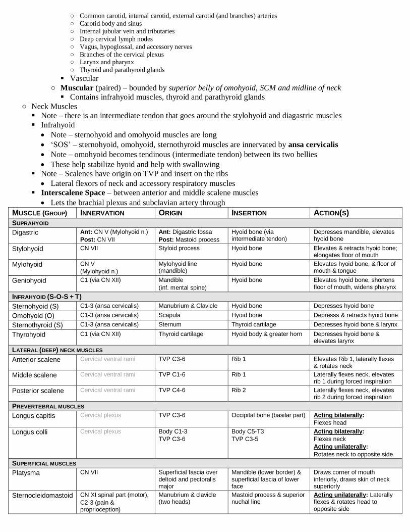
MUSCLE (GROUP) INNERVATION ORIGIN INSERTION ACTION(S)
SUPRAHYOID
Digastric Ant: CN V (Mylohyoid n.)
Post: CN VII
Ant: Digastric fossa
Post: Mastoid process
Hyoid bone (via intermediate tendon)
Depresses mandible, elevates hyoid bone
Stylohyoid CN VII Styloid process Hyoid bone Elevates & retracts hyoid bone; elongates floor of mouth
Mylohyoid CN V
(Mylohyoid n.)
Mylohyoid line (mandible)
Hyoid bone Elevates hyoid bone, & floor of mouth & tongue
Geniohyoid C1 (via CN XII) Mandible
(inf. mental spine)
Hyoid bone Elevates hyoid bone, shortens floor of mouth, widens pharynx
INFRAHYOID (S-O-S + T)
Sternohyoid (S) C1-3 (ansa cervicalis) Manubrium & Clavicle Hyoid bone Depresses hyoid bone
Omohyoid (O) C1-3 (ansa cervicalis) Scapula Hyoid bone Depresss & retracts hyoid bone
Sternothyroid (S) C1-3 (ansa cervicalis) Sternum Thyroid cartilage Depresses hyoid bone & larynx
Thyrohyoid C1 (via CN XII) Thyroid cartilage Hyoid body & greater horn Depresses hyoid bone & elevates larynx
LATERAL (DEEP) NECK MUSCLES
Anterior scalene Cervical ventral rami TVP C3-6 Rib 1 Elevates Rib 1, laterally flexes & rotates neck
Middle scalene Cervical ventral rami TVP C1-6 Rib 1 Laterally flexes neck, elevates rib 1 during forced inspiration
Posterior scalene Cervical ventral rami TVP C4-6 Rib 2 Laterally flexes neck, elevates rib 2 during forced inspiration
PREVERTEBRAL MUSCLES
Longus capitis Cervical plexus TVP C3-6 Occipital bone (basilar part) Acting bilaterally:
Flexes head
Longus colli Cervical plexus Body C1-3
TVP C3-6
Body C5-T3
TVP C3-5
Acting bilaterally:
Flexes neck
Acting unilaterally:
Rotates neck to opposite side
SUPERFICIAL MUSCLES
Platysma CN VII Superficial fascia over deltoid and pectoralis major
Mandible (lower border) & superficial fascia of lower face
Draws corner of mouth inferiorly, draws skin of neck superiorly
Sternocleidomastoid CN XI spinal part (motor),
C2-3 (pain & proprioception)
Manubrium & clavicle (two heads)
Mastoid process & superior nuchal line
Acting unilaterally: Laterally flexes & rotates head to opposite side

Acting bilaterally: Flexes neck
Trapezius CN XI spinal part C3-4 (pain & proprioception)
Occipital bone, lig. nuchae & spinous processes
Scapula and clavicle Elevates, retracts, & rotates scapula to tilt glenoid cavity superiorly
Nerves and Vessels of the Neck ○ Objectives
Blood Vessels
Describe the venous drainage of the neck. What is the relationship between the internal jugular vein and the carotid sheath? Where is the external jugular vein located?
Describe the branches of the external carotid artery. Are there branches of the internal carotid artery in the neck?
Describe the carotid body and carotid sinus
Nerves and Plexuses
Describe the cervical and branchial plexuses in the neck. Cervical plexus (ventral rami C1-C4) cutaneous nerves, ansa cervicalis
(C1-3) and phrenic nerve (C3-5). Brachial plexus (ventral rami C5-T1)
Describe the cranial nerves that course through and/or supply the neck: glossopharyngeal nerve, vagus nerve, accessory nerve,
hypoglossal nerve
Describe the cervical sympathetic trunk, its ganglia and branches
○ Veins of the Neck
Retromandibular Vein – drains into external jugular
Main tributaries
○ Superficial Temporal vein –
○ Maxillary veins – drains the pterygoid plexus (which is
deep to mandible)
External Jugular Vein – is in superficial fascia
Main tributaries (superiorly)
○ Retromandibular vein -
○ Posterior Auricular vein – from behind the ear
Other tributaries (inferiorly)
○ Anterior Jugular vein – is medial to the SCM
○ Transverse Cervical vein –
○ Suprascapular vein –
Terminates on Subclavian vein
Internal Jugular Vein – receives blood from brain
Main Tributaries – most come in anteriorly
○ Sigmoid Sinus – a continuation of IJV receiving brain blood
○ Inferior Petrosal Sinus – comes in anteriorly
○ Occipital Vein – comes in posteriorly
○ Pharyngeal Veins –
○ Common Facial Vein –
○ Lingual Vein –
○ Superior and Middle Thyroid Veins – supply thyroid
Unites with Subclavian to create the brachiocephalic vein
Just before the union with subclavian there is a bicuspid valve
that prevents backflow
○ External Carotid Artery (ECA)
Note – internal carotid doesn‟t branch in the neck
Note – external carotid artery isn’t in the carotid sheath
„SALFOAMS‟ when naming branches inferior to superior
Superficial Temporal Artery – supplies parotid & temporal regions
of skull
Can take a pulse here
Maxillary – supplies tissues around maxilla
Posterior Auricular – supplies tissues around external ear
Occipital – supplies posterior scalp and SCM
Facial – supplies face; ends in angular artery
Can take pulse, right at jaw line where it comes over mandible

Lingual – supplies tongue & floor of mouth
Can share common branch with facial artery
Ascending Pharyngeal Artery – supplies pharynx and SCM
Superior Thyroid Artery – supplies thyroid gland, SCM, infrahyoid muscles, part of larynx
○ Carotid Body & Carotid Sinus
Carotid Sinus Carotid Body
Location Dilation at bifurcation Ovoid mass at bifurcation
Receptors Baroreceptors Chemoreceptors
Response Reacts to ↑ arterial pressure
changes to ↓ HR
Reacts to ↓ O2 or ↑ CO2 → ↑ rate and
depth of respiration, ↑ HR, ↑ BP
Innervation Carotid Sinus Nerve – branch of glossopharyngeal (CN IX)
Secondary – vagus (provides parasympathetic vasodilation)
Secondary – cervical sympathetics (provide sympathetic vasoconstriction)
○ Cervical Plexus
Made of ventral rami of C1-C4
Motor Innervation
Hypoglossal Nerve (CN XII) – not part of cervical plexus
○ Innervates – tongue, geniohyoid and thyrohyoid
C1 Fibers – travel with hypoglossal nerve after coming off of C1
○ Thyrohyoid Nerve, Geniohyoid Nerve
Ansa Cervicalis (C1-C3) – „goose neck‟; lies on carotid sheath; „SOS‟
○ Has inferior root and superior root
○ Supplies the infrahyoid muscles and deep neck muscles
○ Sternohyoid Nerve, Omohyoid Nerve, Sternothyroid Nerve
Sensory Innervation
Ventral Rami – supply skin of anterior and lateral neck
○ Lesser Occipital (C2) – innervates neck and scalp posterosuperior
to auricle
○ Great Auricular (C2-3) – skin over parotid gland; posterior aspect
of auricle; between angle of mandible & mastoid process
○ Transverse Cervical (C2-3) – anterior cervical region
○ Supraclavicular (C3-4) – neck and shoulder
Dorsal Rami – supplies skin of posterior head and neck
○ Suboccipital (C1) –
○ Greater Occipital (C2) –
○ Third Occipital Nerve (C3) –
○ Cranial Nerves of the Neck
Glossopharyngeal (CN IX) – medial to vagus nerve
There is a bunch of extra info he didn‟t cover
Vagus (CN X) – in carotid sheath
There is a bunch of extra info he didn‟t cover
Supplies all the intrinsic laryngeal muscles (muscles of speech)
Superior Laryngeal Nerve – branch of vagus nerve
○ Internal Branch – penetrates the thyrohyoid membrane to be sensory to mucosa of larynx
○ External Branch – motor to cricothyroid muscle (a laryngeal muscle)
○ Note that there is a Superior Laryngeal Artery (branch of superior thyroid) that follows the superior
laryngeal nerve
Recurrent Laryngeal Nerve – goes around an artery then goes superiorly in the tracheoesophageal
groove
○ Motor to all intrinsic laryngeal muscles, except the cricothyroid
○ Right Recurrent Laryngeal – goes under subclavian
○ Left Recurrent Laryngeal – goes under aortic arch
Accessory Nerve (CN XI) – has two very different parts

Spinal Part – motor to SCM and trapezius
○ Fibers originate in cervical spinal cord (C1-5) and form spinal root of CN XI which ascends between the
dorsal and ventral roots then exits through jugular foramen
Cranial Part – motor fibers originate in brainstem, but eventually join up with CN X
○ Fibers join with spinal part of CN XI to exit through the jugular foramen, then split off to join CN X
○ Muscle Supplied – soft palate, pharynx, intrinsic laryngeal muscles, palatoglossus
○ Cervical Sympathetic Trunk and Ganglia????????????????
Anterolateral to vertebral column
Covered by prevertebral fascia
Presynaptic Cells – come from lateral horn of thoracic spinal cord
T1-3 – head and salivary glands
T1-2 – eye
Presynaptic Fibers – get to sympathetic trunk via thoracic spinal nerves and white rami
Fibers then synapse in a cervical ganglia (inferior, middle, superior)
Note – there aren‟t any white rami in the cervical region because the fibers come from thoracic region
Postsynaptic Fibers – leave via gray rami to join cervical spinal nerves
Cardiopulmonary splanchnic nerves – supply thoracic viscera
Use sympathetic plexi?
Superior Cervical Ganglion (C1-2) – large ganglion posterior to ICA
Postsynaptic Fibers????????????????
○ ICA plexus enters cranial cavity to supply cranial vasculature and other structures
○ Contributes fibers to ECA branches
○ Gray rami to C1-4 spinal nerves to cervical plexus
○ Superior cervical cardiac nerve to heart
Middle Cervical Ganglion (C6) – smallest cervical ganglion; anterior or superior to inferior thyroid artery
Postsynaptic Fibers?????????
○ Gray rami to C5-C6 spinal nerves to brachial plexus?
○ Forms periarterial plexuses to thyroid gland
○ Middle cervical cardiac nerve to heart
Inferior Cervical Ganglion (C7) – usually fused with T1 ganglion to form stellate (cervicothoracic)
ganglion
Postsynaptic Fibers????????
○ Gray rami to C7-T1 spinal nerves to brachial plexus
○ Inferior cervical cardiac nerve to deep cardiac plexus
○ Forms plexus on vertebral artery to cranial cavity
Horner Syndrome – caused by lesion to cervical sympathetic trunk
Causes pupilary constriction, drooping of eyelid, sinking in of eye, vasodilation and absence of sweating on
face and neck
Clinical Case
???????????????
Root of the Neck ○ Objectives
Lymphatics of the head and neck: define the major groups of lymph nodes found in the neck. How is lymph returned to the venous
system in the root of the neck?
Root of the Neck
Describe the thyrocervical trunk
Describe the anatomical relationship between the subclavian vessels, brachial plexus, and scalene muscles
Describe the root of the neck, including its boundaries, contents and important anatomical relationships
Describe the relationship between the scalene muscles (especially anterior and middle) and the major vessels and nerves in the
root of the neck
Describe the branches of the subclavian artery
What is the relationship between the subclavian vein, the clavicle, and the first rib
○ Root of Neck
Boundaries
Lateral – Rib 1
Anterior – manubrium
Posterior – T1 vertebral body
Anterior → Posterior
Clavicle → subclavian vein → anterior scalene (with phrenic) → subclavian artery → brachial plexus →
middle scalene muscle
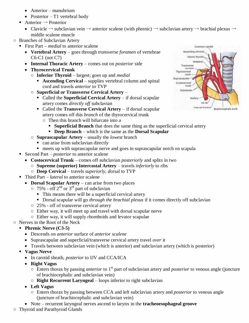
○ Branches of Subclavian Artery
First Part – medial to anterior scalene
Vertebral Artery – goes through transverse foramen of vertebrae
C6-C1 (not C7)
Internal Thoracic Artery – comes out on posterior side
Thyrocervical Trunk ○ Inferior Thyroid – largest; goes up and medial
Ascending Cervical – supplies vertebral column and spinal
cord and travels anterior to TVP
○ Superficial or Transverse Cervical Artery –
Called the Superficial Cervical Artery – if dorsal scapular
artery comes directly off subclavian
Called the Transverse Cervical Artery – if dorsal scapular
artery comes off this branch of the thyrocervical trunk
○ Then this branch will bifurcate into a
Superficial Branch that does the same thing as the superficial cervical artery
Deep Branch – which is the same as the Dorsal Scapular
○ Suprascapular Artery – usually the lowest branch
can arise from subclavian directly
meets up with suprascapular nerve and goes in suprascapular notch on scapula
Second Part – posterior to anterior scalene
Costocervical Trunk – comes off subclavian posteriorly and splits in two
○ Supreme (superior) Intercostal Artery – travels inferiorly to ribs
○ Deep Cervical – travels superiorly, dorsal to TVP
Third Part – lateral to anterior scalene
Dorsal Scapular Artery – can arise from two places
○ 75% - off 2nd
or 3rd
part of subclavian
This means there will be a superficial cervical artery
Dorsal scapular will go through the brachial plexus if it comes directly off subclavian
○ 25% - off of transverse cervical artery
○ Either way, it will meet up and travel with dorsal scapular nerve
○ Either way, it will supply rhomboids and levator scapulae
○ Nerves in the Root of the Neck
Phrenic Nerve (C3-5)
Descends on anterior surface of anterior scalene
Suprascapular and superficial/transverse cervical artery travel over it
Travels between subclavian vein (which is anterior) and subclavian artery (which is posterior)
Vagus Nerve
In carotid sheath, posterior to IJV and CCA/ICA
Right Vagus ○ Enters thorax by passing anterior to 1
st part of subclavian artery and posterior to venous angle (juncture
of brachiocephalic and subclavian vein)
○ Right Recurrent Laryngeal – loops inferior to right subclavian
Left Vagus ○ Enters thorax by passing between CCA and left subclavian artery and posterior to venous angle
(juncture of brachiocephalic and subclavian vein)
Note – recurrent laryngeal nerves ascend to larynx in the tracheoesophageal groove
○ Thyroid and Parathyroid Glands
Thyroid
Goes from C6-T1
2 Lateral Lobes, 1 Isthmus, maybe a Pyramidal Lobe
Surrounded by fibrous capsule
Innervation, arteries and veins are same for thyroid and parathyroid glands
Note – parathyroid glands are on posterior side of thyroid and there can be 2-3 pairs of them
Innervation – sympathetic innervation to arteries
Vasomotor
Arteries
Superior Thyroid – comes off ECA
Inferior Thyroid – comes from thyrocervical trunk
Thyroid Ima – only in 10% of cases
Veins
Superior Thyroid – to IJV
Middle Thyroid – to IJV
Inferior Thyroid – to brachiocephalic; only one of them, usually goes to left
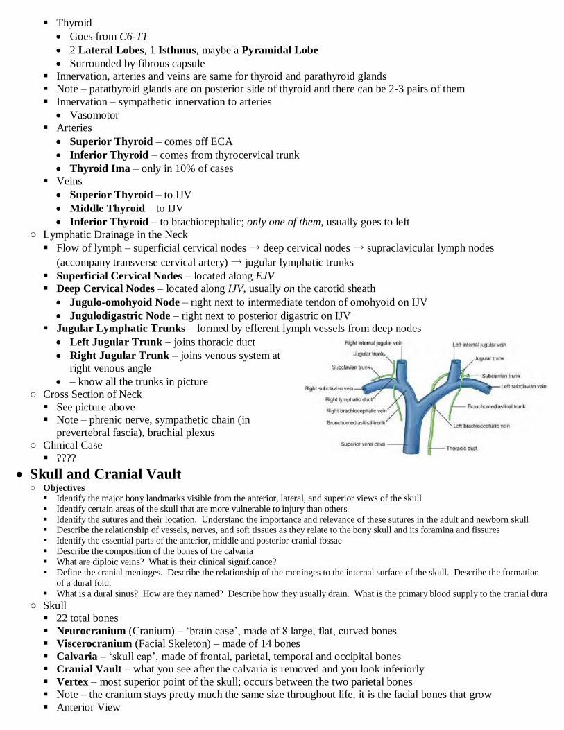
○ Lymphatic Drainage in the Neck
Flow of lymph – superficial cervical nodes → deep cervical nodes → supraclavicular lymph nodes
(accompany transverse cervical artery) → jugular lymphatic trunks
Superficial Cervical Nodes – located along EJV
Deep Cervical Nodes – located along IJV, usually on the carotid sheath
Jugulo-omohyoid Node – right next to intermediate tendon of omohyoid on IJV
Jugulodigastric Node – right next to posterior digastric on IJV
Jugular Lymphatic Trunks – formed by efferent lymph vessels from deep nodes
Left Jugular Trunk – joins thoracic duct
Right Jugular Trunk – joins venous system at
right venous angle
– know all the trunks in picture
○ Cross Section of Neck
See picture above
Note – phrenic nerve, sympathetic chain (in
prevertebral fascia), brachial plexus
○ Clinical Case
????
Skull and Cranial Vault ○ Objectives
Identify the major bony landmarks visible from the anterior, lateral, and superior views of the skull
Identify certain areas of the skull that are more vulnerable to injury than others
Identify the sutures and their location. Understand the importance and relevance of these sutures in the adult and newborn skull Describe the relationship of vessels, nerves, and soft tissues as they relate to the bony skull and its foramina and fissures
Identify the essential parts of the anterior, middle and posterior cranial fossae
Describe the composition of the bones of the calvaria
What are diploic veins? What is their clinical significance?
Define the cranial meninges. Describe the relationship of the meninges to the internal surface of the skull. Describe the formation
of a dural fold.
What is a dural sinus? How are they named? Describe how they usually drain. What is the primary blood supply to the cranial dura
○ Skull
22 total bones
Neurocranium (Cranium) – „brain case‟, made of 8 large, flat, curved bones
Viscerocranium (Facial Skeleton) – made of 14 bones
Calvaria – „skull cap‟, made of frontal, parietal, temporal and occipital bones
Cranial Vault – what you see after the calvaria is removed and you look inferiorly
Vertex – most superior point of the skull; occurs between the two parietal bones
Note – the cranium stays pretty much the same size throughout life, it is the facial bones that grow
Anterior View
Frontal Bone – not paired; helps make root of orbit
○ Glabella – the space between the eyebrows
Glabella Reflex – tap a finger on glabella and person will consistently blink if they have dementia
○ Nasion – junction of nasal bones and the frontal bone
○ Superciliary Arch/Ridge – ridge just deep to eyebrows
○ Supra-orbital Margin – sharp edge of frontal bone going down into orbit
○ Supra-orbital Foramen – transmits supraorbital nerve, artery and vein
Maxilla –
○ Body – main part of it
○ Infra-orbital Margin – sharp edge going down into orbit
○ Infra-orbital Foramen – transmits infraorbital nerve, artery and vein
○ Alveolar Process – forms sockets for teeth
Zygomatic Bone – cheek bones
○ Temporal Process – can maybe be seen anteriorly; it is the part that extends to meet temporal bone
Nasal Bone – the bridge of the nose
Mandible – not paired
○ Body – main part
○ Mental Foramen – transmits mental nerve, artery and vein
○ Alveolar Process – forms sockets for teeth
Lateral View
Parietal Bone – the big ones on side
○ Inferior and Superior Temporal Lines – muscles attach here
Temporal Bone – has many features
○ Squamous Portion – flat portion of the bone
○ Petrous Portion – goes into skull
Internal Auditory Meatus transmits CN 7 & 8
○ Mastoid Portion – posterior to external auditory meatus
○ Tympanic Portion – contains the External Auditory Meatus
○ Styloid Process – inferior to external auditory meatus; has 3 muscles connected to it
○ Zygomatic Process – portion that goes anterior to meet the zygomatic bone
Sphenoid Bone – just the greater wing here; between temporal and frontal bones
Occipital Bone – back part
Pterion – the part where the frontal, sphenoid, temporal and parietal bones meet
○ Bones are really thin here and the middle meningeal artery and vein run deep to it and so it vulnerable
to injury
Posterior View
Sutures – immovable joints of the skull
○ Saggital Suture – runs between the 2 parietal bones
○ Coronal Suture – runs between frontal and 2 parietal bones
○ Lambdoid Suture – runs between 2 parietal and occipital bones
Bregma – intersection of the saggital and coronal sutures
○ Was the anterior fontanelle at birth
Lambda – intersection of saggital and lambdoid sutures
○ Was the posterior fontanelle at birth
Note – fontanelles are membranous and usually close before year 1
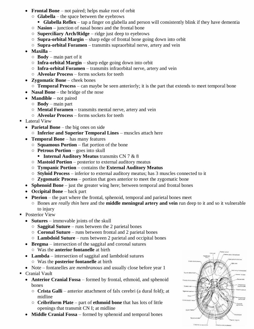
Cranial Vault
Anterior Cranial Fossa – formed by frontal, ethmoid, and sphenoid
bones
○ Crista Galli – anterior attachment of falx cerebri (a dural fold); at
midline
○ Cribriform Plate – part of ethmoid bone that has lots of little
openings that transmit CN I; at midline
Middle Cranial Fossa – formed by sphenoid and temporal bones

○ Cranial Nerve Openings – 5 of them in sphenoid bone
○ Hypophysial Fossa – depression of sphenoid bone that houses pituitary; part of Sella Turcica
Posterior Cranial Fossa – formed by occipital bone
○ Is the deepest and largest
○ Supports the cerebellum, pons and medulla
○ Foramen Magnum – transmits the spinal cord
○ Jugular Foramen – where the internal jugular vein begins
Transmits CN 9, 10, 11
Calvaria – has interesting bone structure
Has an inner and outer layer of compact bone with a layer of diploe in between
○ Diploe – spongy bone layer of calvaria (makes it lighter)
○ Diploic Veins – run through diploe and drain into dural venous sinuses and thus the meninges
Are valveless and go through the meninges
Diploic tributaries drain into 4 diploic veins (with characteristic names)
○ Cranial Meninges and Such
Continuous with meninges of spinal cord
Pia Mater – not innervated; follows every contour of the brain
Subarachnoid Space – contains CSF
Arachnoid Mater – not innervated
Dura Mater – highly innervated; has two layers (unlike spinal dura mater) that are usually fused, except when
they make a dural sinus
Meningeal Layer – in contact with the arachnoid mater
Endocranium (periosteal layer) – outer layer; fixed to bones; creates the periosteum of the bones
Dural Folds – areas where the meningeal dura separates from the
periosteal dura and forms reflections
These help provide support for the brain
Falx Cerebri – divides brain into right and left hemispheres
○ Largest; suspended from superior aspect of the cranial cavity
Falx Cerebelli – divides the cerebellum into hemispheres
○ Smaller; in posterior cranial fossa
Tentorium Cerebelli – separates cerebrum from cerebellum
○ Perpendicular to falx cerebri (thus goes lateral to medial
Diaphragma Sellae – circular fold that covers pituitary fossa
Dural Sinuses – when meningeal dura starts to form a reflection
then it creates a space between it and the periosteal dura
Lined with endothelium (so it is kinda like a blood vessel)
All drain into internal jugular vein
Veins of brain can drain into these
Superior Saggital Sinus – unpaired; in superior margin of falx
cerebri
Inferior Saggital Sinus – unpaired; in inferior margin of falx
cerebri
Straight Sinus – unpaired; in tentorium cerebelli
○ Runs from inferior saggital sinus to the confluence of sinuses
Transverse Sinus – paired; in tentorium cerebelli along posterior
attachment to occipital bone
○ Connects confluence of sinuses to sigmoid sinus
Sigmoid Sinus – paired; a continuation of the transverse
○ Ends as the internal jugular vein
Cavernous Sinus – paired; adjacent to the pituitary
○ Lots of things can drain here (I don‟t know what) and then they
drain into sigmoid sinus via the petrosal

Superior and Inferior Petrosal Sinus - paired; along superior and inferior margins of the petrous portion
of the temporal bone
○ Connects cavernous sinus to the sigmoid sinus (blood runs anterior to posterior)
Confluence of Sinuses – unpaired; where the superior saggital, straight and occipital sinuses meeth and the
transverse sinuses leave from
Blood Supply to Meninges
Middle Meningeal Artery – primary arterial blood supply to the dura mater; branch of maxillary artery
○ Foramen Spinosum – entry point into the cranium
○ Source – ECA
○ Location – runs in what would be the „epidural space‟, but there isn‟t one and so it becomes embedded
in the bone
○ Clinical Correlation
Pterion (the thin spot of skull) lies right above a part of the middle meningeal artery and thus if you
break the skull at the pterion it could cause a huge bleed
Epidural Hematoma – because it is epidural blood leaks in very slowly and pushes the dura away
from the bone (compressing brain)
○ Symptoms occur many hours after the injury
Intro to Cranial Nerves ○ Objectives
Review the definition and characteristics of a cranial nerve Review the components of a spinal nerve; describe the components of cranial nerves
Name the 12 cranial nerves
For each cranial nerve, identify functional components and relative position in the cranial vault
Name and describe ganglia that are associated with the cranial nerves
Be able to differentiate between sensory ganglia and autonomic ganglia
○ Cranial Nerves
12 Pair
Skeletal muscle and viscera of head and neck
Skin of face and scalp
Named foramina
Mixed, motor only, sensory only
May contain parasympathetics only
31 Pair
Skeletal muscle and viscera of trunk and limbs
Skin of trunk and limbs
Intervetebral foramina
All mixed nerves
I – Olfactory
II – Optic
III – Oculomotor
IV – Trochlear – comes out dorsally
V – Trigeminal
VI – Abducens
VII – Facial
VIII – Vestibulocochlear
IX – Glossopharyngeal
X – Vagus
XI – Accessory kinda comes out below XII
XII – Hypoglossal kinda comes out above XI
Functional Components for Cranial Nerves
Possible Efferent Components
○ General Somatic Efferents (GSE) – supply muscles of the head, neck, body wall and extremities that
arise from myotomes associated with embryonic somites
○ Special Visceral Efferents (SVE) – supply muscles of head, neck, body wall and extremities that aris
from pharyngeal arches
This is a terribly non-discriptive and misleading name
○ General Visceral Efferent (GVE) – supply smooth muscle, cardiac muscle and glands
Note – just parasympathetic fibers for cranial nerves
Possible Afferent Components

○ General Somatic Afferents (GSA) – pain, touch temperature, proprioception from skin, head, neck and
body
○ General Visceral Afferents (GVA) – convey sensory information from viscera
○ Special Somatic Afferents (SSA) – convey sensory information about the special senses of vision,
hearing or balance
○ Special Visceral Afferents (SVA) – convey sensory information about the special senses of smell and
taste Motor Parasympathetic Sensory
Nerve Skeletal
Muscle
(somatic)
Skeletal Muscle
(pharyngeal)
Smooth Muscle or
Glands
Pain, touch or
temperature
Pain from
organs or
mucous
membranes
Special (smell
and taste)
Special
(vision,
hearing,
balance)
1 – Olfactory Smell
2 – Optic Vision
3 – Oculomotor 5 muscles of
orbit
Pupillary sphincter
Ciliary muscle
4 – Trochlear (SO4) Superior
oblique eye
muscle
5 – Trigeminal
- V1 Ophthalmic Skin of face
(forehead, upper
eyelid, cornea, part
of nasal cavity,
part of nose)
- V2 Maxillary Skin of face
(lower eyelid,
cheek, lateral nose,
upper lip, upper
teeth)
- V3 Mandibular -muscles of
mastication
-anterior belly of
digastric
-myohyoid
-2 tensors
Skin of face
(cheek, chin, lower
lip, tongue, lower
teeth)
6 – Abducens (LR6) Lateral rectus
eye muscle
7 – Facial -muscles of facial
expression
-posterior belly of
digastric
-stylohyoid
1. mucous glands of
nasal cavity & oral
cavity, lacrimal gland
2. submandibular
salivary gland,
sublingual salivary
gland
-ear
-external auditory
meatus
Taste on anterior
2/3 of tongue
8 – Vestibulocochlear -hearing
-balance
9 – Glossopharyngeal Stylopharyngeus Parotid salivary gland -posterior 1/3 of
tongue
-carotid body
-carotid sinus
Taste on posterior
1/3 of tongue
Mucous membrane of pharynx
10 – Vagus -muscle of soft
palate
-pharynx & larynx
-mucous glands of
pharynx and larynx
-thoracic and abdominal
organs
-ear
-external auditory
meatus
-mucous
membrane of
larynx
-from organs
Taste on epiglottis
11 – Accessory SCM
Trapezius
12 – Hypoglossal Intrinsic and
extrinsic
muscles of
the tongue
○ She didn't talk about the foramina
Face, Scalp and Parotid Region ○ Objectives List the layers of the scalp. Give the importance of each layer
Describe the blood supply and innervation to the scalp. What is the clinical significance of the arrangement?
Review on your own the major bones of the face.
Describe the sensory innervation and blood supply to the skin of the face
Explain the clinical significance of the „danger triangle‟ of the face
Describe the organization of the skin of the face, list the major muscles and give their action and innervation
Discuss the consequences of interrupting CN V or VII innervation to the face
Describe the location of the parotid gland
Give the relationships of the parotid gland to the surrounding structures
Describe the nerve and vascular supply to the gland
○ Scalp –
„SCALP‟
Skin –
Connective Tissue – very dense with blood vessels and nerves
○ Density makes it hard to get infected
○ Density makes it hard for blood vessels to close if they get cut, thus there is a lot of bleeding
Aponeurosis – flat tendinous sheet between two heads of the occipitofrontalis muscle
Loose Areolar Tissue – CT that is loose and thus lets above layers move around
○ Note – the three layers above the loose areolar tissue are fused very tightly
Pericranium – the periosteum of the skull
Veins
Emmisary Veins – drain scalp into dural venous sinuses
○ Valveless and thus can bring bad things into the dural sinuses?
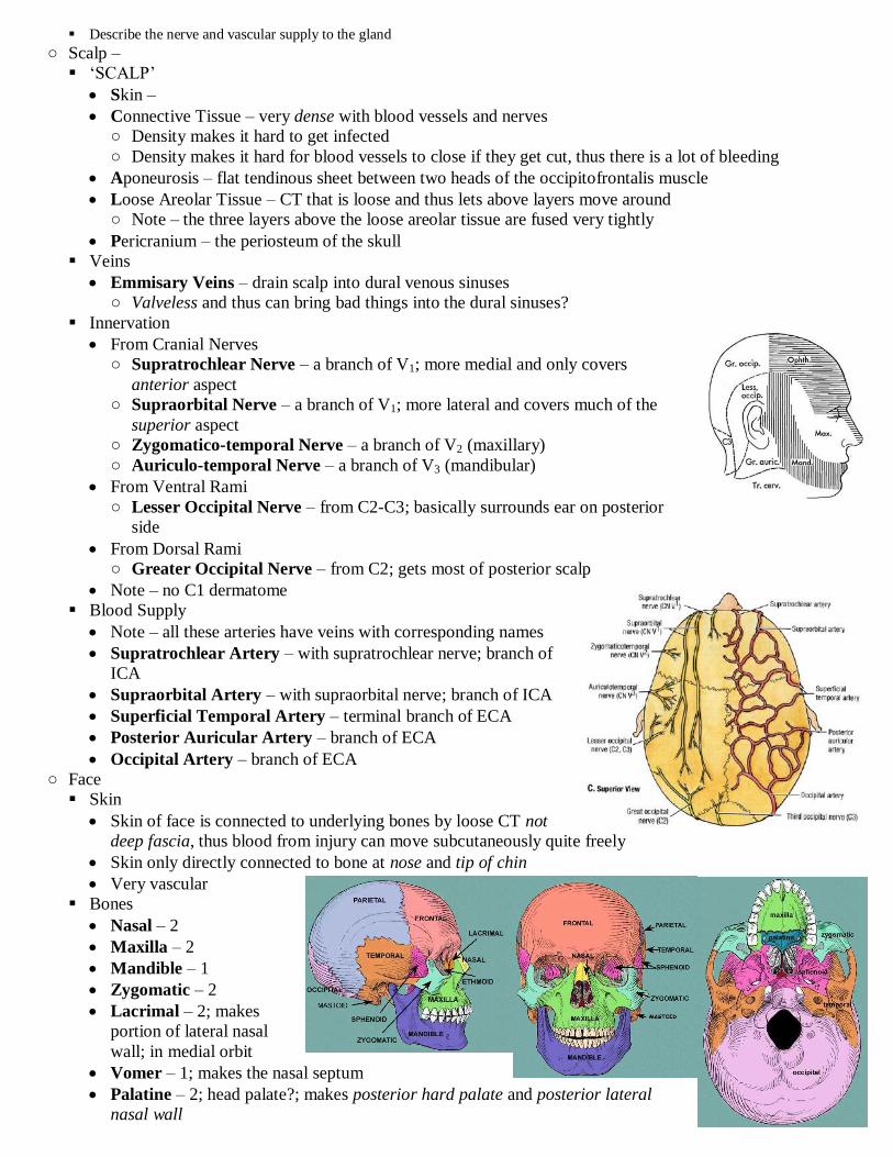
Innervation
From Cranial Nerves
○ Supratrochlear Nerve – a branch of V1; more medial and only covers
anterior aspect
○ Supraorbital Nerve – a branch of V1; more lateral and covers much of the
superior aspect
○ Zygomatico-temporal Nerve – a branch of V2 (maxillary)
○ Auriculo-temporal Nerve – a branch of V3 (mandibular)
From Ventral Rami
○ Lesser Occipital Nerve – from C2-C3; basically surrounds ear on posterior
side
From Dorsal Rami
○ Greater Occipital Nerve – from C2; gets most of posterior scalp
Note – no C1 dermatome
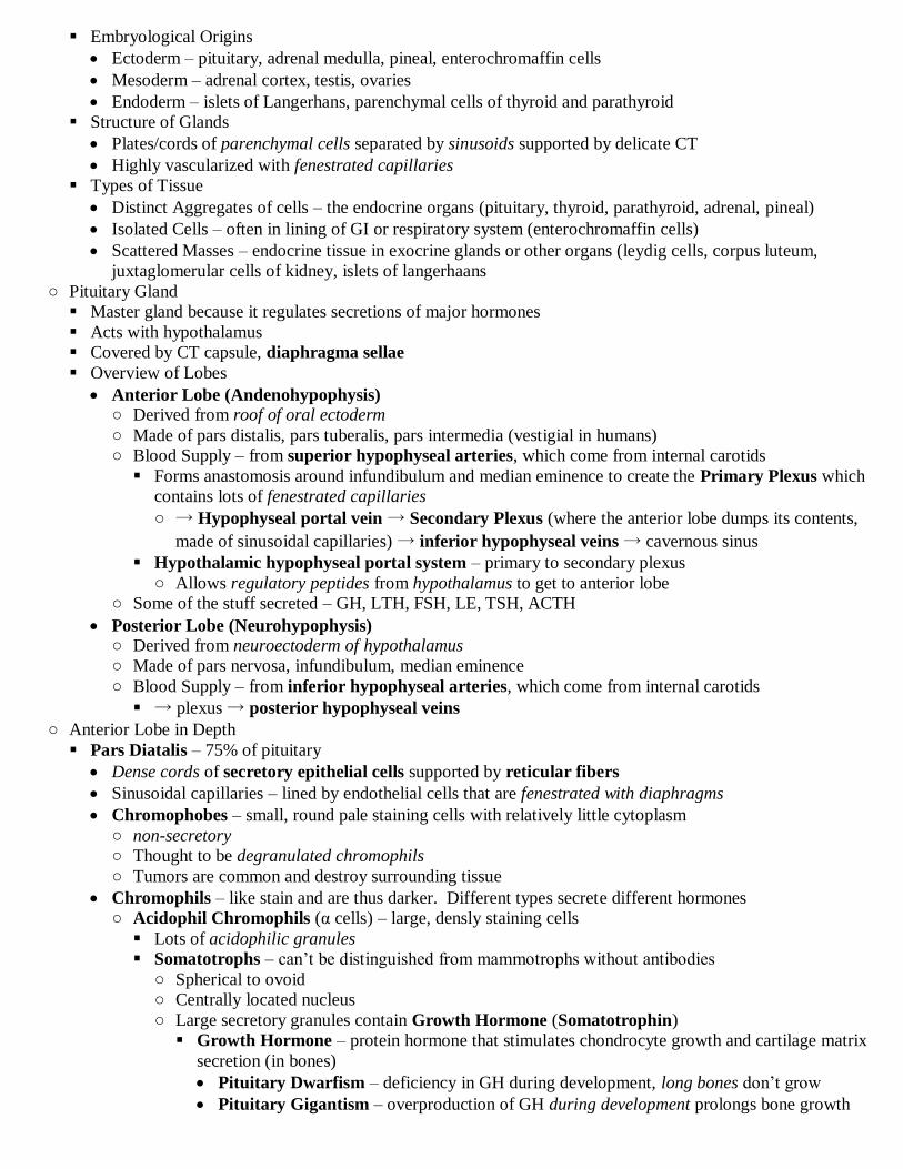
Blood Supply
Note – all these arteries have veins with corresponding names
Supratrochlear Artery – with supratrochlear nerve; branch of
ICA
Supraorbital Artery – with supraorbital nerve; branch of ICA
Superficial Temporal Artery – terminal branch of ECA
Posterior Auricular Artery – branch of ECA
Occipital Artery – branch of ECA
○ Face
Skin
Skin of face is connected to underlying bones by loose CT not
deep fascia, thus blood from injury can move subcutaneously quite freely
Skin only directly connected to bone at nose and tip of chin
Very vascular
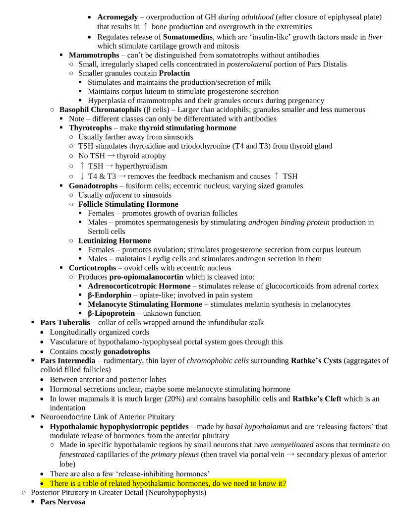
Bones
Nasal – 2
Maxilla – 2
Mandible – 1
Zygomatic – 2
Lacrimal – 2; makes
portion of lateral nasal
wall; in medial orbit
Vomer – 1; makes the nasal septum
Palatine – 2; head palate?; makes posterior hard palate and posterior lateral
nasal wall

Inferior Nasal Conchae – 2; makes lateral nasal cochea
Ethmoid – 1; makes nasal cavity and paranasal sinuses; superior and middle nasal conchae
Sensory Innervation
“know the basic territories of the 3 divisions, not necessarily the individual subdivisions”
Ophthalmic Nerve Branches
○ Supertrochlear nerve, Supra-orbital
nerve, Lacrimal nerve, Infratrochlear
nerve, External nasal nerve
Maxillary Nerve Branches
○ Infraorbital nerve, Zygomatico-facial
nerve (goes down), Zygomatico-
temporal nerve (goes up)
Mandibular Nerve Branches
○ Mental nerve (through mental
foramen), Buccal nerve, Auriculo-
temporal nerve
Blood Supply
Facial Artery – main artery for face;
branch of ECA
○ Submental Artery – a branch; goes to
chin
○ Inferior and Superior Labial branches – goes to lower and upper lips
○ Angular Artery – the end of the facial artery around side of nose
Superficial Temporal Artery –terminal ranch of ECA
Transverse Facial Artery – branch of superficial temporal artery going horizontal
Supraorbital and Supratrochlear Arteries – branches of ophthalmic artery
Veins
○ Veins of face can drain to two places: the facial vein (mainly goes here) or
the dural sinuses
○ Most tributaries of the facial vein can also drain into the dural sinuses, thus
if facial vein gets blocked then the blood can go into the dural sinuses
○ Danger Zone – infection to side of nose between eye and upper lip can
cause venous drainage to go mainly to the dural sinuses and infect the brain
○ Note – facial vein is valveless
○ Note – facial vein has variable termination, but usually to one of the jugular
veins
○ See picture for ophthalmic vein, infraorbital vein, pterygoid plexus and deep facial vein, all of which can
go either to dural sinuses or the facial vein
Muscles of Face
Embedded in superficial fascia
Attachments – skin & loose connection to bones of face
Innervation – CN VII
Derivation – all from 2nd
pharyngeal arch
Platysma – tenses skin of neck and corners of mouth
Occipitofrontalis 1 – no bony attachment
○ Frontal belly – wrinkle forehead and raise eyebrows
○ Occipital belly – over occipital bone
Orbicularis Oculi 3 – closes eye
Zygomaticus Major 7 – draws ↑ corner of mouth, smile
Zygomaticus Minor 8 -
Orbicularis Oris 10 – purses lips
Levator Labii Superioris 11 –
Levator Labii Superioris Alaque Nasi 9 –

Levator Anguli Oris 12 – deep to above
Depressor Anguli Oris 13 – draws corner of mouth ↓
Depressor Labii Inferioris 14 –
Buccinator 16 – keeps cheek from expanding
Muscular Innervation of Face
Facial Nerve – supplies all muscles, but no skin
○ Enters skull at internal acoustic meatus, gives off nerve to petrosal ganglion, ear and tongue then exits
the skull at the stylomastoid foramen (inferomedial to ear)
○ Once it leaves the skull it makes three branches
Muscles of auricle and occipitalis
Stylohyoid and posterior belly of digastric
Parotid gland – once in parotid gland it breaks into 5 branches
○ “Two Zebras Bit My Cookie” (distributed like your 5 fingers)
○ Temporal Branches –
○ Zygomatic Branches –
○ Buccal Branches –
○ Marginal Mandibular Branches –
○ Cervical Branches –
○ Bell’s Palsy – trauma to facial nerve causes loss of function to muscles of facial expression
Can be temporary
Can‟t close eye and eyes can dry out
Can be caused by middle ear infection, since the facial nerve innervates that and goes right by it
○ Parotid Gland
Largest salivary glands; lies posterior to jaw bone superficially
Parotid Duct – goes over masseter muscle → pierces buccinator muscle → opens adjacent to 2nd
upper molar
Things that traverse or lie deep to the parotid gland
Facial Nerve
External Carotid Artery exits gland as the superficial temporal artery
Retromandibular vein
Auriculo-temporal nerve (branch of V3)
Endocrine 1 – Hypophysis and Pineal ○ Objectives
Describe the histology of the pituitary gland; include the infundibular stalk, the four main parts, and its embryology
Identify chromophils and chromophobes of the pars distalis. Indicate which pituitary hormones are made by each, what are the functions of each hormone and what is the target organ, tissue or cell of each.
Identify the components of the neurohypophysis
Describe the two hormones that are liberated from the posterior lobe of the pituitary in terms of their origin, the hypothalamophyseal
tract, their target organs, and their functions
Be able to describe the signals which trigger the release of pituitary hormones and their feedback regulation
Describe the overall histology of the pineal gland and the hormones produced by the pineal gland
○ Hormones
Sites of Action
Circulating – travel through circulation to act on distant tissues
Paracrine – act on neighboring cells and tissues
Autocrine – act on the same cells that secrete them
Chemical Classes - Steroid Hormones (testosterone, work in days), Protein Hormones (prolactin, work in
minutes), Amino Acid Derivatives (norepinephrine, work in seconds)
Effective at low concentrations; often have feedback control
Direct Action Hormones – often lipid soluble and get into cell membrane or nucleus to modulate something
Indirect Action Hormones – often water soluble and bind to a membrane receptor that activates a membrane-
bound enzyme → which activates a secondary messenger (cAMP) → intracellular signaling cascade
Often used by peptide hormones
○ Endocrine System – ductless glands whose secretions are passed directly to blood or lymph
Ex. pituitary, thyroid, parathyroid, adrenals, pancreatic islets, pineal, testis, ovaries
Embryological Origins
Ectoderm – pituitary, adrenal medulla, pineal, enterochromaffin cells
Mesoderm – adrenal cortex, testis, ovaries
Endoderm – islets of Langerhans, parenchymal cells of thyroid and parathyroid
Structure of Glands
Plates/cords of parenchymal cells separated by sinusoids supported by delicate CT
Highly vascularized with fenestrated capillaries
Types of Tissue
Distinct Aggregates of cells – the endocrine organs (pituitary, thyroid, parathyroid, adrenal, pineal)
Isolated Cells – often in lining of GI or respiratory system (enterochromaffin cells)
Scattered Masses – endocrine tissue in exocrine glands or other organs (leydig cells, corpus luteum,
juxtaglomerular cells of kidney, islets of langerhaans
○ Pituitary Gland
Master gland because it regulates secretions of major hormones
Acts with hypothalamus
Covered by CT capsule, diaphragma sellae
Overview of Lobes
Anterior Lobe (Andenohypophysis)
○ Derived from roof of oral ectoderm
○ Made of pars distalis, pars tuberalis, pars intermedia (vestigial in humans)
○ Blood Supply – from superior hypophyseal arteries, which come from internal carotids
Forms anastomosis around infundibulum and median eminence to create the Primary Plexus which
contains lots of fenestrated capillaries
○ → Hypophyseal portal vein → Secondary Plexus (where the anterior lobe dumps its contents,
made of sinusoidal capillaries) → inferior hypophyseal veins → cavernous sinus
Hypothalamic hypophyseal portal system – primary to secondary plexus
○ Allows regulatory peptides from hypothalamus to get to anterior lobe
○ Some of the stuff secreted – GH, LTH, FSH, LE, TSH, ACTH
Posterior Lobe (Neurohypophysis)
○ Derived from neuroectoderm of hypothalamus
○ Made of pars nervosa, infundibulum, median eminence
○ Blood Supply – from inferior hypophyseal arteries, which come from internal carotids
→ plexus → posterior hypophyseal veins
○ Anterior Lobe in Depth
Pars Diatalis – 75% of pituitary
Dense cords of secretory epithelial cells supported by reticular fibers
Sinusoidal capillaries – lined by endothelial cells that are fenestrated with diaphragms
Chromophobes – small, round pale staining cells with relatively little cytoplasm
○ non-secretory
○ Thought to be degranulated chromophils
○ Tumors are common and destroy surrounding tissue
Chromophils – like stain and are thus darker. Different types secrete different hormones
○ Acidophil Chromophils (α cells) – large, densly staining cells
Lots of acidophilic granules
Somatotrophs – can‟t be distinguished from mammotrophs without antibodies
○ Spherical to ovoid
○ Centrally located nucleus
○ Large secretory granules contain Growth Hormone (Somatotrophin)
Growth Hormone – protein hormone that stimulates chondrocyte growth and cartilage matrix
secretion (in bones)
Pituitary Dwarfism – deficiency in GH during development, long bones don‟t grow
Pituitary Gigantism – overproduction of GH during development prolongs bone growth
Acromegaly – overproduction of GH during adulthood (after closure of epiphyseal plate)
that results in ↑ bone production and overgrowth in the extremities
Regulates release of Somatomedins, which are „insulin-like‟ growth factors made in liver
which stimulate cartilage growth and mitosis
Mammotrophs – can‟t be distinguished from somatotrophs without antibodies
○ Small, irregularly shaped cells concentrated in posterolateral portion of Pars Distalis
○ Smaller granules contain Prolactin
Stimulates and maintains the production/secretion of milk
Maintains corpus luteum to stimulate progesterone secretion
Hyperplasia of mammotrophs and their granules occurs during pregenancy
○ Basophil Chromatophils (β cells) – Larger than acidophils; granules smaller and less numerous
Note – different classes can only be differentiated with antibodies
Thyrotrophs – make thyroid stimulating hormone
○ Usually farther away from sinusoids
○ TSH stimulates thyroxidine and triodothyronine (T4 and T3) from thyroid gland
○ No TSH → thyroid atrophy
○ ↑ TSH → hyperthyroidism
○ ↓ T4 & T3 → removes the feedback mechanism and causes ↑ TSH
Gonadotrophs – fusiform cells; eccentric nucleus; varying sized granules
○ Usually adjacent to sinusoids
○ Follicle Stimulating Hormone
Females – promotes growth of ovarian follicles
Males – promotes spermatogenesis by stimulating androgen binding protein production in
Sertoli cells
○ Leutinizing Hormone
Females – promotes ovulation; stimulates progesterone secretion from corpus leuteum
Males – maintains Leydig cells and stimulates androgen secretion in them
Corticotrophs – ovoid cells with eccentric nucleus
○ Produces pro-opiomalanocortin which is cleaved into:
Adrenocorticotropic Hormone – stimulates release of glucocorticoids from adrenal cortex
β-Endorphin – opiate-like; involved in pain system
Melanocyte Stimulating Hormone – stimulates melanin synthesis in melanocytes
β-Lipoprotein – unknown function
Pars Tuberalis – collar of cells wrapped around the infundibular stalk
Longitudinally organized cords
Vasculature of hypothalamo-hypophyseal portal system goes through this
Contains mostly gonadotrophs
Pars Intermedia – rudimentary, thin layer of chromophobic cells surrounding Rathke’s Cysts (aggregates of
colloid filled follicles)
Between anterior and posterior lobes
Hormonal secretions unclear, maybe some melanocyte stimulating hormone
In lower mammals it is much larger (20%) and contains basophilic cells and Rathke’s Cleft which is an
indentation
Neuroendocrine Link of Anterior Pituitary
Hypothalamic hypophysiotropic peptides – made by basal hypothalamus and are „releasing factors‟ that
modulate release of hormones from the anterior pituitary
○ Made in specific hypothalamic regions by small neurons that have unmyelinated axons that terminate on
fenestrated capillaries of the primary plexus (then travel via portal vein → secondary plexus of anterior
lobe)
There are also a few „release-inhibiting hormones‟
There is a table of related hypothalamic hormones, do we need to know it?
○ Posterior Pituitary in Greater Detail (Neurohypophysis)
Pars Nervosa

Three Elements
○ Dense Capillary Plexus
○ Pituicytes – highly branched, non-secretory, glial like cells, provide nutrients; end on capillaries
○ Tons of unmyelinated axons of neurosecretory cells of the supraoptic and paraventricular region of
hypothalamus
Hypothalamic hypohyseal tract – tract of these axons that terminate near the fenestrated capillaries
of the capillary plexus
Stuff secreted by exocytosis
Neurophysin – carrier protein that carries hormones down the axon
Herring Bodies – visible dilations of axons containing vesicles of hormones
No secretory epithelial cells
Hormones
○ Oxytocin – peptide made in the paraventricular nucleus of the hypothalamus
Induces peristaltic contractions of uterine smooth muscle
Induces contraction of myoepithelial cells of mammary gland resulting in excretion of milk
○ Vasopressin (antidiuretic hormone) – peptide made in supraoptic nucleus of hypothalamus
Promotes water resorption through the collecting tubules of the kidney
↑ BP by promoting contraction of vascular smooth muscle resulting in ↑ peripheral resistance
○ Endocrine Feedback Mechanisms
Negative – output of target cells ↓ hormone production
Positive – output of target cells ↑ hormone production
1st Order Regulation – neurohypophyseal hormone acts on non-endocrine target (ie oxytocin, vasopressin)
levels of product from target in blood feed back to hypothalamus regulating the neurohypophyseal hormone
2nd
Order Regulation – hypothalamic releasing factors stimulate release of anterior pituitary hormones
levels of anterior pituitary hormones in blood feed back to hypothalamus or pituitary
Ex growth hormone, prolactin
3rd
Order Regulation - hypothalamic releasing factors stimulate release of anterior pituitary hormones which
then causes release of another hormone from an endocrine organ
levels of this new hormone in blood feed back to hypothalamus or pituitary
Ex thyroid stimulating hormone, ACTH
○ Pineal Gland
Flattened, conical gland attached to diencephalon of CNS
Encapsulated by pia mater of CNS, which also penetrates parenchyma as trabeculae
Pinealocytes – basophilic cytoplasm with long cytoplasmic processes ending in bulb-like expansion in close
proximity to capillaries, which contain vesicles that are exocytosed into the capillaries
Large oval nucleus and clearly distinguishable nucleoli
Melatonin – synthesized from serotonin
○ Causes pigment retention in melanophors
○ May be free radical scavenger; may inhibit growth and metastasis of some tumors
○ Release is inhibited by light; overproduction may be involved in seasonal affective disorder; involved in
sleep/wake cycle
Glial Cells – resemble astrocytes
Brain Sand – characteristic small aggregates of calcium phosphate and calcium carbonate
○ Are radiopaque and ↑ with brain lesions or compression
Endocrine 2 – Thyroid, Parathyroid, Adrenal Glands ○ Objectives
Describe the histology of the thyroid gland
Describe the function of thyroid follicular cells and the synthesis, storage and secretion of thyroid hormone
Describe and identify the parafollicular (C cells)
Describe and identify the histology of the parathyroid glands. Distinguish the chief (principle) cell from the oxyphil cell
Describe the overall histology of the adrenal gland, its blood supply, and the embryological origin of each region
Describe the three zones of the adrenal cortex, their constituent cells, and substances produced by the cells in each zone Describe the functions of each hormone secreted by the adrenal gland and the clinical disorders associated with each
Describe the organization of the adrenal medulla and it‟s constituent cells

Indicate which cells of the adrenal medulla secrete hormones, and identify the hormones secreted by each
Identify the endocrine component of the pancreas, the specific hormone producing cells and the functions of each hormone
○ Thyroid
Regulates tissue metabolism, tissue growth, and secondary regulation of plasma Ca++
Right, left, ishmus and pyramidal lobes
Embryology
Develops as median downgrowth of the base of the tongue and descends to the upper tracheal region
leaving a duct behind, the thyroglossal duct (obliterated during development)
Enclosed by two layers of CT, an external dense CT layer, and an inner, thin layer that penetrates the lobes
Thin layer of CT divides thyroid into lobules
Follicles – functional and structural subunit of thyroid
Spherical cyst-like structures bounded by follicular epithelial cells
Follicular Epithelial Cells – simple cuboidal epithelial cells that form outer, single-layer border of follicle
○ Synthesize the secretions
Also surrounded by a basal lamina and supported by reticular fibers
Enveloped in meshwork of fenestrated capillaries that receive the endocrine secretion
Colloid – gelatinous substance in center of each follicle
○ Contains stored products of the follicular epithelial cells
Cells of Thyroid
Parafollicular (C-Cells) ○ Less numerous cell; found in walls of thyroid follicles (between and within?)
Within the basement membrane of the follicle, but are insulated from the lumen by cytoplasmic
extensions of the follicular cells
○ Originate from neural crest (from APUD cell line)
○ Larger and lighter staining
○ Centrally located nucleus
○ Vesicles contain calcitonin
○ Calcitonin – peptide that ↓ plasma Ca (in direct response to elevated Ca levels) via
Inhibiting osteoclast activity (which ↓ bone resorption and thus the freeing of Ca)
Promoting excretion of Ca (in kidneys)
Not under control of the pituitary
Follicular Epithelial Cells ○ Structure
Cytoplasm is slightly basophilic; centrally-located nucleus; usually simple cuboidal
If hyperactive → cells become columnar, colloid is ↓, inner border will become “scalloped”
If hypoactive → cells become squamous and colloid ↑ a bunch
EM Level – features of a cell making a lot of exported proteins are displayed
○ Tight junctions at lateral borders
○ Dilated cisternae of RER, colloid droplets, numerous coated vesicles, and
microvilli
○ Function
Synthesize thyroid hormone
Thyroglobulin – precursor to thyroid hormone that is a major component of
the colloid
Note – follicular cells are unique endocrine cells since they store their product
extracellularly
○ Synthesis of Thyroid Hormone
Thyroglobulin made in rER and processed in golgi
Tyrosine residues added to it and shipped in vesicles to surface, where it is
exocytosed
Thyroperoxidase – enzyme that stays right near the microvilli of the follicular
epithelial cells (made by same cells) that catalyzes the iodination of thyroglobulin
○ Note – this reaction occurs in the lumen of the follicle within 1-2 µm of microvilli
Note - Iodide Pump on basal side of membrane brings in iodide from the plasma

○ Iodide is oxidized in the cell and forms iodine which is then secreted into the follicular lumen
Iodinated Thyroglobin formed after iodine is added
○ 1 added – MIT; 2 added – DIT (are just iodine carriers; are deiodinated and recycled in cell)
○ MIT + DIT = T3 (active form of thyroid hormone)
○ DIT + DIT = T4/thyroxine (active form of thyroid hormone)
Iodinated thyroglobin is taken up by apical surface and formed into colloid droplets
○ It is then digested by lysosyme and T3, T4, MIT and DIT are made
T3 and T4 then diffuse out basal membrane surface and into capillary circulation where they are
bound by plasma proteins
○ Regulation of Thyroid Hormone
Activated by thyroid stimulating hormone released by the anterior pituitary
○ Causes hypertrophy and hyperplasia of follicular cells
○ ↑ production and iodination of thyroglobulin
○ ↑ re-uptake and lysosomal digestion of iodinated thyroglobulin
○ ↑ secretion of T3 and T4
Plasma levels of T3/T4 are monitored by the hypothalamus
If too low then
○ TSHreleasing hormone released into hypothalamic portal system and stimulates TSH release from
anterior pituitary thyrotrophs
○ Follicular cells in thyroid respond and release more T3/T4
○ Increased levels in blood stop TSHrh release in hypothalamus
○ Function of Thyroid Hormone
Metabolic Effects
○ Controls Basal Metabolic Rate (kilocalories/square meter of body surface/hour) energy
expenditure at rest
○ Regulates water and ion transport
○ Reulates protein, fat and carbohydrate metabolism
Growth Promoting Effects
○ Acts synergistically with growth hormone to promote skeletal development
○ Controls molting and metamorphosis in lower vertebrates
○ Thyroid Dysfunction
Hypothyroid Hyperthyroid
Mentally and physically sluggish Restless, irritable, anxious
Low BMR Elevated BMR
Mental retardation Mentally alert
↓ glucose absorption ↑ glucose absorption
Weak heartbeat Tachycardia
Hypothyroidism
○ If during development → Cretinism – stunted physical and mental growth with big belly
○ If during adulthood → Myxedema – lethargy and mental deficiencty
○ Hashimoto’s Disease – autoimmune destruction of follicular cells
Goiter – enlargement of thyroid gland due to hypertrophy and hyperplasia of follicular cells
○ Can be caused by:
Iodine Deficiency – causes ↓ T3/T4 output → ↑TSH release → follicular hypertrophy
Graves Disease – thyroid stimulating immunoglobulin causes stimulation of follicular cells
Also causes protrusion of eyeballs due to increased water absorption in retro-occular tissues
○ Importance of Calcium
Most abundant cation
Membrane permeability, excitability of muscles and nerves, enzyme activity, blood clotting, acid/base
Absorbed by SI but Vit D is required
Minor fluctuations in plasma concentrations can have large effect
Hypercalcemia – ectopic calcification of soft tissues; kidney stones

Hypocalcemia – hyperexcitability of neurons; prolonged skeletal muscle contractions; aberrant cardiac
muscle contraction
○ Parathyroid Glands
Maintain appropriate plasma concentrations of Ca
4 small glandular masses on posterior thyroid (in pairs)
2 inferior parathyroids derived from 3rd
pharyngeal arch and descend with the thymus
2 superior parathyroids derived from 4th
pharyngeal arch and descend with the thyroid
Lie just outside of thyroid capsule, but enclosed in same fascia
CT septa carry blood into tissue, but don’t separate it into lobes
Cells arranged in cords supported by reticular fibers and separated by fenestrated capillaries
Types of Cells
Chief Cells ○ Structure – most numerous cell; small; centrally located nucleus
If cytoplasm is light → Light Cells – inactive
If cytoplasm is dark → Dark Cells – active and secreting parathyroid hormone
EM Level
○ Irregularly shaped secretory granules, but they are less numerous than in other endocrine glands
because the parathyroid doesn‟t store much PTH
○ Function – secretes parathyroid hormone (PTH)
Three Effects when blood Ca is ↓ (thus not under control of pituitary)
○ ↑ release of osteoclast stimulating factor which stimulates osteoclast activity to ↑ bone
resorptoin and thus ↑ Ca release
○ ↑ Ca resorption – by distal convoluted tubules
○ Promotes synthesis of dihydroxycholecalciferol (a Vit D derivative) to ↑ Ca absorption in SI
Oxyphil Cells – unknown function
○ Less numerous; larger (more cytoplasm), dark staining nuclei, strongly acidophilic cytoplasm
○ EM Level – there is a ton of mitochondria
○ Don‟t appear until age 7
○ Pancreatic Islets of Langerhans
Structure
Endocrine „micro-organs‟/clusters of cells in pancreas
More concentrated in tail region of pancreas
Encapsulated by reticular fibers
Cells are pale and a little smaller than surrounding acini of pancreas
Function
Stimulate digestion
Regulates glucose transfer
Types of Cells
Alpha Cells – secretes Glucagon which ↑ blood glucose by breaking down glycogen
○ 20%; large cells; in periphery of islet; numerous granules
○ EM – eccentrically place electron dense core
Beta Cells – secretes Insulin which ↓ blood glucose by 1. promoting glucose transfer into tissue and 2.
stimulating formation of glycogen
○ 70%; small cells; center of islet
○ EM – electron dense core
○ Diabetes Mellitus – hyperglycemia, glucosuria, polyuria (increased excretion of water)
Type 1 – reduced beta cell secretion
Type 2 – defect of insulin receptors on tissue cells
Delta Cells – secrete Somatostatin and in the pancreas it affects glucagon and insulin release
○ Somatostatin can be a neurotransmitter or a neurohormone
When released by hypothalamus it causes growth hormone release
When released in pancreas it affects glucagon and insulin release
F Cells – secretes pancreatic polypeptide which:
○ Stimulates release of gastric secretions in gut
○ Inhibits bile secretion in the gall bladder
G Cells – secrete Gastrin which 1. increase HCl secretion in stomach 2. ↑ gastric motility
○ Only present during maturation
Note
○ α, β and D cells are connected by gap junctions and thus secretions influenced by local diffusion
(paracrine)
○ Glucagon and insulin levels are changed in response to increased or decreased plasma glucose levels
○ Adrenal Glands
Covered by thick fibroelastic CT capsule with rich blood supply
Cortex and medulla can be considered 2 separate endocrine tissues because they have different:
Embryological origin; hormonal secretion; function
Blood Supply – superior (from inferior phrenic), middle (from aorta), inferior (from renal artery)
Capsular Arteries – continuation of above arteries in the capsule, where they branch to form:
○ Cortical Arterioles – branch a bunch immediately and create
Capillary plexus of cortex and cortical sinusoids
○ Medullary Arterioles – go deep and create capillary plexus of the medulla
○ Note – the capillaries create are fenestrated sinusoid (just like other endocrine organs)
Cortex
Function
○ Secretes different Steroid Hormones
Mineralcorticoids – affect fluid and electrolyte balances by promoting resorption of Na from 1. the
distal convoluted tubules and 2. the sweat and salivary glands
○ Secretion controlled primarily by renin/angiotensin system but also stress related ACTH
○ Aldosterone and deoxycorticosterone
Glucocorticoids – cortisol, cortisone, corticosterone
○ Downregulate the immune system – inhibit lymphocyte production
○ Modulation of carbohydrate metabolism – promotes formation of glucose from protein
○ Suppression of inflammatory response - ↓ production of T-cells and plasma cells
○ Negative feedback control over release
Hypothalamic neurosecretory cells release corticotrophin releasing factor
Corticotrophs in anterior pituitary release ACTH
When glucocorticoid levels are too high, then levels of this stuff decreases??????
Gonadal Steroids – effects are similar to hormones secreted by test?is
Secretion controlled by ACTH of anterior pituitary and the renin/angiotensin system
Structure
○ Made of secretory epithelium supported by reticular fibers
○ Encapsulated by fibroelastic CT which penetrates and brings nerves and BV
○ Layers (distinction between the layers is gradual)
Zona Glomerulosa – 15%; thin outer layer
○ Small columnar cells arranged in spherical aggregates surrounded by capillaries
○ Dark staining nuclei
○ EM Level – numerous mitochondria with lamellar cristae
○ Function – release 2 mineralcorticoids: Aldosterone and Deoxycorticosterone
Zona Fasciculata – 78%; thick middle layer
○ Long, radially oriented cords of secretory epithelial cells separated by capillaries
○ Larger cells; centrally located nuclei, sometimes binucleated
○ Cytoplasm contains numerous lipid droplets
○ Membrane near capillaries will have short microvilli extending into it
○ Numerous mitochondria
○ Function – secretes glucocorticoids: Cortisol
Zona Reticularis – 7%; thin inner layer
○ Irregularly anastomosing cords separated by sinusoids

○ Numerous secondary lysosomes and lipofuscin pigment granules
○ Function
Secretes a little cortisol
Secretes several weak steroidal androgens that aren‟t stored, but synthesized and released
Secretions here aren‟t significant unless there is a tumor (which causes ↑ release)
Male tumor – early development of sex organs and secondary sex characteristics
Female tumor - Adrenogenital Syndrome causing androgenization of genitalia,
development of male secondary sex characteristics and if present in womb then can cause
pseudohermaphroditism
○ Dysfunction
Addison’s Disease – hypoadrenalism due to idiopathic atrophy of cortex
○ Fatigue, weakness and drowsiness – due to low blood glucose (↓ glucocorticoids)
○ ↓ BP & ↓ adsorption of Na – due to ↓ mineralcorticoids
○ ↑ ACTH secretion
○ Darkening of the skin
Cushings Disease – hyperadrenalism can be caused by tumors or excessive synthetic glucocorticoid
use
○ Moon face – redistribution of fat around neck, face and abdomen
○ Muscle wasting – due to antianabolic effects of glucocorticoids (tissue of limb muscle and bones
broken down)
○ Thinning of skin – loss of fat in hypodermis (blood vessels can show through)
○ Hyperglycemia
Medulla
Composed of chromaffin cells arranged in irregular cords between wide
fenestrated capillaries and supported by reticular fibers
Chromaffin Cells ○ Large, ovoid, pale cells
○ EM – dense core granules containing catecholamines
○ Derived from sympathetic ganglion cells of the celiac plexus that migrated in
○ Neurons that have lost their axonal and dendritic processes and are just
secretory
○ Innervated by preganglionic sympathetic fibers with cholinergic synapses
○ Types – distinguished by immunocytochemistry or EM
Epinephrine Cells – 80%; have round granules in the vesicles
○ Cluster around adrenal sinusoids
Norepinephrine Cells – 20%; have flattened or ovoid granules with dense core, but su?rrounded by
less dens?e ring (it seems that epinephrine granules have this too??)
○ Granules are darker
○ Cluster around adrenal arterioles
Are the descriptions for the granules really right?
Reasoning for different distribution
○ Epinephrine is synthesized from norepinephrine and the enzyme that does this is glucocorticoid-
induced
Thus, epinephrine cells are closest to adrenal sinudoids so that they can receive the
glucocorticoids and norepinephrine cells are farther away and don‟t receive the glucocorticoid
○ Catecholamine Function
Released from medulla due to impulses of the sympathetic preganglionic fibers
Reinforce actions of sympathetic nervous system in preparation for stress
○ Elevate blood glucose
○ ↑ BP and CO
○ Dilate coronary and skeletal muscle blood vessels
○ Cutaneous vasoconstriction

Temporal and Infratemporal Fossae ○ Objectives
Define the temporal fossa
What are the contents of the temporal fossa?
How do you define the infratemporal fossa? Do the temporal and infratemporal fossae communicate?
What are the contents of the infratemporal fossae?
What foramina are associated with the infratemporal fossa?
What are the muscles of the fossa?
What are the major blood vessels of the fossa?
What are the nerves of the fossa?
What is the otic ganglion? Where is it located? What is its significance? Are there any other ganglia in the infratemporal fossa?
What is the chorda tympani? How did it acquire its name? How is it associated with the infratemporal fossa?
Can you differentiate between sensory and motor nerves as they relate to the infratemporal region?
○ Mandible
External Structures
Body – the horizontal part
Ramus – the vertical part
Angle – where the body and ramus meet
Head/Condylar Process – posterior point; where mandible articulates with temporal bone (TMJ)
Mandibular Notch – scooped out portion; masseteric N, A & V go through here
Coronoid Process – anterior point; where temporalis muscle attaches
Mental Foramen – transmits mental N, A & V
Internal Structures
Lingula – where sphenomandibular ligament attaches; it is a little spike; on ramus
Mandibular Foramen – entrance to mandibular canal which exits at the mental foramen; houses nerve
for teeth
Mylohyoid Groove – made by nerve to mylohyoid, which branches off of the inferior alveolar nerve
Mylohyoid Line – point of attachment for the mylohyoid; runs along inside body
Mental Spine – below front teeth; attachment of geniohyoid and genioglossus muscles
Mandible and Age
With age your facial skeleton will normally shrink a little
If you loose your teeth then the bone sockets will fill in
○ The mandible will shrink significantly if you loose your teeth; it can shrink so much that it opens the
mental foramen
○ This is why dentures constantly need to be refitted
○ Sphenoid Bone
Lesser Wing –
Superior Orbital Fissure – kinda right in between greater and lesser wings
Greater Wing –
Foramen Rotundum – transmits V2
Foramen Ovale – transmits V3
Foramen Spinosum – transmits middle meningeal artery
Body – stuff in the middle that isn‟t one of the wings
Sella Turcica – where pineal gland lays
Inferior Surface
Lateral Pterygoid Plate – where some muscles of mastication attach; at more of an angle
Medial Pterygoid Plate – where some muscles of the soft palate attach
○ Temporal and Infratemporal Regions
Zygomatic Arch – boundary between the two regions
Made by temporal process of zygomatic bone and zygomatic process of temporal bone
Temporal Fossa – runs deep to zygomatic arch; filled by temporalis muscle
○ Connects the temporal and infratemporal regions
Temporal Region – bounded by temporal lines, frontal and zygomatic bones
Infratemporal Region

Boundaries
○ Anterior – maxilla
○ Superior – greater wing of sphenoid
○ Medial – pterygoid plate of sphenoid
○ Lateral – ramus of mandible
○ Posterior – tympanic portion of temporal bone
Contents
○ Portions of muscles of mastication
○ Branches of the maxillary artery
○ Pterygoid plexus of veins
○ Branches of the mandibular nerve (V3)
Muscles
○ Actions
Protraction – push jaw anteriorly
Retraction – bring jaw posteriorly
Elevation – biting
Depression – actually not an action of muscles of mastication, done by suprahyoid muscles
Note – grinding teeth is done by alternate protraction (right) and retraction (left)
Origin Insertion Action Nerve
Temporalis -inferior temporal line
-temporalis fascia (which attaches to superior temporal line)
-coronoid process to anterior aspect of ramus -Elevation
-Retraction V3
Masseter (main
chewing muscle)
-zygomatic arch
-zygomatic bone
External aspect of ramus -Elevation
-Protraction
Medial Pterygoid (fibers in same
orientation as masseter)
-medial surface of lateral
pterygoid plate
-posterior maxilla (this head goes
over lateral pterygoid muscle)
Internal aspect of ramus (inferior to
mandibular foramen)
-Elevation
-Protraction
Lateral Pterygoid (runs horizontally)
-lateral surface of lateral
pterygoid plate
-sphenoid bone
-mandibular head
-articular disc of TMJ
-Protraction
(strongest)
Blood Supply
○ Maxillary Artery A terminal branch of the ECA
Posterior to ramus
Can lie anterior or posterior to the
lateral pterygoid muscle
○ If maxillary artery is deep to muscle
then temporal and middle meningeal
arteries kind of switch places (thus
make sure you identify arteries by where they go)
2/3rds of it stays in infratemporal fossa (supplies most muscles of mastication) and last 1/3rd
goes to
the posterior aspect of the nasal cavity (pterygopalatine fossa)

Branches
○ Middle Meningeal – goes through foramen spinosum into skull and out middle cranial fossa to
supply dura mater
Frequently encircled by the auriculotemporal nerve
○ Accessory Meningeal – if present, then it goes through foramen ovale and supplies dura mater
○ Inferior Alveolar – enters mandibular canal, gives dental branches to teeth, exits bone through
mental foramen and becomes mental artery
○ Deep Temporals – stay superficial and end in temporalis muscle
○ Posterior Superior Alveolar – pierces maxilla and supplies posterior upper teeth
○ Inferior ophthalmic artery and sphenopalatine artery at end
○ Pterygoid Plexus of Veins
Lies deep to the temporalis muscle but above the pterygoid muscles
Receives veins from – superficial face (facial vein), orbit (ophthalmic
vein), and pharynx
Drains into – either the jugular system or the cavernous sinus (thus the
dural venous sinuses)
Nerves
○ Mandibular Nerve
Motor Branches - all of these muscles are derived from the 1st pharyngeal
arch
○ To muscles of mastication – temporalis, masseter, medial and lateral
pterygoid
○ To tensor muscles – tensor tympani (in middle ear, dampens sound), tensor veli palatini (muscle
of soft palate)
○ Nerve to Mylohyoid – supplies
mylohyoid
○ Branch to anterior belly of digastric -
Sensory Branches – find these in lab
○ Auriculotemporal Supplies skin of anterior ear, temple,
lateral scalp
Often divides to let middle meningeal
artery through
Travels onto skin with superficial
temporal artery
○ Inferior Alveolar
Gums and teeth of lower jaw
Enters mandibular canal, branches to
teeth, goes out of jaw through mental
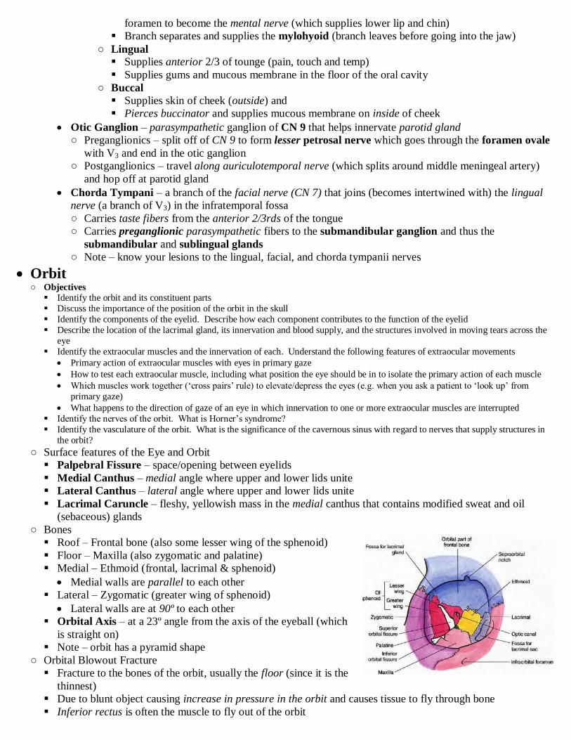
foramen to become the mental nerve (which supplies lower lip and chin)
Branch separates and supplies the mylohyoid (branch leaves before going into the jaw)
○ Lingual Supplies anterior 2/3 of tounge (pain, touch and temp)
Supplies gums and mucous membrane in the floor of the oral cavity
○ Buccal
Supplies skin of cheek (outside) and
Pierces buccinator and supplies mucous membrane on inside of cheek
Otic Ganglion – parasympathetic ganglion of CN 9 that helps innervate parotid gland
○ Preganglionics – split off of CN 9 to form lesser petrosal nerve which goes through the foramen ovale
with V3 and end in the otic ganglion
○ Postganglionics – travel along auriculotemporal nerve (which splits around middle meningeal artery)
and hop off at parotid gland
Chorda Tympani – a branch of the facial nerve (CN 7) that joins (becomes intertwined with) the lingual
nerve (a branch of V3) in the infratemporal fossa
○ Carries taste fibers from the anterior 2/3rds of the tongue
○ Carries preganglionic parasympathetic fibers to the submandibular ganglion and thus the
submandibular and sublingual glands
○ Note – know your lesions to the lingual, facial, and chorda tympanii nerves
Orbit ○ Objectives
Identify the orbit and its constituent parts
Discuss the importance of the position of the orbit in the skull
Identify the components of the eyelid. Describe how each component contributes to the function of the eyelid
Describe the location of the lacrimal gland, its innervation and blood supply, and the structures involved in moving tears across the
eye
Identify the extraocular muscles and the innervation of each. Understand the following features of extraocular movements
Primary action of extraocular muscles with eyes in primary gaze
How to test each extraocular muscle, including what position the eye should be in to isolate the primary action of each muscle
Which muscles work together („cross pairs‟ rule) to elevate/depress the eyes (e.g. when you ask a patient to „look up‟ from
primary gaze)
What happens to the direction of gaze of an eye in which innervation to one or more extraocular muscles are interrupted
Identify the nerves of the orbit. What is Horner‟s syndrome?
Identify the vasculature of the orbit. What is the significance of the cavernous sinus with regard to nerves that supply structures in
the orbit?
○ Surface features of the Eye and Orbit
Palpebral Fissure – space/opening between eyelids
Medial Canthus – medial angle where upper and lower lids unite
Lateral Canthus – lateral angle where upper and lower lids unite
Lacrimal Caruncle – fleshy, yellowish mass in the medial canthus that contains modified sweat and oil
(sebaceous) glands
○ Bones
Roof – Frontal bone (also some lesser wing of the sphenoid)
Floor – Maxilla (also zygomatic and palatine)
Medial – Ethmoid (frontal, lacrimal & sphenoid)
Medial walls are parallel to each other
Lateral – Zygomatic (greater wing of sphenoid)
Lateral walls are at 90º to each other
Orbital Axis – at a 23º angle from the axis of the eyeball (which
is straight on)
Note – orbit has a pyramid shape
○ Orbital Blowout Fracture
Fracture to the bones of the orbit, usually the floor (since it is the
thinnest)
Due to blunt object causing increase in pressure in the orbit and causes tissue to fly through bone
Inferior rectus is often the muscle to fly out of the orbit
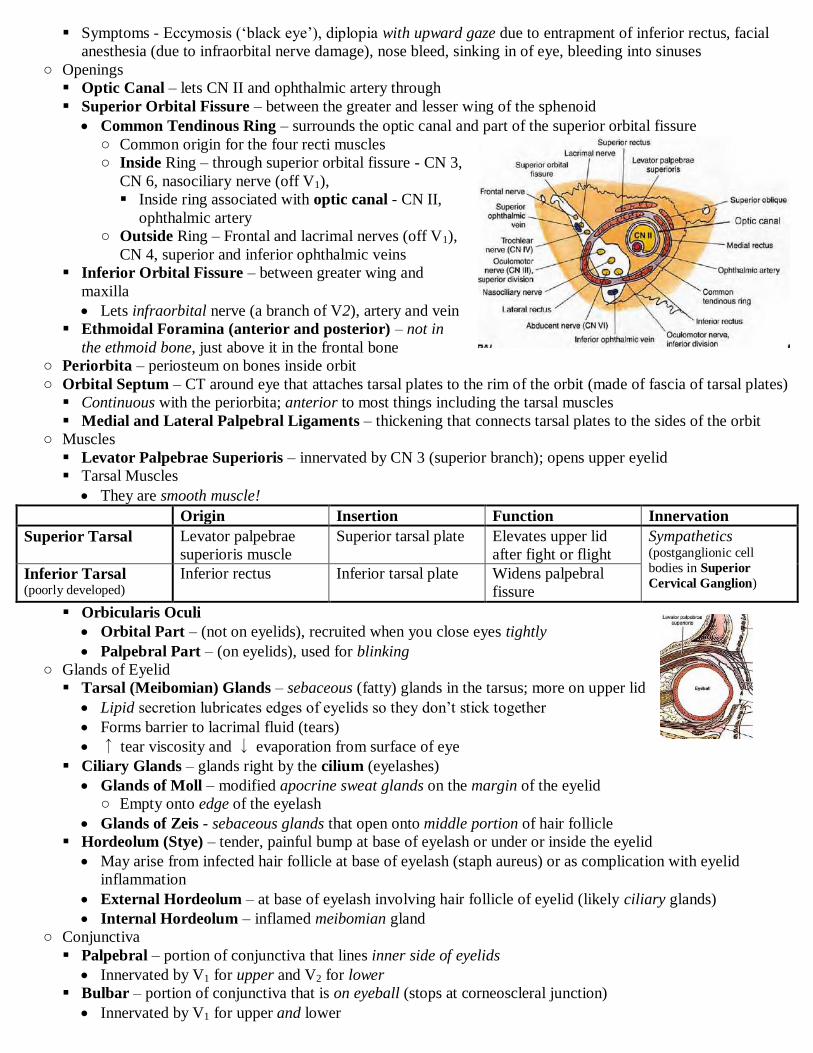
Symptoms - Eccymosis („black eye‟), diplopia with upward gaze due to entrapment of inferior rectus, facial
anesthesia (due to infraorbital nerve damage), nose bleed, sinking in of eye, bleeding into sinuses
○ Openings
Optic Canal – lets CN II and ophthalmic artery through
Superior Orbital Fissure – between the greater and lesser wing of the sphenoid
Common Tendinous Ring – surrounds the optic canal and part of the superior orbital fissure
○ Common origin for the four recti muscles
○ Inside Ring – through superior orbital fissure - CN 3,
CN 6, nasociliary nerve (off V1),
Inside ring associated with optic canal - CN II,
ophthalmic artery
○ Outside Ring – Frontal and lacrimal nerves (off V1),
CN 4, superior and inferior ophthalmic veins
Inferior Orbital Fissure – between greater wing and
maxilla
Lets infraorbital nerve (a branch of V2), artery and vein
Ethmoidal Foramina (anterior and posterior) – not in
the ethmoid bone, just above it in the frontal bone
○ Periorbita – periosteum on bones inside orbit
○ Orbital Septum – CT around eye that attaches tarsal plates to the rim of the orbit (made of fascia of tarsal plates)
Continuous with the periorbita; anterior to most things including the tarsal muscles
Medial and Lateral Palpebral Ligaments – thickening that connects tarsal plates to the sides of the orbit
○ Muscles
Levator Palpebrae Superioris – innervated by CN 3 (superior branch); opens upper eyelid
Tarsal Muscles
They are smooth muscle!
Origin Insertion Function Innervation
Superior Tarsal Levator palpebrae
superioris muscle
Superior tarsal plate Elevates upper lid
after fight or flight
Sympathetics (postganglionic cell
bodies in Superior
Cervical Ganglion) Inferior Tarsal (poorly developed)
Inferior rectus Inferior tarsal plate Widens palpebral
fissure
Orbicularis Oculi
Orbital Part – (not on eyelids), recruited when you close eyes tightly
Palpebral Part – (on eyelids), used for blinking
○ Glands of Eyelid
Tarsal (Meibomian) Glands – sebaceous (fatty) glands in the tarsus; more on upper lid
Lipid secretion lubricates edges of eyelids so they don‟t stick together
Forms barrier to lacrimal fluid (tears)
↑ tear viscosity and ↓ evaporation from surface of eye
Ciliary Glands – glands right by the cilium (eyelashes)
Glands of Moll – modified apocrine sweat glands on the margin of the eyelid
○ Empty onto edge of the eyelash
Glands of Zeis - sebaceous glands that open onto middle portion of hair follicle
Hordeolum (Stye) – tender, painful bump at base of eyelash or under or inside the eyelid
May arise from infected hair follicle at base of eyelash (staph aureus) or as complication with eyelid
inflammation
External Hordeolum – at base of eyelash involving hair follicle of eyelid (likely ciliary glands)
Internal Hordeolum – inflamed meibomian gland
○ Conjunctiva
Palpebral – portion of conjunctiva that lines inner side of eyelids
Innervated by V1 for upper and V2 for lower
Bulbar – portion of conjunctiva that is on eyeball (stops at corneoscleral junction)
Innervated by V1 for upper and lower

Fornices (superior and inferior) – the reflection of palpebral and bulbar conjunctiva; potential space with fluid
○ Lacrimal Apparatus
Lacrimal Gland – in superolateral orbit and secretes lacrimal into superior fornix
Lacrimal Fluid – contains lipid, aqueous and mucin; contains lysosyme (kills bacteria); provides nutrients
and oxygen to cornea
○ Makes up most of the liquid covering of the eye
Lacrimal Lake – area in medial canthus where lacrimal fluid collects
Lacrimal fluid travels from lacrimal gland medially across the conjunctiva to the medial canthus
Lacrimal Puncta – very small duct on medial edge of eyelids that comes into contact with the lacrimal lake
during blinking
Lacrimal Canaliculi – drains lacrimal puncta to lacrimal sac
Lacrimal Sac – top part of nasolacrimal duct that receives the fluids of the lacrimal canaliculi
Nasolacrimal Duct – travels inferiorly down to inferior meatus in the nasal cavity
Clinical Connection
Dry Eye – thin spots in tear film fail to protect eye
○ Causes – can be due to ↓ tear secretion from lacrimal gland or loss of tears due to excess evaporation
○ Symptoms – discomfort, visual fatigue, sensitivity to light, blurred vision
Sjogren’s Syndrome – inflammation of tear-secreting glands causing chronic dry eye and changes in tear
composition
Innervation
Sensory Component – capsule of gland
innervated by lacrimal nerve (V1)
○ Note - lacrimal nerve also carries
sensory fibers from lateral upper eyelid
and conjunctiva (just the V1 parts)
Parasympathetic Component – provides
secretomotor fibers to the parenchyma
○ Presynaptic Fibers – CN 7 gives off
branches to form the greater petrosal
nerve → nerve of pterygoid canal →
pterygopalatine ganglion
○ Postsynaptic Fibers – pterygopalatine ganglion → V2 branches (zygomatic → zygomaticotemporal
nerve) → hops off and goes to lacrimal gland
Sympathetic Branches – innervate vasculature of gland
○ Presynaptic Fibers – from lateral horn of T1-T4 → superior cervical ganglion
○ Postsynaptic Fibers – SCG → ICA plexus → deep petrosal nerve → nerve of pterygoid canal →
pterygopalatine ganglion → follow postsynaptic parasympathetics
○ Orbital Fascia
Fascial Sheath of the Eye (Tenon’s Capsule)
Envelops eye and distal part of EOMs from optic nerve to corneoscleral junction
Pierced by tendons of EOMs and continuous with their deep fascia
Suspensory Ligament of the Eye – continuous with fascial sheath of the eye
○ Attachments – starts at horizontal recti, goes inferior, connects with inferior
oblique and inferior rectus, ends on the other horizontal recti
○ Function – supports eye (forms hammock below eye)
Medial & Lateral Check Ligaments – strong expansions of fascial sheaths of m and l
rectus muscles
Attachments – orbit and lacrimal or zygomatic bones
Function – limits the movement of the m and l rectus muscles, limiting inward and
outward movement of the eye
○ Extraocular Muscles
Insertion – sclera of eye

Note – each of the rectus muscles inserts into sclera at ↑ distances from cornea starting with medial to
inferior to lateral to superior
“LR6 SO4 AO3”
Superior Oblique has a trochlea in the anteriormost and medial part of the orbit
Inferior Oblique originates on the anterior aspect of the orbit
Origin 1º Action 2º Action Innervation
Lateral Rectus Tendinous ring Abduction (primary) None CN 6
Medial Rectus Adduction (primary) None CN 3
Superior Rectus Elevation (primary) Adduction
Medial rotation
CN 3 (superior
branch)
Inferior Rectus Depression (primary) Adduction
Lateral rotation
CN 3
Inferior Oblique Maxilla
(anterior orbit)
Depression
Abduction
Medial rotation CN 3
Superior Oblique Sphenoid Elevation
Abduction
Lateral rotation CN 4
Actions
Except for the horizontal rectus muscles, the EOM actions depend on the orientation of the eye
Cross-Pairs Rule – change to opposite name to get pair
○ You need a pair of EOMs to look „straight up‟ or „straight down‟
○ To look straight up – superior rectus works with inferior oblique
○ To look straight down – inferior rectus works with superior oblique
○ Note – the rotational movements are not voluntary
Note – medial rotation (intorsion); lateral rotators (extorsion)
Actions of EOMs in isolation – see picture (right)
Testing EOMs – bring axis of eye into alignment with axis of the muscle
○ Horizontal rectus muscles are easy
○ Vertical rectus and oblique muscles are more difficult
Palsy Examples
○ LOOK AT HIS HANDOUT pg 153
Innervation
Occulomotor (CN 3)
○ Superior branch - levator palpebrae superioris, superior rectus
○ Inferior branch – medial rectus, inferior rectus, inferior oblique, ciliary ganglion
Trochlear (CN 4) – runs on medial side and dives into superior oblique
Abducent (CN 6) – runs on lateral side and dives into lateral rectus
○ Sensory Innervation
Ophthalmic Nerve (V1)
Lacrimal Nerve – travels on lateral side; (does not pass through tendinous ring)
Frontal Nerve – travels superior to eye; (does not pass through tendinous ring)
○ Becomes Supraorbital and Supratrochlear nerves
Nasociliary – travels medially passes through common tendinous ring;
○ Crosses lateral to medial, superior to CN 2
○ Nasociliary Root of Ciliary Ganglion – fibers pass straight through ciliary ganglion and don‟t synapse
○ Long Ciliary – sensory to eye
Sympathetic to papillary dilator muscle
○ Posterior Ethmoidal – (tough to see), sensory to sphenoidal & ethmoidal sinuses
○ Anterior Ethmoidal – sensory to nasal mucosa and face
Branches about halfway into orbit
Goes in between superior oblique and medial rectus
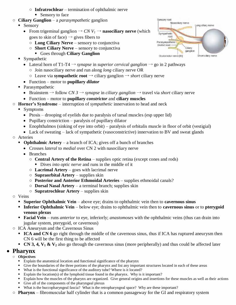
○ Infratrochlear – termination of ophthalmic nerve
Sensory to face
○ Ciliary Ganglion – a parasympathetic ganglion
Sensory
From trigeminal ganglion → CN V1 → nasociliary nerve (which
goes to skin of face) → gives fibers to
○ Long Ciliary Nerve – sensory to conjunctiva
○ Short Ciliary Nerve – sensory to conjunctiva
Goes through Ciliary Ganglion
Sympathetic
Lateral horn of T1-T4 → synapse in superior cervical ganglion → go in 2 pathways
○ Join nasociliary nerve and run along long ciliary nerve OR
○ Leave via sympathetic root → ciliary ganglion → short ciliary nerve
Function – motor to pupillary dilator
Parasympathetic
Brainstem → follow CN 3 → synapse in ciliary ganglion → travel via short ciliary nerve
Function – motor to pupillary constrictor and ciliary muscles
○ Horner’s Syndrome – interruption of sympathetic innervation to head and neck
Symptoms
Ptosis – drooping of eyelids due to paralysis of tarsal muscles (esp upper lid)
Pupillary constriction – paralysis of pupillary dilator
Enophthalmos (sinking of eye into orbit) – paralysis of orbitalis muscle in floor of orbit (vestigial)
Lack of sweating – lack of sympathetic (vasoconstrictive) innervation to BV and sweat glands
○ Arteries
Ophthalmic Artery – a branch of ICA; gives off a bunch of branches
Crosses lateral to medial over CN 2 with nasociliary nerve
Branches
○ Central Artery of the Retina – supplies optic retina (except cones and rods)
Dives into optic nerve and runs in the middle of it
○ Lacrimal Artery – goes with lacrimal nerve
○ Supraorbital Artery – supplies skin
○ Posterior and Anterior Ethmoidal Arteries – supplies ethmoidal canals?
○ Dorsal Nasal Artery – a terminal branch; supplies skin
○ Supratrochlear Artery – supplies skin
○ Veins
Superior Ophthalmic Vein – above eye; drains to ophthalmic vein then to cavernous sinus
Inferior Ophthalmic Vein – below eye; drains to ophthalmic vein then to cavernous sinus or to pterygoid
venous plexus Facial Vein – runs anterior to eye, inferiorly; anastomoses with the ophthalmic veins (thus can drain into
jugular system, pterygoid, or cavernous)
○ ICA Aneurysm and the Cavernous Sinus
ICA and CN 6 go right through the middle of the cavernous sinus, thus if ICA has ruptured aneurysm then
CN 6 will be the first thing to be affected
CN 3, 4, V1 & V2 also go through the cavernous sinus (more peripherally) and thus could be affected later
Pharynx ○ Objectives
Explain the anatomical location and functional significance of the pharynx
Give the boundaries of the three portions of the pharynx and list any important structures located in each of these areas What is the functional significance of the auditory tube? Where is it located?
Explain the location(s) of the lymphoid tissue found in the pharynx. Why is it important?
Explain how the muscles of the pharynx are organized. Give general origins and insertions for these muscles as well as their actions
Give all of the components of the pharyngeal plexus
What is the buccopharyngeal fascia? What is the retropharyngeal space? Why are these important?
○ Pharynx – fibromuscular half cylinder that is a common passageway for the GI and respiratory system

Goes from posterior aperture of the nose to the esophagus
○ Walls of the Pharnyx
Mucous Membrane – has sensory innervation (CN V2 & 9)
Submucosa –
Fibrous Layer (pharyngobasilar fascia) – provides support for muscles
Muscular Layer – constrictors are kinda circular, also longitudinal ones
Loose Connective Tissue - buccopharyngeal fascia
Then retropharyngeal space
○ Subdivisions of the Pharynx
Nasopharynx – base of skull to C2
Lies posterior to the nasal cavity
Choanae – opening between nasal cavity and nasopharynx
Pharyngeal Tonsil (Adenoids) – the superior portion of the nasopharynx abbuted to the occipital bone
Tubal Elevation – around superior entrance of auditory tube; made of cartilage
Auditory Tube (Pharyngotympanic Tube/Eustacian Tube) – connects middle ear to the nasopharynx
○ 2/3rds cartilage and 1/3 bone
○ Otitis Media – middle ear infection
Tubes in Ears – a drain put into the tympanic membrane to fix recurring otitis media
○ Otitis Externa – infection of the skin (which is really thin) of external ear canal
Skin there innervated by CN V3, 7 & 10
Oropharynx – C2-C4
Lies posterior to the oral cavity and is separated from it by the palatoglossal arches
Isthmus – opening between oral cavity and oropharynx
Palatoglossal Arches – fold of mucous membrane over the palatoglossal muscle
○ Right in line with the Uvula
Tonsilar Bed – area between palatoglossal arches (anterior) and palatopharyngeal arches (posterior)
containing the palatine tonsils
○ Innervation – CN 9
○ Blood Supply – facial artery
○ Palatine Tonsils – the „tonsils‟; lymphoid tissue
Palatopharyngeal Arches - fold of mucous membrane over the palatopharyngeal
muscle
Laryngopharynx – C4-C6
Lies posterior to the larynx
Piriform Fossa (recess) – indentation where things can get stuck and cause
irritation
Cricoid Cartilage – bulges into laryngopharnyx
○ Muscles of the Pharynx
Constrictors
Common Aspects
○ Insertion – pharyngeal raphe
○ Action – constrict consecutively and move bolus inferiorly
○ Innervation – CN 10
Superior Constrictor ○ Origin - pterygomandibular raphe (runs from pterygoid hamulus to mandible
posterior to 3rd
molar); is common connection of buccinator
Middle Constrictor ○ Origin – stylohyoid ligament & hyoid bone (superior margin)
Inferior Constrictor ○ Origin – lateral aspect of thyroid cartilage & cricoid cartilage
Longitudinals
Common Aspects
○ Insertion – blend in with constrictors of pharyngeal wall

○ Action – elevate the pharynx
○ Innervation – CN 10 except the stylopharyngeus which is
innervated by CN 9
Salpingopharyngeus
○ Origin – auditory tube
○ Covered in mucous membrane
○ Note – salpinx = tube
Stylopharyngeus ○ Origin – styloid process
○ Innervation – CN 9
Palatopharyngeus ○ Origin – soft palate
○ Covered by mucous membrane and makes the
palatopharyngeus arch
○ Blood Supply
Ascending pharyngeal artery, branches of maxillary artery, branches of facial artery
Also – inferior and superior thyroid arteries, and some lingual artery
○ Innervation
Pharyngeal Plexus – contains CN 10, CN 9 (stylopharyngeus portion), V2, sympathetic fibers (from cervical
sympathetic ganglion to BV), parasympathetic fibers (??to glands?, from CN X?)
Motor Innervation – CN X for constrictors and longitudinals except the stylopharyngeus (CN 9)
Sensory Innervation
Nasopharynx – V2
Oropharynx – CN 9
Laryngopharynx – internal laryngeal branch of CN 10
Note – innervation from cranial branch of accessory is basically called „CN X‟
Accessory Nerve – has two origins
○ Spinal component (cervical) – ventral horn of C1-C4, joins with cranial portion and goes through
jugular foramen and innervates SCM and trapezius
○ Cranial component – begins in nucleus ambiguous, joins with cervical portion and goes through
jugular foramen then joins with CN X
From here we just call it CN X
This stuff innervates pharynx and larynx
○ Temporomandibular Joint (TMJ)
Is a modified hinge joint and a synovial joint
Articular surfaces
condylar process
Postglenoid tubercle (of temporal bone) – limits posterior movement
Mandibular fossa (of temporal bone) – where condylar process rests
Articular tubercle (of temporal bone) – condylar process goes on top of this after jaw opens
Articular Disc – divides joint into two spaces
Inferior Joint Space – allows for hinge movement which is where the condylar process rotates on the
articular disc. This only allows jaw to open about 22 mm, after that protraction of the jaw starts
Superior Joint Space – allows for the gliding movement where the condylar process goes up and rests on
articular tubercle
What occurs when the mouth opens
Depression of the mandible – here the condylar process and articular disc do not move together
Protraction – here the articular disc and condylar process slide as a unit onto the anterior tubercle
More depression – condylar process continues to hinge on the articular disc to open jaw further
TMJ Syndrome – articular disc becomes detached from the chondyle??
This makes 2 clicks (during opening???)
Dislocated jaw – condylar process slides to anterior side of articular tubercle
Branchial and Pharyngeal Apparatus

○ Objectives
Describe a pharyngeal (brachial arch), pharyngeal (branchial) groove, pharyngeal (branchial) pouch, and pharyngeal (branchial)
membrane)
List the four structures that are found in a typical pharyngeal arch
List the skeletal, muscular derivatives and the innervation of each pharyngeal arch
Describe the fate of each of the branchial grooves, including the formation and fate of the cervical sinus Define and/or describe the fate of each of the branchial pouches or derivatives
Define and/or describe the point of origin, the migratory path, and the definitive situation of the thyroid gland
Describe the formation of the tongue
Describe the formation of the face from the five face primordial
Describe the formation of the palate and the congenital abnormalities that result in cleft palate and cleft lip
○ Everything here happens between weeks 4-8
○ Stomadeum – shallow depression that is the primitive mouth
Separated from pharyngeal foregut by the buccopharyngeal membrane
Buccopharyngeal membrane degenerates around 4th
week and forgut opens into amniotic cavity
○ Pharyngeal Apparatus – makes face, nasal cavities, mouth, larynx, pharynx and viscera of neck
Made by pharyngeal pouches, grooves and arches
Pharyngeal Arches – condensations of mesenchyme in between sets of opposed pharyngeal pouches and
grooves
Also contain neural crest cells that invaded in
Each contains
○ An arch artery
○ Cartilagenous rod – (the first cartilaginous rod is called the mandibular
prominence??)
○ Cranial nerve
○ Muscular component – the mesoderm part
Derivatives
○ Note - 1st Arch has two processes, the maxillary and the mandibular
Arch Skeletal Muscles Nerve Blood Vessel
1st
“Mandibular”
- “Meckel‟s cartilage” regresses
- malleus, incus
- sphenomandibular ligament
- muscles of mastication
- two tensors
- anterior belly of digastric
-mylohyoid
V3 Portions of
maxillary artery
2nd
“Hyoid” - “Reichert‟s cartilage” regresses
- stapes, styloid process
- stylohyoid ligament
- superior portion of hyoid
- muscles of facial expression
- stylohyoid
- posterior belly of diastric
- stapedius
CN 7 Stapedial Artery
3rd
- part of hyoid
- small cartilages in larynx (immobile)
- stylopharyngeus CN 9 ICA
4th & 6
th - large laryngeal cartilages - soft palate muscles
- pharynx and larynx muscles
CN 10 - left aortic arch
- right subclavian
Pharyngeal Pouches – out-pouchings of pharyngeal foregut that develop posterior to degenerating b mem.
Lined by endoderm
Each pouch takes the number of the arch above it
Pouch Derivatives
1st Elongates and forms middle ear, internal surface of tympanic membrane, auditory tube
2nd
Doesn‟t develop much
Forms tonsilar fossa, (invaded lymphatic tissue???)
3rd
Dorsal – forms inferior parathyroid (migrates a long way)
Ventral – forms thymus
4th
Dorsal – forms superior parathyroid
Ventral – forms ultimobranchial body (neural crest cells that invade thyroid and makes
parafollicular cells)
5th
Gets combined with 4th
Pharyngeal Grooves – grooves between the pouches, on the outside, opposite the pharyngeal pouches
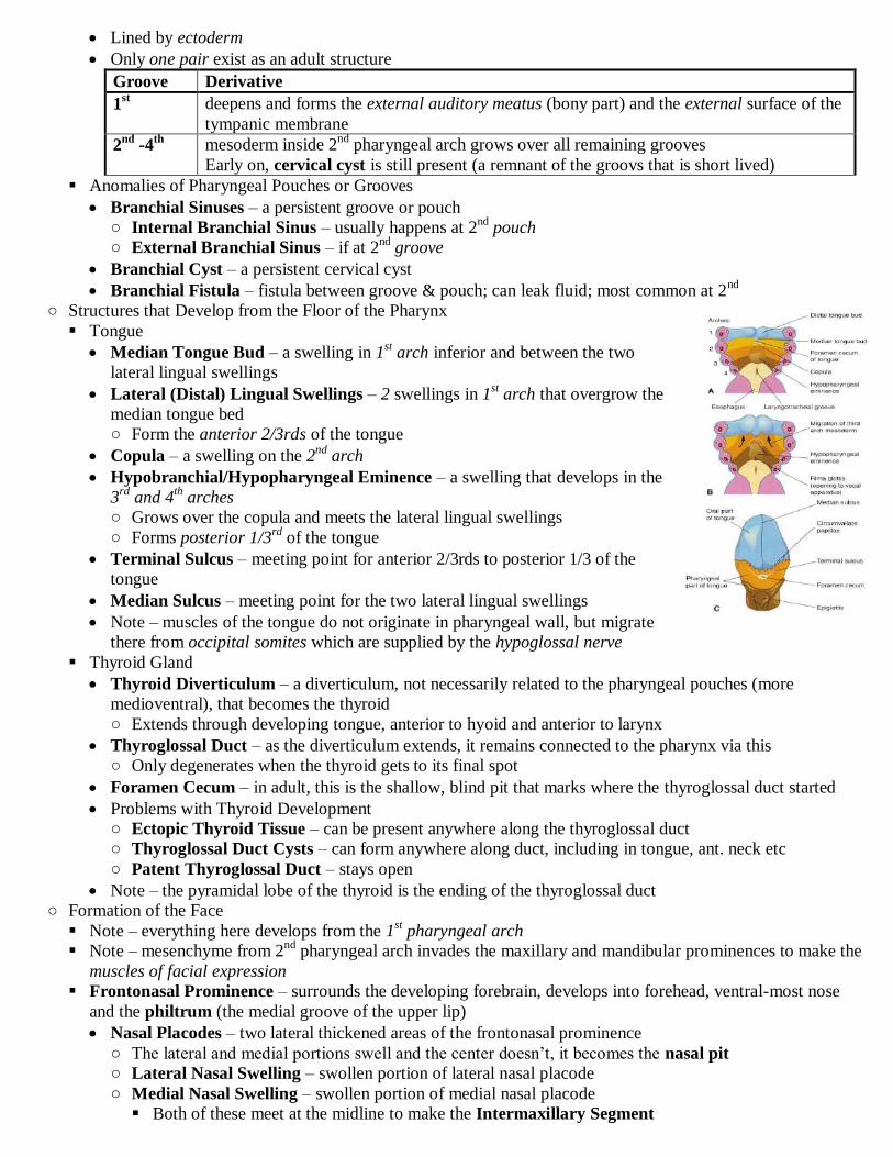
Lined by ectoderm
Only one pair exist as an adult structure
Groove Derivative
1st deepens and forms the external auditory meatus (bony part) and the external surface of the
tympanic membrane
2nd
-4th
mesoderm inside 2nd
pharyngeal arch grows over all remaining grooves
Early on, cervical cyst is still present (a remnant of the groovs that is short lived)
Anomalies of Pharyngeal Pouches or Grooves
Branchial Sinuses – a persistent groove or pouch
○ Internal Branchial Sinus – usually happens at 2nd
pouch
○ External Branchial Sinus – if at 2nd
groove
Branchial Cyst – a persistent cervical cyst
Branchial Fistula – fistula between groove & pouch; can leak fluid; most common at 2nd
○ Structures that Develop from the Floor of the Pharynx
Tongue
Median Tongue Bud – a swelling in 1st arch inferior and between the two
lateral lingual swellings
Lateral (Distal) Lingual Swellings – 2 swellings in 1st arch that overgrow the
median tongue bed
○ Form the anterior 2/3rds of the tongue
Copula – a swelling on the 2nd
arch
Hypobranchial/Hypopharyngeal Eminence – a swelling that develops in the
3rd
and 4th arches
○ Grows over the copula and meets the lateral lingual swellings
○ Forms posterior 1/3rd
of the tongue
Terminal Sulcus – meeting point for anterior 2/3rds to posterior 1/3 of the
tongue
Median Sulcus – meeting point for the two lateral lingual swellings
Note – muscles of the tongue do not originate in pharyngeal wall, but migrate
there from occipital somites which are supplied by the hypoglossal nerve
Thyroid Gland
Thyroid Diverticulum – a diverticulum, not necessarily related to the pharyngeal pouches (more
medioventral), that becomes the thyroid
○ Extends through developing tongue, anterior to hyoid and anterior to larynx
Thyroglossal Duct – as the diverticulum extends, it remains connected to the pharynx via this
○ Only degenerates when the thyroid gets to its final spot
Foramen Cecum – in adult, this is the shallow, blind pit that marks where the thyroglossal duct started
Problems with Thyroid Development
○ Ectopic Thyroid Tissue – can be present anywhere along the thyroglossal duct
○ Thyroglossal Duct Cysts – can form anywhere along duct, including in tongue, ant. neck etc
○ Patent Thyroglossal Duct – stays open
Note – the pyramidal lobe of the thyroid is the ending of the thyroglossal duct
○ Formation of the Face
Note – everything here develops from the 1st pharyngeal arch
Note – mesenchyme from 2nd
pharyngeal arch invades the maxillary and mandibular prominences to make the
muscles of facial expression
Frontonasal Prominence – surrounds the developing forebrain, develops into forehead, ventral-most nose
and the philtrum (the medial groove of the upper lip)
Nasal Placodes – two lateral thickened areas of the frontonasal prominence
○ The lateral and medial portions swell and the center doesn‟t, it becomes the nasal pit
○ Lateral Nasal Swelling – swollen portion of lateral nasal placode
○ Medial Nasal Swelling – swollen portion of medial nasal placode
Both of these meet at the midline to make the Intermaxillary Segment

○ Intermaxillary Segment – gives rise to the philtrum, portions of the upper jaw, nasal septum and
primary palate
Maxillary Prominence – produced by proliferation of neural crest cells
Grow toward the lateral nasal swellings and meet at the nasolacrimal groove
Forms the lateral portion of the upper lip, upper cheek, most of maxilla and the palate
Mandibular Prominence – produced by proliferation of neural crest cells
The two mandibular prominences join together at midline, also meet the maxillary prominence above
Forms the lower lip, lower cheek, chin and mandible
○ Formation of the Palate
Occurs during 5th week
Incisive Foramen – junction of primary and secondary palates
Midline Raphe – marks the junction of the lateral palatine processes
Primary Palate – develops from the intermaxillary segment (part of frontonasal prominence)
Forms anterior portion of hard palate (anterior to incisive foramen)
Secondary Palate – the lateral palatine processes, which are prominences on the internal surface of the
maxillary prominences, enlarge and grow to meet in the midline
They fuse with the nasal septum
The anterior portion of the processes forms the hard palate and the posterior portion forms the soft palate
Cleft Lip – 1 out of 900 births; boys>girls
Occurs at the philtrum
Unilateral – failure of the maxillary and intermaxillary segments to fuse on one side
Bilateral – both sides
Cleft Palate – 1 out of 2500 births; girls>boys
Occurs at incisive foramen or the midline raphe
Primary (anterior) – failure of the primary palate to meet the secondary palate (incisve foramen)
Secondary (posterior) – failure of the lateral palatine processes to fuse together and with primary palate
Complete – no fusion of nothing (how is this different from
secondary??)
A – normal, B – unilateral cleft lip, C – unilateral cleft lip
and primary cleft palate, D – bilateral cleft lip and primary
cleft palate, E – isolated cleft palate, unilateral cleft lip and
secondary cleft palate
Nasal Cavity, Palate and Pterygopalantine Fossa ○ Objectives
Explain the external anatomy of the nose. What is responsible for the different shapes and sizes of noses?
Discuss the parts of the nasal septum. Give the blood supply and innervation to this structure
Explain the irregular contour of the lateral nasal wall. The inferior nasal conchae is a part of what bone? Give the blood supply and
innervation to the lateral nasal wall
Explain the shape of the ethmoid bone. What is the function of the paranasal sinuses? Describe the route of mucus drainage from
these sinuses
Discuss several common clinical problems associated with the nasal cavity (fractures, nosebleeds, chronic sinusitis) What bones make up the hard palate? What muscles make up the soft palate? What is the function of the uvulae?
Describe the complete course of the greater palatine nerve, the nasopalatine nerve. What components are in these nerves?
Give the source of the blood supply to the hard and soft palate
○ Nose – external nose and nasal cavity
External Nose – composed of 5 cartilages (4 paired, 1 unpaired) which determine shape of nose, and
fibrofatty tissue at the lateral end
Septal Cartilage – the 1 unpaired cartilage that runs on dorusm of external nose
Nares – anterior nasal openings
Dorsum – the „superior‟ surface
Root – where the bridge of the glasses sit
Apex – the tip of the nose
Ala – the winged, lateral portion
Nasal Septum – divides the two nares

Bony portion – made up of the two nasal, maxillary and frontal bones; superior to cartilages
Innervation
○ Dorsum → apex = CN V1
○ Ala – CN V2
Nasal Cavity – begins at the nares and ends at the posterior openings of the choana where it opens into
nasopharynx
Vestibule – entrance into nasal cavity (where you pick your nose); has course hair
○ Transition from skin of face to mucus membrane of nasal cavity
○ Innervation – nasal branches of infraorbital nerve (V2)
Mucoperiosteum – the mucus membrane of the nasal cavity
○ Special name because it is fused with the periosteum
○ respiratory mucus membrane with lots of glands and is highly vascular
Nasal Septum – composed of cartilage and very weak bone
○ Composition
Bony Portion
○ Perpendicular plate of Ethmoid – superior and anterior
○ Vomer – inferior and posterior
○ Nasal crest of maxilla (anterior) and palatine (posterior) – inferiormost part
Cartilagenous portion
○ Septal Cartilage – forms anterior-most portion of nasal septum
Note – the cribriform plate of the ethmoid bone forms a superior border of the nasal cavity
○ Innervation
Olfactory Nerve (CN 1) – goes through the cribriform plate and sends to olfactory bulb
○ Neurons are bipolar and have chemoreceptors in olfactory mucus membrane
○ Located in upper 1/3rd
of nasal cavity
Sensory – portions of CN V
○ Anterior Ethmoidal Nerve (septal branch) – V1 (a branch of the nasociliary nerve)
Courses anteriorly to almost the tip of the nose
○ Nasopalatine Nerve – V2
Comes out posterior to the olfactory nerve and courses obliquely on nasal septum
Goes into Incisive Foramen to supply the anterior hard palate
○ Blood Supply
Branches of Ophthalmic artery
○ Anterior Ethmoidal artery (septal branches) – follows anterior ethmoidal nerve
○ Posterior Ethmoidal Artery (septal branches) – comes out in the middle of the olfactory nerve
area and supplies middle septum
Branches of Maxillary artery
○ Sphenopalatine Artery – follows course of nasopalatine nerve (through incisive foramen)
A terminal branch of the maxillary artery
Note – each of those arteries has a corresponding vein
Lateral Nasal Wall – not flat to ↑ surface area to warm and moisten air (along with mucoperiosteum)
○ Concha – curved shelves of bone that extend from the lateral nasal wall
Inferior Nasal Concha – largest and most anterior; formed by seperate bone
Middle Nasal Concha – part of ethmoid bone
Superior Nasal Concha – part of ethmoid bone; smallest and most posterior
Note – in a vertical cross section you will often only see two concha at a time
○ Nasal Meatus – passageway deep to each concha
Inferior Meatus – (under inferior nasal concha) has opening for nasolacrimal duct
Middle Meatus – lots of openings
○ Hiatus Semilunaris – a semilunar groove with holes
Infundibulum – the anterior-superior most portion of it that receives the frontonasal duct
which drains the frontal sinus
Has an opening for the anterior ethmoidal air cells (more anterior superior)
Has an opening for the maxillary sinus (more posterior inferior)

○ Hiatus semilunaris is bounded by bulge superior to it, the ethmoid bulla, which is a protrusion of
the middle ethmoidal air sacs
Has openings from middle ethmoidal air sacs on it
Superior Meatus – has opening for posterior ethmoidal air cells
Sphenothmoidal Recess – above the superior nasal concha
○ Has opening for the sphenoidal sinus
○ Innervation
Olfactory Nerve – upper 1/3rd
Sensory – branches of CN V
○ Anterior Ethmoidal Nerve (septal branch) – V1, innervates anterior lateral nasal wall
○ Posterior Lateral Nasal Nerves – V2, come out posterior to olfactory nerve area and then goes
lateral across the concha
○ Inferior Nasal Branches of Infraorbital Nerve – V2, supplies inferior-anterior portion and
comes in inferiorly
○ Blood Supply
Exactly the same as the blood supply for nasal septum
○ except the sphenopalatine artery (a branch of the maxillary artery) gives off posterior nasal
branches that follow the posterior lateral nasal nerves and supply the concha
○ Paranasal Sinuses
Diverticula of the nasal cavity; mucus membrane is continuous
Enlarge with age
Named by the bone that contains them
Innervation – CN V
Frontal Sinus – most superior; drained by frontonasal duct, which opens
into the hiatus semilunaris at the infundibulum
Ethmoidal Sinuses (Air Cells) – the anterior and middle ones open into
middle meatus, the posterior one opens into the superior meatus
There are 8-12 air cells on each side
Sphenoid Sinus – most posterior; drains into sphenoethmoidal recess
(above superior nasal concha)
Maxillary Sinus – most inferior; drains into hiatus semilunaris (of middle meatus)
Inferior portion of sinus is very close to the upper molars
○ Maxillary Sinusitis – can manifest as upper molar pain
○ Absess of Upper Molars – can spread into maxillary sinus
Note – only the frontal sinus can drain into the nasal cavity easily by gravity
Thus mucus can build up and cause sinus headache or infection
If recurrent problem then a common surgery is to drill a hole in the maxillary sinus at the bottom of the
nasal floor to allow it to drain more easily (there will still be a potential place for mucus to pool however)
○ Palate
Separates the nasal cavity from the oral cavity
Hard Palate – bony portion that is more anterior
Lined by mucoperiosteum (continuous with surroundings)
Contains minor salivary glands (in mucous membrane)
Ruggae – ridges on top of mouth
Bones
○ Palatine Process of maxilla – forms the anterior portion
Most made by secondary palate
○ Horizontal portion of palatine bone – forms the inferior portion
Is this made by secondary palate too??
Incisive Foramen – marks juncture of the primary palate (anterior) and the two sides of the secondary
palate
○ Transmits the nasopalatine nerve (from nasal septum) and anastomosis of sphenopalatine and greater
palatine artery
Greater Palatine Foramen – transmits greater palatine N. A. & V.
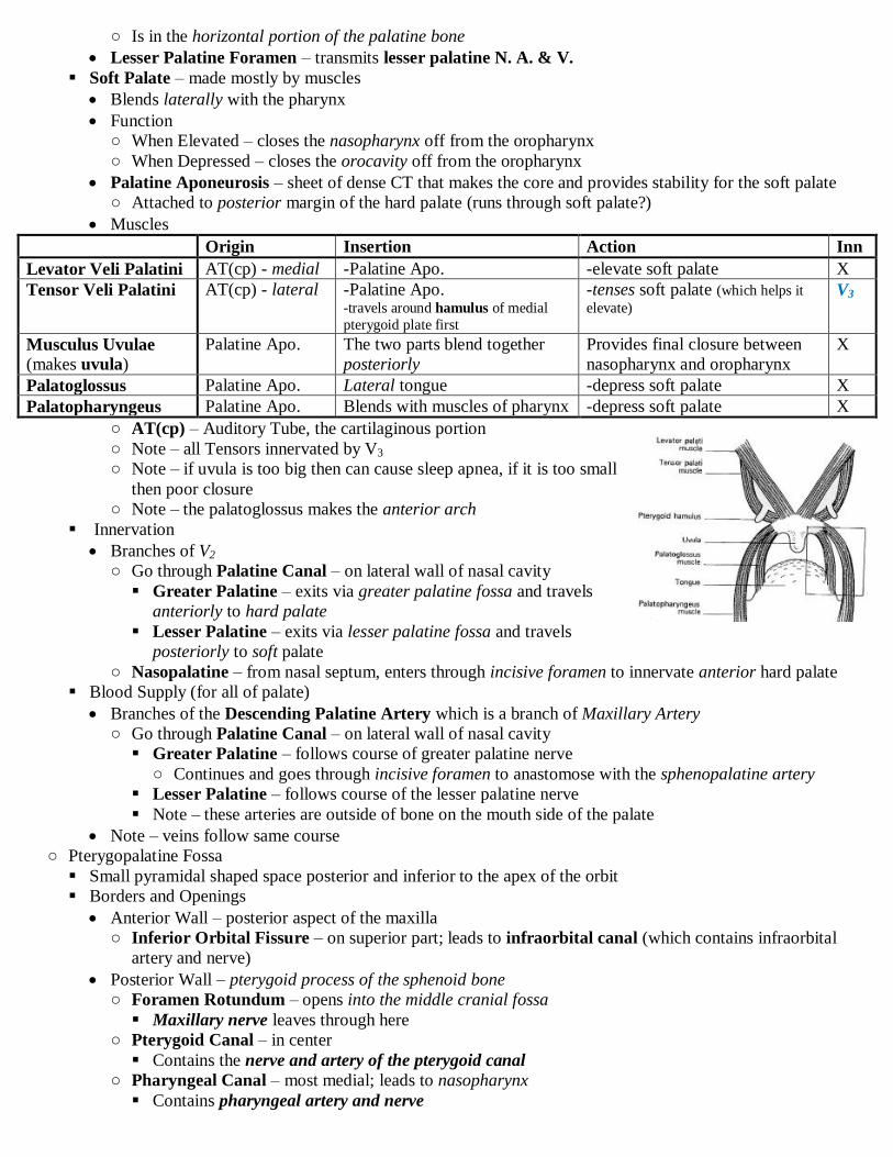
○ Is in the horizontal portion of the palatine bone
Lesser Palatine Foramen – transmits lesser palatine N. A. & V.
Soft Palate – made mostly by muscles
Blends laterally with the pharynx
Function
○ When Elevated – closes the nasopharynx off from the oropharynx
○ When Depressed – closes the orocavity off from the oropharynx
Palatine Aponeurosis – sheet of dense CT that makes the core and provides stability for the soft palate
○ Attached to posterior margin of the hard palate (runs through soft palate?)
Muscles
Origin Insertion Action Inn
Levator Veli Palatini AT(cp) - medial -Palatine Apo. -elevate soft palate X
Tensor Veli Palatini AT(cp) - lateral -Palatine Apo. -travels around hamulus of medial
pterygoid plate first
-tenses soft palate (which helps it
elevate) V3
Musculus Uvulae (makes uvula)
Palatine Apo. The two parts blend together
posteriorly
Provides final closure between
nasopharynx and oropharynx
X
Palatoglossus Palatine Apo. Lateral tongue -depress soft palate X
Palatopharyngeus Palatine Apo. Blends with muscles of pharynx -depress soft palate X
○ AT(cp) – Auditory Tube, the cartilaginous portion
○ Note – all Tensors innervated by V3
○ Note – if uvula is too big then can cause sleep apnea, if it is too small
then poor closure
○ Note – the palatoglossus makes the anterior arch
Innervation
Branches of V2
○ Go through Palatine Canal – on lateral wall of nasal cavity
Greater Palatine – exits via greater palatine fossa and travels
anteriorly to hard palate
Lesser Palatine – exits via lesser palatine fossa and travels
posteriorly to soft palate
○ Nasopalatine – from nasal septum, enters through incisive foramen to innervate anterior hard palate
Blood Supply (for all of palate)
Branches of the Descending Palatine Artery which is a branch of Maxillary Artery
○ Go through Palatine Canal – on lateral wall of nasal cavity
Greater Palatine – follows course of greater palatine nerve
○ Continues and goes through incisive foramen to anastomose with the sphenopalatine artery
Lesser Palatine – follows course of the lesser palatine nerve
Note – these arteries are outside of bone on the mouth side of the palate
Note – veins follow same course
○ Pterygopalatine Fossa
Small pyramidal shaped space posterior and inferior to the apex of the orbit
Borders and Openings
Anterior Wall – posterior aspect of the maxilla
○ Inferior Orbital Fissure – on superior part; leads to infraorbital canal (which contains infraorbital
artery and nerve)
Posterior Wall – pterygoid process of the sphenoid bone
○ Foramen Rotundum – opens into the middle cranial fossa
Maxillary nerve leaves through here
○ Pterygoid Canal – in center
Contains the nerve and artery of the pterygoid canal
○ Pharyngeal Canal – most medial; leads to nasopharynx
Contains pharyngeal artery and nerve

Medial Wall – vertical portion of the palatine bone
○ Sphenopalatine Foramen – opens into the nasal cavity
Contains sphenopalatine artery and nasopalatine nerve
Lateral Wall – opens to the infratemporal fossa through the pterygomaxillary fissue
○ Pterygomaxillary Fissue – the inferior edges of the pterygopalatine fossa that come to a point
Maxillary artery comes in here, comes in lateral and goes medial
Posterior superior alveolar nerve and artery leave through here
Inferiorly
○ (Greater) Palatine Canal – goes to oral cavity
Transmits the greater and lesser palatine nerves & descending palatine artery
Contents
Third portion of maxillary artery and its branches
Maxillary nerve and its branches
Pterygopalatine ganglion and the nerve of the pterygoid canal
Maxillary Artery Branches
Note – comes in through the pterygomaxillary fissure
Posterior Superior Alveolar Artery – 1st branch
○ Exits PT fossa via pterygomaxillary fissure and pierces maxilla to supply upper teeth
Descending Palatine Artery – exits via palatine canal and divides into the greater and lesser palatine
arteries
Artery of the Pterygoid Canal – exits via pterygoid canal and supplies the nerve of the pterygoid canal
Pharyngeal Artery – exits via the pharyngeal canal and supplies nasopharynx
Infraorbital Artery – traverses the inferior orbital fissure and then exits via the infraorbital canal
○ Supplies the middle and anterior upper teeth and then supplies the face
Sphenopalatine Artery – exits via sphenopalatine foramen to supply the nasal cavity to supply nasal
septum (remember it gives off posterior nasal branches for lateral nasal wall)
○ Is the terminal branch
Maxillary Nerve (V2) Branches
Pure sensory
Exits PT fossa through the foramen rotundum
Nasopalatine – enters the sphenopalatine foramen
and innervates the nasal septum and anterior hard
palate
Infraorbital – enters the infraorbital canal and
innervates face, lateral nasal, cheek, lower eyelid
and upper lip
○ Posterior Superior Alveolar Nerve – branches
before infraorbital canal and innervates
posterior upper teeth
○ Anterior Superior Alveolar Nerve – goes
through infraorbital canal and then branches to
innervate anterior upper teeth
Pharyngeal – enters the pharyngeal canal and innervate mucus membrane of nasopharynx
Exit via Palatine Canal
○ Greater Palatine – exits palatine canal via the greater palatine foramen and innervates hard palate
○ Lesser Palatine – exits palatine canal via the lesser palatine
foramen and innervates soft palate
Pterygopalatine Ganglion
In PT fossa, suspended from maxillary nerve by two roots
Sensory Component
○ Fibers of V2 go through it to supply nasopalatine, posterior nasal,
greater palatine, lesser palatine and pharyngeal nerves
Parasympathetic Component
○ CN 7 → greater petrosal nerve → enters pterygoid canal to make nerve of the pterygoid canal →
enters pterygopalatine ganglion → synapses there → postganglionics are distributed via maxillary nerve
(V2) and also go up to V1 to innervate lacrimal gland
Sympathetic Component
○ lateral horn of T1-T4 → synapses in SCG → deep petrosal nerve → enters pterygoid canal to make
nerve of the pterygoid canal → enters pterygopalatine ganglion → is distributed with branches of V2
Extra Notes
Maxillary artery comes in through lateral side of PT fossa and exits on medial side
Maxillary nerve enters PT fossa from posterior side and exits as the infraorbital nerve
Functional Components of Cranial Nerves ○ Objectives
Review the components of a typical spinal nerve. How do those components compare and contrast to those of a cranial nerve?
Review the general plan for the sympathetic division of the ANS. Describe the primary pathways for sympathetic fibers to the head
and neck
Review the general plan for the organization of the parasympathetic division of the ANS. Which cranial nerves are associated with
the parasympathetic division of the ANS
Diagram all of the cranial nerves with a parasympathetic component. Identify the course of the preganglionic and postganglionic fibers, as well as the location and name of the ganglion associated with each nerve
What role does the trigeminal nerve serve in the autonomic control of structures in the head and neck? Identify portions of the
trigeminal nerve associated with the autonomic components of other cranial nerves.
○ General Types of Nerves in Neck
Afferents (Sensory) - pseudounipolar
Special Sense
Somatic – pain, touch and temp (can be mucus membrane in nasal cavity)
Visceral – „vague‟; in neck mostly innervation of mucus membrane
Efferents (Motor) – unipolar
Skeletal muscle – (from pharyngeal arch or somite, but this doesn‟t matter much)
Parasympathetic – smooth muscle, glands (cardiac muscle)
○ Have preganglionic and postganglionic fibers with an autonomic ganglia
○ Found with CN 3, 7, 9 & 10
○ General Sympathetic Visceral Efferent (in neck)
Effector – sweat glands
Pathway – lateral horn T1-T4 → ventral root → white ramus → paravertebral ganglion → cervical
sympathetic chain → synapse in any of the cervical ganglia → go to the plexus of one of the major arteries
(ECA, ICA) → travel along one of the branches of that artery → effector (often sweat glands)
○ Cranial Nerves
See extra table
CN Sensory Ganglion Parasympathetic Ganglion
3 Ciliary
5 Semilunar
7 Geniculate Pterygopalatine
Submandibular
9 Superior & Inferior Glossopharyngeal Otic
10 Superior & Inferior Vagal (Jugular) Terminal
Oral Cavity ○ Objectives
Define the boundaries of the oral cavity Review the innervation of the following structures: the lips, cheeks and gums
Review the five areas where lymphoid tissue forms “tonsils” around regions of the oral cavity and the formation of the tonsilar ring
Explain the anatomical considerations associated with the tongue and its sensory innervation
Know the organization, action, blood supply and innervation of the intrinsic and extrinsic muscles of the tongue
Review the suprahyoid muscles. What are their separate innervations? What is their collective action?
Review the three main pairs of salivary glands. Explain their location, the location of their duct(s) and their innervation
○ Boundaries
Lateral – cheeks

Superior – palate
Inferior - sublingual fossa and tongue
Anterior – lips and rima oris (opening of lips)
Posterior – oropharynx
○ Parts of Oral Cavity
Vestibule – space on buccal side of cheek and gums
Oral Cavity Proper – area on lingual side of teeth and gums; normally filled with the tongue
○ Review – Oropharynx contains palatoglossal arch (more anterior), palatopharyngeal arch (more posterior),
tonsilar bed and palatine tonsil and is innervated by CN 9
But the blood supply is the tonsilar branch of the facial artery
○ Lips
Vermillion Border – transition from red skin to normal skin
Philtrum – shallow vertical groove; the portion of the upper lip derived from the frontonasal prominence
(cleft lip occurs here)
Labial Frenulum – small fold of mucus membrane that is an attachment of lips to gums
Innervation
Upper Lip - Infraorbital Nerve (V2)
Lower Lip - Mental Nerve (V3)
○ Cheek
Boundaries – Medial boundary = Nasolabial Groove (the smile crease); Lateral Boundary = across zygo arch
Contents
Buccal Fat Pad – padding
Buccinator – a muscle of facial expression supplied by CN 7
Parotid Duct - pierces buccinator and opens as a papillae adjacent to the 2nd
upper molar in vestibule
Innervation
Infraorbital (V2)
(Long) Buccal Nerve (V3) – innervates inside and outside of cheek
○ pierces the buccinator
○ Gums (Gingivae) – fibrous CT covered with mucus membrane
Alveolar Gingivae – unattached part; the part between and encircling each tooth
Gingiva Proper – attached part; tightly affixed to the mandible or maxilla
Innervation – basically any nerve that passes the gums (don‟t focus on this)
External
○ V2 - posterior superior alveolar, anterior superior alveolar, infraorbital
○ V3 - inferior alveolar, mental, buccal
Internal
○ V2 – greater palatine, nasopalatine
○ V3 – lingual
○ Review – Innervation of Teeth
Upper Teeth = V2 → infraorbital → anterior superior alveolar & posterior superior alveolar (sometimes
middle)
Lower Teeth = V3 → inferior alveolar (then sometimes mental nerve)
○ Tongue
Function
Mechanical Digestion – keeps food between teeth, shape food into bolus, force bolus against soft palate
Move food into pharynx
Aid in phonation (formation of words) – if problem with tongue then slurred speech
Parts
Oral Portion (Body) – the unattached/movable part; in oral cavity; basically the anterior 2/3
Pharyngeal Portion (Root) – the attached (to hyoid) part; faces oropharynx; basically the posterior 1/3
Surfaces
Inferior Surface – covered by a thin layer of mucous membrane; can see blood vessels through it
○ Contains deep lingual veins and sublingual veins which are useful for drug delivery

○ Lingual Frenulum – line of mucous membrane from sublingual fossa to inferior tongue
If too short – tongue tied and can be surgically cut
If too long – often can touch nose with tongue
Dorsum (Superior Surface) – covered by thick mucous membrane and papillae
○ Papillae – specializations of mucous membrane; on anterior 2/3 of tongue
Filiform Papillae – increase texture of tongue for grip; most numerous, parallel to sulcus terminalis
○ No taste buds
Fungiform Papillae - club-shaped; have a singular taste bud; randomly placed
Folate Papillae – on lateral tongue; contain numerous taste buds
Vallate Papillae – 8-12 of them just anterior to sulcus terminalis; contains numerous taste buds
Note – no papillae on posterior 1/3, but there are taste buds there
○ Other Features
Median Sulcus – joining of the lateral tongue buds
Sulcus Terminalis – junction of anterior 2/3 (lateral tongue buds) with posterior 1/3
(hypopharyngeal eminence)
Foramen Cecum – old opening of the thyroglossal duct; right at point of sulcus terminalis
Lingual Tonsil – lymphoid tissue covered by mucous membrane (in posterior 1/3rd
)
Innervation
Anterior 2/3
○ Sensory – trigeminal ganglion → Lingual Nerve (V3)
○ Taste – geniculate ganglion → chorda tympani (CN 7) → Lingual nerve (V3 + CN 7)
Posterior 1/3
○ Sensory – Superior or inferior petrosal ganglia → lingual branch of CN 9
○ Taste
Superior or inferior petrosal ganglia → lingual branch of CN 9 → vallate papillae and taste buds of
posterior 1/3
Vagus (Lingual Branch) (superior & inferior vagal ganglion) → taste buds near epiglottis and very
back of tongue
Muscles
Innervation – CN 12 (Hypoglossal)
Intrinsic Muscles – create the body of the tongue
○ Arranged in bands (horizontal, longitudinal, vertical)
External Muscles – move body of tongue
○ Embryological Origin - Somites
Origin Insertion Action Inn
Hyoglossus Hyoid (superior part) Lateral tongue body Depress posterior and lateral
tongue
CN 12
Styloglossus Styloid Process Posterior-Superior
tongue body
Retract & elevate posterior
tongue
Genioglossus Mental Spine Fans out and attaches to
body of tongue
Protudes tongue
○ Clinical Application
Test function of CN 12 by asking patient to stick out tongue and see if it deviates to one side
○ Note – palatoglossus also helps move tongue? It is innervated by CN X
Blood Supply
Lingual Artery – from ECA; landmark – dives deep to the hyoglossus
○ Dorsal Lingual – to posterior tongue
○ Deep Lingual – termination of lingual artery; supplies body of tongue
○ Sublingual – anterior branch of lingual artery; supplies the sublingual
gland
○ Salivary Glands
Parotid Glands – largest salivary gland
Parotid Duct – crosses masseter → pierces buccinator → opens into

vestibule adjacent to 2nd
upper molar
Innervation
○ Parasympathetic – CN 9 → lesser petrosal → synapse in otic ganglion → joins with auriculotemporal
(V3) → jumps off and goes to parotid
○ Sympathetic – T1-T4 → synapse in SCG → follows BVs to parotid
Can go through otic ganglion if it wants
Submandibular Glands – located along body of mandible on either side of mylohyoid
Submandibular Duct – crosses under lingual nerve (nice landmark) in sublingual fossa → touches
sublingual gland → opens in sublingual papilla (adjacent to lingual frenulum)
Sublingual Glands – in floor of mouth, deep to tongue
Opens via many ducts directly into the sublingual fossa
Innervation to Submandibular and Sublingual Glands
Parasympathetic – CN 7 → chorda tympani → joins with lingual nerve (V3) → synapses in submandibular
ganglion
○ Postsynaptics then either go to submandibular gland or to the sublingual gland (where they meet with
lingual nerve again?)
Sympathetic – T1-T4 → synapse in SCG → follows BVs to parotid
○ Can go through submandibular ganglion if it wants
○ Structures of the Sublingual Fossa
Sublingual Gland
Submandibular Duct
Lingual Nerve (V3) – landmark – crosses over submandibular duct
Hypoglossal Nerve
Sublingual Artery – in anterior part
○ Muscles to Suprahyoid Region
Action
raise hyoid, tongue and floor
steady hyoid for independent tongue movements
If hyoid fixed then they can help open mouth
Names – stylohyoid, mylohyoid, geniohyoid, digastric
Larynx ○ Objectives
Describe the cartilaginous skeleton of the larynx
Explain the orientation of the membranes that act to support the cartilages of the larynx Describe the internal anatomy of the larynx. Where are the vestibular folds? Where are the vocal folds?
Explain the location, innervation and action of each of the intrinsic muscles of the larynx
Review the blood supply and innervation to the mucous membrane that lines the larynx
Explain how the larynx works during phonation. What anatomical features of the larynx may affect the quality of your voice?
○ General
Larynx – organ of speech; holds airway open
Anteriorly covered by infrahyoid muscles and the thyroid gland
Adjacent to C3-C6; visible as laryngeal prominence
Anterior to laryngopharynx
Larger in men
Completely covered in mucous membrane (inside and out)
○ Anatomy
Cartilages
Unpaired
○ Thyroid Cartilage – largest; is open posteriorly
Made by 2 plates that come together and form superior thyroid notch and superiorly the laryngeal
prominence Superior Horn – thyrohyoid membrane attaches here
Inerior Horn – articulates with the cricoid cartilage

○ Cricoid Cartilage – only part that forms complete ring
Narrow anterior arch; wider and flatter posterior lamina (like signet ring)
Posterior superior portion articulates with arytenoid cartilages
Articulates with inferior horn of thyroid cartilage
○ Epiglottis – leaf shaped, elastic cartilage
Articulates with internal anterior surface of thyroid cartilage
Paired – (made of hyaline cartilage?)
○ Arytenoid Cartilages – pyramidal shape
Muscular Process – lateral extension; attachment for posterior and lateral cricoarytenoid muscles
Vocal Ligament – anterior extension; attachment for vocal ligament
○ Corniculate Cartilages – small cartilages that lie on the apex of the arytenoid cartilages
○ Cuneiform Cartilages – small nodules of cartilage in the aryepiglottic fold
Membranes
Outside
○ Thyrohyoid Membrane – causes movement of the hyoid to translate to movement of the thyroid
Pierced by the internal laryngeal nerve and the superior laryngeal artery
○ Cricothyroid Membrane
Medial Cricothyroid Membrane – exterior; just connects thyroid to cricoid
Lateral Cricothyroid Membrane (Conus Elasticus) – interior; connects vocal ligament to cricoid
○ Cricotracheal Membrane – goes all the way around
Inside
○ Quadrangular Membrane – creates tube called the Laryngeal Inlet
Attachments – lateral margins of epiglottic cartilage & anterolateral margin of arytenoid cartilage
Aryepiglottic Fold – superior margin of quadrangular membrane covered in mucous membrane
Vestibular Ligament – inferior margin of quadrangular membrane
Vestibular Fold – vestibular ligament covered in mucous membrane
○ Conus Elasticus (Lateral Cricothyroid Membrane) – body of it connects to cricoid
Vocal Ligament – superior margin of conus elasticus; connects arytenoids to anterior thyroid
Vocal Fold – vocal ligament covered in mucous membrane
Chambers of Interior Larynx
Rima Glottis – space between the two vocal ligaments
Vestibule – interior area of larynx superior to the vestibular fold
Ventricle – diverticulum of mucous membrane between the vestibular and vocal folds
Infraglottic Cavity – interior area of larynx inferior to the vocal fold
Muscles
All are skeletal muscle
Extrinsic – move the entire larynx
○ Suprahyoid Muscles – elevate larynx
○ Infrahyoid Muscles – depress larynx
Intrinsic – act on laryngeal cartilages to change vocal ligament for speech
○ All are covered by mucous membrane
○ Innervated by recurrent laryngeal
Posterior Cricoarytenoid – causes abduction of the vocal ligaments by pivoting arytenoid
Lateral Cricoarytenoid – causes adduction of the vocal ligaments by pivoting arytenoid
Transverse & Oblique Arytenoids – causes adduction of vocal ligaments by sliding arytenoid
Thyroarytenoid – slackens vocal ligament
○ Fibers follow the vocal ligament and thus connect arytenoid to the thyroid cartilage
○ When they contract it brings the arytenoid and thyroid cartilages closer together
○ Innervated by external branch of superior laryngeal/external laryngeal
Cricothyroid – pivots thyroid cartilage (pivots at posterior horn) down towards the cricoid cartilage
and thus lengthens/tightens the vocal ligament
Swallowing – larynx elevates (extrinsic muscles) and epiglottis is pushed into the posterior 1/3rd
of the
tongue causing it to be passively bent to close the laryngeal inlet
Blood Supply
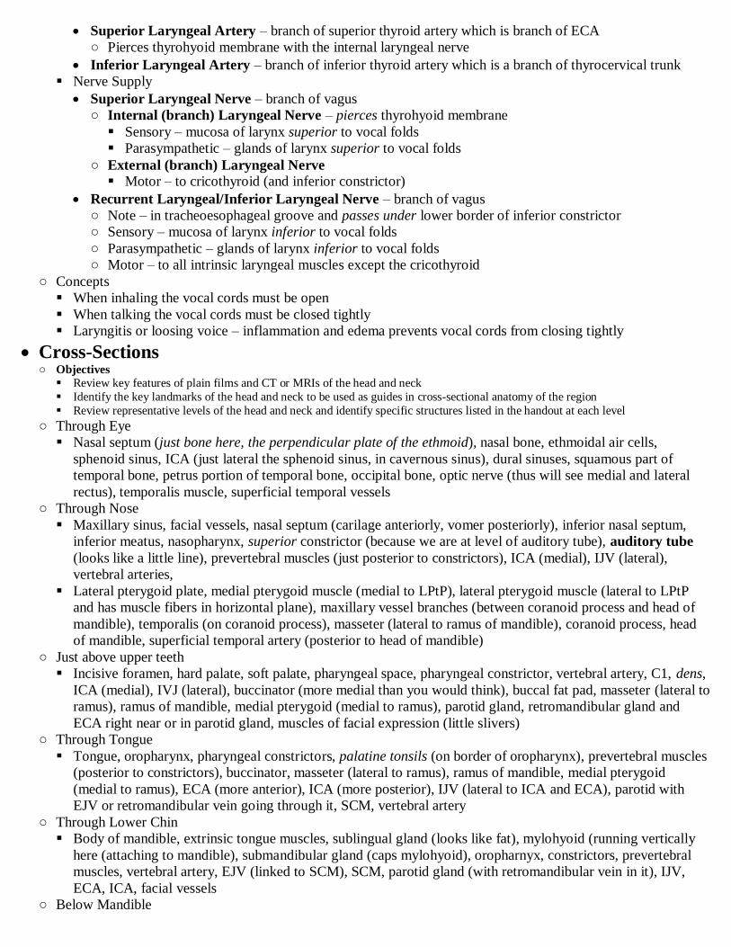
Superior Laryngeal Artery – branch of superior thyroid artery which is branch of ECA
○ Pierces thyrohyoid membrane with the internal laryngeal nerve
Inferior Laryngeal Artery – branch of inferior thyroid artery which is a branch of thyrocervical trunk
Nerve Supply
Superior Laryngeal Nerve – branch of vagus
○ Internal (branch) Laryngeal Nerve – pierces thyrohyoid membrane
Sensory – mucosa of larynx superior to vocal folds
Parasympathetic – glands of larynx superior to vocal folds
○ External (branch) Laryngeal Nerve Motor – to cricothyroid (and inferior constrictor)
Recurrent Laryngeal/Inferior Laryngeal Nerve – branch of vagus
○ Note – in tracheoesophageal groove and passes under lower border of inferior constrictor
○ Sensory – mucosa of larynx inferior to vocal folds
○ Parasympathetic – glands of larynx inferior to vocal folds
○ Motor – to all intrinsic laryngeal muscles except the cricothyroid
○ Concepts
When inhaling the vocal cords must be open
When talking the vocal cords must be closed tightly
Laryngitis or loosing voice – inflammation and edema prevents vocal cords from closing tightly
Cross-Sections ○ Objectives
Review key features of plain films and CT or MRIs of the head and neck
Identify the key landmarks of the head and neck to be used as guides in cross-sectional anatomy of the region
Review representative levels of the head and neck and identify specific structures listed in the handout at each level
○ Through Eye
Nasal septum (just bone here, the perpendicular plate of the ethmoid), nasal bone, ethmoidal air cells,
sphenoid sinus, ICA (just lateral the sphenoid sinus, in cavernous sinus), dural sinuses, squamous part of
temporal bone, petrus portion of temporal bone, occipital bone, optic nerve (thus will see medial and lateral
rectus), temporalis muscle, superficial temporal vessels
○ Through Nose
Maxillary sinus, facial vessels, nasal septum (carilage anteriorly, vomer posteriorly), inferior nasal septum,
inferior meatus, nasopharynx, superior constrictor (because we are at level of auditory tube), auditory tube
(looks like a little line), prevertebral muscles (just posterior to constrictors), ICA (medial), IJV (lateral),
vertebral arteries,
Lateral pterygoid plate, medial pterygoid muscle (medial to LPtP), lateral pterygoid muscle (lateral to LPtP
and has muscle fibers in horizontal plane), maxillary vessel branches (between coranoid process and head of
mandible), temporalis (on coranoid process), masseter (lateral to ramus of mandible), coranoid process, head
of mandible, superficial temporal artery (posterior to head of mandible)
○ Just above upper teeth
Incisive foramen, hard palate, soft palate, pharyngeal space, pharyngeal constrictor, vertebral artery, C1, dens,
ICA (medial), IVJ (lateral), buccinator (more medial than you would think), buccal fat pad, masseter (lateral to
ramus), ramus of mandible, medial pterygoid (medial to ramus), parotid gland, retromandibular gland and
ECA right near or in parotid gland, muscles of facial expression (little slivers)
○ Through Tongue
Tongue, oropharynx, pharyngeal constrictors, palatine tonsils (on border of oropharynx), prevertebral muscles
(posterior to constrictors), buccinator, masseter (lateral to ramus), ramus of mandible, medial pterygoid
(medial to ramus), ECA (more anterior), ICA (more posterior), IJV (lateral to ICA and ECA), parotid with
EJV or retromandibular vein going through it, SCM, vertebral artery
○ Through Lower Chin
Body of mandible, extrinsic tongue muscles, sublingual gland (looks like fat), mylohyoid (running vertically
here (attaching to mandible), submandibular gland (caps mylohyoid), oropharnyx, constrictors, prevertebral
muscles, vertebral artery, EJV (linked to SCM), SCM, parotid gland (with retromandibular vein in it), IJV,
ECA, ICA, facial vessels
○ Below Mandible

Mylohyoid (forming floor of oral cavity), anterior belly of digastric, root of tongue, epiglottis (anterior to
pharynx), laryngopharynx, constrictor, vertebral arteries, platysma, submandibular gland (with facial vessels
in it), ECA, ICA, IJV, SCM (with EJV linked)
○ Below C4
Epiglottis, vestibule of larynx, thyroid cartilage (looks different), infrahyoid muscles, laryngopharynx, inferior
constrictor, EJV, SCM (with EJV), IJV, CCA, anterior jugular (anteriorly)
○ C6
Trachea, esophagus, thyroid gland, infrahyoid muscles, SCM, vertebral artery, anterior scalene, brachial
plexus (white tissue), middle scalene, EJV (near brachial plexus), IJV, CCA
Development of the Eye and Ear ○ Objectives
Eye
Define and/or describe: optic vesicle, optic cup, optic fissure (choroid fissue), lens placode, lens pit, lens vesicle
Define and/or describe: hyaloid artery (central artery of the retina), hyaloid canal
Define and/or describe the three major layers of the eye and list the component structures found in each
Describe the major steps that occur in the formation of the lens. What induces formation of the lens?
Describe the formation of the anterior chamber of the cornea. What induces the differentiation of the cornea?
Describe the formation of the iris, ciliary body, retina
Ear
Describe the formation of the internal ear and external ear
Define and/or describe the three anatomical subdivisions of the ear; the otic placod and list the structures derived from it; the
membranous labyrinth and the bony labyrinth; endolymph and perilymph
Describe the relationships between the utricle and related semicircular ducts and endolymphatic duct. Describe the relationship
of the saccule and the cochlear duct
Briefly describe the function or the neural transducers found in the semicircular ducts, the utricle, and the cochlear duct
Briefly describe the formation of the tympanic cavity from the 1st pharyngeal arch. What is the developmental history of the
middle ear ossicles?
Briefly describe the formation of the external auditory canal from the 1st branchial cleft
Define pinna (auricle). What is the developmental history of the auricle?
Distinguish between “nerve deafness” and “conduction deafness”. List at least one congenital cause of each
What is congenital cholesteatoma?
When is the ear most susceptible to teratogens?
Explain the developmental history of any/all structures observed in microanatomy lab exercises
○ Eye
Summary of Derivations
Note – adult eye is composed of 3 coats, but each has multiple embryological origins
Surface Ectoderm (from head) – makes corneal epithelium, lens, epithelium of eyelids
Neuroectoderm (from forebrain) – retina, optic nerve, pupillary muscle, epithelium of iris and ciliary body
Mesoderm – extrinsic eye muscles, internal structures of the eyelids
Neural Crest (migrated in) – ciliary muscles, choroid, sclera, corneal endothelium
Development
Apparent at day 22 (week 3)
Optic Grooves – shallow grooves on the anterolateral surface of developing forebrain; become:
Optic Vesicles – further evagination of the optic grooves that project outward? towards the surface
ectoderm of the head
○ Optic Stalk – remaining connection of forebrain to optic vesicle
○ Eventually, the optic vesicle contacts the surface ectoderm of the head and this initiates formation of:
Lens Placode – thickening of surface ectoderm
Optic Cup – invagination of the optic vesicle
○ Anterior portion of vesicle ends up touching the posterior portion
○ Intra-retinal Space – small space between the anterior and posterior parts of cup
Lens Development
○ Lens placode grows inward toward the optic vesicle and forms the Lens Pit which continues to
invaginate and forms the Lens Vesicle which detaches from the surface ectoderm
○ Thus, lens development depends on interaction between the optic vesicle and surface ectoderm
Note – removal of optic vesicle prevents lens formation

Note – you can make optic vesicle tissue from other stuff and use the new stuff to stimulate lens dev
Pax-6 is important for lens development
○ Lens forms in concavity of the optic cup
Lentiretinal Space – space between developing lens and anterior portion of the optic vesicle
○ Becomes filled with gelatinous matrix that eventually forms the vitreous body
○ Cytodifferentiation
Fibroblast Growth Factor – causes non-specialized surface ectoderm cells of lens placode to
become lens cells
○ Secreted by the developing retina around WK 6
○ Causes cells to exit cell cycle and become postmitotic
Elongate along rostro-caudal axis of lens
Synthesize crystallins and become transparent
Cells closest to retina (posterior) become Primary Lens Fibers and form the Lens Nucleus
Lens continues to elongate and grow via division of lens fibers in the equatorial margin and fibers
from here are called Secondary Lens Fibers
○ Blood Supply
Hyaloid Artery – supplies developing optic cup and lens
○ From ICA → ophthalmic artery → hyaloid artery
○ Grows into the ventral surface of the optic stalk in the Choroidal Fissure
○ As eye grows, the vitreous separates the lens from the retina, but the hyaloid artery still supplies
lens until it no longer needs blood and then the anterior portion of the hyaloid artery degenerates
The posterior portion becomes the Central Artery of the Retina
Retinal Development
○ Outer (Posterior) Wall of optic cup – becomes the RPE
Becomes heavily pigmented around 5 weeks
○ Inner (Anterior) Wall of optic cup – thicker; becomes the 9 layers of neural retina
Cells in this layer differentiate between 6 and 7 weeks
Initial Differentiation
○ External Limiting Membrane – separates outer neuroblastic cells
from the intraretinal space
○ Outer Neuroblastic Layer – ↑ cell density; becomes photoreceptors
and such
○ Inner Neuroblastic Layer – ↓ cell density; becomes RGCs and such
○ Internal Limiting Membrane – separates inner neuroblastic cells from vitreous
Later Differentiation
○ RGCs develop first and photoreceptors develop last
○ The cells of the other layers differentiate
○ Axons of RGCs collect on inner surface and grow towards optic stalk, and exit the eyeball
This makes the optic stalk larger and it then becomes called the Optic Nerve
○ Note – the two walls become „fused‟ by week 7 (RPE cells contact outer segments of photoreceptors)
Development of the Choroid and Sclera
○ Mesenchymal Jacket – loose mesenchyme largely derived from neural crest cells that covers the
external surface of the optic cup
These cells respond to inductive signals from the RPE and form two layers
○ Inner Layer – forms the vascular choroid coat
○ Outer Layer – forms the fibrous scleral coat
Development of the Iris and Ciliary Body
○ Develop from anterior rim of optic cup
○ Iris develops from the ciliary body?
○ Three Cellular Origins (from anterior rim of optic cup)
Cells from the anterior rim of the mesenchymal jacket proliferate and form a bulge near lateral lens
→ form the Ciliary Muscles
Pigmented cells of posterior wall of optic cup (the RPE) cover the bulge → forms pigmented layer
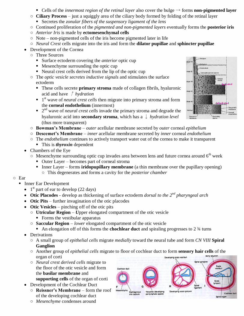
Cells of the innermost region of the retinal layer also cover the bulge → forms non-pigmented layer
○ Ciliary Process – just a squiggly area of the ciliary body formed by folding of the retinal layer
Secretes the zonular fibers of the suspensory ligament of the lens
○ Continued proliferation of the pigmented and non-pigmented layers eventually forms the posterior iris
○ Anterior Iris is made by ectomesenchymal cells
○ Note – non-pigmented cells of the iris become pigmented later in life
○ Neural Crest cells migrate into the iris and form the dilator pupillae and sphincter pupillae
Development of the Cornea
○ Three Sources
Surface ectoderm covering the anterior optic cup
Mesenchyme surrounding the optic cup
Neural crest cells derived from the lip of the optic cup
○ The optic vesicle secretes inductive signals and stimulates the surface
ectoderm
These cells secrete primary stroma made of collagen fibrils, hyaluronic
acid and have ↑ hydration
1st wave of neural crest cells then migrate into primary stroma and form
the corneal endothelium (innermost)
2nd
wave of neural crest cells invade the primary stroma and degrade the
hyaluronic acid into secondary stroma, which has a ↓ hydration level
(thus more transparent)
○ Bowman’s Membrane – outer acellular membrane secreted by outer corneal epithelium
○ Descemet’s Membrane – inner acellular membrane secreted by inner corneal endothelium
○ The endothelium continues to actively transport water out of the cornea to make it transparent
This is thyroxin dependent
Chambers of the Eye
○ Mesenchyme surrounding optic cup invades area between lens and future cornea around 6th week
Outer Layer – becomes part of corneal stroma
Inner Layer – forms iridopupillary membrane (a thin membrane over the pupillary opening)
○ This degenerates and forms a cavity for the posterior chamber
○ Ear
Inner Ear Development
1st part of ear to develop (22 days)
Otic Placodes - develop as thickening of surface ectoderm dorsal to the 2nd
pharyngeal arch
Otic Pits – further invagination of the otic placodes
Otic Vesicles – pinching off of the otic pits
○ Utricular Region – Upper elongated compartment of the otic vesicle
Forms the vestibular apparatus
○ Saccular Region – lower elongated compartment of the otic vesicle
An elongation off of this forms the chochlear duct and spiraling progresses to 2 ¾ turns
Derivations
○ A small group of epithelial cells migrate medially toward the neural tube and form CN VIII Spiral
Ganglion ○ Another group of epithelial cells migrate to floor of cochlear duct to form sensory hair cells of the
organ of corti
○ Neural crest derived cells migrate to
the floor of the otic vesicle and form
the basilar membrane and
supporting cells of the organ of corti
Development of the Cochlear Duct
○ Reissner’s Membrane – form the roof
of the developing cochlear duct
○ Mesenchyme condenses around

cochlear duct forming a cartilaginous capsule
Dorsal and ventral regions begin to vacualize to become perilymph filled scala vestibule and tympani
Poles of the cochlear duct become anchored to the cochlear canal at the spiral lamina and spiral
ligament
The cartilaginous capsule becomes a bony encasement at around 16 weeks
Development of the Vestibular Apparatus
○ Begins around 6 weeks with the cochlear duct
○ Portions of the wall of the utriclar compartment flatten and fuse
○ Adhesion points undergo apoptosis and form the 3 semicircular canals
○ Ampullae develop too
Middle Ear Development
Mesenchyme adjacent to the ectoderm of the 1st and 2
nd arches condense to form the cartilaginous
precursors of the auditory ossicles (bones)
1st pharyngeal arch elongates to form tubotympanic recess, which engulfs the ossicles and forms the
middle ear cavity
1st pharyngeal groove ectoderm invaginates
inwards to touch tubotympanic recess
This forms the tympanic membrane which is
thus made by outer ectoderm, mesenchyme and
inner endoderm
External Ear Development
Derived from the initial invagination that was
dorsal to the 1st pharyngeal arch
That area develops 6 nodular masses of
mesenchyme called auricular hillocks
Hillocks 1-3 – derived from 1st arch
○ Form anterior external ear
Hillocks 4-6 – derived from 2nd
arch and form
posterior external ear
Eye ○ Objectives
Identify the three tunics (major layers) of the eye and sup-components of each
Identify the chambers of the eye; boundary structures for each and contents of each
Identify the layers of the cornea and cellular composition of each
Identify the layers of the choroids region of the eye
Identify the ciliary body and detail its function
Identify the components of the iris and pupil and relate their structure to their function Identify the Canal of Schlemm (scleral venous sinus), its function and its importance in clinical diseases of the eye
Identify the lens of the eye and zonules of Zinn
Describe the production, flow pathway and absorption of the aqueous humor
Identify the retina and its ten layers
Identify the photoreceptors of the eye
○ I‟m abbreviating this lecture
○ Refractile Elements – cornea, aqueous humor, lens, vitreous
○ Note – photoreceptor cells send out graded? neural impulses
○ Development of the Eye
Optic Stalk – part of outpocketing that isn‟t the optic vesicle
Forms from outpocketing of neural tube that has an enlargement at the end called the optic vesicle
Optic Cup – optic vesicle invaginates and the anterior wall smushes up against the posterior wall
○ Posterior Wall – becomes the pigment epithelium of the retina, ciliary body and iris
○ Anterior Wall – becomes the neural retina
Lens Vesicle – an inbudding of surface epithelium that buds off; forms lens
Mesoderm between the anterior and posterior wall and forms the supporting coat and vascular coat
Surface ectoderm – forms eyelids, conjunctiva, outer epithelial layer of cornea
○ Layers of Eye
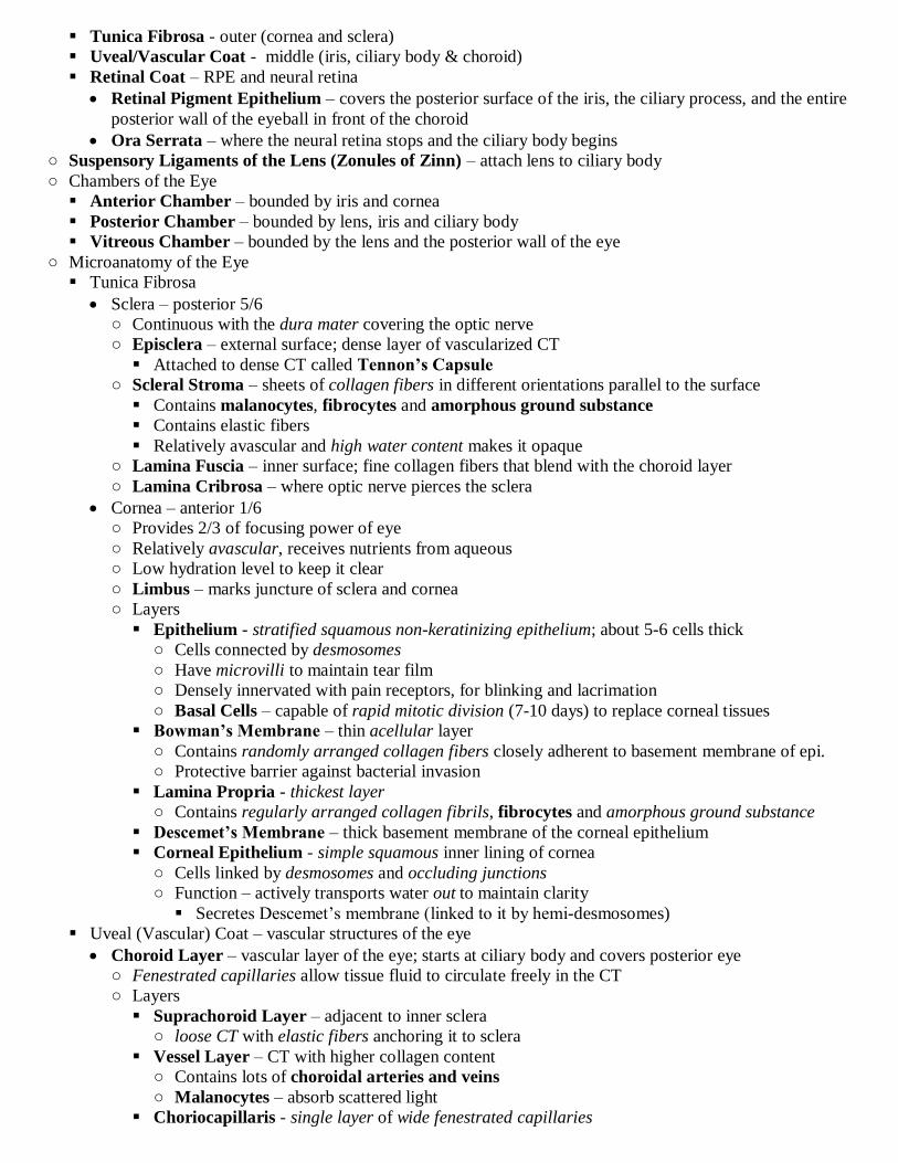
Tunica Fibrosa - outer (cornea and sclera)
Uveal/Vascular Coat - middle (iris, ciliary body & choroid)
Retinal Coat – RPE and neural retina
Retinal Pigment Epithelium – covers the posterior surface of the iris, the ciliary process, and the entire
posterior wall of the eyeball in front of the choroid
Ora Serrata – where the neural retina stops and the ciliary body begins
○ Suspensory Ligaments of the Lens (Zonules of Zinn) – attach lens to ciliary body
○ Chambers of the Eye
Anterior Chamber – bounded by iris and cornea
Posterior Chamber – bounded by lens, iris and ciliary body
Vitreous Chamber – bounded by the lens and the posterior wall of the eye
○ Microanatomy of the Eye
Tunica Fibrosa
Sclera – posterior 5/6
○ Continuous with the dura mater covering the optic nerve
○ Episclera – external surface; dense layer of vascularized CT
Attached to dense CT called Tennon’s Capsule
○ Scleral Stroma – sheets of collagen fibers in different orientations parallel to the surface
Contains malanocytes, fibrocytes and amorphous ground substance
Contains elastic fibers
Relatively avascular and high water content makes it opaque
○ Lamina Fuscia – inner surface; fine collagen fibers that blend with the choroid layer
○ Lamina Cribrosa – where optic nerve pierces the sclera
Cornea – anterior 1/6
○ Provides 2/3 of focusing power of eye
○ Relatively avascular, receives nutrients from aqueous
○ Low hydration level to keep it clear
○ Limbus – marks juncture of sclera and cornea
○ Layers
Epithelium - stratified squamous non-keratinizing epithelium; about 5-6 cells thick
○ Cells connected by desmosomes
○ Have microvilli to maintain tear film
○ Densely innervated with pain receptors, for blinking and lacrimation
○ Basal Cells – capable of rapid mitotic division (7-10 days) to replace corneal tissues
Bowman’s Membrane – thin acellular layer
○ Contains randomly arranged collagen fibers closely adherent to basement membrane of epi.
○ Protective barrier against bacterial invasion
Lamina Propria - thickest layer
○ Contains regularly arranged collagen fibrils, fibrocytes and amorphous ground substance
Descemet’s Membrane – thick basement membrane of the corneal epithelium
Corneal Epithelium - simple squamous inner lining of cornea
○ Cells linked by desmosomes and occluding junctions
○ Function – actively transports water out to maintain clarity
Secretes Descemet‟s membrane (linked to it by hemi-desmosomes)
Uveal (Vascular) Coat – vascular structures of the eye
Choroid Layer – vascular layer of the eye; starts at ciliary body and covers posterior eye
○ Fenestrated capillaries allow tissue fluid to circulate freely in the CT
○ Layers
Suprachoroid Layer – adjacent to inner sclera
○ loose CT with elastic fibers anchoring it to sclera
Vessel Layer – CT with higher collagen content
○ Contains lots of choroidal arteries and veins
○ Malanocytes – absorb scattered light
Choriocapillaris - single layer of wide fenestrated capillaries

○ Nourish tissue, including outer 1/3 of retina
Bruch’s (Glassy) Membrane – network of collagen and elastic fibers
○ Between basement membrane of choriocapilaris and RPE
Iris
○ Layers
Anterior Limiting Layer – discontinuous layer of stromal cells
○ Contains fibroblasts and malanocytes
Stroma – vascularized loose CT
○ Contains fibroblasts and malanocytes
Muscular Layer – 2 bands of muscle
○ Sphincter Pupillae – circular band of smooth muscle near posterior border of iris
↓ size of pupil
Under parasympathetic control
○ Dilator Pupillae – radially oriented smooth muscle near posterior border of iris
↑ size of pupil
Under sympathetic control (look for person‟s pupils to enarge if excited)
Posterior Epithelium - double layer of cuboidal pigmented cells
○ Cells heavily pigmented with melanin to only let light through pupil
○ Eye Color – caused by varying amounts of melanin in the stroma of the iris
Reduced melanin in stroma → blue eyes
Increased melanin in stroma → brown eyes
Ciliary Body – thickening of choroid that forms a ring around the eye, just behind the iris
○ Loose CT, fenestrated capillaries and smooth muscle
○ Epithelium - double cuboidal epithelium
Superficial non-pigmented layer
Deep Pigmented layer Apical surfaces of each layer face each other and thus the two basement membranes abut each other
Two layers connected by desmosomes
○ Ciliary Processes – ridges at the inner edge of the ciliary body
Zonules of Zinn – connect here
○ Ciliary muscles attach to choroid anteriorly (huh?)
○ Functions
Produce Aqueous Humor
○ Aqueous Humor – filtered out of capillaries of the ciliary processes into the posterior chamber
Transported out of the interior of the ciliary body by pigment epithelial cells which are
surrounded by a basement membrane that forms blood-aqueous barrier
Microtubular Meshwork – fibrous channels at lateral aspect of ciliary body which receive
aqueous about to be drained from the eye
Canal of Schlemm – drains aqueous; located near attachment of ciliary body to sclera
Lined with simple squamous epithelia
Fluid goes into aqueous veins then to the conjunctiva then venous blood
Flow – posterior chamber → anterior chamber → microtubular meshwork → canal of schlemm
→ aqueous veins → conjunctiva → venous blood
Regulate Shape of Lens
○ Ciliary Muscles
Controlled by parasympathetic system
Contraction causes release of tension on zonules of Zinn
Attach at the limbus
○ Eyes at rest (focused at distance) – elasticity of choroid pulls the zonules tight and backwards
○ Eyes focusing on something near – ciliary muscles pull things forwards and release tension
This ↑ focusing power of the lens by ↑ its curvature
The Retinal Coat
Note – photoreceptors point away from incoming light

Cell Types – require special stains to distinguish
○ Photoreceptors –
○ Bipolar Cells – 2nd
order neurons; transmit to 3rd
order
○ Horizontal Cells – 2nd
order neurons; interconnect photoreceptors laterally
○ Amacrine Cells – send signal from many bipolar cells to fewer RGCs
○ Ganglion Cells – 3rd
order neurons
Encode whether a contour or edge is present, its color, brightness and exact position in space
○ Muller Cells – glial cells that extend through all 10 layers of the retina
Nuclei found in inner nuclear layer; processes wrap around photoreceptors
Layers
○ Retinal Pigment Epithelium – makes blood/retina barrier
Single layer of cuboidal-columnar cells
○ Apical surface has microvilli and cylindrical cytoplasmic sheaths
○ enclose photoreceptors to nourish and phagocytose pieces shed pieces of their outer segments
○ Contain melanin (prevent light scatter)
Goes over ciliary body and posterior iris
Bruch’s Membrane – attaches base of RPE cells to the choroid
Detached Retina – no firm connection between RPE and retina and so this is where retina can detach
○ Photoreceptor Layer – contains rods and cones
Parts
○ Outer Segment – the light-sensitive region
Constantly turned over by being sloughed off
Dense vertical stacks of membrane bound discs that are
infoldings of the cell membrane
Contain Vit A derivative photopigment made of retinene
and opsin
Rhodopsin – rod pigment; for dim light
Photopisin – 3 types of cone pigments
The shape in cones → broad and tapered
The shape in rods → long, narrow and straight
○ Cilium – connects outer segment to inner segment
Made of non-motile cilia
Microtubules of cilia form 9 peripheral doublets
○ Inner Segment – contains most of the organelles
Membrane of this region contains a lot of K+ channels that are
active during phototransduction
○ Outer Segment – thinner segment between the inner segment
and the cell body
Muller cell processes surround this portion and attach by tight junctions
More prominent in rods
Sometimes called external limiting membrane
○ Cell Body – contains the nucleus
○ Inner Fiber Layer – thin part that expands at the end and makes contact
with other layers
Spherule – expansion of this layer in rods
Pedicle – expansion of this layer in cones
Distribution
○ Fovea Centralis – depression in retina with a huge concentration of cones
Devoid of blood vessels
Other layers of retina are thinner here
○ Rods mainly in periphery
Transduction
○ Light causes hyperpolarization of the photoreceptors
○ cGMP and Ca+ dependent modulation of Na+ channels in photoreceptor membrane

○ External Limiting Membrane – not really a membrane, just the zonula adherens junctions of the
cytoplasmic processes of Muller cells with the photoreceptors
○ Outer Nuclear Layer – where the nuclei of photoreceptor cells are
Nuclei are at different distances and so it looks like it is thick stratified, but it isn‟t
○ Outer Plexiform Layer – contains the synaptic processes of the photoreceptors (in contact with bipolar
or horizontal cells)
No cell bodies, relatively unstained
○ Inner Nuclear Layer (Bipolar Layer) – contains cell bodies of bipolar, horizontal and amacrine cells
○ Inner Plexiform Layer – contains processes of bipolar, horizontal, amacrine and ganglion cells
○ Ganglion Cell Layer – contains cell bodies of retinal ganglion cells
○ Retinal Axon Layer – contains the unmyelinated RGC axons
They become myelinated when they get to the optic nerve
○ Internal Limiting Membrane – not a true membrane
Basement membrane of the Muller cells
Neural Activity
○ Information sent in retina is mainly graded hyperpolarization and depolarization, not real APs
○ RGCs are the only things to make full APs (frequency changes)
○ Remember – photoreceptors are hyperpolarized by light and thus chronically release neurotransmitter
Light brighter than environment causes a ↓ in rate of transmitter release (and visa vers)
Optic Disc (Papilla) – a blind spot caused by the axons of RGCs leaving the retina to form optic nerve
Fovea – visual acuity at its best
○ ↑ density of RGCs and cones; ↓ thickness of other layers
○ little diffusion; appears yellow (macula lutea)
Blood Supply
○ Retinal Artery and Vein – enter eyeball at optic disc and split to go either supierioly or inferiorly
Supplies the inner 2/3rds of the retina with capillaries (the rest is supplied by choroid)
○ Diabetic Retinopathy – uncontrolled plasma glucose in eye causes vascular edema and altered retinal
and choroidal vasculature
Can cause scar tissue induced retinal detachment, edema, vascular leakage, neovascularization
○ The Lens
1/3 of the focusing power of the eye
Zonules of Zinn – composed of microfibrils and are attached to the equator of the lens and ciliary body
Avascular and contains no CT
Anterior Surface
Lens Capsule – made of collagen and proteoglycans
Simple cuboidal epithelium
Lens Fibers – cells that lost their nucleus and organells and form fibers
○ Packed with crystallins
○ Fibers connected together by knob and socket like depressions, tight junctions and gap junctions
○ Fibers at center of the lens persist throughout life (not replaced)
Germinal Zone – at the equatorial rim; here cells divide slowly and are added to the rim
○ Vitreous
Contains amorphous ground substance (GAGS) and thin, randomly oriented collagen fibrils and water
Adheres to the peripheral retina and the ciliary epithelium
Ear ○ Objectives
Identify the three components of the ear (ie outer, middle and inner ear). Identify the boundaries and components of each. Relate
structure to function
Identify the structure of the tympanic membrane (eardrum)
Differentiate between the components of bony labyrinth and membranous labyrinth
Identify the region of the cochlea, including the basilar membrane, helicotrema, osseous spiral laminal and spiral ligament
Describe the fluid that is found within bony labyrinth (perilymph) and that found in the membranous labyrinth (endolymph) and how and where each is formed and removed
Identify the three major divisions of the cochlear canal
Identify the boundaries of the cochlear duct
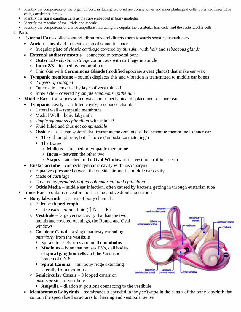
Identify the components of the organ of Corti including: tectorial membrane, outer and inner phalangeal cells, outer and inner pillar
cells, cochlear hair cells
Identify the spiral ganglion cells as they are embedded in bony modiolus
Identify the maculae of the utricle and saccule
Identify the components of cristae ampullaris, including the cupula, the vestibular hair cells, and the sustentacular cells
○ Parts
External Ear – collects sound vibrations and directs them towards sensory transducers
Auricle – involved in locatization of sound in space
○ Irregular plate of elastic cartilage covered by thin skin with hair and sebaceous glands
External auditory meatus – connected to temporal bone
○ Outer 1/3 - elastic cartilage continuous with cartilage in auricle
○ Inner 2/3 – formed by temporal bone
○ Thin skin with Ceruminous Glands (modified apocrine sweat glands) that make ear wax
Tympanic membrane – sounds displaces this and vibration is transmitted to middle ear bones
○ 2 layers of collagen
○ Outer side – covered by layer of very thin skin
○ Inner side – covered by simple squamous epithelium
Middle Ear – transduces sound waves into mechanical displacement of inner ear
Tympanic cavity – air filled cavity; resonance chamber
○ Lateral wall – tympanic membrane
○ Medial Wall – bony labyrinth
○ simple squamous epithelium with thin LP
○ Fluid filled and thus not compressible
○ Ossicles – a „lever system‟ that transmits movements of the tympanic membrane to inner ear
They ↓ amplitude, but ↑ force („impedance matching‟)
The Bones
○ Malleus – attached to tympanic membrane
○ Incus – between the other two
○ Stapes – attached to the Oval Window of the vestibule (of inner ear)
Eustacian tube – connects tympanic cavity with nasopharynx
○ Equalizes pressure between the outside air and the middle ear cavity
○ Made of cartilage
○ Covered by pseudostratified columnar ciliated epithelium
○ Otitis Media – middle ear infection, often caused by bacteria getting in through eustacian tube
Inner Ear – contains receptors for hearing and vestibular sensation
Bony labyrinth – a series of bony channels
○ Filled with perilymph
Like extracellular fluid (↑Na, ↓K)
○ Vestibule – large central cavity that has the two
membrane covered openings, the Round and Oval
windows
○ Cochlear Canal – a single pathway extending
anteriorly from the vestibule
Spirals for 2.75 turns around the modiolus
Modiolus – bone that houses BVs, cell bodies
of spiral ganglion cells and the *acoustic
branch of CN 8
Spiral Lamina – thin bony ridge extending
laterally from modiolus
○ Semicircular Canals – 3 looped canals on
posterior side of vestibule
Ampulla – dilation at portions connecting to the vestibule
Membranous Labyrinth – membranes suspended in the perilymph in the canals of the bony labyrinth that
contain the specialized structures for hearing and vestibular sense

○ Delicate CT lined with simple squamous epithelium
○ Utricle and Saccule are membranous sacs in the vestibule
○ All membranous ducts are continuous and contain endolymph
Endolymph – similar to intracellular fluid (↓Na, ↑K)
○ Endolyphatic Duct – where old endolymph drains into; off of utricle and saccule
○ Auditory Sensory Apparatus
Cochlear Duct – suspended medially in cochlear canal
Roughly triangular in cross section and divides canal into three sections
○ Scala Vestibuli (above) & Scala Tympani (below) – filled with perilymph
Lined with thin CT that is continuous with periosteum of cochlear canal
Helicotrema – connection between scala vestibule and scala tympani at apex of cochlear canal
○ Scala Media (Cochlea) – filled with endolymph; central partition of cochlear canal
Walls
○ Reissner’s (Vestibular) Membrane – roof
2 layers of simple squamous epithelium separated by a basement membrane
○ Stria Vascularis - lateral wall; source of endolymph
Thin layer of CT and highly vascular epithelium
○ Basilar Membrane – floor
Amorphous ground substance with transversely oriented filaments
Lower surface covered by columnar epithelium
Supports organ of Corti (spiral ligament and osseous spiral lamina support it)
Has different width and stiffness at different points along its length which determine which
frequency a specific point along its legth is most sensitive to
High frequency sounds – localize near base (beginning) of cochlea where it is narrowest
and most taught
Low frequency sounds – localize near apex (end) of cochlea where it is narrowest and
least taught
Organ of Corti – rests atop the basilar membrane
Tunnel of Corti – triangular shaped tunnel midway across the basilar membrane
○ Base formed by basilar membrane but walls formed by supporting cells
Pillar Cells – form walls of tunnel of Corti
○ Cone-shaped columnar cells with basilar nuclei
○ Rigid due to lots of microtubules
Phalangeal Cells – tall columnar cells
○ Phalanx – cytoplasmic process that extends along side the hair cells and support the base of them
○ Sensory Hair Cells
Inner Hair Cells – sensitive to small changes in sound intensity
○ About 3500, short, goblet-shaped cells
○ Arranged in a single-row
○ Each has <70 stereocilia
○ Innervated by CN 8 (each cell innervated by about 20 fibers)
Outer Hair Cells – respond best to low intensity sounds
○ About 20,000, cylindrical, columnar cells
○ Arranged in 3 rows kinda like a „W‟
○ Each has >100 stereocilia
○ Tips of tallest stereocilia are embedded in the the tectorial membrane
○ Innervated by CN 8 (lots of cells innervated by a single nerve fiber)
Note – CN 8 supplies spiral ganglion???
○ Tectorial Membrane – sheering of this membrane triggers electrical impulses in embedded hair cells
Gelatinous membrane composed of keratin-like protein
Made by cells of spiral laminae
Vibrations to Nerve Impulses
Pressure waves travel down external auditory meatus and displace tympanic membrane
Vibrations of tympanic membrane are ↓ in amplitude and ↑ in force by ossicles (impedence matching)

Vibrations transferred to oval window which causes wave in perilymph to cochlear canal in scala vestibule
→ helicotrema → scala tympani and dissipate at round window
○ During this pathway vibrations are transferred to the basilar membrane
Movement of the basilar membrane causes „shearing motion‟ of hair cell stereocilia with respect to
the tectorial membrane
This causes change in polarization in the hair cells and sends an impulse in CN 8 Spiral Ganglion
cells in the modiolus
Impulses go to the auditory cortex of CNS
Coding Attributes of Sound
Sound Amplitude – loud sounds produce larger amplitude waves that stimulate more hair cells
Sound Pitch
○ Volley Theory – sound frequency encoded by frequency of APs in 8th
nerve
For sounds >200 Hz fibers still fire in phase, but skip groups to take turns
○ Place Theory – sound frequencies have a different optimal spot to be received on the basilar membrane
Higher frequencies → base; low frequencies (which can travel farther) → apex
Prolonged exposure to sounds at a certain frequency can cause degredation of the cells receiving it
○ Vestibular Sensory Apparatus
Composed of 2 endolymph filled sacs and 3 endolymph filled semicircular ducts arising from ampulla
Semicircular Canals and Apulla
All connect to the utricle
Each has an expanded region called the ampulla at one of its junctions with the utricle
Cristae Ampullaris – a raised transverse ridge in the ampulla
○ Types of Cells in Cristae
Sustentacular Cells – tall columnar supporting cells
Sensory Hair Cells
○ Innervated by neurons of Vestibular (Scarpa’s) Ganglion in the modiolus
These neurons terminate in vestibular nuclei of the brainstem
○ Type I Cells – flask-shaped cells with stereocilia similar to inner hair cells
Flanked by a single non-motile kinocilium
Receive funnel-shaped nerve endings
○ Type II Cells – cylindrical cells with stereocilia similar to outer hair cells
Flanked by a single non-motile kinocilium
Receive bouton-like nerve endings
Cupula – gelatinous glycoprotein that covers the sensory hair cells
○ Movement in the plane of a given semicircular canal causes stereocilia to bend against the inertia
of the endolymph and the cupula
This causes change in polarization of hair cells and APs to vestibular ganglion cells
Utricle and Saccule
Both in the vestibule of the bony labyrinth
Macula – 3x3mm patch of sensory epithelium
○ Structure
Columnar Supporting Cells Sensory Hair Cells – similar to those in cristae
Otolithic Membrane – gelatinous glycoprotein that covers the surface of the hair cells
Otoliths – crystalline bodies of calcium carbonate that are suspended in the otolithic membrane
○ Function
Inertia of otolithic membrane and otoliths causes bending of stereocilia and change in polarization
Senses head position
Senses linear acceleration
Saccule – vertical plane
Utricle – horizontal plane
○ Basic Structure of Hair Cells
Similar in auditory and vestibular system and between cell types
Features
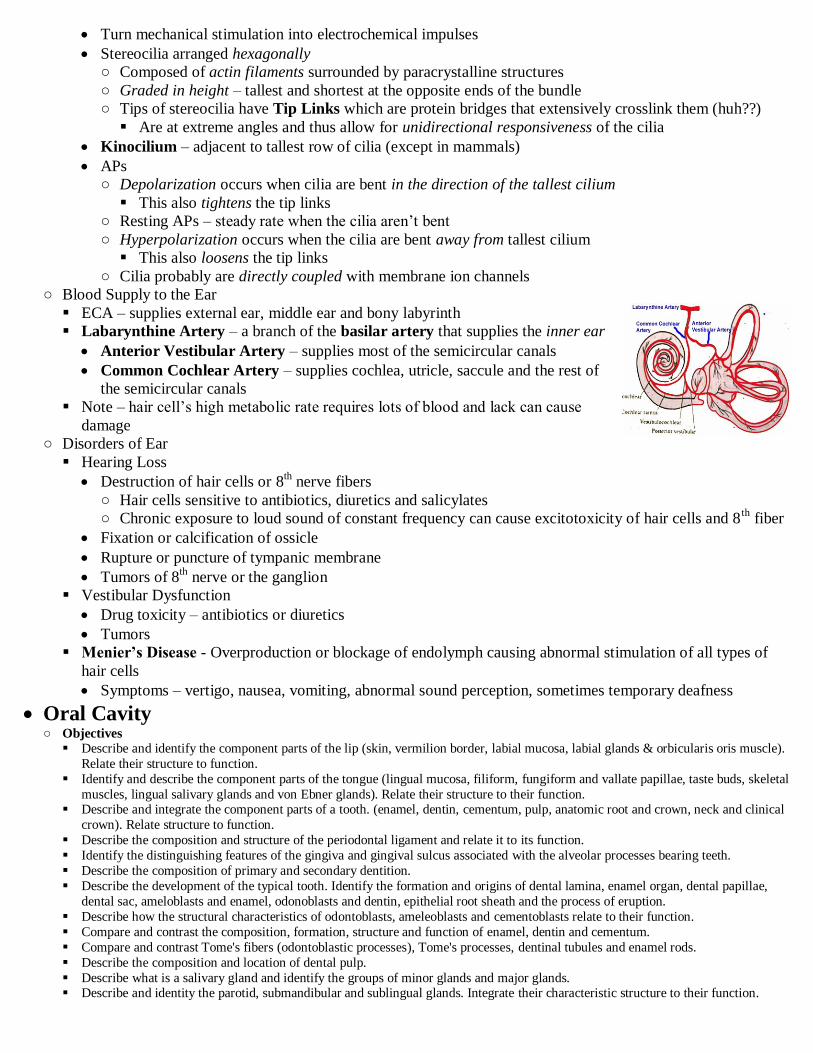
Turn mechanical stimulation into electrochemical impulses
Stereocilia arranged hexagonally
○ Composed of actin filaments surrounded by paracrystalline structures
○ Graded in height – tallest and shortest at the opposite ends of the bundle
○ Tips of stereocilia have Tip Links which are protein bridges that extensively crosslink them (huh??)
Are at extreme angles and thus allow for unidirectional responsiveness of the cilia
Kinocilium – adjacent to tallest row of cilia (except in mammals)
APs
○ Depolarization occurs when cilia are bent in the direction of the tallest cilium
This also tightens the tip links
○ Resting APs – steady rate when the cilia aren‟t bent
○ Hyperpolarization occurs when the cilia are bent away from tallest cilium
This also loosens the tip links
○ Cilia probably are directly coupled with membrane ion channels
○ Blood Supply to the Ear
ECA – supplies external ear, middle ear and bony labyrinth
Labarynthine Artery – a branch of the basilar artery that supplies the inner ear
Anterior Vestibular Artery – supplies most of the semicircular canals
Common Cochlear Artery – supplies cochlea, utricle, saccule and the rest of
the semicircular canals
Note – hair cell‟s high metabolic rate requires lots of blood and lack can cause
damage
○ Disorders of Ear
Hearing Loss
Destruction of hair cells or 8th nerve fibers
○ Hair cells sensitive to antibiotics, diuretics and salicylates
○ Chronic exposure to loud sound of constant frequency can cause excitotoxicity of hair cells and 8th
fiber
Fixation or calcification of ossicle
Rupture or puncture of tympanic membrane
Tumors of 8th nerve or the ganglion
Vestibular Dysfunction
Drug toxicity – antibiotics or diuretics
Tumors
Menier’s Disease - Overproduction or blockage of endolymph causing abnormal stimulation of all types of
hair cells
Symptoms – vertigo, nausea, vomiting, abnormal sound perception, sometimes temporary deafness
Oral Cavity ○ Objectives
Describe and identify the component parts of the lip (skin, vermilion border, labial mucosa, labial glands & orbicularis oris muscle).
Relate their structure to function.
Identify and describe the component parts of the tongue (lingual mucosa, filiform, fungiform and vallate papillae, taste buds, skeletal
muscles, lingual salivary glands and von Ebner glands). Relate their structure to their function. Describe and integrate the component parts of a tooth. (enamel, dentin, cementum, pulp, anatomic root and crown, neck and clinical
crown). Relate structure to function.
Describe the composition and structure of the periodontal ligament and relate it to its function.
Identify the distinguishing features of the gingiva and gingival sulcus associated with the alveolar processes bearing teeth.
Describe the composition of primary and secondary dentition.
Describe the development of the typical tooth. Identify the formation and origins of dental lamina, enamel organ, dental papillae,
dental sac, ameloblasts and enamel, odonoblasts and dentin, epithelial root sheath and the process of eruption.
Describe how the structural characteristics of odontoblasts, ameleoblasts and cementoblasts relate to their function.
Compare and contrast the composition, formation, structure and function of enamel, dentin and cementum.
Compare and contrast Tome's fibers (odontoblastic processes), Tome's processes, dentinal tubules and enamel rods.
Describe the composition and location of dental pulp.
Describe what is a salivary gland and identify the groups of minor glands and major glands. Describe and identity the parotid, submandibular and sublingual glands. Integrate their characteristic structure to their function.
Describe and integrate serous and mucous acini, serous demilunes, intralobular ducts (intercalated and striated) and interlobular
ducts into the structure of all salivary glands.
○ Oral Cavity
Boundaries
Anterioly – lips
Posteriorly – palatoglossal folds
Laterally – buccal cavities
Contents – lips, tongue, teeth, salivary glands
○ Lips
Orbicularis Oris – allows them to move
Regions
Cutaneous - stratified squamous keratinized epithelium with hair follicles and sweat glands
Vermillion Border – red region due to tall dermal papillae with capillaries
○ stratified squamous para-keratinized epithelium
○ Lacks sweat glands, needs to be moistened occasionally
Oral Mucosa – internal
○ Lining Mucosa – lines most of oral cavity (including soft palate)
mucosal stratified squamous epithelium on moderately dense irregular CT
Have submucosa and glands
○ Masticatory Mucosa – lines regions exposed to pressure and shear forces (ex. gingiva and hard palate)
parakeratinized to keratinized stratified squamous epithelium on tightly bound dense irregular CT
Lack submucosa and glands
○ Tongue
stratified squamous non-keratinizing epithelium and thin LP
Contains serous and mucous glands and lingual tonsils
Muscles
Extrinsic
○ Responsible for moving tongue in and out of mouth and side to side
○ Genioglossus, hyoglossus, styloglossus and palatoglossus
Intrinsic
○ Arranged in intertwining bundles: longitudinal, transverse and oblique
○ Responsible for changing shape of tongue (phonation)
Surface
Sulcus Terminalis – V-shaped groove that divides anterior 2/3rds from posterior 1/3rd
Foramen Cecum – depression at point of V that marks old thyroglossal duct
Posterior 1/3 – contains root of tongue and lingual tonsils
Lingual Papillae – mucosal projections on dorsum
○ Have a vascular core of CT covered by stratified squamous non-keratinizing epithelium
○ Types
Filiform Papillae – narrow conical; most common
○ Give tongue its texture (increase friction); tip has keratinized epithelium
○ no taste buds
Fungiform Papillae – mushroom shaped portion contains taste buds
○ Location – lateral edges of tongue
○ non-keratinized epithelium with redder color
Foliate Papillae – leaf shaped
○ Not well developed in humans (lose taste buds by age 3)
○ Location – margin of tongue
Circumvallate Papillae – like a mushroom cloud; there is a moat surrounding each
○ 9-12 of them just anterior to the sulcus terminalis
○ The protrusion has central core of CT covered by non-keratinized epithelium
○ Taste buds located in groove created by protrusion and on its sides
○ Serous Glands of von Ebner – located deep to the protrusion and drain into the moat
○ Taste Pore – narrow opening formed by surrounding epithelial cells

○ Taste Buds – spherical structure composed of 60-80 cells; approximately 3000 of them
Taste Hair – long, slender microvilli of receptor cells that extend out of taste pore
Receptor Cells – life span of 10-14 days
○ Progression – basal cells (IV) → intermediate cells (III) → light cells (II) → dark cells (I)
○ Taste receptors located on microvilli
○ In synaptic contact with nerve fibers of CN 7 (anterior 2/3) and CN 9 (posterior 1/3)
And Vagus
Each nerve fiber receives from about 5 taste buds
Each taste bud is innervated by about 50 nerve fibers
Taste Sensations – caused by specific taste receptor cells
Note – each taste bud can discern all tastes, but some kinda specialize
Note – tastants are continually washed away by salivary glands
Sweet – tip of tongue; 5 different types: for sugars, alcohols, ketones, amino acids, and sucrose
○ Caused by binding of G-Protein coupled receptors → ↑cAMP → ↓ permeability of K+
Salty – front sides; by salt and inorganic salts: NaCl, NaF, NH4Cl, MgCl2
○ Caused by direct entry of Na+ into receptor cell
Sour – sides; H+ concentration
○ Caused by decreased permeability of K+
Bitter – back of tongue; stimulated by quinine or organic alkaloids of toxic plants
○ Mainly received by circumvallate papillae
○ Caused by binding of G-Protein coupled receptors → ↑IP3 → ↑ intracellular Ca++
Umami – for monosodium glutamate
○ Salivary Glands
Make 1-1.5 quarts of saliva a day
Function – initiates digestion, lubricates swallowing, protection, aids food tasting
Contents – mucus, proteins, salts, salivary amylase, lingual lipase, lysosyme (antibacterial), lactoferrin, IgA
Minor Salivary Glands – reside within oral mucosa and tongue
Small short tubular glands
Major Salivary Glands – the Parotid, Submandibular and Sublingual glands
Have ducts that drain directly into oral cavity
Innervated by both sympathetics and parasympathetics
General Structure
○ All are compound tubuloalveolar
○ Organized into lobes and lobules separated by CT septa
○ Secretory Portion – serous, mucus or mixed
○ Acinus – come in tubes or alveoli; surrounded by myoepithelial cells that aid in secretion
○ Duct System
Intralobular Ducts ○ Function – resorb Na & Cl ions
Secrete bicarbonate ions
○ Intercalated Ducts – drain acini and tubules
simple cuboidal and some myoepithelial cells
Hard to recognize; smallest are same size
as acini
○ Striated Ducts – merge together at end
simple cuboidal to low columnar
More reddish due to more mitochondria
and membrane infoldings
Interlobar Ducts - between lobules
○ surrounded by CT – diagnostic
Interlobar Ducts – larger ducts; between lobes
of gland
○ Surrounded by CT of large septa

Main Terminal Duct – drains entire gland
Parotid Gland
○ Largest; 30% of volume; located over angle of mandible
○ Secretory portion is tubule and acini
○ Secretion – serous – diagnostic
Reddish staining
Granules rich in: salivary amylase, IgA (inactivates antigens), antimicrobial proteins, lactoferrin,
Na, Cl
○ White adipocytes mixed in - ↑ with age
○ Ducts
Intercalated ducts – drain acini and tubules; same size as acini and therefore difficult to see
Striated ducts - light pink staining, striations visible (due to mitochondria)
○ Not surrounded by CT
Interlobular Ducts – normal; surrounded by CT, located between lobules
Main Terminal Duct (Stetsen‟s Duct)
Submandibular Gland ○ Produces 60% of saliva; Located under mandible in floor of mouth
○ Similar in structure to parotid gland
○ Main excretory duct (Wharton‟s) exits on frenulum of tongue
○ Serous acini : Mucus acini – 5:1 – diagnostic
Mucus cells secrete mucin rich in sialic acids and sulfates
Pellicles – mucins and proteins in saliva form protective coat on teeth to protect against acids, retain
moisture, and regulate adherence of bacteria
Lack of saliva causes tooth decay, yeast infections and inflammation
Sublingual Gland ○ Located inerior to tongue
○ Similar in structure to others
○ Secretes through multiple ducts on sublingual fold
○ Predominantly mucus, but some serous demilunes – diagnostic
○ Function – mucin helps lubricate food and also to form the pellicle
○ Duct system – shorter and harder to find
○ Teeth
Permanent – 32; Deciduous (baby teeth) – 20
Types – incisors (for cutting), canines (for puncturing & holding), molars (for crushing and grinding)
Structure
Crown – part visible in oral cavity
Root – part buried in alveolus of bone
Neck – junction between the crown and root
Pulp Chamber – space in center of tooth; filled with vascular areolar CT
○ Rich in proteoglycans and glycoproteins
○ Odontoblasts – line the pulp and produce dentin
○ Root Canal – longer portion; carries BVs and nerves
○ Apical Foramen – opening at root tip for entry of BVs and nerves
Periodontal Ligament – dense irr. CT that holds tooth in socket by connecting alveolar bone to cementum
Sharpey’s Fibers – bundles of collage in the ligament
Structurally it is a fibrous joint (a gomphosis), functionally it is a synarthroses
Gingiva – supports tooth and seals it off oral cavity from CT
parakeratinized → keratinized epithelium (Masticator mucosa)
Gingival Groove – space adjacent to the tooth, deep to the gingival
Junctional Epithelium – where epithelium attaches to enamel surface by hemidesmosomes (where?)
○ Forms sealing barrier around neck of tooth
Body of Tooth
Enamel – covers crown; hardest surface in body
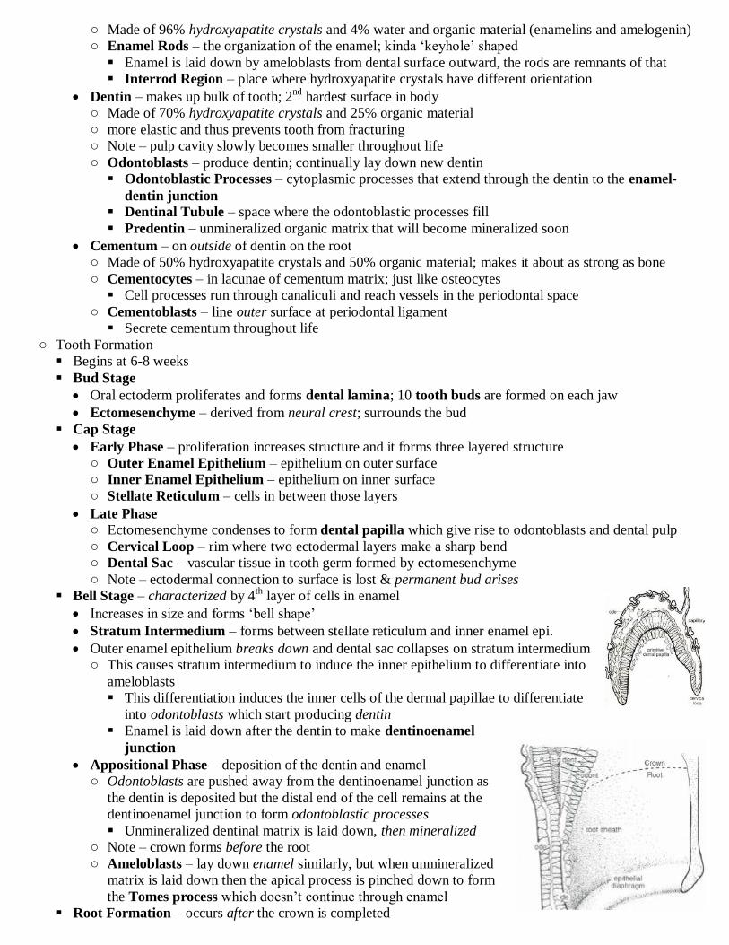
○ Made of 96% hydroxyapatite crystals and 4% water and organic material (enamelins and amelogenin)
○ Enamel Rods – the organization of the enamel; kinda „keyhole‟ shaped
Enamel is laid down by ameloblasts from dental surface outward, the rods are remnants of that
Interrod Region – place where hydroxyapatite crystals have different orientation
Dentin – makes up bulk of tooth; 2nd
hardest surface in body
○ Made of 70% hydroxyapatite crystals and 25% organic material
○ more elastic and thus prevents tooth from fracturing
○ Note – pulp cavity slowly becomes smaller throughout life
○ Odontoblasts – produce dentin; continually lay down new dentin
Odontoblastic Processes – cytoplasmic processes that extend through the dentin to the enamel-
dentin junction Dentinal Tubule – space where the odontoblastic processes fill
Predentin – unmineralized organic matrix that will become mineralized soon
Cementum – on outside of dentin on the root
○ Made of 50% hydroxyapatite crystals and 50% organic material; makes it about as strong as bone
○ Cementocytes – in lacunae of cementum matrix; just like osteocytes
Cell processes run through canaliculi and reach vessels in the periodontal space
○ Cementoblasts – line outer surface at periodontal ligament
Secrete cementum throughout life
○ Tooth Formation
Begins at 6-8 weeks
Bud Stage
Oral ectoderm proliferates and forms dental lamina; 10 tooth buds are formed on each jaw
Ectomesenchyme – derived from neural crest; surrounds the bud
Cap Stage
Early Phase – proliferation increases structure and it forms three layered structure
○ Outer Enamel Epithelium – epithelium on outer surface
○ Inner Enamel Epithelium – epithelium on inner surface
○ Stellate Reticulum – cells in between those layers
Late Phase ○ Ectomesenchyme condenses to form dental papilla which give rise to odontoblasts and dental pulp
○ Cervical Loop – rim where two ectodermal layers make a sharp bend
○ Dental Sac – vascular tissue in tooth germ formed by ectomesenchyme
○ Note – ectodermal connection to surface is lost & permanent bud arises
Bell Stage – characterized by 4th layer of cells in enamel
Increases in size and forms „bell shape‟
Stratum Intermedium – forms between stellate reticulum and inner enamel epi.
Outer enamel epithelium breaks down and dental sac collapses on stratum intermedium
○ This causes stratum intermedium to induce the inner epithelium to differentiate into
ameloblasts
This differentiation induces the inner cells of the dermal papillae to differentiate
into odontoblasts which start producing dentin
Enamel is laid down after the dentin to make dentinoenamel
junction
Appositional Phase – deposition of the dentin and enamel
○ Odontoblasts are pushed away from the dentinoenamel junction as
the dentin is deposited but the distal end of the cell remains at the
dentinoenamel junction to form odontoblastic processes
Unmineralized dentinal matrix is laid down, then mineralized
○ Note – crown forms before the root
○ Ameloblasts – lay down enamel similarly, but when unmineralized
matrix is laid down then the apical process is pinched down to form
the Tomes process which doesn‟t continue through enamel
Root Formation – occurs after the crown is completed

Hertwig Epithelial Root Sheath – forms the root
○ Created from cervical loop epithelium that extends downward
○ Stratum intermedium does not form below dentinoenamel junction
Thus the inner layer below does not form ameloblasts
○ Odontoblasts form and deposit dentin
○ Outer and inner epithelial layers disintegrate in cervical loop region and ectomesenchyme cells come in
contact with the dentin and differentiated into cementoblasts to deposit cementum on outside of root
Recommended