Postępy Dermatologii i Alergologii XXIX; 2012/5390
AAddddrreessss ffoorr ccoorrrreessppoonnddeennccee:: Anna Rosińska-Więckowicz MD, PhD, Department of Dermatology, Poznan University of Medical Sciences, 49 Przybyszewskiego St, 60-355 Poznan, phone: +48 604 906 464, e-mail: [email protected]
Bloch-Sulzberger syndrome: a case report
Anna Rosińska-Więckowicz, Magdalena Czarnecka-Operacz
Department of Dermatology, Poznan University of Medical Sciences, PolandHead: Prof. Wojciech Silny MD, PhD
Postep Derm Alergol 2012; XXIX, 5: 390-394
DOI: 10.5114/pdia.2012.31494
Case report
AbstractIncontinentia pigmenti (IP, Bloch-Sulzberger syndrome) is a very rare genodermatosis characterized by typical skinlesions accompanied by dental, central nervous system, bone and ocular abnormalities. Incontinentia pigmenti isusually observed among women, as this X-linked dominantly inherited disorder is lethal in males. The hallmark fea-ture of IP is cutaneous eruption along the lines of Blaschko, usually accompanied by neurological disorders. Apartfrom clinical features of the disease, skin biopsy is the best diagnostic tool to confirm the diagnosis. We presenta case of a newborn with typical vesicular and then verrucous lesions affecting the lower legs.
KKeeyy wwoorrddss:: incontinentia pigmenti, Bloch-Sulzberger syndrome.
Introduction
Incontinentia pigmenti (IP, Bloch-Sulzberger syndrome)is a very rare X-linked dominantly inherited genoder-matosis predominant among females as it is usually lethalin males [1-6]. There are however single reports of malepatients with IP and XXY karyotype [7, 8]. Incontinentiapigmenti was first described by Garrod in 1906 [9], andfurther defined by Bardach [10], Bloch in 1926 [11],Sulzberger in 1928 [12] and Siemens in 1929 [13], howev-er only the names of Bloch and Sulzberger feature in theeponym. Incontinentia pigmenti is a multisystem, ecto-dermal disorder characterized by skin lesions (100%)accompanied by dental (90%), central nervous system(CNS) (40%), bone (40%) and ocular (35%) abnormalities.In 1993, Landy and Donnai [4], after they had evaluateda group of over 100 pa tients with IP, proposed the diagnos-tic criteria for this neurodermatosis, as shown in Table 1.
Dermatologic manifestations are among the mostimportant signs of IP as skin lesions observed in almostall individuals with IP are relatively easy to diagnose [1-4].Fortunately, skin lesions are the least damaging aspect ofthe disease and actually do not require any treatment asspontaneous resolution of lesions is one of the featuresof the disease. The hallmark feature of IP is cutaneouseruption along the lines of Blaschko that evolves in fourdistinct stages: • 1st stage: inflammatory, erythematous, vesiculobullous
lesions, usually configured in a linear pattern (birth to1-2 weeks),
• 2nd stage: papules, verrucous lesions with hyperkerato-sis (2-6 weeks),
• 3rd stage: hyperpigmentation of the skin (3-6 months),• 4th stage: hypopigmentation and atrophy of the skin
(2-3 decade). One of the late manifestations of IP are subungual
tumors of IP (STIPs), which usually appear after puberty,between 15 and 40 years of life [14, 15]. Subungual tumorsof IPs are usually observed on fingers rather than on toes,and clinically resemble plain warts, epidermoid cysts, fibro-mas, keratoacanthoma (KA) and squamous cell carcino-ma (SCC) [16]. Subungual tumors of IPs tend to destroythe underlying bone of the distal phalanx, due to pres-sure necrosis. Subungual tumors of IPs may be associat-ed with a very intense pain due to fast growth. Althoughthe histological picture of STIPs may cause misdiagnosisas it resembles KA or SCC, radiographic appearance withbone destruction in the distal phalanx without accom-panying sclerosis or periosteal reaction may help to makethe right diagnosis [16].
Case report
We present a full-term infant with cutaneous mani-festation of IP. The girl was born by uncomplicated deliv-ery as the first child of unrelated parents in the 40th weekof pregnancy (Hbd 40+6), Apgar 9 points and signs ofintrauterine hypotrophy (body mass at birth 2360 g). Onthe third day of life, she developed linear rash on the skin
Postępy Dermatologii i Alergologii XXIX; 2012/5 391
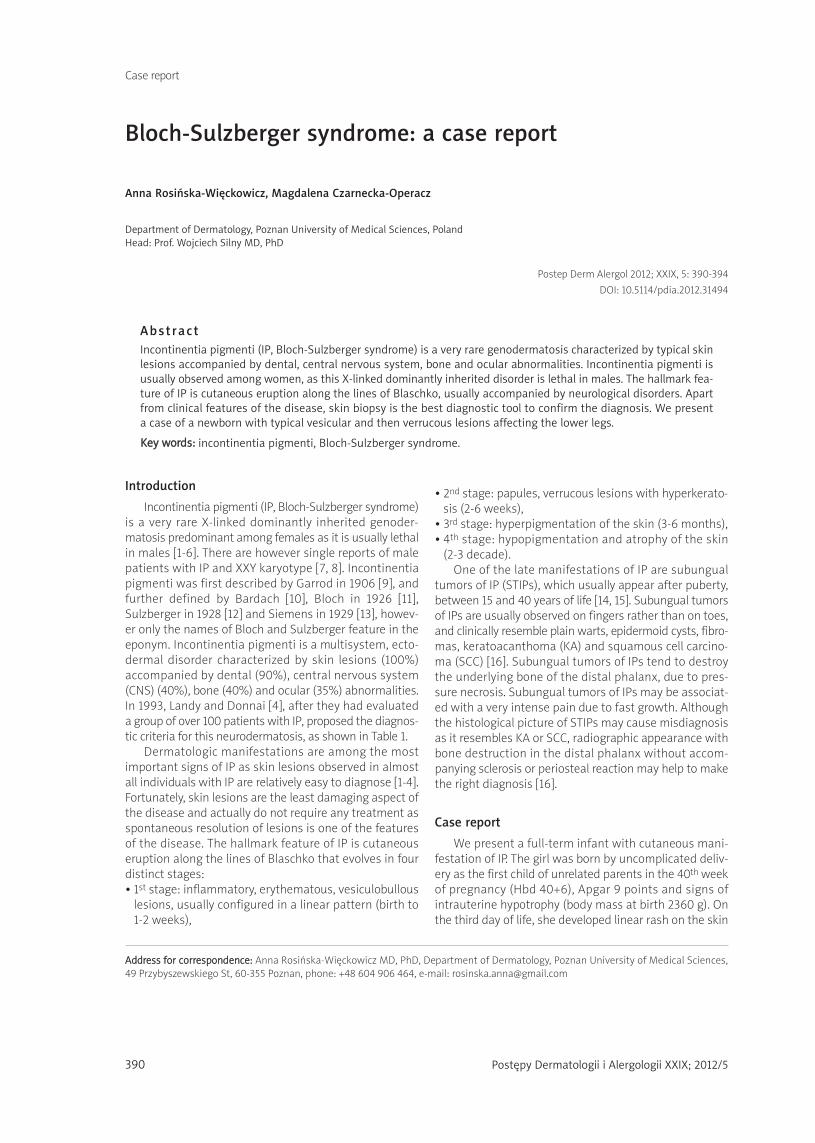
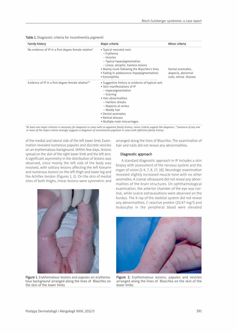
of the medial and lateral side of the left lower limb. Exam-ination revealed numerous papules and discrete vesicleson an erythematous background. Within few days, lesionsspread on the skin of the right lower limb and the left arm.A significant asymmetry in the distribution of lesions wasobserved, since mainly the left side of the body wasinvolved, with solitary lesions affecting the left forearmand numerous lesions on the left thigh and lower leg andthe Achilles tendon (Figures 1, 2). On the skin of medialsites of both thighs, linear lesions were symmetric and
arranged along the lines of Blaschko. The examination ofhair and nails did not reveal any abnormalities.
DDiiaaggnnoossttiicc aapppprrooaacchh
A standard diagnostic approach in IP includes a skinbiopsy with assessment of the nervous system and theorgan of vision [1-4, 7, 8, 17, 18]. Neurologic examinationrevealed slightly increased muscle tone with no otheranomalies. A cranial ultrasound did not reveal any abnor-malities of the brain structures. On ophthalmologicalexamination, the anterior chamber of the eye was nor-mal, while scarce extravasations were observed on thefundus. The X-ray of the skeletal system did not revealany abnormalities. C-reactive protein (20.47 mg/l) andleukocytes in the peripheral blood were elevated
TTaabbllee 11.. Diagnostic criteria for incontinentia pigmenti
FFaammiillyy hhiissttoorryy MMaajjoorr ccrriitteerriiaa MMiinnoorr ccrriitteerriiaa
No evidence of IP in a first degree female relative* • Typical neonatal rash:– Erythema – Vesicles– Typical hyperpigmentation – Linear, atrophic, hairless lesions
• Mainly trunk following the Blaschko’s lines Dental anomalies, • Fading in adolescence (hypopigmentation) alopecia, abnormal • Eosinophilia nails, retinal disease
Evidence of IP in a first degree female relative** • Suggestive history or evidence of typical rash• Skin manifestations of IP
– Hyperpigmentation– Scarring
• Hair abnormalities– Hairless streaks– Alopecia at vertex – Woolly hair
• Dental anomalies• Retinal disease• Multiple male miscarriages
*At least one major criterion is necessary for diagnosis in cases with no apparent family history; minor criteria support the diagnosis, **presence of any oneor more of the major criteria strongly suggests a diagnosis of incontinentia pigmenti in cases with definitive family history
FFiigguurree 22.. Erythematous lesions, papules and vesiclesarranged along the lines of Blaschko on the skin of the lower limbs
FFiigguurree 11.. Erythematous lesions and papules on erythema-tous background arranged along the lines of Blaschko onthe skin of the lower limbs
Bloch-Sulzberger syndrome: a case report
Postępy Dermatologii i Alergologii XXIX; 2012/5392
(14.9-29.6 K/µl) as signs of pneumonia were observed inthe first days of life. Additionally, glucose blood levelsshowed hypoglycemia (25-60 mg/dl). Intrauterine bloodsmear did not reveal eosinophilia, which is a significantsign in IP.
HHiissttoollooggiiccaall eexxaammiinnaattiioonn ooff tthhee sskkiinn bbiiooppssyy
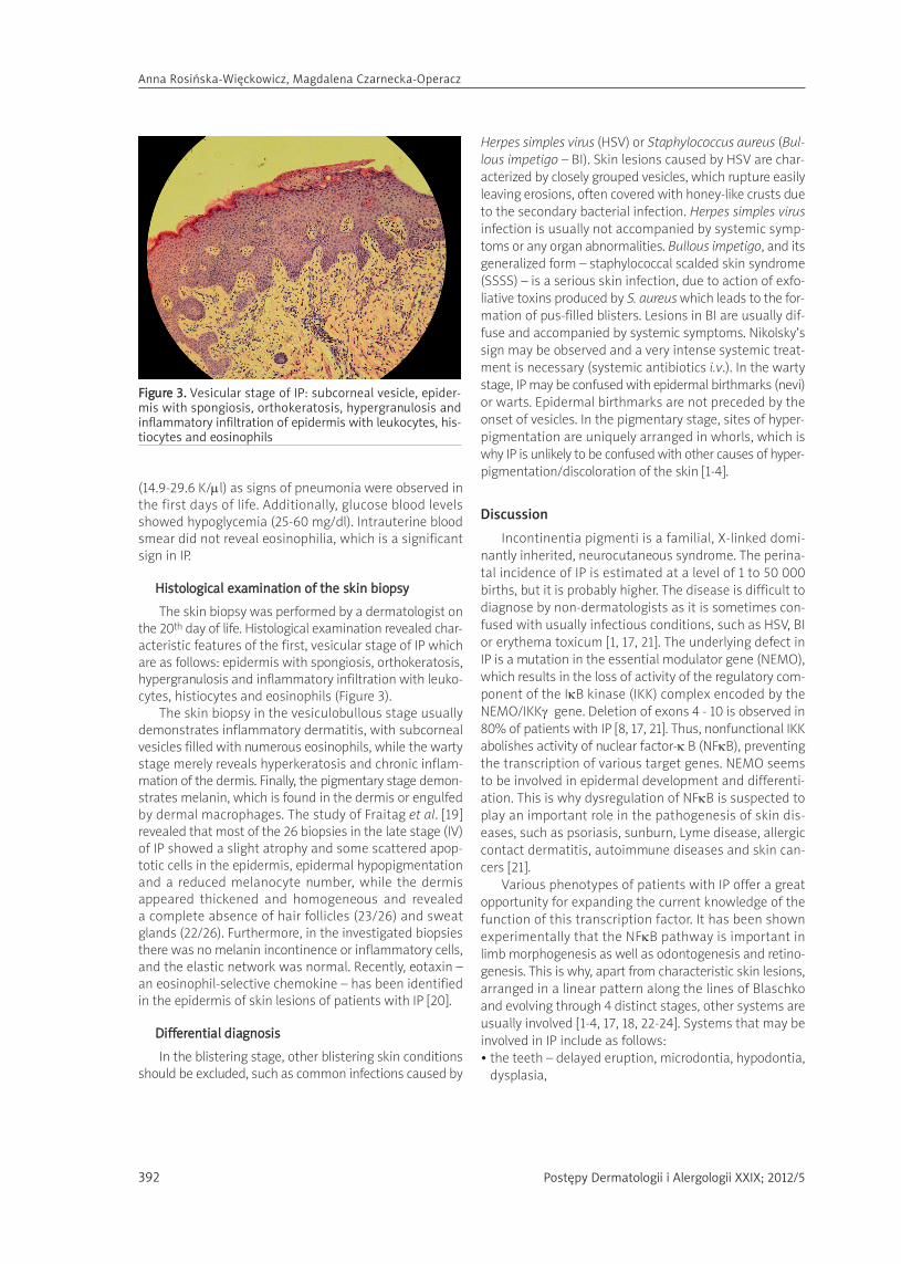
The skin biopsy was performed by a dermatologist onthe 20th day of life. Histological examination revealed char-acteristic features of the first, vesicular stage of IP whichare as follows: epidermis with spongiosis, orthokeratosis,hypergranulosis and inflammatory infiltration with leuko-cytes, histiocytes and eosinophils (Figure 3).
The skin biopsy in the vesiculobullous stage usuallydemonstrates inflammatory dermatitis, with subcornealvesicles filled with numerous eosinophils, while the wartystage merely reveals hyperkeratosis and chronic inflam-mation of the dermis. Finally, the pigmentary stage demon-strates melanin, which is found in the dermis or engulfedby dermal macrophages. The study of Fraitag et al. [19]revealed that most of the 26 biopsies in the late stage (IV)of IP showed a slight atrophy and some scattered apop-totic cells in the epidermis, epidermal hypopigmentationand a reduced melanocyte number, while the dermisappeared thickened and homogeneous and revealeda complete absence of hair follicles (23/26) and sweatglands (22/26). Furthermore, in the investigated biopsiesthere was no melanin incontinence or inflammatory cells,and the elastic network was normal. Recently, eotaxin –an eosinophil-selective chemokine – has been identifiedin the epidermis of skin lesions of patients with IP [20].
DDiiffffeerreennttiiaall ddiiaaggnnoossiiss
In the blistering stage, other blistering skin conditionsshould be excluded, such as common infections caused by
Herpes simples virus (HSV) or Staphylococcus aureus (Bul-lous impetigo – BI). Skin lesions caused by HSV are char-acterized by closely grouped vesicles, which rupture easilyleaving erosions, often covered with honey-like crusts dueto the secondary bacterial infection. Herpes simples virusinfection is usually not accompanied by systemic symp-toms or any organ abnormalities. Bullous impetigo, and itsgeneralized form – staphylococcal scalded skin syndrome(SSSS) – is a serious skin infection, due to action of exfo-liative toxins produced by S. aureus which leads to the for-mation of pus-filled blisters. Lesions in BI are usually dif-fuse and accompanied by systemic symptoms. Nikolsky’ssign may be observed and a very intense systemic treat-ment is necessary (systemic antibiotics i.v.). In the wartystage, IP may be confused with epidermal birthmarks (nevi)or warts. Epidermal birthmarks are not preceded by theonset of vesicles. In the pigmentary stage, sites of hyper-pigmentation are uniquely arranged in whorls, which iswhy IP is unlikely to be confused with other causes of hyper-pigmentation/discoloration of the skin [1-4].
Discussion
Incontinentia pigmenti is a familial, X-linked domi-nantly inherited, neurocutaneous syndrome. The perina-tal incidence of IP is estimated at a level of 1 to 50 000births, but it is probably higher. The disease is difficult todiagnose by non-dermatologists as it is sometimes con-fused with usually infectious conditions, such as HSV, BIor erythema toxicum [1, 17, 21]. The underlying defect inIP is a mutation in the essential modulator gene (NEMO),which results in the loss of activity of the regulatory com-ponent of the IκB kinase (IKK) complex encoded by theNEMO/IKKγ� gene. Deletion of exons 4 - 10 is observed in80% of patients with IP [8, 17, 21]. Thus, nonfunctional IKKabolishes activity of nuclear factor-κ B (NFκB), preventingthe transcription of various target genes. NEMO seemsto be involved in epidermal development and differenti-ation. This is why dysregulation of NFκB is suspected toplay an important role in the pathogenesis of skin dis-eases, such as psoriasis, sunburn, Lyme disease, allergiccontact dermatitis, autoimmune diseases and skin can-cers [21].
Various phenotypes of patients with IP offer a greatopportunity for expanding the current knowledge of thefunction of this transcription factor. It has been shownexperimentally that the NFκB pathway is important inlimb morphogenesis as well as odontogenesis and retino-genesis. This is why, apart from characteristic skin lesions,arranged in a linear pattern along the lines of Blaschkoand evolving through 4 distinct stages, other systems areusually involved [1-4, 17, 18, 22-24]. Systems that may beinvolved in IP include as follows:• the teeth – delayed eruption, microdontia, hypodontia,
dysplasia,
FFiigguurree 33.. Vesicular stage of IP: subcorneal vesicle, epider-mis with spongiosis, orthokeratosis, hypergranulosis andinflammatory infiltration of epidermis with leukocytes, his-tiocytes and eosinophils
Anna Rosińska-Więckowicz, Magdalena Czarnecka-Operacz
Postępy Dermatologii i Alergologii XXIX; 2012/5 393
Bloch-Sulzberger syndrome: a case report
• CNS – seizures, spasticity, mental deficiency, micro-cephaly,
• the eyes – uveitis, keratitis, cataract, retinal dysplasia,strabismus, retinal detachment, retrolental dysplasia,blue sclerae, pigment retinopathy,
• the musculoskeletal system – hemiatrophy, extra rib,hemivertebrae, kyphoscoliosis, syndactyly, short armsand legs,
• the hair – alopecia.The high rate of neurological disturbances and blind-
ness in the population of neonates with IP remains themost important challenge for clinicians. For that reason,newborns with the suspicion of IP should be carefullydiagnosed by the ophthalmologist and neurologist asthese disorders decrease the quality of life significantly.The variety of neurological symptoms is very wide, includ-ing recurrent strokes and acute disseminated encephalo -myelitis [22-25]. Current results of neuroradiologic andhistopathologic observations indicate that vascular anom-alies in CNS might be responsible for neurologic compli-cations in IP [23, 26, 27].
Chromosomal instability seen in IP patients mayincrease the risk of malignancy in young children [28]. Dueto mutation in the NEMO gene, which protects againstTNF-α-induced apoptosis, IP is considered as a pre-apop-totic state leading to male lethality and cell destructionin females. This may account for the dyskeratosisobserved in the histological examination of verrucouslesions in the course of IP. Moreover, the late manifesta-tion of IP, STIP, may clinically resemble keratoacanthoma,which is a pre-malignant condition, or even SCC [14-16].In adolescents and young adults with IP, recurrent casesof SCC have also been described [29, 30].
TTrreeaattmmeenntt ooppttiioonnss
There is no specific treatment. Most of the therapeu-tic methods are claimed to be ineffective as they do nothasten the resolution of any of phases in the course of IP.However, vesicubullous lesions which appear due toinflammatory infiltration of the epidermis (mainly witheosinophils) are expected to respond to topical treatmentwith corticosteroids. It was proved that topical steroidsreduced the expression of eotaxin in the epidermis ofpatients with IP [20, 31]. Topical use of steroids and anti-septic agents (diflucortolone valerate, chlorquinaldol 1%)was observed to contribute to resolution of vesicularlesions [20, 31]. Furthermore, tacrolimus (0.1% ointment),a topical calcineurin inhibitor, has been reported recent-ly to be an effective agent in the treatment of IP [32, 33].According to Jessup et al., tacrolimus halted the progres-sion of the disease through its subsequent disfiguringstages [32]. Even though systemic and topical antibioticsmay show anti-inflammatory effects on the level of theskin, they are not effective in the course of IP, except forlesions with secondary bacterial infection [1, 8]. In patients
with solitary STIP, the first-line treatment is surgical exci-sion, though multiple new lesions appear in other loca-tions. Moreover, the treatment with 5-fluorouracil injec-tions with a good clinical outcome has also been reported[20]. Finally, therapy with retinoids is also worth consid-ering as reports of resolution of lesions and growth of nailsafter the systemic treatment with acitretin (25 mg for 2 months) as well as topical application of retinoic acidwere reported [16, 34, 35]. All-trans-retinoic acid (ATRA)regulates synthesis of NFκB components and activatesapoptosis of various cell lines. Acitretin, which is a syn-thetic analog of retinoic-acid-receptor, prevents the for-mation of STIPs probably by inducing NEMO-independentmechanisms [36, 37].
Although skin lesions are the least damaging aspectof Bloch-Sulzberger syndrome, the proper diagnosis isvery important, thus a careful head-to-toe clinical exam-ination is critical in the evaluation of a child with sus-pected IP. Dermatological examination and diagnosis isthe first step in the multidisciplinary approach includingpediatricians, ophthalmologists, neurologists, dermatol-ogists and dental consultants, which is recommended inpatients with the suspicion of IP.
Photographs were used courtesy of Dr. AleksandraDańczak-Pazdrowska and Dr. Leszek Bartoszak.
References
1. Chang JT, Chiu PC, Chen YY, et al. Multiple clinical manife-stations and diagnostic challenges of incontinentia pigmen-ti: 12 years' experience in 1 medical center. J Chin Med Assoc2008; 71: 455-60.
2. Jentarra G, Snyder SL, Narayanan V. Genetic aspects of neu-rocutaneous disorders. Semin Pediatr Neurol 2006; 13: 43-7.
3. Ehrenreich M, Tarlow MM, Godlewska-Janusz E, et al. Incon-tinentia pigmenti (Bloch-Sulzberger syndrome): a systemicdisorder. Cutis 2007; 79: 355-62.
4. Landy SJ, Donnai D. Incontinentia pigmenti (Bloch-Sulzber-ger syndrome). J Med Genet 1993; 30: 53-9.
5. Hallas TE, Gislason T, Gislason D. Mite allergy and mite expo-sure in Iceland. Ann Agric Environ Med 2011; 18: 13-7.
6. Stankiewicz-Choroszucha BL, Wawrzyniak ZM, Lipiec A, et al.Consequences of smoke inhalation in the ‘Epidemiology ofAllergic Diseases in Poland’ project (ECAP). Ann Agric Envi-ron Med 2011; 18: 420-8.
7. Aradhya S, Courtois G, Rajkovic A, et al. Atypical forms ofincontinentia pigmenti in male individuals result from muta-tions of a cytosine tract in exon 10 of NEMO (IKK-gamma).Am J Hum Genet 2001; 68: 765-71.
8. Berlin AL, Paller AS, Chan LS. Incontinentia pigmenti: a reviewand update on the molecular basis of pathophysiology. J AmAcad Dermatol 2002; 47: 169-87.
9. Garrod A. Peculiar pigmentation of a skin of an infant. TransClin Soc Lond 1906; 39: 216.
10. Bardach M. Systematislette Naevubildungen bei einem eine-iigen Zwillingspoor. Z Kinderheilkd 1925; 39: 542.
11. Bloch B. Eigentumliche bisher nicht beschriebene pigmen-taffektion (incontinentia pigmenti). Schweiz Med Wehnschi1926; 7: 404-5.
Postępy Dermatologii i Alergologii XXIX; 2012/5394
12. Sulzberger F. Uber eine bisher nicht beschriebene congeni-tale Pigmentanomalie. Arch Dermatol Syph 1928; 154: 19-32.
13. Siemens H. Die Melanosis cori degenerativa eine neue Pig-mentdermatose. Arch Dermatol Syph 1929; 157: 382-91.
14. Adeniran A, Townsend PL, Peachey RD. Incontinentia pigmenti(Bloch-Sulzberger syndrome) manifesting as painful periun-gual and subungual tumours. J Hand Surg Br 1993; 18: 667-9.
15. Simmons DA, Kegel MF, Scher RK, et al. Subungual tumors inincontinentia pigmenti. Arch Dermatol 1986; 122: 1431-4.
16. Young A, Manolson P, Cohen B, et al. Painful subungal dys-keratotic tumors in incontinentia pigmenti. J Am Acad Der-matol 2005; 52: 726-9.
17. Nelson DL. NEMO, NFkappaB signaling and incontinentia pig-menti. Curr Opin Genet Dev 2006; 16: 282-8.
18. Kitakawa D, Fontes PC, Magalhaes FA, et al. Incontinentia pig-menti presenting as hypodontia in a 3-year-old girl: a casereport. J Med Case Reports 2009; 3: 116.
19. Fraitag S, Rimella A, de Prost Y, et al. Skin biopsy is helpful forthe diagnosis of incontinentia pigmenti at late stage (IV):a series of 26 cutaneous biopsies. J Cutan Pathol 2009; 36:966-71.
20. Jean-Baptiste S, O'Toole EA, Chen M, et al. Expression of eota-xin, an eosinophil-selective chemokine, parallels eosinophilaccumulation in the vesiculobullous stage of incontinentiapigmenti. Clin Exp Immunol 2002; 127: 470-8.
21. Bell S, Degitz K, Quirling M, et al. Involvement of NF-kappaBsignalling in skin physiology and disease. Cell Signal 2003;15: 1-7.
22. Cartwright MS, White DL, Miller LM 3rd, et al. Recurrent stro-ke in a child with incontinentia pigmenti. J Child Neurol 2009;24: 603-5.
23. Avrahami E, Harel S, Jurgenson U, et al. Computed tomogra-phic demonstration of brain changes in incontinentia pig-menti. Am J Dis Child 1985; 139: 372-4.
24. Shah SN, Gibbs S, Upton CJ, et al. Incontinentia pigmentiassociated with cerebral palsy and cerebral leukomalacia:a case report and literature review. Pediatr Dermatol 2003;20: 491-4.
25. Matsumoto N, Takahashi S, Toriumi N, et al. Acute dissemi-nated encephalomyelitis in an infant with incontinentia pig-menti. Brain Dev 2009; 31: 625-8.
26. Mangano S, Barbagallo A. Incontinentia pigmenti: clinicaland neuroradiologic features. Brain Dev 1993; 15: 362-6.
27. Pascual-Castroviejo I, Roche MC, Martinez Fernandez V, et al.Incontinentia pigmenti: MR demonstration of brain changes.AJNR Am J Neuroradiol 1994; 15: 1521-7.
28. Roberts WM, Jenkins JJ, Moorhead EL 2nd, et al. Incontinen-tia pigmenti, a chromosomal instability syndrome is asso-ciated with childhood malignancy. Cancer 1988; 62: 2370-2.
29. Korstanje MJ, Bessems PJ. Incontinentia pigmenti with hyper-keratotic lesions in adulthood and possible squamous cellcarcinoma. Dermatologica 1991; 183: 234-6.
30. Jamnadas B, Agarwal R, Caddy CM. A rare case of SCC ina young patient with incontinentia pigmenti. J Plast ReconstrAesthet Surg 2008; 61: 973-4.
31. Kaya TI, Tursen U, Ikizoglu G. Therapeutic use of topical cor-ticosteroids in the vesiculobullous lesions of incontinentiapigmenti. Clin Exp Dermatol 2009; 34: e611-3.
32. Jessup C, Morgan S, Cohen L, et al. Incontinentia pigmenti:treatment of IP with topical tacrolimus. J Drugs Dermatol2009; 10: 944-6.
33. Silny W, Sadowska A, Dańczak-Pazdrowska A, Polańska A. Appli-cation of tacrolimus in the treatment of skin diseases otherthan atopic dermatitis. Postep Derm Alergol 2011; 28: 41-5.
34. Malvehy J, Palou J, Mascaro JM. Painful subungual tumour inincontinentia pigmenti. Response to treatment with etreti-nate. Br J Dermatol 1998; 138: 554-5.
35. Donati P, Muscardin L, Amantea A, et al. Detection of HPV-15 in painful subungual tumors of incontinentia pigmenti:successful topical therapy with retinoic acid. Eur J Dermatol2009; 19: 243-7.
36. Manna SK, Aggarwal BB. All-trans-retinoic acid upregulatesTNF receptors and potentiates TNF-induced activation of nuc-lear factors-kappaB, activated protein-1 and apoptosis inhuman lung cancer cells. Oncogene 2000; 19: 2110-9.
37. Farina AR, Masciulli MP, Tacconelli A, et al. All-trans-retinoic acid induces nuclear factor kappaB activation andmatrix metalloproteinase-9 expression and enhances base-ment membrane invasivity of differentiation-resistant humanSK-N-BE 9N neuroblastoma Cells. Cell Growth Differ 2002;13: 343-54.
Anna Rosińska-Więckowicz, Magdalena Czarnecka-Operacz
Recommended