ARTICLE
Structural mechanism of bivalent histoneH3K4me3K9me3 recognition by the Spindlin1/C11orf84 complex in rRNA transcription activationYongming Du 1, Yinxia Yan1, Si Xie1, Hao Huang2, Xin Wang 2, Ray Kit Ng 1, Ming-Ming Zhou 3 &
Chengmin Qian 1✉
Spindlin1 is a unique multivalent epigenetic reader that facilitates ribosomal RNA transcrip-
tion. In this study, we provide molecular and structural basis by which Spindlin1 acts in
complex with C11orf84 to preferentially recognize non-canonical bivalent mark of trimethy-
lated lysine 4 and lysine 9 present on the same histone H3 tail (H3K4me3K9me3). We
demonstrate that C11orf84 binding stabilizes Spindlin1 and enhances its association with
bivalent H3K4me3K9me3 mark. The functional analysis suggests that Spindlin1/C11orf84
complex can displace HP1 proteins from H3K4me3K9me3-enriched rDNA loci, thereby
facilitating the conversion of these poised rDNA repeats from the repressed state to the
active conformation, and the consequent recruitment of RNA Polymerase I for rRNA tran-
scription. Our study uncovers a previously unappreciated mechanism of bivalent
H3K4me3K9me3 recognition by Spindlin1/C11orf84 complex required for activation of rRNA
transcription.
https://doi.org/10.1038/s41467-021-21236-x OPEN
1 School of Biomedical Sciences, The University of Hong Kong, Hong Kong Island, Hong Kong. 2 Department of Biomedical Sciences, The City University ofHong Kong, Kowloon, Hong Kong. 3 Department of Pharmacological Sciences, Icahn School of Medicine at Mount Sinai, New York, NY, USA.✉email: [email protected]
NATURE COMMUNICATIONS | (2021) 12:949 | https://doi.org/10.1038/s41467-021-21236-x | www.nature.com/naturecommunications 1
1234
5678
90():,;
Transcription of ribosomal DNA, the first key step ofribosome biogenesis, is tightly regulated by multi-layerepigenetic mechanisms involving DNA methylations, his-
tone modifications, chromatin remodeling, and non-coding RNAregulation1,2. Although ribosomal RNA production represents themost active transcription, only a subset of rRNA genes is activelytranscribed at a given time, and the inactive rRNA genes exist inthe compact heterochromatic state that is typically associatedwith repressive histone marks such as methylated histone H3lysine 9 (H3K9), H3K27, and H4K20. Intriguingly, rDNA chro-matin is metastable, and can readily respond to environmentalstimuli and developmental cues. Dynamic changes of epigeneticmodifications can help shift the balance between repressed andactive rRNA genes. For instance, chromatin remodeling complexNoRC recruits corepressors such as SETDB1 and HDAC1/2 torDNA promoters to establish trimethylated H3K9 (H3K9me3)for rRNA gene silencing3. On the other hand, overexpression ofthe H3K9 demethylase PHF8 facilitates rRNA transcription4, andthe H3K4me3 demethylase KDM2B was found to localize in thenucleolus and repress rRNA transcription5.
To meet the increasing demand for ribosomes in fast growingcells, a higher level of rRNA transcription is achieved through themore copies of actively transcribed rRNA genes and the highertranscription rate. To convert the rRNA genes in repressed andclosed conformation into the open and transcription-permissivestate, methylation on H3K4 and demethylation on H3K9 at theribosomal RNA gene promoter region needs to be established.Although H3K4me3 and H3K9me3 are generally perceived to bemutually exclusive, bivalent H3K4me3 and H3K9me3 chromatindomains have been reported in different cell types6–9. Interest-ingly, it was reported that binding of KDM4A and KDM4C toH3K4me3 through their double Tudor domains greatly facilitatesthese enzymes to efficiently demethylate H3K9me310,11, sug-gesting the coexistence of H3K4me3 and H3K9me3 on the samehistone H3 and likely a functional crosstalk between these twoseemingly opposing epigenetic marks. It has been suggested thatsome euchromatic rRNA genes may exist in a poised state har-boring canonical bivalent mark H3K4me3K27me36,12, or non-canonical bivalent mark H3K4me3K9me313,14, thus allowingtheir timely activation in response to environmental changes.However, our current mechanistic understanding of how bivalenthistone marks function with respect to corresponding singularhistone marks is very limited. In this study, we provided newstructural and functional insights into a previously unappreciatedmechanism by which Spindlin1 in complex with C11orf84recognizes K4me3 and K9me3 dual marks on the same histoneH3 tail. This unique bivalent histone recognition results in dis-sociation of HP1 from condensed rDNA chromatin to relax thechromatin structure and facilitate the recruitment of RNA Poly-merase I, leading to productive transcription of rRNA.
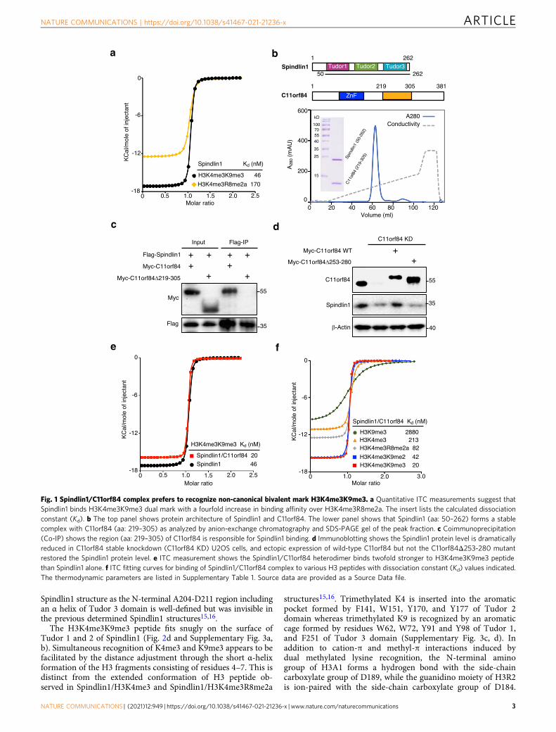
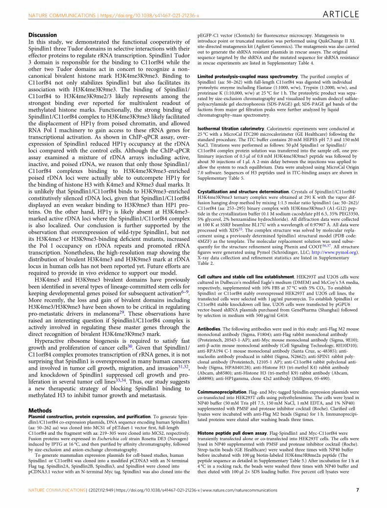
ResultsNon-canonical bivalent H3K4me3K9me3 recognition by Spin-dlin1/C11orf84 complex. Human Spindlin1 was initially char-acterized as a reader of H3K4me3 and recently reported torecognize H3K4me3R8me2a dual mark15,16. Given that Spindlin1facilitates ribosomal RNA transcription15,17, we explored whetherSpindlin1 binds to H3K4me3K9me3 at poised rRNA genes. Wefirst carried out quantitative ITC measurement to show thatSpindlin1 binds to H3K4me3K9me3 with a Kd of 46 nM, aboutfourfold higher than that to H3K4me3R8me2a (Fig. 1a and Sup-plementary Fig. 1a).
Because C11orf84 was recently reported to bind Spindlin1 andcause it to dissociate from the chromatin18, we investigated and
found that full-length C11orf84 and Spindlin1 (aa: 50–262) forma stable heterodimer as shown by analytical gel filtration(Supplementary Fig. 1b). We mapped the region spanningresidues 219–305 in C11orf84 responsible for Spindlin1 bindingusing limited proteolysis-coupled mass spectrometry. The resultwas confirmed by observation of a stable protein complex ofSpindlin1 (aa: 50–262) and C11orf84 (aa: 219–305) in anion-exchange chromatography (Fig. 1b), and by coimmunoprecipita-tion (co-IP) assay showing that C11orf84Δ219-305 truncationmutant lost Spindlin1 binding (Fig. 1c). Furthermore, we foundthat the protein level of Spindlin1 is substantially reduced inC11orf84 knockdown U2OS cells, and ectopic expression of wild-type C11orf84 but not the C11orf84Δ253-280 mutant can restorethe Spindlin1 protein level (Fig. 1d). These results suggested thatC11orf84 is likely a bona fide binding partner of Spindlin1 andcan stabilize Spindlin1 through the direct interaction.
In contrast to the previous report18, we showed that over-expression of C11orf84 in HEK293T cells does not block thebinding of Spindlin1 to methylated histone H3 (SupplementaryFig. 1c). Our observation is consistent with a previous proteomic-based study that C11orf84 can be pulled out from H3K4me3peptide19, and also in good agreement with a more recent study thata Spindlin1 inhibitor bound on H3K4me3 pocket of Tudor 2 canpull down both Spindlin1 and C11orf8420. Moreover, we performedITC measurement to reveal that Spindlin1/C11orf84 heterodimerbinds to H3K4me3K9me3 peptide with a Kd of 20 nM, abouttwofold higher than that of Spindlin1 alone, suggesting thatC11orf84 enhanced Spindlin1 binding to H3K4me3K9me3 peptide(Fig. 1e). Interestingly, Spindlin1/C11orf84 complex also showedthe strong binding to H3K4me3K9me2 which is a preferredsubstrate for PHF8 (Fig. 1f)4. ITC data also suggested thatH3K4me3 is a primary mark responsible for the binding, theaddition of H3K9me2/3 mark significantly enhanced the binding,indicative of a multivalent readout of dual H3K4me3K9me2/3 markby the Spindlin1/C11orf84 complex (Supplementary Table 1). It isalso worth mentioning that the interaction of Spindlin1/C11orf84with H3K4me3K9me3 represents one of the strongest bindings inmethylated histone H3 recognition reported so far21, provided thatmost reported methyl histone binding is in the micromolar or sub-micromolar range.
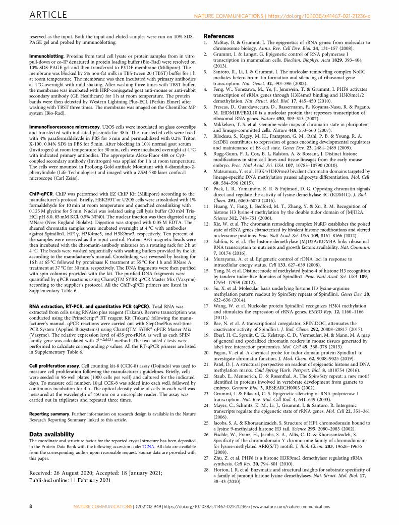
Crystal structure of Spindlin1/C11orf84 complex bound toH3K4me3K9me3. To gain mechanistic insight into the bivalentH3K4me3K9me3 recognition, we determined the crystal structureof Spindlin1/C11orf84/H3K4me3K9me3 ternary complex at1.60 Å resolution (crystallographic statistics is given in Supple-mentary Table 2). The ternary structure revealed that methylhistone peptide lies across Tudor 1 and 2 domains, whereas theC11orf84 fragment associates with Tudor 3 domain (Fig. 2a andSupplementary Fig. 2a).
The electron density map of C11orf84 amino-acid sequenceT255-Q283 is clearly defined and adopts a unique stem–loopconformation (Supplementary Fig. 2b). This segment inserts intothe cleft between the strand β1 and β4 of Spindlin1 Tudor 3to form a closed β barrel-like structure (Fig. 2b). Structuralcomparison with the previous determined Spindlin1/H3K4me3R8-me2a structure (PDB: 4MZF) showed that binding to C11orf84caused obvious conformational changes in Tudor 3 domain: (i)Y256-K260 segment of Spindlin1 Tudor 3 adopts a β-strandconformation and forms an antiparallel β-sheet with the β-strandadopted by the F274-L278 segment of C11orf84; (ii) part of Tudor3 β1–loop–β2 region is pushed outward with ~6 Å in distance toaccommodate the C11orf84 fragment (Fig. 2c and SupplementaryFig. 2c). In addition, the binding to C11orf84 appears to stabilize
ARTICLE NATURE COMMUNICATIONS | https://doi.org/10.1038/s41467-021-21236-x
2 NATURE COMMUNICATIONS | (2021) 12:949 | https://doi.org/10.1038/s41467-021-21236-x | www.nature.com/naturecommunications
Spindlin1 structure as the N-terminal A204-D211 region includingan α helix of Tudor 3 domain is well-defined but was invisible inthe previous determined Spindlin1 structures15,16.
The H3K4me3K9me3 peptide fits snugly on the surface ofTudor 1 and 2 of Spindlin1 (Fig. 2d and Supplementary Fig. 3a,b). Simultaneous recognition of K4me3 and K9me3 appears to befacilitated by the distance adjustment through the short α-helixformation of the H3 fragments consisting of residues 4–7. This isdistinct from the extended conformation of H3 peptide ob-served in Spindlin1/H3K4me3 and Spindlin1/H3K4me3R8me2a
structures15,16. Trimethylated K4 is inserted into the aromaticpocket formed by F141, W151, Y170, and Y177 of Tudor 2domain whereas trimethylated K9 is recognized by an aromaticcage formed by residues W62, W72, Y91 and Y98 of Tudor 1,and F251 of Tudor 3 domain (Supplementary Fig. 3c, d). Inaddition to cation-π and methyl-π interactions induced bydual methylated lysine recognition, the N-terminal aminogroup of H3A1 forms a hydrogen bond with the side-chaincarboxylate group of D189, while the guanidino moiety of H3R2is ion-paired with the side-chain carboxylate group of D184.
a
c
ZnFC11orf84
1 219 305 381
Spindlin11 262
50 262Tudor1 Tudor2 Tudor3
e
Molar ratio
KC
al/m
ole
of in
ject
ant
-12
-6
-18
0
1.0 2.0 2.50 1.50.5
H3K4me3K9me3 46
H3K4me3R8me2a 170
Spindlin1 Kd (nM)
Molar ratio
KC
al/m
ole
of in
ject
ant
-12
-6
-18
0
1.0 2.0 2.50 1.50.5
Spindlin1/C11orf84 20Spindlin1 46
H3K4me3K9me3 Kd (nM)
Molar ratio
-12
-6
-18
0
1.0 2.0 3.00
H3K9me3 2880H3K4me3 213H3K4me3R8me2a 82
H3K4me3K9me2 42 H3K4me3K9me3 20
KC
al/m
ole
of in
ject
ant
Spindlin1/C11orf84 Kd (nM)
A28
0(m
AU
)
A280 Conductivity
0
200
400
600
0 20 40 60 80 100 120 Volume (ml)
Spin
dlin
1 (5
0-26
2)
C11
of84
(219
-305
)
15
25
35
405570
kD
100
Myc
Flag
Input
Myc-C11orf84
Myc-C11orf84 219-305
Flag-Spindlin1
Flag-IP
+ + + ++ +
+ +
55
35
C11orf84
Spindlin1
-Actin
Myc-C11orf84 WT
Myc-C11orf84 253-280
++
C11orf84 KD
55
35
40
b
d
f
Fig. 1 Spindlin1/C11orf84 complex prefers to recognize non-canonical bivalent mark H3K4me3K9me3. a Quantitative ITC measurements suggest thatSpindlin1 binds H3K4me3K9me3 dual mark with a fourfold increase in binding affinity over H3K4me3R8me2a. The insert lists the calculated dissociationconstant (Kd). b The top panel shows protein architecture of Spindlin1 and C11orf84. The lower panel shows that Spindlin1 (aa: 50–262) forms a stablecomplex with C11orf84 (aa: 219–305) as analyzed by anion-exchange chromatography and SDS-PAGE gel of the peak fraction. c Coimmunoprecipitation(Co-IP) shows the region (aa: 219–305) of C11orf84 is responsible for Spindlin1 binding. d Immunoblotting shows the Spindlin1 protein level is dramaticallyreduced in C11orf84 stable knockdown (C11orf84 KD) U2OS cells, and ectopic expression of wild-type C11orf84 but not the C11orf84Δ253-280 mutantrestored the Spindlin1 protein level. e ITC measurement shows the Spindlin1/C11orf84 heterodimer binds twofold stronger to H3K4me3K9me3 peptidethan Spindlin1 alone. f ITC fitting curves for binding of Spindlin1/C11orf84 complex to various H3 peptides with dissociation constant (Kd) values indicated.The thermodynamic parameters are listed in Supplementary Table 1. Source data are provided as a Source Data file.
NATURE COMMUNICATIONS | https://doi.org/10.1038/s41467-021-21236-x ARTICLE
NATURE COMMUNICATIONS | (2021) 12:949 | https://doi.org/10.1038/s41467-021-21236-x | www.nature.com/naturecommunications 3
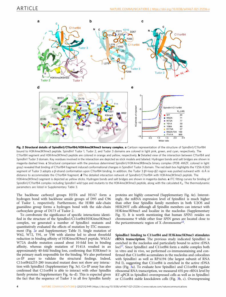
The backbone carbonyl groups H3T6 and H3A7 form ahydrogen bond with backbone amide groups of D95 and C96of Tudor 1, respectively. Furthermore, the H3R8 side-chainguanidino group forms a hydrogen bond with the side-chaincarboxylate group of D173 of Tudor 2.
To corroborate the significance of specific interactions identi-fied in the structure of the Spindlin1/C11orf84/H3K4me3K9me3complex, we generated a number of Spindlin1 mutants andquantitatively evaluated the effects of mutation by ITC measure-ment (Fig. 2e and Supplementary Table 3). Single mutation ofW62, W72, Y91, or Y98 with alanine led to about threefoldreduction in binding affinity of H3K4me3K9me3 peptide, W62A/W72A double mutation caused about 10-fold loss in bindingaffinity, whereas single mutation of F141A resulted in anapproximately 40-fold binding loss, confirming that H3K4me3 isthe primary mark responsible for the binding. We also performedco-IP assay to validate the structural findings. Indeed,C11orf84Δ253-280 truncated mutant does not show any interac-tion with Spindlin1 (Supplementary Fig. 3e). Co-IP assay has alsoconfirmed that C11orf84 is able to interact with other Spindlinfamily proteins (Supplementary Fig. 4a–d). This is expected giventhe fact that the sequence of Tudor 3 in all five Spindlin family
proteins are highly conserved (Supplementary Fig. 4e). Interest-ingly, the mRNA expression level of Spindlin1 is much higherthan other four Spindlin family members in both U2OS andHEK293T cells although all Spindlin members can interact withH3K4me3K9me3 and localize in the nucleolus (SupplementaryFig. 5). It is worth mentioning that human SPIN1 resides onchromosome 9 while other four SPIN genes are located close tothe pericentromeric region of X chromosome22.
Spindlin1 binding to C11orf84 and H3K4me3K9me3 stimulatesrRNA transcription. The previous study indicated Spindlin1 isenriched in the nucleolus and particularly bound to active rDNAloci17. Since Spindlin1 and C11orf84 form a stable complex bothin vitro and in vivo, we performed co-immunostaining and con-firmed that C11orf84 accumulates in the nucleolus and colocalizeswith Spindlin1 as well as RPA194 (the largest subunit of RNAPol I), suggesting that C11orf84 is enriched in the active rDNAsites (Fig. 3a). To evaluate how Spindlin1 and C11orf84 regulateribosomal RNA transcription, we measured 45S pre-rRNA level byRT-qPCR in Spindlin1-overexpressed cells as well as in Spindlin1or C11orf84 stable knockdown cells (Fig. 3b, c). Overexpressing
a
d
Tudor 1Tudor 2H3K4me3K9me3
N
N
C
C
C11orf84
Tudor 3
C
N
KC
al/m
ole
of in
ject
ant
-12
-6
-18
0
Molar ratio1.0 2.0 2.50 1.50.5
WT 20W62A 71W72A 77Y91A 59Y98A 80
F141A 869
D173A 57W62A/72A 199
Kd (nM)
A1 R2
T3
Q5
A7
R8S10 T11
W72
W62
Y98
Y91
W151
F141Y170
D173
Y177
K9me3
D184
D95
C96
T6D189
K4me3
F251
Spindlin1/C11orf84
b c
e
Fig. 2 Structural details of Spindlin1/C11orf84/H3K4me3K9me3 ternary complex. a Cartoon representation of the structure of Spindlin1/C11orf84bound to H3K4me3K9me3 peptide. Spindlin1 Tudor 1, Tudor 2, and Tudor 3 domains are colored in light pink, green, and cyan, respectively. TheC11orf84 segment and H3K4me3K9me3 peptide are colored in orange and yellow, respectively. b Detailed view of the interaction between C11orf84 andSpindlin1 Tudor 3 domain. Key residues involved in the interaction are depicted as stick models and labeled. Hydrogen bonds and salt bridges are shown inmagenta dashed lines. c Structural comparison with the previous determined Spindlin1/H3K4me3R8me2a binary complex (PDB: 4MZF, colored in lightgray) revealed that binding of C11orf84 fragment induced conformational changes in Spindlin1 Tudor 3 domain. The red dash box highlights the Y256-K260segment of Tudor 3 adopts a β-strand conformation upon C11orf84 binding. In addition, the Tudor 3 β1–loop–β2 region was pushed outward with ~6 Å indistance to accommodate the C11orf84 fragment. d The detailed interaction network of Spindlin1/C11orf84 with H3K4me3K9me3 peptide. TheH3K4me3K9me3 segment is depicted as yellow sticks. Hydrogen bonds and salt bridges are shown in magenta dashes. e ITC fitting curves for binding ofSpindlin1/C11orf84 complex including Spindlin1 wild type and mutants to the H3K4me3K9me3 peptide, along with the calculated Kd. The thermodynamicparameters are listed in Supplementary Table 3.
ARTICLE NATURE COMMUNICATIONS | https://doi.org/10.1038/s41467-021-21236-x
4 NATURE COMMUNICATIONS | (2021) 12:949 | https://doi.org/10.1038/s41467-021-21236-x | www.nature.com/naturecommunications
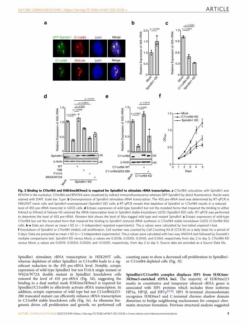
Spindlin1 stimulates rRNA transcription in HEK293T cells,whereas depletion of either Spindlin1 or C11orf84 leads to a sig-nificant reduction in the 45S pre-rRNA level. Notably, ectopicexpression of wild-type Spindlin1 but not F141A single mutant orW62A/W72A double mutant in Spindlin1 knockdown cellsrestored the level of 45S pre-rRNA (Fig. 3d), suggesting thebinding to a dual methyl mark H3K4me3K9me3 is required forSpindlin1/C11orf84 to effectively activate rRNA transcription. Inaddition, ectopic expression of wild type but not C11orf84Δ253-280 truncated mutant can efficiently enhance rRNA transcriptionin C11orf84 stable knockdown cells (Fig. 3e). As ribosome bio-genesis drives cell proliferation and growth, we performed cell
counting assay to show a decreased cell proliferation in Spindlin1-or C11orf84-depleted cells (Fig. 3f).
Spindlin1/C11orf84 complex displaces HP1 from H3K4me-3K9me3-enriched rDNA loci. The majority of H3K9me2/3marks in constitutive and temporary silenced rRNA genes isassociated with HP1 proteins which includes three isoformsHP1α, HP1β, and HP1γ23,24. HP1 N-terminal chromodomainrecognizes H3K9me3 and C-terminal chromo shadow domaindimerizes to bridge neighboring nucleosomes for compact chro-matin structure formation. Previous structural analysis suggested
Fig. 3 Binding to C11orf84 and H3K4me3K9me3 is required for Spindlin1 to stimulate rRNA transcription. a C11orf84 colocalizes with Spindlin1 andRPA194 in the nucleolus. C11orf84 and RPA194 were visualized by indirect immunofluorescence whereas GFP-Spindlin1 by direct fluorescence. Nuclei werestained with DAPI. Scale bar: 5 μm. b Overexpression of Spindlin1 stimulates rRNA transcription. The 45S pre-rRNA level was determined by RT-qPCR inHEK293T mock cells and Spindlin1-overexpressed (Spindlin1 OE) cells. c RT-qPCR reveals that depletion of Spindlin1 or C11orf84 results in a reducedlevel of 45S pre-rRNA transcript in U2OS cells. d Ectopic expression of wild-type Spindlin1 but not the mutated forms that impaired the binding to eitherK4me3 or K9me3 of histone H3 restored the rRNA transcription level in Spindlin1 stable knockdown U2OS (Spindlin1 KD) cells. RT-qPCR was performedto determine the level of 45S pre-rRNA. Western blot shows the level of Myc-tagged wild type and mutant Spindlin1. e Ectopic expression of wild-typeC11orf84 but not the truncated form that impaired the binding to Spindlin1 restored rRNA synthesis in C11orf84 stable knockdown U2OS (C11orf84 KD)cells. b–e Data are shown as mean ± SD (n= 3 independent repeated experiments). The p values were calculated by two-tailed unpaired t-test.f Knockdown of Spindlin1 or C11orf84 inhibits cell proliferation. Cell number was counted by Cell Counting Kit-8 (CCK-8) on a daily basis for a period of5 days. Data are presented as mean ± SD (n= 5 independent experiments). The p values were calculated with two-way ANOVA test followed by Dunnett’smultiple comparisons test. Spindlin1 KD versus Mock, p values are 0.0036, 0.0005, 0.0045, and 0.004, respectively from day 2 to day 5; C11orf84 KDversus Mock, p values are 0.0019, 0.0004, 0.0004, and <0.0001, respectively, from day 2 to day 5. Source data are provided as a Source Data file.
NATURE COMMUNICATIONS | https://doi.org/10.1038/s41467-021-21236-x ARTICLE
NATURE COMMUNICATIONS | (2021) 12:949 | https://doi.org/10.1038/s41467-021-21236-x | www.nature.com/naturecommunications 5
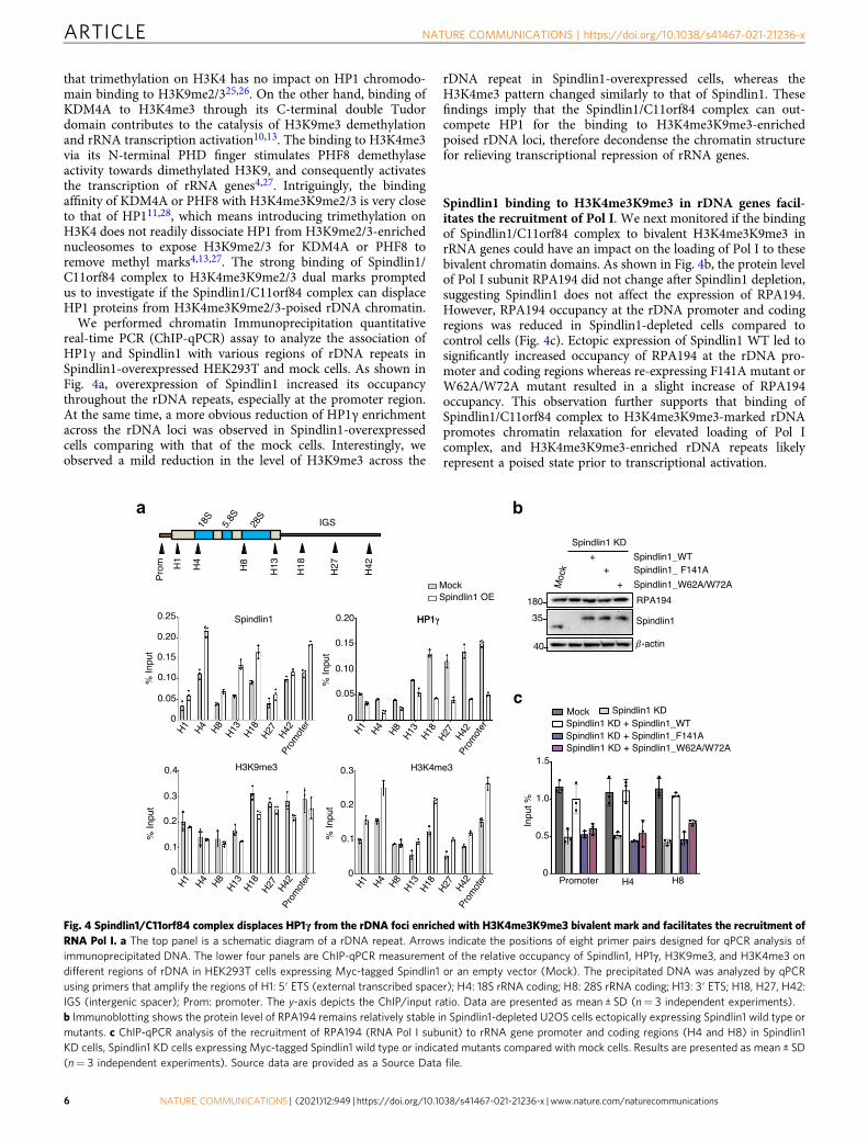
that trimethylation on H3K4 has no impact on HP1 chromodo-main binding to H3K9me2/325,26. On the other hand, binding ofKDM4A to H3K4me3 through its C-terminal double Tudordomain contributes to the catalysis of H3K9me3 demethylationand rRNA transcription activation10,13. The binding to H3K4me3via its N-terminal PHD finger stimulates PHF8 demethylaseactivity towards dimethylated H3K9, and consequently activatesthe transcription of rRNA genes4,27. Intriguingly, the bindingaffinity of KDM4A or PHF8 with H3K4me3K9me2/3 is very closeto that of HP111,28, which means introducing trimethylation onH3K4 does not readily dissociate HP1 from H3K9me2/3-enrichednucleosomes to expose H3K9me2/3 for KDM4A or PHF8 toremove methyl marks4,13,27. The strong binding of Spindlin1/C11orf84 complex to H3K4me3K9me2/3 dual marks promptedus to investigate if the Spindlin1/C11orf84 complex can displaceHP1 proteins from H3K4me3K9me2/3-poised rDNA chromatin.
We performed chromatin Immunoprecipitation quantitativereal-time PCR (ChIP-qPCR) assay to analyze the association ofHP1γ and Spindlin1 with various regions of rDNA repeats inSpindlin1-overexpressed HEK293T and mock cells. As shown inFig. 4a, overexpression of Spindlin1 increased its occupancythroughout the rDNA repeats, especially at the promoter region.At the same time, a more obvious reduction of HP1γ enrichmentacross the rDNA loci was observed in Spindlin1-overexpressedcells comparing with that of the mock cells. Interestingly, weobserved a mild reduction in the level of H3K9me3 across the
rDNA repeat in Spindlin1-overexpressed cells, whereas theH3K4me3 pattern changed similarly to that of Spindlin1. Thesefindings imply that the Spindlin1/C11orf84 complex can out-compete HP1 for the binding to H3K4me3K9me3-enrichedpoised rDNA loci, therefore decondense the chromatin structurefor relieving transcriptional repression of rRNA genes.
Spindlin1 binding to H3K4me3K9me3 in rDNA genes facil-itates the recruitment of Pol I. We next monitored if the bindingof Spindlin1/C11orf84 complex to bivalent H3K4me3K9me3 inrRNA genes could have an impact on the loading of Pol I to thesebivalent chromatin domains. As shown in Fig. 4b, the protein levelof Pol I subunit RPA194 did not change after Spindlin1 depletion,suggesting Spindlin1 does not affect the expression of RPA194.However, RPA194 occupancy at the rDNA promoter and codingregions was reduced in Spindlin1-depleted cells compared tocontrol cells (Fig. 4c). Ectopic expression of Spindlin1 WT led tosignificantly increased occupancy of RPA194 at the rDNA pro-moter and coding regions whereas re-expressing F141A mutant orW62A/W72A mutant resulted in a slight increase of RPA194occupancy. This observation further supports that binding ofSpindlin1/C11orf84 complex to H3K4me3K9me3-marked rDNApromotes chromatin relaxation for elevated loading of Pol Icomplex, and H3K4me3K9me3-enriched rDNA repeats likelyrepresent a poised state prior to transcriptional activation.
Fig. 4 Spindlin1/C11orf84 complex displaces HP1γ from the rDNA foci enriched with H3K4me3K9me3 bivalent mark and facilitates the recruitment ofRNA Pol I. a The top panel is a schematic diagram of a rDNA repeat. Arrows indicate the positions of eight primer pairs designed for qPCR analysis ofimmunoprecipitated DNA. The lower four panels are ChIP-qPCR measurement of the relative occupancy of Spindlin1, HP1γ, H3K9me3, and H3K4me3 ondifferent regions of rDNA in HEK293T cells expressing Myc-tagged Spindlin1 or an empty vector (Mock). The precipitated DNA was analyzed by qPCRusing primers that amplify the regions of H1: 5′ ETS (external transcribed spacer); H4: 18S rRNA coding; H8: 28S rRNA coding; H13: 3′ ETS; H18, H27, H42:IGS (intergenic spacer); Prom: promoter. The y-axis depicts the ChIP/input ratio. Data are presented as mean ± SD (n= 3 independent experiments).b Immunoblotting shows the protein level of RPA194 remains relatively stable in Spindlin1-depleted U2OS cells ectopically expressing Spindlin1 wild type ormutants. c ChIP-qPCR analysis of the recruitment of RPA194 (RNA Pol I subunit) to rRNA gene promoter and coding regions (H4 and H8) in Spindlin1KD cells, Spindlin1 KD cells expressing Myc-tagged Spindlin1 wild type or indicated mutants compared with mock cells. Results are presented as mean ± SD(n= 3 independent experiments). Source data are provided as a Source Data file.
ARTICLE NATURE COMMUNICATIONS | https://doi.org/10.1038/s41467-021-21236-x
6 NATURE COMMUNICATIONS | (2021) 12:949 | https://doi.org/10.1038/s41467-021-21236-x | www.nature.com/naturecommunications
DiscussionIn this study, we demonstrated the functional cooperativity ofSpindlin1 three Tudor domains in selective interactions with theireffector proteins to regulate rRNA transcription. Spindlin1 Tudor3 domain is responsible for the binding to C11orf84 while theother two Tudor domains act in concert to recognize a non-canonical bivalent histone mark H3K4me3K9me3. Binding toC11orf84 not only stabilizes Spindlin1 but also facilitates itsassociation with H3K4me3K9me3. The binding of Spindlin1/C11orf84 to H3K4me3K9me2/3 likely represents among thestrongest binding ever reported for multivalent readout ofmethylated histone marks. Functionally, the strong binding ofSpindlin1/C11orf84 complex to H3K4me3K9me3 likely facilitatedthe displacement of HP1γ from poised chromatin, and allowedRNA Pol I machinery to gain access to these rRNA genes fortranscriptional activation. As shown in ChIP-qPCR assay, over-expression of Spindlin1 reduced HP1γ occupancy at the rDNAloci compared with the control cells. Although the ChIP-qPCRassay examined a mixture of rDNA arrays including active,inactive, and poised rDNA, we reason that only those Spindlin1/C11orf84 complexes binding to H3K4me3K9me3-enrichedpoised rDNA loci were actually able to outcompete HP1γ forthe binding of histone H3 with K4me3 and K9me3 dual marks. Itis unlikely that Spindlin1/C11orf84 binds to H3K9me3-enrichedconstitutively silenced rDNA loci, given that Spindlin1/C11orf84displayed an even weaker binding to H3K9me3 than HP1 pro-teins. On the other hand, HP1γ is likely absent at H3K4me3-marked active rDNA loci where the Spindlin1/C11orf84 complexis also localized. Our conclusion is further supported by theobservation that overexpression of wild-type Spindlin1, but notits H3K4me3 or H3K9me3-binding deficient mutants, increasedthe Pol I occupancy on rDNA repeats and promoted rRNAtranscription. Nonetheless, the high-resolution map showing thedistribution of bivalent H3K4me3 and H3K9me3 mark at rDNAlocus in human cells has not been reported yet. Future efforts arerequired to provide in vivo evidence to support our model.
H3K4me3 and H3K9me3 bivalent domains have previouslybeen identified in several types of lineage-committed stem cells forkeeping developmental genes poised for subsequent activation6–9.More recently, the loss and gain of bivalent domains includingH3K4me3/H3K9me3 have been shown to be critical in regulatingpro-metastatic drivers in melanoma29. These observations haveraised an interesting question if Spindlin1/C11orf84 complex isactively involved in regulating these master genes through thedirect recognition of bivalent H3K4me3K9me3 mark.
Hyperactive ribosome biogenesis is required to satisfy fastgrowth and proliferation of cancer cells30. Given that Spindlin1/C11orf84 complex promotes transcription of rRNA genes, it is notsurprising that Spindlin1 is overexpressed in many human cancersand involved in tumor cell growth, migration, and invasion31,32,and knockdown of Spindlin1 suppressed cell growth and pro-liferation in several tumor cell lines33,34. Thus, our study suggestsa new therapeutic strategy of blocking Spindlin1 binding tomethylated H3 to inhibit tumor growth and metastasis.
MethodsPlasmid construction, protein expression, and purification. To generate Spin-dlin1/C11orf84 co-expression plasmids, DNA sequence encoding human Spindlin1(aa: 50–262 aa) was cloned into MCS1 of pETduet-1 vector first, full-lengthC11orf84 and the fragment with aa: 219–305 were cloned into MCS2, respectively.Fusion proteins were expressed in Escherichia coli strain Rosetta DE3 (Novagen)induced by IPTG at 16 °C, and then purified by affinity chromatography, followedby size-exclusion and anion-exchange chromatography.
To generate mammalian expression plasmids for cell-based studies, humanSpindlin1 or C11orf84 was cloned into a modified pCDNA3 with an N-terminalFlag tag. Spindlin2A, Spindlin2B, Spindlin3, and Spindlin4 were cloned intopCDNA3.1 vector with an N-terminal Myc tag. Spindlin1 was also cloned into the
pEGFP-C1 vector (Clontech) for fluorescence microscopy. Mutagenesis tointroduce point or truncated mutation was performed using QuikChange II XLsite-directed mutagenesis kit (Agilent Genomics). The mutagenesis was also carriedout to generate the shRNA resistant plasmids in rescue assays. The originalsequence targeted by the shRNA and the mutated sequence for shRNA resistancein rescue experiments are listed in Supplementary Table 4.
Limited proteolysis-coupled mass spectrometry. The purified complex ofSpindlin1 (aa: 50–262) with full-length C11orf84 was digested with individualproteolytic enzyme including Elastase (1:1000, w/w), Trypsin (1:2000, w/w), andproteinase K (1:10,000, w/w) at 25 °C for 1 h. The proteolytic product was sepa-rated by size-exclusion chromatography and visualized by sodium dodecyl sulfate-polyacrylamide gel electrophoresis (SDS-PAGE) gel; SDS-PAGE gel bands of thefactions from major gel filtration peaks were further analyzed by liquidchromatography–mass spectrometry.
Isothermal titration calorimetry. Calorimetric experiments were conducted at25 °C with a MicroCal ITC200 microcalorimeter (GE Healthcare) following thestandard procedure. The ITC buffer contains 20 mM HEPES pH 7.5 and 150 mMNaCl. Titrations were performed as follows: 50 μM Spindlin1 or Spindlin1/C11orf84 complex protein solution was transferred into the sample cell, one pre-liminary injection of 0.5 μl of 0.8 mM H3K4me3K9me3 peptide was followed byabout 30 injections of 1 μl. A 2-min delay between the injections was applied toallow the system to reach equilibrium. Data were analyzed using MicroCal Origin7.0 software. Sequences of H3 peptides used in ITC-binding assays are shown inSupplementary Table 5.
Crystallization and structure determination. Crystals of Spindlin1/C11orf84/H3K4me3K9me3 ternary complex were obtained at 291 K with the vapor dif-fusion hanging drop method by mixing 1:1.5 molar ratio Spindlin1 (aa: 50–262)/C11orf84 (aa: 253–295) binary complex with H3K4me3K9me3 (A1-G12) pep-tide in the crystallization buffer (0.1 M sodium cacodylate pH 6.5, 35% PEG3350,5% glycerol, 2% benzamidine hydrochloride). All diffraction data were collectedat 100 K at SSRF beamline BL17U with a wavelength of 0.97907 Å. All data wereprocessed with XDS35. The complex structure was solved by molecular repla-cement using a previously determined Spindlin1 structural model (PDB code:4MZF) as the template. The molecular replacement solution was used subse-quently for the structure refinement using Phenix and COOT36,37. All structurefigures were generated using Pymol (Schrödinger, LLC; http://www.pymol.org).X-ray data collection and refinement statistics are listed in SupplementaryTable 2.
Cell culture and stable cell line establishment. HEK293T and U2OS cells werecultured in Dulbecco’s modified Eagle’s medium (DMEM) and McCoy’s 5A media,respectively, supplemented with 10% FBS at 37 °C with 5% CO2. To establishSpindlin1 or C11orf84 stably overexpressed HEK293T and U2OS cell lines, thetransfected cells were selected with 1 μg/ml puromycin. To establish Spindlin1 orC11orf84 stable knockdown cell line, U2OS cells were transfected by pGPU6vector-based shRNA plasmids purchased from GenePharma (Shanghai) followedby selection in medium with 500 μg/ml G418.
Antibodies. The following antibodies were used in this study: anti-Flag M2 mousemonoclonal antibody (Sigma, F1804); anti-Flag rabbit monoclonal antibody(Proteintech, 20543-1-AP); anti-Myc mouse monoclonal antibody (Sigma, 9E10);anti-β-actin mouse monoclonal antibody (Cell Signaling Technology, 8H10D10);anti-RPA194 C-1 mouse monoclonal antibody (Santa Cruz, sc-48385); anti-nucleolin antibody produced in rabbit (Sigma, N2662); anti-SPIN1 rabbit poly-clonal antibody (Proteintech, 12105-1 AP); anti-C11orf84 rabbit polyclonal anti-body (Sigma, HPA040128); anti-Histone H3 (tri-methyl K4) rabbit antibody(Abcam, ab8580); anti-Histone H3 (tri-methyl K9) rabbit antibody (Abcam,ab8898); anti-HP1gamma, clone 42s2 antibody (Millipore, 05-690).
Coimmunoprecipitation. Flag- and Myc-tagged Spindlin expression plasmids wereco-transfected into HEK293T cells using polyethylenimine. The cells were lysed inNP40 buffer (50 mM Tris pH 7.5, 150 mM NaCl, 1 mM EDTA, and 1% NP40)supplemented with PMSF and protease inhibitor cocktail (Roche). Clarified celllysates were incubated with anti-Flag M2 beads (Sigma) for 1 h. Immunoprecipi-tated proteins were eluted after washing beads three times.
Histone peptide pull down assay. Flag-Spindlin1 and Myc-C11orf84 weretransiently transfected alone or co-transfected into HEK293T cells. The cells werelysed in NP40 supplemented with PMSF and protease inhibitor cocktail (Roche).Strep-tactin beads (GE Healthcare) were washed three times with NP40 bufferbefore incubated with 100 μg biotin-labeled H3K4me3R8me2a peptide (Thepeptide sequence as detailed in Supplementary Table 5.) After incubation for 1 h at4 °C in a rocking rack, the beads were washed three times with NP40 buffer andthen eluted with 100 μl 2× SDS loading buffer. Five percent cell lysates were
NATURE COMMUNICATIONS | https://doi.org/10.1038/s41467-021-21236-x ARTICLE
NATURE COMMUNICATIONS | (2021) 12:949 | https://doi.org/10.1038/s41467-021-21236-x | www.nature.com/naturecommunications 7
reserved as the input. Both the input and eluted samples were run on 10% SDS-PAGE gel and probed by immunoblotting.
Immunoblotting. Proteins from total cell lysate or protein samples from in vitropull-down or co-IP denatured in protein loading buffer (Bio-Rad) were resolved on10% SDS-PAGE gel and then transferred to PVDF membrane (Millipore). Themembrane was blocked by 5% non-fat milk in TBS-tween 20 (TBST) buffer for 1 hat room temperature. The membrane was then incubated with primary antibodiesat 4 °C overnight with mild shaking. After washing three times with TBST buffer,the membrane was incubated with HRP-conjugated goat anti-mouse or anti-rabbitsecondary antibody (GE Healthcare) for 1 h at room temperature. The proteinbands were then detected by Western Lightning Plus-ECL (Perkin Elmer) afterwashing with TBST three times. The membrane was imaged on the ChemiDoc MPsystem (Bio-Rad).
Immunofluorescence microscopy. U2OS cells were inoculated on glass coverslipsand transfected with indicated plasmids for 48 h. The transfected cells were fixedwith 4% paraformaldehyde in PBS for 5 min and permeabilized with 0.2% TritonX-100, 0.04% SDS in PBS for 5 min. After blocking in 10% normal goat serum(Invitrogen) at room temperature for 30 min, cells were incubated overnight at 4 °Cwith indicated primary antibodies. The appropriate Alexa-Fluor 488 or Cy3-coupled secondary antibody (Invitrogen) was applied for 1 h at room temperature.The cells were mounted in ProLong-Gold antifade Mountant with 6-diamidino-2-phenylindole (Life Technologies) and imaged with a ZSM 780 laser confocalmicroscope (Carl Zeiss).
ChIP-qPCR. ChIP was performed with EZ ChIP Kit (Millipore) according to themanufacturer’s protocol. Briefly, HEK293T or U2OS cells were crosslinked with 1%formaldehyde for 10 min at room temperature and quenched crosslinking with0.125 M glycine for 5 min. Nuclei was isolated using cell lysis buffer (20 mM Tris-HCl pH 8.0, 85 mM KCl, 0.5% NP40). The nuclear fraction was then digested usingMNase (New England Biolabs). Digestion was stopped with 0.05M EDTA. Thesheared chromatin samples were incubated overnight at 4 °C with antibodiesagainst Spindlin1, HP1γ, H3K4me3, and H3K9me3, respectively. Ten percent ofthe samples were reserved as the input control. Protein A/G magnetic beads werethen incubated with the chromatin-antibody mixtures on a rotating rack for 2 h at4 °C. The beads were washed sequentially with washing buffers provided by the kitaccording to the manufacturer’s manual. Crosslinking was reversed by heating for16 h at 65 °C followed by proteinase K treatment at 55 °C for 1 h and RNase Atreatment at 37 °C for 30 min, respectively. The DNA fragments were then purifiedwith spin columns provided with the kit. The purified DNA fragments werequantified by qPCR reactions using ChamQTM SYBR qPCR Master Mix (Vazyme)according to the supplier’s protocol. All the ChIP-qPCR primers are listed inSupplementary Table 6.
RNA extraction, RT-PCR, and quantitative PCR (qPCR). Total RNA wasextracted from cells using RNAiso plus reagent (Takara). Reverse transcription wasconducted using the PrimeScript® RT reagent Kit (Takara) following the manu-facturer’s manual. qPCR reactions were carried out with StepOnePlus real-timePCR System (Applied Biosystems) using ChamQTM SYBR® qPCR Master Mix(Vazyme). The relative expression level of 45S pre-rRNA as well as each SPINfamily gene was calculated with 2(−ΔΔCt) method. The two-tailed t-tests wereperformed to calculate corresponding p values. All the RT-qPCR primers are listedin Supplementary Table 6.
Cell proliferation assay. Cell counting kit-8 (CCK-8) assay (Dojindo) was used tomeasure cell proliferation following the manufacturer’s guidelines. Briefly, cellswere seeded in 96-well plates (1000 cells per well) and cultured for the indicateddays. To measure cell number, 10 µl CCK-8 was added into each well, followed bycontinuous incubation for 4 h. The optical density value of cells in each well wasmeasured at the wavelength of 450 nm on a microplate reader. The assay wascarried out in triplicates and repeated three times.
Reporting summary. Further information on research design is available in the NatureResearch Reporting Summary linked to this article.
Data availabilityThe coordinate and structure factor for the reported crystal structure has been depositedin the Protein Data Bank with the following accession code: 7CNA. All data are availablefrom the corresponding author upon reasonable request. Source data are provided withthis paper.
Received: 26 August 2020; Accepted: 18 January 2021;
References1. McStay, B. & Grummt, I. The epigenetics of rRNA genes: from molecular to
chromosome biology. Annu. Rev. Cell Dev. Biol. 24, 131–157 (2008).2. Grummt, I. & Langst, G. Epigenetic control of RNA polymerase I
transcription in mammalian cells. Biochim. Biophys. Acta 1829, 393–404(2013).
3. Santoro, R., Li, J. & Grummt, I. The nucleolar remodeling complex NoRCmediates heterochromatin formation and silencing of ribosomal genetranscription. Nat. Genet. 32, 393–396 (2002).
4. Feng, W., Yonezawa, M., Ye, J., Jenuwein, T. & Grummt, I. PHF8 activatestranscription of rRNA genes through H3K4me3 binding and H3K9me1/2demethylation. Nat. Struct. Mol. Biol. 17, 445–450 (2010).
5. Frescas, D., Guardavaccaro, D., Bassermann, F., Koyama-Nasu, R. & Pagano,M. JHDM1B/FBXL10 is a nucleolar protein that represses transcription ofribosomal RNA genes. Nature 450, 309–313 (2007).
6. Mikkelsen, T. S. et al. Genome-wide maps of chromatin state in pluripotentand lineage-committed cells. Nature 448, 553–560 (2007).
7. Bilodeau, S., Kagey, M. H., Frampton, G. M., Rahl, P. B. & Young, R. A.SetDB1 contributes to repression of genes encoding developmental regulatorsand maintenance of ES cell state. Genes Dev. 23, 2484–2489 (2009).
8. Rugg-Gunn, P. J., Cox, B. J., Ralston, A. & Rossant, J. Distinct histonemodifications in stem cell lines and tissue lineages from the early mouseembryo. Proc. Natl Acad. Sci. USA 107, 10783–10790 (2010).
9. Matsumura, Y. et al. H3K4/H3K9me3 bivalent chromatin domains targeted bylineage-specific DNA methylation pauses adipocyte differentiation. Mol. Cell60, 584–596 (2015).
10. Pack, L. R., Yamamoto, K. R. & Fujimori, D. G. Opposing chromatin signalsdirect and regulate the activity of lysine demethylase 4C (KDM4C). J. Biol.Chem. 291, 6060–6070 (2016).
11. Huang, Y., Fang, J., Bedford, M. T., Zhang, Y. & Xu, R. M. Recognition ofhistone H3 lysine-4 methylation by the double tudor domain of JMJD2A.Science 312, 748–751 (2006).
12. Xie, W. et al. The chromatin remodeling complex NuRD establishes the poisedstate of rRNA genes characterized by bivalent histone modifications and alterednucleosome positions. Proc. Natl Acad. Sci. USA 109, 8161–8166 (2012).
13. Salifou, K. et al. The histone demethylase JMJD2A/KDM4A links ribosomalRNA transcription to nutrients and growth factors availability. Nat. Commun.7, 10174 (2016).
14. Murayama, A. et al. Epigenetic control of rDNA loci in response tointracellular energy status. Cell 133, 627–639 (2008).
15. Yang, N. et al. Distinct mode of methylated lysine-4 of histone H3 recognitionby tandem tudor-like domains of Spindlin1. Proc. Natl Acad. Sci. USA 109,17954–17959 (2012).
16. Su, X. et al. Molecular basis underlying histone H3 lysine-argininemethylation pattern readout by Spin/Ssty repeats of Spindlin1. Genes Dev. 28,622–636 (2014).
17. Wang, W. et al. Nucleolar protein Spindlin1 recognizes H3K4 methylationand stimulates the expression of rRNA genes. EMBO Rep. 12, 1160–1166(2011).
18. Bae, N. et al. A transcriptional coregulator, SPIN.DOC, attenuates thecoactivator activity of Spindlin1. J. Biol. Chem. 292, 20808–20817 (2017).
19. Eberl, H. C., Spruijt, C. G., Kelstrup, C. D., Vermeulen, M. & Mann, M. A mapof general and specialized chromatin readers in mouse tissues generated bylabel-free interaction proteomics. Mol. Cell 49, 368–378 (2013).
20. Fagan, V. et al. A chemical probe for tudor domain protein Spindlin1 toinvestigate chromatin function. J. Med. Chem. 62, 9008–9025 (2019).
21. Patel, D. J. A structural perspective on readout of epigenetic histone and DNAmethylation marks. Cold Spring Harb. Perspect. Biol. 8, a018754 (2016).
22. Staub, E., Mennerich, D. & Rosenthal, A. The Spin/Ssty repeat: a new motifidentified in proteins involved in vertebrate development from gamete toembryo. Genome Biol. 3, RESEARCH0003 (2002).
23. Grummt, I. & Pikaard, C. S. Epigenetic silencing of RNA polymerase Itranscription. Nat. Rev. Mol. Cell Biol. 4, 641–649 (2003).
24. Mayer, C., Schmitz, K. M., Li, J., Grummt, I. & Santoro, R. Intergenictranscripts regulate the epigenetic state of rRNA genes. Mol. Cell 22, 351–361(2006).
25. Jacobs, S. A. & Khorasanizadeh, S. Structure of HP1 chromodomain bound toa lysine 9-methylated histone H3 tail. Science 295, 2080–2083 (2002).
26. Fischle, W., Franz, H., Jacobs, S. A., Allis, C. D. & Khorasanizadeh, S.Specificity of the chromodomain Y chromosome family of chromodomainsfor lysine-methylated ARK(S/T) motifs. J. Biol. Chem. 283, 19626–19635(2008).
27. Zhu, Z. et al. PHF8 is a histone H3K9me2 demethylase regulating rRNAsynthesis. Cell Res. 20, 794–801 (2010).
28. Horton, J. R. et al. Enzymatic and structural insights for substrate specificity ofa family of jumonji histone lysine demethylases. Nat. Struct. Mol. Biol. 17,38–43 (2010).
ARTICLE NATURE COMMUNICATIONS | https://doi.org/10.1038/s41467-021-21236-x
8 NATURE COMMUNICATIONS | (2021) 12:949 | https://doi.org/10.1038/s41467-021-21236-x | www.nature.com/naturecommunications
29. Terranova, C. et al. Bivalent and broad chromatin domains regulate pro-metastatic drivers in melanoma. Preprint at https://www.biorxiv.org/content/10.1101/721480v1 (2019).
30. Pelletier, J., Thomas, G. & Volarevic, S. Ribosome biogenesis in cancer: newplayers and therapeutic avenues. Nat. Rev. Cancer 18, 51–63 (2018).
31. Wang, J. X. et al. SPINDLIN1 promotes cancer cell proliferation throughactivation of WNT/TCF-4 signaling. Mol. Cancer Res. 10, 326–335 (2012).
32. Chen, X. et al. Suppression of SPIN1-mediated PI3K-Akt pathway by miR-489increases chemosensitivity in breast cancer. J. Pathol. 239, 459–472 (2016).
33. Chew, T. G. et al. A tudor domain protein SPINDLIN1 interacts with themRNA-binding protein SERBP1 and is involved in mouse oocyte meioticresumption. PLoS ONE 8, e69764 (2013).
34. Fang, Z. et al. SPIN1 promotes tumorigenesis by blocking the uL18 (universallarge ribosomal subunit protein 18)-MDM2-p53 pathway in human cancer.Elife 7, e31275 (2018).
35. Kabsch, W. XDS. Acta Crystallogr. D Biol. Crystallogr. 66, 125–132 (2010).36. Emsley, P. & Cowtan, K. Coot: model-building tools for molecular graphics.
Acta Crystallogr. D Biol. Crystallogr. 60, 2126–2132 (2004).37. Adams, P. D. et al. PHENIX: a comprehensive Python-based system for
macromolecular structure solution. Acta Crystallogr. D Biol. Crystallogr. 66,213–221 (2010).
AcknowledgementsWe wish to acknowledge the use of the Shanghai synchrotron radiation facility (beamlineBL17U) for X-ray data collection. We thank Dr. Kangsheng Li at Shantou UniversityMedical College for providing the C11orf84 construct. This study was supported byHong Kong Research Grants Council, General Research Fund 17127917, 17160016, and17129218 (to C.Q.).
Author contributionsY.D. and C.Q. designed the study. C.Q. supervised this study. Y.D., S.X. and C.Q. didstructural and biochemical analyses. Y.D., Y.Y., H.H., X.W. and R.N. performed the cell-based assays. C.Q. wrote the manuscript with comments from M.-M.Z.
Competing interestsThe authors declare no competing interests.
Additional informationSupplementary information The online version contains supplementary materialavailable at https://doi.org/10.1038/s41467-021-21236-x.
Correspondence and requests for materials should be addressed to C.Q.
Peer review information Nature Communications thanks Tatiana Kutateladze and theother, anonymous, reviewer(s) for their contribution to the peer review of this work.
Reprints and permission information is available at http://www.nature.com/reprints
Publisher’s note Springer Nature remains neutral with regard to jurisdictional claims inpublished maps and institutional affiliations.
Open Access This article is licensed under a Creative CommonsAttribution 4.0 International License, which permits use, sharing,
adaptation, distribution and reproduction in any medium or format, as long as you giveappropriate credit to the original author(s) and the source, provide a link to the CreativeCommons license, and indicate if changes were made. The images or other third partymaterial in this article are included in the article’s Creative Commons license, unlessindicated otherwise in a credit line to the material. If material is not included in thearticle’s Creative Commons license and your intended use is not permitted by statutoryregulation or exceeds the permitted use, you will need to obtain permission directly fromthe copyright holder. To view a copy of this license, visit http://creativecommons.org/licenses/by/4.0/.
© The Author(s) 2021
NATURE COMMUNICATIONS | https://doi.org/10.1038/s41467-021-21236-x ARTICLE
NATURE COMMUNICATIONS | (2021) 12:949 | https://doi.org/10.1038/s41467-021-21236-x | www.nature.com/naturecommunications 9
Recommended


