
Journal of Immunological Methods 375 (2012) 138–147
Contents lists available at SciVerse ScienceDirect
Journal of Immunological Methods
j ourna l homepage: www.e lsev ie r .com/ locate / j im
Research paper
The development of a sensitive and specific ELISA for mouse eosinophilperoxidase: Assessment of eosinophil degranulation ex vivo and in models ofhuman disease
Sergei I. Ochkur a, John Dongil Kim a,b, Cheryl A. Protheroe c, Dana Colbert c, Redwan Moqbel d,Paige Lacy b, James J. Lee a,⁎, Nancy A. Lee c,⁎a Division of Pulmonary Medicine, Department of Biochemistry and Molecular Biology, Mayo Clinic Arizona, Scottsdale, AZ, United Statesb Pulmonary Research Group, Department of Medicine, University of Alberta, Edmonton, AB, Canadac Division of Hematology/Oncology, Department of Biochemistry and Molecular Biology, Mayo Clinic Arizona, Scottsdale, AZ, United Statesd Department of Immunology, University of Manitoba, Winnipeg, MB, Canada
a r t i c l e i n f o
⁎ Corresponding authors at: Department of BiocheBiology, Mayo Clinic Collaborative Research Building13400 E. Shea Blvd., Scottsdale, AZ 85259, United Sta7183; fax: +1 480 3017017.
E-mail addresses: [email protected] (J.J. Lee), nlee@m
0022-1759/$ – see front matter © 2011 Elsevier B.V. Adoi:10.1016/j.jim.2011.10.002
a b s t r a c t
Article history:Received 7 September 2011Received in revised form 3 October 2011Accepted 5 October 2011Available online 12 October 2011
Mouse models of eosinophilic disorders are often part of preclinical studies investigating theunderlying biological mechanisms of disease pathology. The presence of extracellular eosino-phil granule proteins in affected tissues is a well established and specific marker of eosinophilactivation in both patients and mouse models of human disease. Unfortunately, assessments ofgranule proteins in the mouse have been limited by the availability of specific antibodies and areliance on assays of released enzymatic activities that are often neither sensitive nor eosino-phil specific. The ability to detect immunologically and quantify the presence of a mouse eosin-ophil granule protein in biological fluids and/or tissue extracts was achieved by the generationof monoclonal antibodies specific for eosinophil peroxidase (EPX). This strategy identifiedunique pairs of antibodies with high avidity to the target protein and led to the developmentof a unique sandwich ELISA for the detection of EPX. Full factorial design was used to developthis ELISA, generating an assay that is eosinophil-specific and nearly 10 times more sensitivethan traditional OPD-based detection methods of peroxidase activity. The added sensitivityafforded by this novel assay was used to detect and quantify eosinophil degranulation in sev-eral settings, including bronchoalveolar fluid from OVA sensitized/challenged mice (an animalmodel of asthma), serum samples derived from peripheral blood recovered from the tail vas-culature, and from purified mouse eosinophils stimulated ex vivowith platelet activating factor(PAF) and PAF+ionomycin. This ability to assess mouse eosinophil degranulation represents aspecific, sensitive, and reproducible assay that fulfills a critical need in studies of eosinophil-associated pathologies in mice.
© 2011 Elsevier B.V. All rights reserved.
Keywords:EPXEosinophiliaGranule proteinsAllergic inflammation
1. Introduction
In both mice and humans, eosinophils generally comprise1–3% of circulating peripheral blood leukocytes with absolute
mistry and Molecular, Mayo Clinic Arizonates. Tel.: +1 480 301
ayo.edu (N.A. Lee).
ll rights reserved.
,
counts below 300 cells/mm3 of blood. However, elevations inthe numbers of eosinophils, and/or their activation leading tothe release of granule proteins (i.e., degranulation), are linkedwith a variety of inflammatory disease states. Indeed, the listof diseases and their associated mouse models is significant,including allergic diseases of the skin (e.g., atopic dermatitis;human (Leiferman, 1989 vs. mouse (Herz et al., 1998)), thelung (e.g., asthma; human (Lacoste et al., 1993) vs. mouse(Kips et al., 2003)), and the gastrointestinal tract (e.g., eosin-ophil esophagitis; human (Rothenberg et al., 2001) vs.mouse

139S.I. Ochkur et al. / Journal of Immunological Methods 375 (2012) 138–147
(Mishra et al., 2001)), as well as autoimmune neurologic dis-orders (e.g., multiple sclerosis; human (Correale and Fiol,2004) vs. mouse (Gladue et al., 1996)), cancer (human(Samoszuk, 1997) vs. mouse (Cormier et al., 2006)), trans-plantation rejection (human (Goldman et al., 2001) vs.mouse (Le Moine et al., 1999)), and infection with parasitic(human (Klion and Nutman, 2004) vs. mouse (Behm andOvington, 2000; Fabre et al., 2009)) and fungal (human(Schubert, 2006) vs. mouse (Kobayashi et al., 2009)) patho-gens. Evaluations of human subjects with eosinophil-mediated diseases often include assessments of eosinophildegranulation (i.e., release of granule proteins), which arecommonly performed tasks utilizing commercially availablereagents (see for example Jang and Choi, 2000). However,similar evaluations in mouse models of human disease arefar from commonplace and are often assumed not to beeven possible (see for example Stelts et al., 1998). This per-ception results from two logistical issues: (i) antibodies spe-cific for mouse eosinophils and/or eosinophils granuleproteins are limited in number and (ii) many investigatorshave noted a significantly attenuated degranulation responsein mouse vs. human eosinophils (see for example Perssonand Erjefalt, 1999). Consequently, to date only histologicalassessments (see for example Lee et al., 2004), single-dimensional immunoblot assays (Mould et al., 2000; Ochkuret al., 2007), and non-specific enzymatic assays (Strath etal., 1985) have been reported as assessments of mouse eosin-ophil degranulation. That is, there are currently no availableeosinophil-specific assays using an ELISA format capable ofassessing mouse eosinophil numbers or levels of degranula-tion in fluid samples. This lack of a sensitive high throughputassay has severely limited the ability to evaluate availablemouse models and, in turn, limited the utility of the mouseas a model of eosinophil-mediated human diseases.
We describe in this report the development and utility of aneasy to perform eosinophil-specific ELISA based on a pair of eo-sinophil peroxidase (EPX)-specific monoclonal antibodies wehave generated by sensitizing eosinophil peroxidase knockoutmice with purified mouse EPX. The EPX-based ELISA that wasdeveloped functions in high-throughput formats, representinga sensitive and quantitative assay for the detection of eosino-phils and, more importantly, eosinophil degranulation. Thestudies presented also demonstrate that this ELISA affords a10-fold increase in sensitivity relative to the widely usedOPD-based peroxidase activity assay. Thus, EPX-based ELISAsolves the logistical problems faced by investigators using themouse as a model system of human disease — the availabilityof an easy to perform high throughput assay of biologicalfluid samples from mice that is both eosinophil-specific andsensitive enough to quantify the lower levels of eosinophil de-granulation observed in this animal.
2. Materials and methods
2.1. Mice
Wild type and transgenic/gene knockout mice(6–12 weeks of age) on C57BL/6 background were used inexperiments. The following transgenic and gene knockoutanimals were used as part of these studies: (i) transgenicline of mice (I5) constitutively over-expressing IL-5 from T
cells (NJ.1638 (Lee et al., 1997)); (ii) double transgenicmouse model of severe asthma (I5/E2) constitutively over-expressing IL-5 from T cells and human eotaxin-2 fromClara cells (Ochkur et al., 2007); (iii) transgenicmice congenital-ly devoid of eosinophils (PHIL (Lee et al., 2004)); (iv) tripletransgenic mice over-expressing IL-5 and human eotaxin 2 onan eosinophil-less background (I5/E2/PHIL (Ochkur et al.,2007)); (v) eosinophil peroxidase deficient gene knockoutmice (EPX−/− (Denzler et al., 2001)); and (vi) compound dou-ble transgenic mice expressing IL-5 from T cells and humaneotaxin-2 from Clara cells that are also devoid of eosinophil per-oxidase (I5/E2/EPX−/−). Mice were maintained in ventilatedmicro-isolator cages housed in the specific pathogen-free animalfacility at the Mayo Clinic Arizona. Protocols and studies involv-ing animals were performed in accordance with National Insti-tutes of Health and Mayo Foundation institutional guidelines.
2.2. Antibodies
EPX specific monoclonal antibodies were generatedthrough the immunization of EPX knockout mice (EPX−/−
(Denzler et al., 2001)), with purified EPX protein derivedfrom eosinophils recovered from hyper-eosinophilic IL-5transgenic mouse (NJ.1638 (Lee et al., 1997)). The productionand screening of EPX monoclonal antibodies was performedas previously described (Protheroe et al., 2009). Briefly, pe-ripheral blood eosinophils were isolated from NJ.1638 miceand eosinophil secondary granules were recovered as asource for the purification of eosinophil peroxidase. EPX−/−
mice were repeatedly sensitized by injections with 25 μg ofpurified EPX with RIBI™ adjuvant (RIBI ImmunoChem Re-search Inc., Hamilton, MT). Mice were screened for high anti-body titers and corresponding spleens were harvested togenerate antibody secreting hybridomas (Myeloma FusionPartner, P3X63-Ag8.653; ATCC, Manassas, VA). Approximate-ly 2000 total hybridomas were generated and subsequentlyassessed for use in an ELISA format, yielding seven monoclo-nal antibodies that were capable of binding cell-free EPX inthis format. Additional pair-wise binding assays to EPXshowed that each antibody pairing among these seven dis-played equal binding to EPX relative to the binding observedwhen using individual antibodies alone (data not shown).Two monoclonal antibodies were randomly selected for usein the development of an EPX sandwich ELISA assay (cloneMM25-429.1.1 as the capture antibody and clone MM25-82.2.1 as the detection antibody). The detection antibody(MM25-82.2.1) was biotinylated using the EZ-Link NHS-LC-Biotin kit (Pierce, Cat # 21336). The efficiency and batch tobatch reproducibility of this process were remarkably high,generating biotin-labeled EPX-specific detection antibodywith 8–12 molecules of biotin per molecule of immunoglob-ulin. The purification of EPX, the generation of specificmouse monoclonal antibodies, and the subsequent identifica-tion of an antibody pair for use in an EPX-specific ELISA areschematically summarized in Fig. 1.
2.3. Eosinophil standards
Mouse eosinophil extracts were prepared for use as definedsamples in the creation of assay standard curves. Briefly,peripheral blood eosinophils were purified from NJ.1638

Fig. 1. The generation of mouse anti-EPX monoclonal antibodies and the development of an EPX-specific sandwich ELISA.(A) Eosinophil peroxidase is purifiedusing mouse peripheral blood eosinophils from NJ.1638 transgenic mice constitutively over-expressing the eosinophilopoietic cytokine IL-5 (Lee et al., 1997)The strategy used a combination of physical methodologies to pre-select for eosinophils from blood (Percoll gradient centrifugation) and magnetic bead separa-tion using cell surface specific markers (MACS). The details of this cell isolation strategy, as well as the purification of eosinophil peroxidase, are described in aprevious study (Protheroe et al., 2009). (B) EPX-specific monoclonal antibodies with utilities in an ELISA format were generated by the sensitization of EPX knock-out (EPX−/−) with purified EPX. The generation and screening of EPX-specific monoclonal antibodies were described earlier (Protheroe et al., 2009). Antibodiesuseful in a soluble sandwich ELISA format were identified on the basis of pair-wise comparisons of avidity to EPX relative to the binding of the individual mono-clonal antibodies alone.
140 S.I. Ochkur et al. / Journal of Immunological Methods 375 (2012) 138–147
mice, yielding cell populations with >98% purity (Borchers etal., 2002). The recovered eosinophils (suspended in 1× PBS)were centrifuged at 10,000×g for 10 min (4 °C). This cell pelletwas initially suspended in 1× PBS at a concentration of60,000 eosinophils/μl, after which 4 volumes (i.e., 80% of thetotal suspended volume) of a solution containing 0.22% hexa-decyltrimethylammonium bromide (Sigma-Aldrich, Cat # H-5882)/0.3 M sucrose were added and the tubes mixed thor-oughly (~1 min) on a vortex. Typically, 100 μl aliquots ofeosinophil extract, now at a concentration equivalent of12,000 eosinophils/μl, were prepared, flash frozen in liquid ni-trogen, and stored at −80 °C until use. Individual tubes ofthis eosinophil extract was insensitive to cycles of freeze–thaw (10 total were tested) and without loss of signal in thisELISA format. Serial dilutions of this eosinophil extract wereused for assay optimization, standard curves, and quality con-trols in the described experiments.
2.4. ELISA detection system
We developed an ELISA using a non-peroxidase based de-tection system to rule out any interference from the enzymaticactivity of eosinophil peroxidase. An alkaline phosphatase
.
detection system was chosen as an alternative in combinationwith BluePhos substrate from KPL (Gaithersburg, MD, USA).This substrate provides exceptionally high signal intensity perunit of enzyme activity. The assay developed utilized the KPLpre-optimized ELISA reagent system that includes: Coating So-lution Concentrate 10× (KPL, Cat # 50-84-00), 10%BSADiluent/Blocking Solution Kit (KPL, Cat # 50-61-00), Wash SolutionConcentrate 20× (KPL, Cat # 50-63-00), and BluePhos Micro-well Phosphatase Substrate System (KPL, Cat # 50-88-00),APstop Solution (KPL, Cat # 50-89-00). The other reagents uti-lized in the development of this ELISA assay include,Streptavidin-Alkaline Phosphatase (Strep-AP) from RD (R&DSystems, Minneapolis, MN, Cat # AR001) and Trizma hydro-chloride buffer solution (Sigma-Aldrich, Cat # T2319-1L),which was used to prepare Streptavidin-AP Diluent. Solidphase 96 well Nunc-Immuno Plates with MaxiSorp surface(Thermo Scientific-Nunc, Cat # 439454) were used as the sup-port structure to perform this ELISA as a high throughput assay.
2.5. ELISA protocol
The basic Sandwich/Capture ELISA protocol was createdas per the manufacturer's instructions (i.e., KPL — www.KPL.

141S.I. Ochkur et al. / Journal of Immunological Methods 375 (2012) 138–147
com). This initial protocol was used as a starting point for thedevelopment of the final optimized assay:
1. A micro-titer plate is pre-treated with 2 μg/ml anti-EPXmonoclonal antibody MM25-429.1.1 (capture antibody)in 100 μl of Coating Solution at 4 °C overnight.
2. Coated wells of the micro-titer plate are cleared of un-bound antibody with three cycles of rinsing using WashSolution. Potential areas of non-specific binding in eachwell of the plate are blocked by a 30 min room tempera-ture pre-incubation with 300 μl of Blocking Solution.
3. Following the incubation with Blocking Solution, 100 μl ofsample (and/or standard) is added and the plate is incu-bated at room temperature without shaking for 1.5 h.
4. The wells of the micro-titer plate are cleared of unboundtarget antigen with three cycles of rinsing using Wash So-lution. 100 μl of biotylated anti-EPX monoclonal antibodyMM25-82.2.1 (detection antibody) is added to each wellat a final concentration of 0.8 μg/ml and the plate is incu-bated at room temperature for 1.5 h.
5. The wells of the micro-titer plate are cleared of unbounddetection antibody with three cycles of rinsing usingWash Solution. 100 μl of Strep-AP (diluted 1/500 in 1%BSA, 0.05% Tween 20, 0.025 M Tris, 0.5 M NaCl (pH 7.4))are added to each well and the plate is incubated atroom temperature for 20 min.
6. The wells of the micro-titer plate are cleared of unboundStrep-AP with three cycles of rinsing using Wash Solution.100 μl of BluePhos substrate are added to each well andthe plate is incubated at 37 °C for 1 h with gentle rotation.
7. The colorimetric reaction is terminated with the addition of100 μl of Stop Solution. Absorbance of individual wells ofthe plate is determined at a wavelength of 610–630 nmwith a BioTek μQuant Microplate Spectrophotometer withKC4™ Data Analysis Software from Bio-Tek (Winooski, VT).
2.6. Eosinophil stimulation ex vivo
Mouse peripheral blood eosinophils were collected andpurified from NJ.1638 mice as previously described (Borcherset al., 2002). These cells were counted and re-suspended in Phe-nol Red-free RPMI at 106cells/ml. 200 μl aliquots (2×10⁵cells) ofthis suspension were incubated for 6 h in an atmosphere of 5%CO2 and 95%humidity to assess the release of EPX in the culturedsupernatant following exposure to 50 ng/ml PAF-C18 (AlexisBiochemicals ALX-301-008) or 50 ng/ml PAF-C18 plus 1 μMIonomycin (Sigma-Aldrich, Cat # I0634); DMSO (Sigma-Aldrich,Cat#D5879) alonewasused as a vehicle control. Following incu-bation, the cells were centrifuged at 1300×g for 5 min. The re-covered supernatants were re-spun at 13,000×g for 5 mingenerating final cell and organelle-free supernatants that werestored at−80 °C until used.
2.7. Ovalbumin sensitization/aerosol challenge model of allergicrespiratory inflammation
Wild type mice were subjected to an established OVA sen-sitization/respiratory challenge protocol as previously de-scribed (Lee et al., 2004). Briefly, mice were sensitized onday 0 and day 14 by intraperitoneal injection (i.p.) of 20 μgOVA (grade V; Sigma-Aldrich, Cat # A-5503) and 2.25 mg of
Imject® Alum (Pierce, Cat # 77161) in 100 μl of 1× PBS.These OVA sensitized animals were subsequently challengedon days 24, 25, and 26 of the protocol with an aerosol gener-ated from a 1% (w/v) OVA solution in sterile saline using acollision nebulizer; control animals were challenged withan aerosol generated using saline alone. On protocol day 28,the mice were euthanized and bronchoalveolar lavage fluid(BAL) was collected as previously described (Lee et al.,2004). BAL fluid was flashed frozen in liquid nitrogen andstored at −80 °C until used.
2.8. Preparation of peripheral blood serum
Peripheral blood (200–400 μl) derived from tail bleeds ofmice was collected directly in microcentrifuge tubes andallowed to clot on ice (i.e., 4 °C) during a 30 min incubation.Following this incubation, serum was derived from thesesamples by high-speed centrifugation (10,000×g) at 4 °Cfor 10 min. The serum samples collected were and flash-frozen in liquid nitrogen and stored at −80 °C until used.
2.9. Eosinophil peroxidase enzymatic activity assay
Eosinophil peroxidase activity was assessed in a micro-titerplate as a 125 μl final volume assay. Specifically, in each well75 μl of OPD-substrate solution (50 mM Tris–HCl, pH 8, 0.1%Triton X-100 (Sigma-Aldrich, Cat # T9284), 8.8 mM H2O2
(Sigma-Aldrich, Cat # H1009), 6 mM KBr (Sigma-Aldrich, Cat# P0838), and 10 mM OPD (o-phenylenediamine, Sigma-Aldrich, Cat # P8412))were combinedwith 50 μl of the sampleto be assayed and incubated at 37 °C for 30 min; negative con-trol samples also contained 10 mM of the peroxidase inhibitorresorcinol (1,3-Benzenediol, Sigma-Aldrich, Cat # R-5645).Following incubation, 50 μl of 2 N H2SO4 was added to stop thereactions and the absorbance of each sample was measured at490 nm using a BioTek μQuant Microplate Spectrophotometerwith KC4™Data Analysis Software fromBio-Tek (Winooski, VT).
2.9.1. Statistical analysisGraphPad Prism 5 (GraphPad Software, Inc. La Jolla, CA)
was used for plotting and basic analysis. JMP (SAS Institute,Cary, NC)was used as a designof experiment statistical platform.Data are expressed as the mean±SEM. Statistical analysis forcomparisons between groups was performed pair-wise using aStudent's T test. Differences between mean values were consid-ered significant when pb0.01.
3. Results
3.1. The development and subsequent optimization of an EPX-specific sandwich ELISA
A full-factorial approach was taken in the development ofan EPX-based ELISA (Fig. 2). Specifically, the initial optimiza-tion of capture and detection EPX antibody concentrationswas designed as a full (3×4×4) factorial experiment (Fig. 2(A)) centered on initial ELISA conditions (2 μg/ml capture an-tibody (coating the plates overnight, ~15 h) and 0.2 μg/ml ofbiotinylated detection antibody) that generated the dose re-sponse curve in the lower right-hand panel. Capture antibody(factor 1) was varied on 3 levels 1, 2, and 3 μg/ml. Detection

142 S.I. Ochkur et al. / Journal of Immunological Methods 375 (2012) 138–147
antibody (factor 2) had 4 levels 0.05, 0.10, 0.20, 0.40 μg/mland eosinophil extract (factor 3) was assessed over a 0, 30,60, and 120 eosinophils/μl range derived from a manual celldifferential (Microscopy). These factorial analyses showedthat variation of the capture antibody had little effect on theassay's signal, whereas the concentration of detection anti-body and the eosinophil extract each had significant effects.
The amount of noise (i.e., non-specific background) wasthe same at different antibody concentrations (data notshown); therefore, the goal of the experiments was simplyto maximize the specific signal (i.e., optical density reading).In a similar fashion, we also conducted experiments that op-timized time of plate coating with the capture antibody, thetemperature of enzymatic (alkaline phosphatase) reaction,
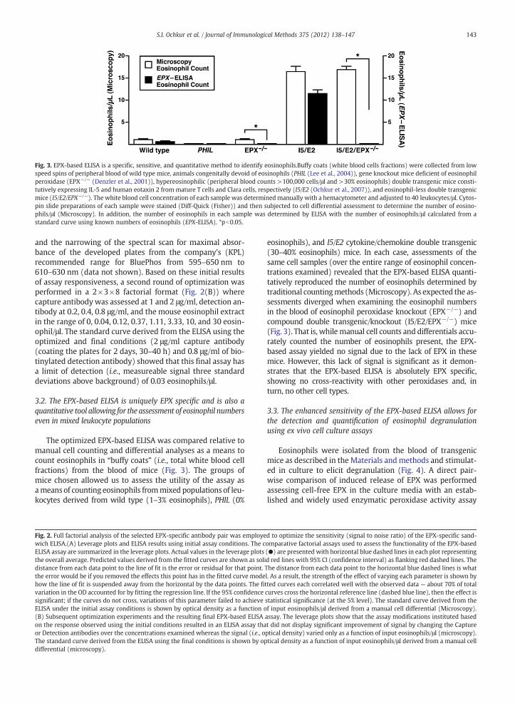
Fig. 3. EPX-based ELISA is a specific, sensitive, and quantitative method to identify eosinophils.Buffy coats (white blood cells fractions) were collected from lowspeed spins of peripheral blood of wild type mice, animals congenitally devoid of eosinophils (PHIL (Lee et al., 2004)), gene knockout mice deficient of eosinophilperoxidase (EPX−/− (Denzler et al., 2001)), hypereosinophilic (peripheral blood counts >100,000 cells/μl and >30% eosinophils) double transgenic mice consti-tutively expressing IL-5 and human eotaxin 2 from mature T cells and Clara cells, respectively (I5/E2 (Ochkur et al., 2007)), and eosinophil-less double transgenicmice (I5/E2/EPX−/−). The white blood cell concentration of each sample was determined manually with a hemacytometer and adjusted to 40 leukocytes/μl. Cytos-pin slide preparations of each sample were stained (Diff-Quick (Fisher)) and then subjected to cell differential assessment to determine the number of eosino-phils/μl (Microscopy). In addition, the number of eosinophils in each sample was determined by ELISA with the number of eosinophils/μl calculated from astandard curve using known numbers of eosinophils (EPX-ELISA). *pb0.05.
143S.I. Ochkur et al. / Journal of Immunological Methods 375 (2012) 138–147
and the narrowing of the spectral scan for maximal absor-bance of the developed plates from the company's (KPL)recommended range for BluePhos from 595–650 nm to610–630 nm (data not shown). Based on these initial resultsof assay responsiveness, a second round of optimization wasperformed in a 2×3×8 factorial format (Fig. 2(B)) wherecapture antibody was assessed at 1 and 2 μg/ml, detection an-tibody at 0.2, 0.4, 0.8 μg/ml, and the mouse eosinophil extractin the range of 0, 0.04, 0.12, 0.37, 1.11, 3.33, 10, and 30 eosin-ophil/μl. The standard curve derived from the ELISA using theoptimized and final conditions (2 μg/ml capture antibody(coating the plates for 2 days, 30–40 h) and 0.8 μg/ml of bio-tinylated detection antibody) showed that this final assay hasa limit of detection (i.e., measureable signal three standarddeviations above background) of 0.03 eosinophils/μl.
3.2. The EPX-based ELISA is uniquely EPX specific and is also aquantitative tool allowing for the assessment of eosinophil numberseven in mixed leukocyte populations
The optimized EPX-based ELISA was compared relative tomanual cell counting and differential analyses as a means tocount eosinophils in “buffy coats” (i.e., total white blood cellfractions) from the blood of mice (Fig. 3). The groups ofmice chosen allowed us to assess the utility of the assay asameans of counting eosinophils frommixed populations of leu-kocytes derived from wild type (1–3% eosinophils), PHIL (0%
Fig. 2. Full factorial analysis of the selected EPX-specific antibody pair was employewich ELISA.(A) Leverage plots and ELISA results using initial assay conditions. The cELISA assay are summarized in the leverage plots. Actual values in the leverage plotsthe overall average. Predicted values derived from the fitted curves are shown as solidistance from each data point to the line of fit is the error or residual for that point.the error would be if you removed the effects this point has in the fitted curve modehow the line of fit is suspended away from the horizontal by the data points. The fivariation in the OD accounted for by fitting the regression line. If the 95% confidencesignificant; if the curves do not cross, variations of this parameter failed to achieveELISA under the initial assay conditions is shown by optical density as a function o(B) Subsequent optimization experiments and the resulting final EPX-based ELISAon the response observed using the initial conditions resulted in an ELISA assay thaor Detection antibodies over the concentrations examined whereas the signal (i.e., oThe standard curve derived from the ELISA using the final conditions is shown by opdifferential (microscopy).
eosinophils), and I5/E2 cytokine/chemokine double transgenic(30–40% eosinophils) mice. In each case, assessments of thesame cell samples (over the entire range of eosinophil concen-trations examined) revealed that the EPX-based ELISA quanti-tatively reproduced the number of eosinophils determined bytraditional countingmethods (Microscopy). As expected the as-sessments diverged when examining the eosinophil numbersin the blood of eosinophil peroxidase knockout (EPX−/−) andcompound double transgenic/knockout (I5/E2/EPX−/−) mice(Fig. 3). That is, while manual cell counts and differentials accu-rately counted the number of eosinophils present, the EPX-based assay yielded no signal due to the lack of EPX in thesemice. However, this lack of signal is significant as it demon-strates that the EPX-based ELISA is absolutely EPX specific,showing no cross-reactivity with other peroxidases and, inturn, no other cell types.
3.3. The enhanced sensitivity of the EPX-based ELISA allows forthe detection and quantification of eosinophil degranulationusing ex vivo cell culture assays
Eosinophils were isolated from the blood of transgenicmice as described in the Materials andmethods and stimulat-ed in culture to elicit degranulation (Fig. 4). A direct pair-wise comparison of induced release of EPX was performedassessing cell-free EPX in the culture media with an estab-lished and widely used enzymatic peroxidase activity assay
d to optimize the sensitivity (signal to noise ratio) of the EPX-specific sand-omparative factorial assays used to assess the functionality of the EPX-based(●) are presented with horizontal blue dashed lines in each plot representingd red lines with 95% CI (confidence interval) as flanking red dashed lines. TheThe distance from each data point to the horizontal blue dashed lines is whatl. As a result, the strength of the effect of varying each parameter is shown bytted curves each correlated well with the observed data — about 70% of totalcurves cross the horizontal reference line (dashed blue line), then the effect isstatistical significance (at the 5% level). The standard curve derived from thef input eosinophils/μl derived from a manual cell differential (Microscopy).assay. The leverage plots show that the assay modifications instituted basedt did not display significant improvement of signal by changing the Captureptical density) varied only as a function of input eosinophils/μl (microscopy).tical density as a function of input eosinophils/μl derived from a manual cell
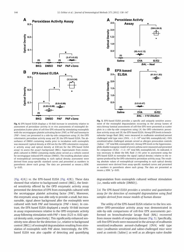
Fig. 4. EPX-based ELISA displays a 10-fold increase in sensitivity relative toassessment of peroxidase activity in ex vivo assessments of eosinophil de-granulation.Scatter plots of cell-free EPX released by stimulating eosinophilswith the secretagogues platelet activating factor (PAF) or PAF and Ionomycin(PAF+Iono) are presented in a side-by-side comparison using (A) the OPDcolorimetric peroxidase activity assay and (B) the EPX-based ELISA. The ab-sorbance of DMSO containing media prior to incubation with eosinophilswas measured (optical density at 450 nm for the OPD colorimetric enzymat-ic activity assay and optical density at 630 nm for the EPX-based ELISAassay) to assess the assay's background (BKG). Supernatants from eosino-phils cultured in DMSO containing media alone served as a vehicle controfor secretagogue-induced EPX release (DMSO). The resulting absolute valuesof eosinophils/μl corresponding to each optical density assessment werederived from assay-specific standard curves and presented as numbers inparenthesis above each group. The data are presented as means±SEM*pb0.05.
Fig. 5. EPX-based ELISA provides a specific and uniquely sensitive assess-ment of the eosinophil degranulation occurring in the airway lumen omice.Airway luminal assessments of cell-free EPX were presented as scatterplots in a side-by-side comparison using (A) the OPD colorimetric peroxi-dase activity assay and (B) the EPX-based ELISA. Airway EPX levels in bronch-oalveolar lavage fluid (BAL) were measured in ovalbumin sensitized/aerosochallenged wild type mice (OVA; ~1–2×10⁶ total BAL eosinophils/ml); OVAsensitized/saline challenged animals served as allergen provocation controls(Saline; b102 total BAL eosinophils/ml). Airway EPX levels in the hypereosino-philic double transgenicmodel of severe asthmaweremeasured and presentedfor comparison (I5/E2; ~1–2×10⁷ total BAL eosinophils/ml). As indicated, iwas necessary to dilute the BAL fluid (1:10) prior to assessment using theEPX-based ELISA to normalize the signal (optical density) relative to the re-sponse produced by the OPD colorimetric peroxidase activity assay. The result-ing absolute values of eosinophils/μl corresponding to each optical densityassessment were derived from assay-specific standard curves and presentedas numbers in parenthesis above each group. The data are presented as
144 S.I. Ochkur et al. / Journal of Immunological Methods 375 (2012) 138–147
l
.
means±SEM. *pb0.05.
(Fig. 4(A)) vs. the EPX-based ELISA (Fig. 4(B)). These datashowed that relative to background control (BKG), the limit-ed sensitivity offered by the OPD enzymatic activity assayprevented the detection of EPX from eosinophils cultured withthe secretagogue platelet activating factor (PAF). Moreover,this enzymatic assay was able only to provide a small, but mea-sureable, signal above background after the eosinophils werecultured with both PAF and Ionomycin (PAF+Iono). In con-trast, the EPX-based ELISA displayed a nearly 10-fold increasein assay responsiveness relative to the OPD enzymatic activityassay following stimulation with PAF+Iono (0.23 vs. 0.02 opti-cal density units, respectively). This significantly enhanced sen-sitivity now allows for the detection (i.e., relative to backgroundcontrol (BKG)) of eosinophil degranulation resulting from stim-ulation of eosinophils with PAF alone. Interestingly, the EPX-based ELISA was also capable of detecting and quantifying
f
l
t
degranulation from eosinophils cultured without stimulation(i.e., media with vehicle (DMSO)).
3.4. The EPX-based ELISA provides a sensitive and quantitativeassay for the detection of eosinophil degranulation using fluidsamples derived from mouse models of human disease
The utility of the EPX-based ELISA relative to the less sen-sitive OPD-peroxidase activity assay was demonstrated inside-by-side comparisons of cell-free EPX assessments per-formed on bronchoalveolar lavage fluid (BAL) recoveredfrom mouse models of respiratory disease (Fig. 5). Specifically,cell-free EPX levelsweremeasured in the airways of ovalbuminsensitized/ovalbumin aerosol-challenged (OVA) wild typemice (ovalbumin sensitized and saline-challenged mice wereused as controls (Saline)) as well as an allergen naïve double

145S.I. Ochkur et al. / Journal of Immunological Methods 375 (2012) 138–147
transgenicmodel of severe asthma (I5/E2 (Ochkur et al., 2007)).These data showed that while both the OPD-peroxidase enzy-matic assay (Fig. 5(A)) and the EPX-based ELISA (Fig. 5(B))were capable of quantifying the release of EPX in the airwaysof OVA aero-challenged mice (OVA), the EPX-based ELISAagain displayed a 10-fold increase in sensitivity, displaying thesame signal (optical density units) with BAL fluid diluted 1 to10. This same signal enhancement was observed at the higherrange of the scale with the EPX-based ELISA again displaying a10-fold enhanced detection of cell-free EPX in the BAL fromthe double transgenic model of severe asthma (I5/E2).
3.5. The sensitive and quantitative characters of the EPX-ELISAallow for the assessments of even low levels of EPX in serumderived from peripheral blood of unmanipulated wild type mice
The EPX-based ELISA assessments of ex vivo cultured eo-sinophils described above suggested that even “resting”blood eosinophils displayed a detectable level of continualrelease of EPX. As a result, the EPX-based ELISA was used todetermine whether the blood sera of unmanipulated wildtype mice with 1–3% eosinophils (~8000 leukocytes/mm3 ofblood) also displayed low levels of released EPX (Fig. 6).These data showed that as expected, knockout mice devoidof eosinophil peroxidase (I5/E2/EPX−/−) displayed no evi-dence of circulating serum EPX. In contrast, the ELISA assaywas easily capable of detecting significant levels of cell-freeEPX in the sera of the double transgenic severe asthma mice(I5/E2). More significantly, low levels of cell-free EPX werealso detected in the sera of unmanipulated wild type mice.
4. Discussion
The creation, and now availability, of an EPX-based ELISA inthe mouse to detect and quantify eosinophil degranulationprovides logistical solutions to issues surrounding studies ofthe role(s) of eosinophils in this animal. These issues include
Fig. 6. EPX-based ELISA of serum provides a sensitive and quantitative ability to assesof serum samples (using saline) from wild type, hypereosinophilic double transgenknockout (I5/E2/EPX−/−) mice were assessed for eosinophil peroxidase using the Elution is plotted together with the resulting absolute values of Eosinophil-Equivalentstandard curves.
technical/strategic advances as well as scientific observationsrelevant to our understanding of mouse eosinophil effectorfunction(s):
4.1 Technical/strategic advances:
4.1.1 The previously available assays assessing eosino-phil degranulation are based on enzymatic activi-ties (i.e., peroxidase or ribonuclease activities)that are not absolutely eosinophil-specific. In con-trast, the EPX-based ELISA is an assay that is inde-pendent of enzymatic activity (even the lackthereof). Moreover, EPX-based ELISA assessmentsfrom wild type vs. EPX−/− mice demonstratedthat the assay is target protein specific.
4.1.2 The ELISA developed is an easy to use protocoltaking advantage of commonly available disposableplasticware and reagents. In addition, this ELISAwas shown to function as a high throughput assayexecutable in micro-titer plate venues.
4.1.3 The utility of the assay is highlighted by the increasedsensitivity it affords relative to established assays thatuniversally detect cell-free EPX by assessing the per-oxidase activity associated with this secondary gran-ule protein (more often than not as a colorimetricenzymatic assay). Direct comparisons showed thatthe EPX-based ELISA is 10-fold more sensitive thanmeasurements made via these peroxidase enzymaticassays. This increased sensitivity promotes a lowerthreshold of detection in a given sample and, inturn, the quantification of this granule protein evenin small and/or dilute samples. An additional contrib-uting factor to both the utility and sensitivity of theEPX-based ELISA is that unlike the peroxidase enzy-matic assays, the ELISA doesn't require the presenceof intact and enzymatically active protein. That is,even partially degraded samples with limited peroxi-dase activity nonetheless may contain epitope-
s levels of cell-free eosinophil peroxidase in the blood of mice.Serial dilutionsic (I5/E2), and eosinophil peroxidase deficient compound double transgenic-PX-based ELISA. Optical density derived from assessments at each serum di-s/μl corresponding to optical density assessments derived from assay-specific

146 S.I. Ochkur et al. / Journal of Immunological Methods 375 (2012) 138–147
bearing fragments that are sufficient for the detec-tion/quantification of EPX by the ELISA.
4.2 Scientific observations relevant to mouse eosinophileffector function(s):
4.2.1 Although from a technical standpoint we haveshown that the EPX-based ELISA is target-protein(i.e., EPX) specific, the question of whether EPX it-self is absolutely eosinophil-specific is still unre-solved. The data presented, however, addressesthis issue. EPX-based ELISA of extracts derivedfrom peripheral white blood cell fractions of wildtype vs. eosinophil-less PHIL mice showed that inthe absence of eosinophils EPX was not detectablefrom the remaining white blood cells. These datasuggest that eosinophils are the only significantsource of EPX among the various peripheral leuko-cyte subtypes in circulation and thus represent avalid target of an eosinophil-specific assay.
4.2.2 Relative to human samples, eosinophil degranula-tion frommouse sources has been difficult to assessbecause of the limited capacity ofmouse eosinophilsto execute this effector function (Stelts et al., 1998;Persson and Erjefalt, 1999; Malm-Erjefält et al.,2001). However, the 10-fold enhancement in sensi-tivity of the EPX-based ELISA vs. the established en-zymatic activity assay for EPX demonstrated thatwhile low on an absolute scale, EPX release bymouse eosinophils in ex vivo assays of degranulationwas measureable and could be quantified. Signifi-cantly, these ex vivo data showed that even in theabsence of stimuli to promote degranulation, “rest-ing” peripheral blood eosinophils were nonethelesssources of released EPX. A similar observation wasnoted in vivo where even otherwise healthy unma-nipulatedwild type animals displayed a low, but de-tectable, level of serumEPX. Although this release ofEPX in both the ex vivo and in vivo studies may haveoccurred as a consequence of sample preparation,these results nonetheless suggest that the releaseof EPX, and presumably the other granule proteins,is not restricted to responses directly linked to oneor more specific stimuli. Instead, peripheral bloodeosinophils appear to undergo a continual piece-meal release of EPX while in circulation; the func-tionality, if any, of this steady-state circulating cell-free EPX remains unknown.
4.2.3 The apparent inability of mouse eosinophils to un-dergo significant degranulation relative to theirhuman counterparts has been a frustrating aspectof working with mouse models of disease, particu-larly mouse models of asthma where eosinophilgranule protein release is an easily measureableevent in the airways of patients (see for exampleFilley et al., 1982; Erjefält et al., 1999). The in-creased sensitivity of EPX-based ELISA, togetherwith the eosinophil specificity of the assay, pre-sents a solution to this issue. Specifically, our datashowed that like human subjects eosinophil de-granulation in BAL fluid from aero-allergen chal-lenged mice is now an easily measureableendpoint assessment that is also quantifiable.
The significance of this EPX-based ELISA will extend be-yond studies of mouse models of human disease. Sequencecomparisons of the mouse EPX gene with its human ortholo-gue demonstrated that the encoded mammalian peroxidasesderived from these two genes are >86% identical at theamino acid level (Horton et al., 1996). Thus, when the issueof cross-reactivity was examined, the mouse anti-mouseEPX monoclonal antibodies used to develop this EPX-basedELISA recognized epitopes in mouse EPX that are identicalto epitopes in human EPX. This cross-reactivity has allowedus to develop an equally specific, sensitive, and quantitativeELISA recognizing human EPX. The availability of this assayhas permitted direct side-by-side comparisons of mouse vs.human degranulation as well as the assessment of biologicalfluid samples from patients as a diagnostic tool in diseasemanagement (manuscript in preparation).
Acknowledgments
The authors wish to thank the members of Lee Laboratoriesas well as colleagues within the greater eosinophil communityfor insightful discussions and critical comments that directlyled to the development of the EPX-based ELISA and, in turn,the preparation of this manuscript. We also wish to acknowl-edge the invaluable assistance of the Mayo Clinic Arizona Sta-tistical support group (Amylou Dueck, PhD, Yu-Hui J. Chang,and Joseph Hentz), our staff medical graphic artist (MarvRuona), and the excellent administrative support provided toLee Laboratories by Linda Mardel and Shirley (“Charlie”)Kern. The Mayo Foundation and grants from the United StatesNational Institutes of Health [NAL (HL058723) and JJL(HL065228, RR0109709)], the American Heart Association[NAL (05556392) and JJL (0855703)], the Canadian Institutesof Health Research [RM (MOP89748)], and the Lung Associa-tion of Alberta [JDK] were the sources of funding used in theperformance of studies as well as data analysis. These fundingsources had no involvement in study design, data collection(including analysis and interpretation), thewriting of theman-uscript, or the decision to submit for publication.
References
Behm, C.A., Ovington, K.S., 2000. The role of eosinophils in parasitic helminthinfections: insights from genetically modified mice. Parasitol. Today 16,202–209.
Borchers, M.T., Ansay, T.L., DeSalle, R., Daugherty, B.L., Shen, H.H., Metzger,M., Lee, N.A., Lee, J.J., 2002. In vitro assessment of chemokine receptor-ligand interactions mediating mouse eosinophil migration. J. Leukoc.Biol. 71, 1033–1041.
Cormier, S.A., Taranova, A.G., Bedient, C., Nguyen, T., Protheroe, C., Pero, R.,Dimina, D., Ochkur, S.I., O'Neill, K., Colbert, D., Lombari, T.R., Constant,S., McGarry, M.P., Lee, J.J., Lee, N.A., 2006. Pivotal advance: eosinophilinfiltration of solid tumors is an early and persistent inflammatory hostresponse. J. Leukoc. Biol. 79, 1131–1139.
Correale, J., Fiol, M., 2004. Activation of humoral immunity and eosinophilsin neuromyelitis optica. Neurology 63, 2363–2370.
Denzler, K.L., Borchers, M.T., Crosby, J.R., Cieslewicz, G., Hines, E.M., Justice,J.P., Cormier, S.A., Lindenberger, K.A., Song, W., Wu, W., Hazen, S.L.,Gleich, G.J., Lee, J.J., Lee, N.A., 2001. Extensive eosinophil degranulationand peroxidase-mediated oxidation of airway proteins do not occurin a mouse ovalbumin-challenge model of pulmonary inflammation.J. Immunol. 167, 1672–1682.
Erjefält, J.S., Greiff, L., Andersson, M., Matsson, E., Petersen, H., Linden, M.,Ansari, T., Jeffery, P.K., Persson, C.G., 1999. Allergen-induced eosinophilcytolysis is a primary mechanism for granule protein release in humanupper airways. Am. J. Respir. Crit. Care Med. 160, 304–312.

147S.I. Ochkur et al. / Journal of Immunological Methods 375 (2012) 138–147
Fabre, V., Beiting, D.P., Bliss, S.K., Gebreselassie, N.G., Gagliardo, L.F., Lee, N.A.,Lee, J.J., Appleton, J.A., 2009. Eosinophil deficiency compromises parasitesurvival in chronic nematode infection. J. Immunol. 182, 1577–1583.
Filley, W.V., Kephardt, G.M., Holley, L.E., Gleich, G.J., 1982. Identification byimmunofluorescence of eosinophil granule major basic protein in lungtissues of patients with bronchial asthma. Lancet ii, 11–16.
Gladue, R.P., Carroll, L.A., Milici, A.J., Scampoli, D.N., Stukenbrok, H.A., Pettipher,E.R., Salter, E.D., Contillo, L., Showell, H.J., 1996. Inhibition of leukotriene B4-receptor interaction suppresses eosinophil infiltration anddisease pathologyin a murine model of experimental allergic encephalomyelitis. J. Exp. Med.183, 1893–1898.
Goldman, M., Le Moine, A., Braun, M., Flamand, V., Abramowicz, D., 2001. Arole for eosinophils in transplant rejection. Trends Immunol. 22,247–251.
Herz, U., Bunikowski, R., Renz, H., 1998. Role of T cells in atopic dermatitis.new aspects on the dynamics of cytokine production and the contributionof bacterial superantigens. Int. Arch. Allergy Immunol. 115, 179–190.
Horton,M.A., Larson, K.A., Lee, J.J., Lee, N.A., 1996. Cloning of themurine eosinophilperoxidase gene (mEPO): characterization of a conserved subgroup ofmammalian hematopoietic peroxidases. J. Leukoc. Biol. 60, 285–294.
Jang, A.S., Choi, I.S., 2000. Eosinophil activation markers in induced sputumin asthmatics. Korean J. Intern. Med. 15, 1–7.
Kips, J.C., Anderson, G.P., Fredberg, J.J., Herz, U., Inman, M.D., Jordana, M.,Kemeny, D.M., Lotvall, J., Pauwels, R.A., Plopper, C.G., Schmidt, D., Sterk,P.J., Van Oosterhout, A.J., Vargaftig, B.B., Chung, K.F., 2003. Murinemodels of asthma. Eur. Respir. J. 22, 374–382.
Klion, A.D., Nutman, T.B., 2004. The role of eosinophils in host defenseagainst helminth parasites. J. Allergy Clin. Immunol. 113, 30–37.
Kobayashi, T., Iijima, K., Radhakrishnan, S., Mehta, V., Vassallo, R., Lawrence,C.B., Cyong, J.C., Pease, L.R., Oguchi, K., Kita, H., 2009. Asthma-relatedenvironmental fungus, Alternaria, activates dendritic cells and producespotent Th2 adjuvant activity. J. Immunol. 182, 2502–2510.
Lacoste, J.Y., Bousquet, J., Chanez, P., Van Vyve, T., Simony-Lafontaine, J.,Lequeu, N., Vic, P., Enander, I., Godard, P., Michel, F.B., 1993. Eosinophilicand neutrophilic inflammation in asthma, chronic bronchitis, and chronicobstructive pulmonary disease. J. Allergy Clin. Immunol. 92, 537–548.
Le Moine, A., Surquin, M., Demoor, F.X., Noel, J.C., Nahori, M.A., Pretolani, M.,Flamand, V., Braun,M.Y., Goldman,M., Abramowicz, D., 1999. IL-5mediateseosinophilic rejection of MHC class II-disparate skin allografts in mice.J. Immunol. 163, 3778–3784.
Lee, N.A., McGarry, M.P., Larson, K.A., Horton, M.A., Kristensen, A.B., Lee, J.J.,1997. Expression of IL-5 in thymocytes/T cells leads to the developmentof a massive eosinophilia, extramedullary eosinophilopoiesis, andunique histopathologies. J. Immunol. 158, 1332–1344.
Lee, J.J., Dimina, D., Macias, M.P., Ochkur, S.I., McGarry, M.P., O'Neill, K.R.,Protheroe, C., Pero, R., Nguyen, T., Cormier, S.A., Lenkiewicz, E., Colbert,D., Rinaldi, L., Ackerman, S.J., Irvin, C.G., Lee, N.A., 2004. Defining a linkwith asthma in mice congenitally deficient in eosinophils. Science 305,1773–1776.
(needs correction!)Leiferman, K.M., 1989. Eosinophils in atopic dermatitis.Allergy 9, 20–26.
Malm-Erjefält, M., Persson, C.G.A., Erjefalt, J.S., 2001. Degranulation status ofairway tissue eosinophils inmousemodels of allergic airway inflammation.Am. J. Respir. Cell Mol. Biol. 24, 352–359.
Mishra, A., Hogan, S.P., Brandt, E.B., Rothenberg, M.E., 2001. An etiologicalrole for aeroallergens and eosinophils in experimental esophagitis.J. Clin. Invest. 107, 83–90.
Mould, A.W., Ramsay, A.J., Matthaei, K.I., Young, I.G., Rothenberg, M.E., Foster,P.S., 2000. The effect of IL-5 and eotaxin expression in the lung oneosinophil trafficking and degranulation and the induction of bronchialhyperreactivity. J. Immunol. 164, 2142–2150.
Ochkur, S.I., Jacobsen, E.A., Protheroe, C.A., Biechele, T.L., Pero, R.S., McGarry,M.P., Wang, H., O'Neill, K.R., Colbert, D.C., Colby, T.V., Shen, H., Blackburn,M.R., Irvin, C.C., Lee, J.J., Lee, N.A., 2007. Co-expression of IL-5 andeotaxin-2 in mice creates an eosinophil-dependent model of respiratoryinflammation with characteristics of severe asthma. J. Immunol. 178,7879–7889.
Persson, C.G., Erjefalt, J.S., 1999. Degranulation in eosinophils in human, butnot in mouse, airways. Allergy 54, 1230–1232.
Protheroe, C.A., Woodruff, S.A., DePetris, G., Mukkada, V., Ochkur, S.I.,Janarthanan, S., Lewis, J.C., Pasha, S., Lunsford, T., Harris, L., Sharma, V.K.,McGarry, M.P., Lee, N.A., Furuta, G.T., Lee, J.J., 2009. A novel histologicalscoring system to evaluate mucosal biopsies from patients with eosin-ophilic esophagitis. Clin. Gastroenterol. Hepatol. 7, 749–755.
Rothenberg, M.E., Mishra, A., Collins, M.H., Putnam, P.E., 2001. Pathogenesisand clinical features of eosinophilic esophagitis. J. Allergy Clin. Immunol.108, 891–894.
Samoszuk, M., 1997. Eosinophils and human cancer. Histol. Histopathol. 12,807–812.
Schubert, M.S., 2006. Allergic fungal sinusitis. Clin. Rev. Allergy Immunol. 30,205–216.
Stelts, D., Egan, R.W., Falcone, A., Garlisi, C.G., Gleich, G.J., Kreutner, W., Kung,T.T., Nahrebne, D.K., Chapman, R.W., Minnicozzi, M., 1998. Eosinophilsretain their granule major basic protein in a murine model of allergicpulmonary inflammation. Am. J. Respir. Cell Mol. Biol. 18, 9463–9470.
Strath, M., Warren, D.J., Sanderson, C.J., 1985. Detection of eosinophils using aneosinophil peroxidase assay. Its use as an assay for eosinophil differentiationfactors. J. Immunol. Methods 83, 209–215.
Recommended


