Embed Size (px)
Citation preview
A Carbohydrate-Derived Splice ModulatorSachin Dhar,† James J. La Clair,*,† Brian Leon,† Justin C. Hammons,† Zhe Yu,‡ Manoj K. Kashyap,‡
Januario E. Castro,‡ and Michael D. Burkart*,†
†Department of Chemistry and Biochemistry, University of California−San Diego, 9500 Gilman Drive, La Jolla, California92093-0358, United States‡Moores Cancer Center, University of California−San Diego, La Jolla, California 92093-0358, United States
*S Supporting Information
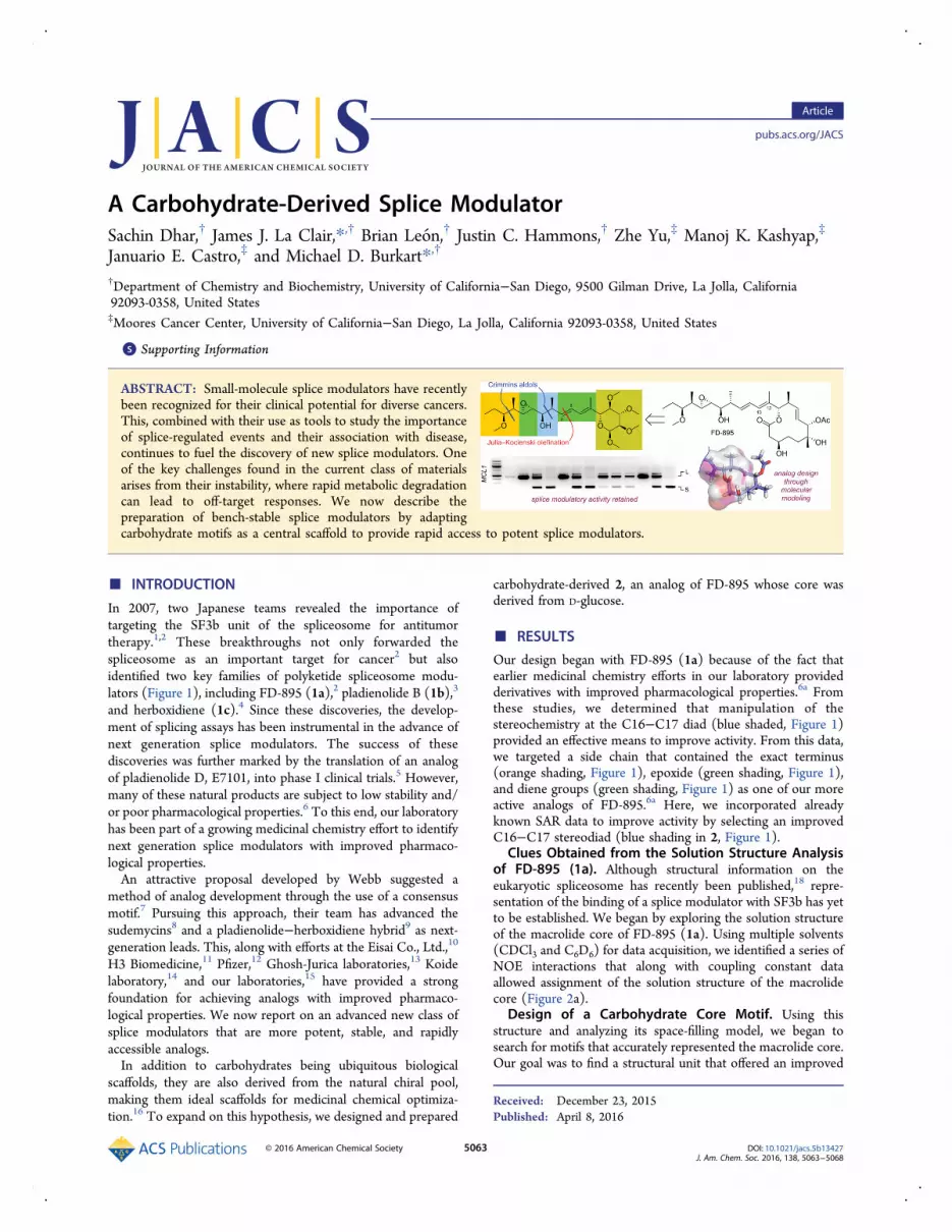
ABSTRACT: Small-molecule splice modulators have recentlybeen recognized for their clinical potential for diverse cancers.This, combined with their use as tools to study the importanceof splice-regulated events and their association with disease,continues to fuel the discovery of new splice modulators. Oneof the key challenges found in the current class of materialsarises from their instability, where rapid metabolic degradationcan lead to off-target responses. We now describe thepreparation of bench-stable splice modulators by adaptingcarbohydrate motifs as a central scaffold to provide rapid access to potent splice modulators.
■ INTRODUCTION
In 2007, two Japanese teams revealed the importance oftargeting the SF3b unit of the spliceosome for antitumortherapy.1,2 These breakthroughs not only forwarded thespliceosome as an important target for cancer2 but alsoidentified two key families of polyketide spliceosome modu-lators (Figure 1), including FD-895 (1a),2 pladienolide B (1b),3
and herboxidiene (1c).4 Since these discoveries, the develop-ment of splicing assays has been instrumental in the advance ofnext generation splice modulators. The success of thesediscoveries was further marked by the translation of an analogof pladienolide D, E7101, into phase I clinical trials.5 However,many of these natural products are subject to low stability and/or poor pharmacological properties.6 To this end, our laboratoryhas been part of a growing medicinal chemistry effort to identifynext generation splice modulators with improved pharmaco-logical properties.An attractive proposal developed by Webb suggested a
method of analog development through the use of a consensusmotif.7 Pursuing this approach, their team has advanced thesudemycins8 and a pladienolide−herboxidiene hybrid9 as next-generation leads. This, along with efforts at the Eisai Co., Ltd.,10
H3 Biomedicine,11 Pfizer,12 Ghosh-Jurica laboratories,13 Koidelaboratory,14 and our laboratories,15 have provided a strongfoundation for achieving analogs with improved pharmaco-logical properties. We now report on an advanced new class ofsplice modulators that are more potent, stable, and rapidlyaccessible analogs.In addition to carbohydrates being ubiquitous biological
scaffolds, they are also derived from the natural chiral pool,making them ideal scaffolds for medicinal chemical optimiza-tion.16 To expand on this hypothesis, we designed and prepared
carbohydrate-derived 2, an analog of FD-895 whose core wasderived from D-glucose.
■ RESULTSOur design began with FD-895 (1a) because of the fact thatearlier medicinal chemistry efforts in our laboratory providedderivatives with improved pharmacological properties.6a Fromthese studies, we determined that manipulation of thestereochemistry at the C16−C17 diad (blue shaded, Figure 1)provided an effective means to improve activity. From this data,we targeted a side chain that contained the exact terminus(orange shading, Figure 1), epoxide (green shading, Figure 1),and diene groups (green shading, Figure 1) as one of our moreactive analogs of FD-895.6a Here, we incorporated alreadyknown SAR data to improve activity by selecting an improvedC16−C17 stereodiad (blue shading in 2, Figure 1).
Clues Obtained from the Solution Structure Analysisof FD-895 (1a). Although structural information on theeukaryotic spliceosome has recently been published,18 repre-sentation of the binding of a splice modulator with SF3b has yetto be established. We began by exploring the solution structureof the macrolide core of FD-895 (1a). Using multiple solvents(CDCl3 and C6D6) for data acquisition, we identified a series ofNOE interactions that along with coupling constant dataallowed assignment of the solution structure of the macrolidecore (Figure 2a).
Design of a Carbohydrate Core Motif. Using thisstructure and analyzing its space-filling model, we began tosearch for motifs that accurately represented the macrolide core.Our goal was to find a structural unit that offered an improved
Received: December 23, 2015Published: April 8, 2016
Article
pubs.acs.org/JACS
© 2016 American Chemical Society 5063 DOI: 10.1021/jacs.5b13427J. Am. Chem. Soc. 2016, 138, 5063−5068

synthesis entry and diverse means of tailoring while alsoaddressing previous pharmacological issues (stability andsolubility). We began by inspecting the overlap between thesmall herboxidiene (1c) and large FD-895 (1a) cores. Althougha clear overlay existed (Figure 1b,c), it was evident that spaceoccupied by the C4−C7 unit within 1a may not be arequirement for activity. We soon realized that simplemonosaccharides such as D-glucose could provide a strongoverlap with these cores. After evaluating several structures, weturned our attention to explore α-methyl-2,3,4-trimethoxy-D-glucopyranoside (3) as a core mimetic.19
Structurally, glycoside 2 offered a functional map that laybetween the core sizes of 1a and 1c (Figure 2). As illustrated inFigure 2d,e, the C2−C4 methoxy groups in 2 provided a fitapproximating 1a relative to that of herboxidiene (Figure 2b,c).Specifically, the C2 methoxy group of 2 filled the space taken bythe C24 methyl group in 1a. Likewise, the C3 methoxy of 2 wasable to partially fill the space of the C29−C30 acetate, a regionthat was already shown to tolerate functional modifications.20
With this support, we turned our attention to synthesize andbiologically evaluate analog 2.Synthetic Strategy. Our plan focused on using a late-stage
Julia−Kocienski olefination to couple a side chain derived fromFD-895 to the monosaccharide core (see Abstract graphic).Here, we envisioned that we could leverage the extensive effortsfrom our own laboratory for development of pladienolide/FD-895 side chain6a,15b as a means to probe the structure−activityrelationships (SARs) of side-chain-incorporating analogs. Wetherefore set out toward preparation of core component 8(Scheme 1) and side-chain component 16 (Scheme 2).Synthesis of a Carbohydrate Core Component 8.
Synthesis of 8 was accomplished in five steps from 3, amonosaccharide derivative that in turn was prepared in three
Figure 1. Structures of FD-895 (1a), pladienolide B (1b), herboxidiene(1c), and carbohydrate-derived splicing modulator 2. Compound 2contains the terminus (orange) of 1a, the epoxide and diene of 1a(green), and a core derived from 1c (gold).
Figure 2. Models of FD-895 (1a, blue), herboxidiene (1c, magenta),and carbohydrate-derived analog 2 (green). (a) Derived solutionstructure of the FD-895 core motif as determined by evaluation of 1Hcoupling constants and NOE interactions. NOE interactions wereobserved both on the top face (red) and bottom face (blue). Datashown was collected from 1H,1H NOESY spectral data of 1a in CDCl3and C6D6 at 23 °C. (b) Model depicting the structure of 1a (blue)superimposed on the surface of 1c (magenta). (c) Model depicting thestructure of 1c (magenta) superimposed on the surface of 1a (blue).(d) Models depicting the structure of 1a (blue) superimposed on thesurface of 2 (green). (e) Model depicting the structure of 2 (green)superimposed on the surface of 1a (blue). The structure of the coremotif in 1a was determined via NMR spectroscopy. Structures of 1band 1c were modeled by energy minimization.17
Scheme 1. Synthesis of Core Component 8
Journal of the American Chemical Society Article
DOI: 10.1021/jacs.5b13427J. Am. Chem. Soc. 2016, 138, 5063−5068
5064

steps (54% yield) from α-methyl-D-glucopyranoside.19 Webegan by oxidizing to aldehyde 4. Advantageously, we wereable to reduce chromatographic purification efforts by using acombination of an IBX oxidation followed by solvent-dependentfiltration through a pad of SiO2. The addition of MeLi to crude 4occurred with minimal byproduct formation, providing alcohol5 in 82% yield from 3.Repetition of the IBX oxidation procedure provided ketone 6,
which could be rapidly purified by passage through a SiO2 plug.Although small samples of 6 were purified for characterization,the crude product was readily subjected to a Horner−Wadsworth−Emmons olefination to afford ester 7. We wereable to convert 5 to 7 in two steps and 80% yield with a singlepurification. Finally, ester 7 was reduced with DIBAL-H toafford core unit 8, which could be stored for months at −20 °Cwithout decomposition. Overall, this process provides access to8 in 66% in five steps from 3 (36% in 8 steps from α-methyl-D-glucopyranoside).Synthesis of the Side Chain Component 16. The
synthesis of the side chain component began with allylic alcohol9, an intermediate that was prepared at >10 g scale for thesynthesis of FD-895 (1a).6a Allylic oxidation with MnO2provided aldehyde 10, which was purified through a SiO2 plugand directly submitted to a Crimmins aldol reaction withauxiliary 11, to afford adduct 12 in 87% yield over two steps.NMR analyses on the crude product indicated that this reactionoccurred without detectable formation of other isomericproducts.With 12 in hand, the carbinol was protected as TBS ether 13,
and the auxiliary was removed by NaBH4 reduction.Optimization studies indicated that a 5:1 ratio of THF:CH3OHprovided a reproducibly high yield of 14. Completion of thiscomponent was accomplished by a two-step installation of the 1-phenyl-1H-tetrazol-5-yl (PT) sulfone. Although Mitsunobuconditions were effective at producing sulfone 15 in 82% yieldat a gram scale, oxidation often provided significant quantities ofthe corresponding sulfoxides (incomplete oxidation byprod-ucts). Typically, samples of these sulfoxides were collected afterchromatographic purification and resubmitted to the oxidationconditions to afford a combined yield of 89% of component 16.To date, we have completed the synthesis of gram quantities of
16 in 6 steps and 58% overall yield from 9 and 12 steps and 21%overall yield from auxiliary 11.6a
Component Assembly and Completion of the Syn-thesis of 2. With components 8 and 16 in hand, thecomponent coupling began by oxidizing allylic alcohol 8 tothe corresponding aldehyde 17. Purification via dry columnvacuum chromatography using Geduran 60 silica gel wassufficient to afford pure 17 for coupling. After initial screening,we found the optimum yield (68%) of 18 was obtained whenapplying 1.05 equiv NaHMDS to 1.0 equiv of sulfone 8 andfollowing this by addition of 1.1 equiv of aldehyde 17. The yieldof this reaction also included recovery of 8% of sulfone 16 and11% of the chromatographically separable cis isomer of 18(structure not shown). With samples of pure 18 on hand,deprotection with TBAF afforded alcohol 19 (Scheme 3).
Next, we turned to the stereoselective VO(acac)2-catalyzedepoxidation of allylic alcohols21 to install the C12−C13 epoxide.As shown in Figure 3a, treatment of 19 with 4.5 equiv oftBuOOH and 3 equiv of VO(acac)2 at −20 °C for 6 h providedthe desired stereoisomer with ∼4:1 diastereoselectivity.Although this was the major product, over-warming or notstarting the reaction at −78 °C resulted in epoxidation at C8−C9. This product was not as stable because the C11 carbinol wascapable of attacking the C8 center of the epoxide, forming thecorresponding furan analog (structure not shown). Althoughadditional optimization efforts will be required, the currentconditions returned 2 in 52% yield with only traces of theundesired C8−C9 epoxidation (5−10% yield).
Validation of the Structural Assignment of 2. Althoughthe mechanism of the VO(acac)2/tBuOOH epoxidation
Scheme 2. Synthesis of Side Chain Component 16
Scheme 3. Component Assembly
Journal of the American Chemical Society Article
DOI: 10.1021/jacs.5b13427J. Am. Chem. Soc. 2016, 138, 5063−5068
5065

suggests delivery of the correct stereochemical outcome, wewanted to gain further confirmation that the product was indeed2. We began by exploring other epoxidation conditions toprepare samples of the alternate isomer. Treatment of 19 with1.2 equiv of m-CPBA at −78 °C in NaHCO3-buffered CH2Cl2followed by slow warming to room temperature afforded a ∼1:1mixture of both epoxides (Figure 3b) in 62% yield. Afterchromatographic purification, we were able to obtain samples ofboth isomers.Comparative NMR analyses on the epoxides from VO-
(acac)2/tBuOOH and m-CPBA provided a solution (Figure 3c).Using 2D NMR data set (Supporting Information), we wereable to fully assign the proton assignments in this product. Weidentified that the chemical shift and coupling patterns of theepoxide protons at C12−C13 provided the closest match to theconfiguration of corresponding C16−C17 isomer 1e (Figure 4),with comparable chemical shifts and coupling constants.Stability and Solubility of 2. Early studies indicated that 2
was more soluble and stable in aqueous buffers than FD-895
(1a). Using capillary NMR and solvent 13C satellites (QSCS) asa means of quantitation,22 we found that 2 was soluble up to 95± 5 mM D2O/DMSO-d6 (10:1) at 23 °C, whereas FD-895 (1a)was limited to 15 ± 5 mM under the same conditions.Additionally, we used NMR monitoring to show that although1a had a half-life of ∼70 h in D2O/DMSO-d6 (10:1) at 37 °C,
6a
carbohydrate analog 2 showed no signs of decomposition evenafter 744 h (1 month) under the same conditions. Althoughdetailed partitioning, permeability, and metabolic stabilityanalyses are ongoing, this initial evidence suggests that 2 offersthe potential to improve pharmacological properties of this classof splice modulator.
Epoxide 2 Displays Potent Activity in Primary CLL-BCells in Contrast to Its Precursor Alkene 19. We thenscreened the activity of 19 and 2 in primary CLL-B cellsobtained from two CLL patients that displayed potent in vitrocytotoxic response (Figure 1), measured in the form of specificinduced apoptosis (% SI), to 1a (IC50 value of 54.1 ± 7.5 nM)and 1b (IC50 value of 84.4 ± 1.2 nM). Interestingly, thesestudies indicated that alkene 19 did not display any activity (IC50value >50 μM in both sets of cells). Epoxide 2, however, wasactive (IC50 value of 153.0 ± 11.8 nM) with ∼3-fold loss ofactivity when compared to FD-895 (1a).
Compound 2Modulates Splicing.We then turned to RT-PCR analysis to determine if the activity of 2 also included asplice response comparable to 1a and 1b in CLL-B cells. Usingunspliced RNU6A and unmodulated GAPDH (top, Figure 5) ascontrols, we were able to obtain complementary splicemodulation in all six genes selected from our prior studies toprovide a diversity of intron retention (IR) response.15a Thisincluded comparable modification of IR in DNAJB1, ARF4,PRPF4, RIOK3, SF3A1, and U2AF2 (Figure 4 and S2) fromprimary CLL-B cells. In contrast, alkene precursor 19 wasinactive for all six genes (Figure 5), indicating that the epoxide in2 is essential for activity.Additionally, we were able to confirm that compound 2 also
induced alternate splicing (AS) events. As shown in Figure 6,
Figure 3. Stereoselectivity of the C12−C13 epoxidation. (a) ExpandedLC-MS trace via UV monitoring at 288 nm depicting the reactionproduct from treatment of 19 with VO(acac)2/tBuOOH (blue). (b)Comparable LC-MS trace depicting the formation of two isomers bym-CBPA epoxidation (red). (c) Expansion of the 1H NMR spectra ofepoxide 2 from VO(acac)2/tBuOOH in CDCl3 (blue). (d) Expansionof the 1H NMR spectra of epoxide 20 in CDCl3 from m-CBPA (red).Traces of 2 denoted by a blue asterisk could not be completely removedfrom 20 because of close retention times.
Figure 4. Coupling constant analyses. NMR analysis in C6D6 provideda coupling pattern at C12−C13 in 2 (blue) that matched that from thecorresponding FD-895 analog 1e (green) but not analogs 1a, 1f, or 1g(black).6a,15b
Journal of the American Chemical Society Article
DOI: 10.1021/jacs.5b13427J. Am. Chem. Soc. 2016, 138, 5063−5068
5066

treatment of CLL-B cells with 1a, 1b, and 2 resulted in adecrease in the long (anti-apoptotic) form and increased theexpression of short (pro-apoptotic) form of MCL1. Similarly,BCL-X demonstrates comparable AS events (Figure 6). Thisindicates that cell death induced by 1a, 1b, and 2 is notexclusively derived through AS of MCL1 but may also includeother players. This fact is further supported by RNAseqanalyses,15a which implicates the involvement of other pathways.Efforts are now underway to explore the roles of IR and ASevents as well as to identify structural motifs that enrich specificevents.
■ CONCLUSIONSWe now describe the preparation of a carbohydrate-derivedspliceosome inhibitor 2 in a total of 22 steps, with the 12 longestlinear steps from common building blocks (auxiliary 11, D-glucose and propionaldehyde). Although complete in vitro andin vivo pharmacological analyses are underway, this analog'simproved solubility, along with improved solubility in aqueousbuffers, suggests an important advance in optimization of splicemodulators.Overall, this development offers several medicinal chemical
benefits. First, it demonstrates that the pyran or macrolide corecommonly associated with the known spliceosome inhibitorscan be replaced with materials directly available frommonosaccharides in the chiral pool. Second, it provides ameans to prepare highly stable materials that lack rapiddegradation and/or metabolic instability. Third, the use of acarbohydrate motif offers ready access to explore the SARcontained within the core unit (macrolide of 1a and 1b or pyranin 1c, Figure 1). Here, one can envision application of thedescribed route to provide derivatives with expanded function-ality at the anomeric C1 center and stereoisomers at C1−C4positions and to explore the use of deoxygenated materials andalternative ethers at C1−C4. Although to date there has been nostructural information on the complex of any established splicemodulator with the Sf3b component of the spliceosome, thesubsequent integration of a rational design could deliver acarbohydrate-derived splice modulator with enhanced bindingand reduced pharmacological risk.
■ ASSOCIATED CONTENT*S Supporting InformationThe Supporting Information is available free of charge on theACS Publications website at DOI: 10.1021/jacs.5b13427.
Experimental procedures, characterization data for all newcompounds, and full acknowledgments. (PDF)
Figure 5. Compound 2 induces splice modulation via intron retention(IR) comparable to FD-895 (1a) and pladienolide B (1b) in CLL-Bcells. The alkene precursor 19, which was shown inactive in screeningassays, did not induce IR. Genes GAPDH and RNU6A were used asunspliced and loading controls, respectively. Complementary qRT-PCR data has been provided in Figure S2.
Figure 6. Compound 2 modulates alternate splicing (AS) in CLL-Bcells, which is comparable to FD-895 (1a) and pladienolide B (1b).The alkene precursor 19, which was shown inactive in screening assays,did not induce AS. H2A gene was used as unspliced loading control asdepicted. S and L denote short and long isoforms, respectively.
Journal of the American Chemical Society Article
DOI: 10.1021/jacs.5b13427J. Am. Chem. Soc. 2016, 138, 5063−5068
5067

■ AUTHOR INFORMATION
Corresponding Authors*[email protected]*[email protected]
NotesThe authors declare no competing financial interest.
■ ACKNOWLEDGMENTSThis work was support by financial support from the LymphomaResearch Foundation (#285871), the NIH (PO1-CA081534),the University of California−San Diego Foundation BloodCancer Research Fund and the Bennett Family Foundation. B.L. was supported by the NIH IRACDA K12 (GM068524)award. We thank Dr. Yongxuan Su for mass spectral analysesand Drs. Anthony Mrse and Xuemei Huang for assistance withacquiring NMR spectral data. We thank Prof. Thomas J. Kippsfor providing the CLL patient-derived cells.
■ REFERENCES(1) (a) Kotake, Y.; Sagane, K.; Owa, T.; Mimori-Kiyosue, Y.; Shimizu,H.; Uesugi, M.; Ishihama, Y.; Iwata, M.; Mizui, Y.Nat. Chem. Biol. 2007,3, 570. (b) Kaida, D.; Motoyoshi, H.; Tashiro, E.; Nojima, T.;Hagiwara, M.; Ishigami, K.; Watanabe, H.; Kitahara, T.; Yoshida, T.;Nakajima, H.; Tani, T.; Horinouchi, S.; Yoshida, M. Nat. Chem. Biol.2007, 3, 576. (c) Hasegawa, M.; Miura, T.; Kuzuya, K.; Inoue, A.; WonKi, S.; Horinouchi, S.; Yoshida, T.; Kunoh, T.; Koseki, K.; Mino, K.;Sasaki, R.; Yoshida, M.; Mizukami, T. ACS Chem. Biol. 2011, 6, 229.(d) Bonnal, S.; Vigevani, L.; Valcarcel, J. Nat. Rev. Drug Discovery 2012,11, 847.(2) Seki-Asano, M.; Okazaki, T.; Yamagishi, M.; Sakai, N.; Takayama,Y.; Hanada, K.; Morimoro, S.; Takatsuki, A.; Mizoue, K. J. Antibiot.1994, 47, 1395.(3) (a) Mizui, Y.; Sakai, T.; Iwata, M.; Uenaka, T.; Okamoto, K.;Shimizu, H.; Yamori, T.; Yoshimatsu, K.; Asada, M. J. Antibiot. 2004,57, 188. (b) Asai, N.; Kotake, Y.; Niijima, J.; Fukuda, Y.; Uehara, T.;Sakai, T. J. Antibiot. 2007, 60, 364.(4) Salient examples of this family also include spliceostatin A,FR901465, meayamycin, and herboxidiene (GEX1A, 1c), as describedwithin the following: (a) Miller-Wideman, M.; Makkar, N.; Tran, M.;Isaac, B.; Biest, N.; Stonard, R. J. Antibiot. 1992, 45, 914. (b) Sakai, Y.;Yoshida, T.; Ochiai, K.; Uosaki, Y.; Saitoh, Y.; Tanaka, F.; Akiyama, T.;Akinaga, S.; Mizukami, T. J. Antibiot. 2002, 55, 855. (c) Liu, X.; Biswas,S.; Berg, M. G.; Antapli, C. M.; Xie, F.; Wang, Q.; Tang, M. C.; Tang, G.L.; Zhang, L.; Dreyfuss, G.; Cheng, Y. Q. J. Nat. Prod. 2013, 76, 685.(d) Nakajima, H.; Sato, B.; Fujita, T.; Takase, S.; Terano, H.; Okuhara,M. J. Antibiot. 1996, 49, 1196−203. (e) Meng, F.; McGrath, K. P.;Hoveyda, A. H. Nature 2014, 513, 367.(5) For reports on the clinical trials on E7101, consult (a) Hong, D.S.; Kurzrock, R.; Naing, A.; Wheler, J. J.; Falchook, G. S.; Schiffman, J.S.; Faulkner, N.; Pilat, M. J.; O’Brien, J.; LoRusso, P. Invest. New Drugs2014, 32, 436. (b) Dehm, S. M. Clin. Cancer Res. 2013, 19, 6064.(6) (a) Villa, R.; Kashyap, M. K.; Kumar, D.; Kipps, T. J.; Castro, J. E.;La Clair, J. J.; Burkart, M. D. J. Med. Chem. 2013, 56, 6576. (b) Lagisetti,C.; Palacios, G.; Goronga, T.; Freeman, B.; Caufield, W.; Webb, T. R. J.Med. Chem. 2013, 56, 10033. (c) Albert, B. J.; Sivaramakrishnan, A.;Naka, T.; Czaicki, N. L.; Koide, K. J. Am. Chem. Soc. 2007, 129, 2648.(7) Lagisetti, C.; Pourpak, A.; Jiang, Q.; Cui, X.; Goronga, T.; Morris,S. W.; Webb, T. R. J. Med. Chem. 2008, 51, 6220.(8) (a) Convertini, P.; Shen, M.; Potter, P. M.; Palacios, G.; Lagisetti,C.; de la Grange, P.; Horbinski, C.; Fondufe-Mittendorf, Y. N.; Webb,T. R.; Stamm, S. Nucleic Acids Res. 2014, 42, 4947. (b) Webb, T. R.;Joyner, A. S.; Potter, P. M. Drug Discovery Today 2013, 18, 43. (c) Fan,L.; Lagisetti, C.; Edwards, C. C.; Webb, T. R.; Potter, P. M. ACS Chem.Biol. 2011, 6, 582−9. (d) Lagisetti, C.; Palacios, G.; Goronga, T.;Freeman, B.; Caufield, W.; Webb, T. R. J. Med. Chem. 2013, 56, 10033.
(9) Lagisetti, C.; Yermolina, M. V.; Sharma, L. K.; Palacios, G.;Prigaro, B. J.; Webb, T. R. ACS Chem. Biol. 2014, 9, 643.(10) Kanada, R. M.; Itoh, D.; Nagai, M.; Niijima, J.; Asai, N.; Mizui, Y.;Abe, S.; Kotake, Y. Angew. Chem., Int. Ed. 2007, 46, 4350.(11) Arai, K.; Buonamici, S.; Chan, B.; Corson, L.; Endo, A.; Gerard,B.; Hao, M. H.; Karr, C.; Kira, K.; Lee, L.; Liu, X.; Lowe, J. T.; Luo, T.;Marcaurelle, L. A.; Mizui, Y.; Nevalainen, M.; O’Shea, M. W.; Park, E.S.; Perino, S. A.; Prajapati, S.; Shan, M.; Smith, P. G.; Tivitmahaisoon,P.; Wang, J. Y.; Warmuth, M.; Wu, K. M.; Yu, L.; Zhang, H.; Zheng, G.Z.; Keaney, G. F. Org. Lett. 2014, 16, 5560.(12) (a) He, H.; Ratnayake, A. S.; Janso, J. E.; He, M.; Yang, H. Y.;Loganzo, F.; Shor, B.; O’Donnell, C. J.; Koehn, F. E. J. Nat. Prod. 2014,77, 1864−70. (b) Eustaquio, A. S.; Janso, J. E.; Ratnayake, A. S.;O’Donnell, C. J.; Koehn, F. E. Proc. Natl. Acad. Sci. U. S. A. 2014, 111,E3376.(13) (a) Ghosh, A. K.; Veitschegger, A. M.; Sheri, V. R.; Effenberger,K. A.; Prichard, B. E.; Jurica, M. S. Org. Lett. 2014, 16, 6200. (b) Ghosh,A. K.; Chen, Z. H.; Effenberger, K. A.; Jurica, M. S. J. Org. Chem. 2014,79, 5697. (c) Ghosh, A. K.; Ma, N.; Effenberger, K. A.; Jurica, M. S. Org.Lett. 2014, 16, 3154−7. (d) Effenberger, K. A.; Anderson, D. D.; Bray,W. M.; Prichard, B. E.; Ma, N.; Adams, M. S.; Ghosh, A. K.; Jurica, M. S.J. Biol. Chem. 2014, 289, 1938.(14) (a) Larrayoz, M.; Blakemore, S. J.; Dobson, R. C.; Blunt, M. D.;Rose-Zerilli, M. J.; Walewska, R.; Duncombe, A.; Oscier, D.; Koide, K.;Forconi, F.; Packham, G.; Yoshida, M.; Cragg, M. S.; Strefford, J. C.;Steele, A. J. Leukemia 2016, 30, 351. (b) Schreiber, C. A.; Sakuma, T.;Izumiya, Y.; Holditch, S. J.; Hickey, R. D.; Bressin, R. K.; Basu, U.;Koide, K.; Asokan, A.; Ikeda, Y. PLoS Pathog. 2015, 11, e1005082.(c) Gao, Y.; Trivedi, S.; Ferris, R. L.; Koide, K. Sci. Rep. 2014, 4, 6098.(d) Gao, Y.; Koide, K. ACS Chem. Biol. 2013, 8, 895. (e) Gao, Y.; Vogt,A.; Forsyth, C. J.; Koide, K. ChemBioChem 2013, 14, 49. (f) Visconte,V.; Rogers, H. J.; Singh, J.; Barnard, J.; Bupathi, M.; Traina, F.;McMahon, J.; Makishima, H.; Szpurka, H.; Jankowska, A.; Jerez, A.;Sekeres, M. A.; Saunthararajah, Y.; Advani, A.S.; Copelan, E.; Koseki,H.; Isono, K.; Padgett, R. A.; Osman, S.; Koide, K.; O’Keefe, C.;Maciejewski, J. P.; Tiu, R. V. Blood 2012, 120, 3173. (g) Osman, S.;Albert, B. J.; Wang, Y.; Li, M.; Czaicki, N. L.; Koide, K. Chem. - Eur. J.2011, 17, 895. (h) Albert, B. J.; McPherson, P. A.; O’Brien, K.; Czaicki,N. L.; Destefino, V.; Osman, S.; Li, M.; Day, B. W.; Grabowski, P. J.;Moore, M. J.; Vogt, A.; Koide, K. Mol. Cancer Ther. 2009, 8, 2308.(15) (a) Kashyap, M. K.; Kumar, D.; Villa, R.; La Clair, J. J.; Benner,C.; Sasik, R.; Jones, H.; Ghia, E. M.; Rassenti, L. Z.; Kipps, T. J.;Burkart, M. D.; Castro, J. E. Haematologica. 2015, 100, 945. (b) Villa,R.; Mandel, A. L.; Jones, B. D.; La Clair, J. J.; Burkart, M. D. Org. Lett.2012, 14, 5396.(16) (a) Ernst, B.; Magnani, J. L. Nat. Rev. Drug Discovery 2009, 8,661. (b) Jensen, K. J.; Brask, J. Biopolymers 2005, 80, 747. (c) Schweizer,F. Angew. Chem., Int. Ed. 2002, 41, 230.(17) Structures were generated by positioning the atoms within themolecule using the known NOE correlations and minimizing withMM2 energy minimization. Efforts are now underway to complete adetailed solution structure of 1a, 1b, and 2.(18) Yan, C.; Hang, J.; Wan, R.; Huang, M.; Wong, C. C.; Shi, Y.Science 2015, 349, 1182.(19) (a) Noel, A.; Delpech, B.; Crich, D. Org. Lett. 2012, 14, 4138.(b) Collins, D. J.; Hibberd, A. I.; Skelton, B. W.; White, A. H. Aust. J.Chem. 1998, 51, 681. (c) Pinilla, I. M.; Martınez, M. B.; Galbis, J. A.Carbohydr. Res. 2003, 338, 549. (c) Matwiejuk, M.; Thiem, J. Chem.Commun. 2011, 47, 8379.(20) For instance, the first clinical lead, E-7101, bears a carbamate inplace of the C29-C30 acetate; see Eskens, F. A.; Ramos, F. J.; Burger,H.; O’Brien, J. P.; Piera, A.; de Jonge, M. J.; Mizui, Y.; Wiemer, E. A.;Carreras, M. J.; Baselga, J.; Tabernero, J. Clin. Cancer Res. 2013, 19,6296.(21) (a) Nunes, C. D.; Vaz, P. D.; Felix, V.; Veiros, L. F.; Moniz, T.;Rangel, M.; Realista, S.; Mourato, A. C.; Calhorda, M. J. Dalton Trans.2015, 44, 5125. (b) Rodríguez-Berríos, R. R.; Torres, G.; Prieto, J. A.Tetrahedron 2011, 67, 830.(22) Dalisay, D. S.; Molinski, T. F. J. Nat. Prod. 2009, 72, 739.
Journal of the American Chemical Society Article
DOI: 10.1021/jacs.5b13427J. Am. Chem. Soc. 2016, 138, 5063−5068
5068