Embed Size (px)
Citation preview

Crystal Structure of the UbiquitinBinding Domains of Rabex-5 Reveals TwoModes of Interaction with UbiquitinLorenza Penengo,1,4 Marina Mapelli,1,4 Andrea G. Murachelli,1,4 Stefano Confalonieri,1 Laura Magri,1
Andrea Musacchio,2 Pier Paolo Di Fiore,1,2,3,* Simona Polo,1,2,* and Thomas R. Schneider1,2,*1 IFOM, the FIRC Institute for Molecular Oncology Foundation, Via Adamello 16, 20139 Milan, Italy2European Institute of Oncology, Via Ripamonti 435, 20141 Milan, Italy3University of Milan, 20122 Milan, Italy4These authors contributed equally to this work.
*Contact: [email protected] (P.P.D.F.); [email protected] (S.P.);[email protected] (T.R.S.)
DOI 10.1016/j.cell.2006.02.020
SUMMARY
The interaction between ubiquitinated proteinsand intracellular proteins harboring ubiquitinbinding domains (UBDs) is critical to a multitudeof cellular processes. Here, we report thatRabex-5, a guanine nucleotide exchange factorfor Rab5, binds to Ub through two independentUBDs. These UBDs determine a number ofproperties of Rabex-5, including its coupledmonoubiquitination and interaction in vivo withubiquitinated EGFRs. Structural and biochemi-cal characterization of the UBDs of Rabex-5 re-vealed that one of them (MIU, motif interactingwith ubiquitin) binds to Ub with modes superim-posable to those of the UIM (ubiquitin-interact-ing motif):Ub interaction, although in the oppo-site orientation. The other UBD, RUZ (Rabex-5ubiquitin binding zinc finger) binds to a surfaceof Ub centered on Asp58Ub and distinct fromthe ‘‘canonical’’ Ile44Ub-based surface. Thetwo binding surfaces allow Ub to interact simul-taneously with different UBDs, thus openingnew perspectives in Ub-mediated signaling.
INTRODUCTION
Ubiquitination regulates protein stability and function, with
impact on numerous cellular phenotypes. One paramount
function of ubiquitin (Ub) is to determine proteolysis of in-
tracellular proteins. In this process, a Ub chain, composed
of at least four Ub branching from K48, is appended to tar-
get proteins that are then delivered to the proteasome for
degradation (Pickart, 2001). Nonproteolytic functions of
Ub rely on monomeric Ub or poly-Ub chains branched
from lysines other than K48. In this case, ubiquitination af-
fects the structure, the activity, or the localization of the
C
target protein, thereby regulating processes, such as
DNA repair (Sun and Chen, 2004), NF-kB-mediated tran-
scription (Chen, 2005), and endocytosis and trafficking
(Hicke and Dunn, 2003). The functions of Ub largely de-
pend on its protein-interaction ability. Many proteins (Ub
receptors) harbor Ub binding domains/motifs (UBD),
which interact with mono- and/or polyubiquitin chains
(Hicke et al., 2005). Thus, ubiquitinated proteins and Ub
receptors constitute a signaling network within the cell.
Some UBDs (CUE, UIM, GAT) can also sustain a process,
termed coupled monoubiquitination, whereby a Ub recep-
tor becomes itself monoubiquitinated, through mecha-
nisms that are still largely obscure (see Hicke et al.
[2005] for proposed models).
To date, eleven families of UBDs have been identified
(Bienko et al., 2005; Hicke et al., 2005). For six of them,
the three-dimensional structure of at least one member
has been solved, either alone or in complex with Ub. Three
UBDs, CUE, UBA, and GAT, although unrelated at se-
quence level, display similar structural features, as they
fold into a three helix bundle with two helices contacting
Ub (Kang et al., 2003; Kawasaki et al., 2005; Ohno et al.,
2005; Prag et al., 2005). The UIM is a single a helix that
packs against Ub (Fisher et al., 2003; Swanson et al.,
2003); while the UEV folds into an a/b structure contacting
Ub through two loops and part of the b sheet (Pornillos
et al., 2002; Sundquist et al., 2004). Finally, the NZF is
a zinc finger domain that contacts Ub through residues in-
tercalating the cysteines that coordinate the metal atom
(Alam et al., 2004; Wang et al., 2003).
Despite these structural differences and their likely un-
related evolutionary origins, UBDs show remarkable con-
vergence in the mode of interaction with Ub, which is de-
termined by contacts between a hydrophobic surface on
the UBD and a hydrophobic pocket centered on Ile44 on
Ub. In almost all characterized cases, Ile44 contacts an
extremely conserved residue that is specific for each
type of UBD (Hicke et al., 2005). Variations on this theme
are known. In the case of UBM, a recently described
ell 124, 1183–1195, March 24, 2006 ª2006 Elsevier Inc. 1183

UBD, the binding to the ‘‘classical’’ hydrophobic patch on
Ub is displaced toward Leu8 and apparently does not in-
volve Ile44 (Bienko et al., 2005). These findings explain
why the Ile44 ‘‘face’’ of Ub has been totally conserved
from yeast to mammals. The extreme conservation of
Ub, however, extends to the entire molecule, suggesting
that other portions of its surface might be involved in inter-
actions.
The S. cerevisiae protein Vps9p is a guanine nucleotide
exchange factor (GEF) that regulates the activity of the
Rab-like GTPase Vps21 (Hama et al., 1999). Its mamma-
lian ortholog, Rabex-5, regulates the activity of the Rab5
GTPase (Horiuchi et al., 1997). Yeast Vps9p harbors
a CUE domain, which interacts with Ub (Kang et al.,
2003; Prag et al., 2003) and determines coupled monoubi-
quitination of Vps9p (Davies et al., 2003; Shih et al., 2003).
Moreover, the CUE domain of Vps9 appears to inhibit in
cis its GEF activity (Donaldson et al., 2003; Shih et al.,
2003). Mammalian Rabex-5 displays a region of limited
sequence similarity with the CUE of yeast Vps9p (Donald-
son et al., 2003); however, it is doubtful whether this region
is a bona fide CUE domain and whether Rabex-5 is a true
Ub interactor and/or a ubiquitinated protein. Thus, while
models for Ub-mediated regulation of Vps9p activity can
be put forward (Davies et al., 2003; Di Fiore et al., 2003;
Donaldson et al., 2003; Shih et al., 2003), it is unclear
whether Rabex-5 is subjected to the same kind of regula-
tion. The present studies were undertaken to shed light on
the Ub binding abilities of Rabex-5 and their regulatory
role.
RESULTS
Ub Binding Ability of Rabex-5
Rabex-5 displays from N to C terminus (Figure 1A) a zinc
finger module, belonging to the ZnF_A20 family (Opipari
et al., 1990); a Vps9 domain, which encodes for the
Rab5-GEF catalytic activity; and a putative CUE domain.
Using a panel of deletion mutants in in vitro pull-down ex-
periments, we found that Rabex-5 binds to Ub through
two independent regions, while its putative CUE is not
a UBD (Figure 1A and see Figure S1 in the Supplemental
Data available with this article online). The first Ub binding
region (aa 2–49) corresponds to the ZnF_A20 domain.
Thus, a third type of zinc finger domain is able to bind to
Ub, besides the NZF and the PAZ domains (Hicke et al.
[2005] and references within). Henceforth, we refer to
this domain as RUZ domain (Rabex-5 ubiquitin binding
zinc finger). The second region interacting with Ub (aa
48–74) is adjacent to the RUZ and does not contain any
evident sequence similarity with known domains.
Characterization of the MIU
The region from aa 48 to 74 of Rabex-5 is well conserved
in vertebrates (Figures 1B and S2; Table S1). Secondary
structure prediction highlights a putative amphipathic
a helix from aa 51 to 70, wherein a signature can be iden-
tified (D-f-x-LA-x-x-L-x-x-E-E) that resembles the signa-
1184 Cell 124, 1183–1195, March 24, 2006 ª2006 Elsevier Inc.
ture of a UIM (Hofmann and Falquet, 2001; Polo et al.,
2003) in a reversed primary sequence orientation (Fig-
ure 1B). This suggested that this region of Rabex-5 repre-
sents a novel UBD. Within the 48–74 region, Ala58 lays in
a position (Figure 1B) similar to that occupied in UIMs by
an Ala residue critical for Ub binding (Swanson et al.,
2003). Mutagenesis of this residue to Gly, in full-length
(FL) Rabex-5, reduced binding to Ub (Figure 1C). In addi-
tion, the RUZ-less 48–491 mutant, bearing the additional
A58G mutation, showed no detectable Ub binding
(Figure 1C). Thus, the 48–74 region of Rabex-5 corre-
sponds to a UBD, which we named MIU (motif interacting
with Ub).
We searched for other MIU-containing proteins, and
found 73 putative MIUs in 62 proteins from all species
(Figure S3; Table S2). Prompted by these findings, we
tested three putative MIUs from Myosin VI and RNF168
and found that they all bind to polyUb chains linked by
K48 or K63 (Figure 1D, left and right panels, respectively).
Mutation of the critical Ala to Gly strongly reduced the
binding of all tested MIUs (Figure 1D). Interestingly, bind-
ing to K48- or K63-linked chains was differently affected
by the mutations (Figure 1D). Whether this reflects selec-
tive preferences of various MIUs for different Ub chains,
as shown for other UBDs (Raasi et al., 2004; Varadan
et al., 2004), remains to be established.
The MIU Sustains Coupled Monoubiquitination
of Rabex-5
A characteristic of some UBD-containing proteins is to un-
dergo monoubiquitination in a process that depends on
the integrity of the UBD itself, named coupled monoubi-
quitination (Hicke et al., 2005). To check whether the
UBDs of Rabex-5 can sustain this process, we coex-
pressed FLAG-tagged Rabex-5, together with HA-tagged
Ub, and found that Rabex-5 is indeed monoubiquitinated
in vivo (Figure 1E). Next, we tested mutants displaying ei-
ther a mutation in the MIU (Rabex-5-A58G), the deletion of
the RUZ (48–491), or the double mutation (48–491-A58G).
The deletion of RUZ did not significantly affect the ability of
Rabex-5 to undergo monoubiquitination. Conversely, the
A58G mutation severely reduced monoubiquitination of
the FL protein or of the RUZ-deleted mutant (Figure 1E).
Thus, the MIU sustains coupled monoubiquitination of
Rabex-5.
Rabex-5 Binds to Ubiquitinated EGFR through
its UBDs
We searched for physiological partners of the UBDs of Ra-
bex-5. As shown in Figures 2A and 2B, both FLAG-tagged
Rabex-5 and endogenous Rabex-5 could coimmunopre-
cipitate endogenous EGFR, in a ligand-dependent fash-
ion. In addition, GFP-tagged Rabex-5 was recruited to
the plasma membrane upon EGF treatment and remained
colocalized with EGFR at several stations of the endocytic
pathway (Figures 2C and S4).
Rabex-5 preferentially associated with high-molec-
ular weight forms of EGFR, probably representing the

Figure 1. Rabex-5 Binds to Ub
(A) Top, schematic of Rabex-5. Amino acid positions and domains are indicated. Bottom, in vitro pull-down assay using the indicated GST-tagged
Rabex-5 constructs (see also Figure S1). GST-fusion proteins were incubated with synthetic polyUb2–7 linked by K48 and analyzed in immunoblot (IB)
as indicated.
(B) Alignment of human Rabex-5 (aa 48–74) with its orthologs from different organisms (see also Figure S2; Table S1). The secondary structure pre-
diction by SAM-T02 (Karplus et al., 1998) of Rabex-5 fragment 48–74 is depicted above the alignment (a helix, curly line). The signatures of MIU, UIM,
and of a reversed UIM are shown; f, large hydrophobic; #, acidic; x, any aa. Abbreviations are as in Figure S3.
(C) In vitro pull-down assay with the indicated Rabex-5 constructs performed as in (A).
(D) In vitro pull-down assay of polyUb2–7 linked by K48 (left) or K63 (right) with the indicated GST-MIU fusion proteins (RNF168 MIU-1, aa 168–191;
RNF168 MIU-2, aa 439–462; Myosin VI, aa 998–1031; see Supplemental Experimental Procedures for details). IB was as indicated.
(E) HeLa cells were transfected with the indicated FLAG-tagged Rabex-5-based constructs, together with HA-tagged Ub. Twenty-four hours after
transfection, cellular lysates (0.5 mg, obtained in a buffer containing 1% Triton X-100, 1% sodium deoxycholate, 0.1% SDS, to reduce coIP of un-
desired Ub-containing proteins) were immunoprecipitated (IP) and IB as indicated.
ubiquitinated receptor (Figure 2D). To verify this possibil-
ity, we overexpressed, in HeLa cells, either Cbl—the E3 li-
gase that ubiquitinates EGFR—or its dominant-negative
version 70Z-Cbl, that markedly reduces ubiquitination of
the receptor (Yokouchi et al., 1999). Overexpression of
Cbl significantly increased the Rabex-5:EGFR interaction,
while the presence of 70Z-Cbl severely reduced it (Fig-
ure 2E). Finally, we tested the impact of the Rabex-5
UBDs on the interaction. The double mutant, Rabex-5
48–491 A58G, did not show appreciable interaction with
EGFR, whereas the presence of either a functional RUZ
(Rabex-5 A58G) or a functional MIU (Rabex-5 48–491)
was sufficient for binding to the EGFR (Figure 2F). We
concluded that, in vivo, both the RUZ and the MIU are
able to engage ubiquitinated EGFR.
Structure of the Tandem RUZ-MIU in Complex
with Ub
We determined the crystal structure of the RUZ-MIU
fragment of Rabex-5 (aa 2–74, henceforth RUZ-MIU) in
Cell 124, 1183–1195, March 24, 2006 ª2006 Elsevier Inc. 1185

Figure 2. Rabex-5 Binds EGFR through Its UBDs
(A) HeLa cells, transfected with FLAG-Rabex-5, were stimulated with EGF (100 ng/ml) for the indicated time points (min). Lysates (1 mg) were IP and IB
as indicated. TCL, total cellular lysate.
(B) HeLa cells, not transfected, were processed as in (A).
(C) HeLa cells, transfected with GFP-Rabex-5, were incubated (1 hr, on ice) with the 13A9 anti-EGFR antibody. After wash and addition of EGF (100
ng/ml), cells were shifted to 37ºC for the indicated time points (T, min). 13A9 was detected with Cy3-conjugated secondary antibody (red). Central
sections obtained by confocal microscopy are shown. Merged channels are shown (see also Figure S4). The boxed areas are magnified in the insets.
(D) HeLa cells, transfected as indicated, where stimulated with EGF (100 ng/ml, 5 min). Lysates (1 mg) were IP and IB as indicated.
(E) HeLa cells, stably transfected with Cbl, 70Z-Cbl, or empty vector, were treated with EGF (as in [D]). IP and IB were as indicated.
(F) HeLa cells, transfected with the indicated FLAG-tagged Rabex-5 constructs, were treated with EGF (as in [D]). IP and IB were as indicated. Note
that, in this experiment, coIP of FL-Rabex-5 with EGFR appears reduced, as compared to that of the Rabex-5 A58G and Rabex-5 48–491 mutants,
because less Rabex-5 was IP (see IB: FLAG panel). However, when corrected for the amount of protein IP, all three proteins display comparable in-
teraction with EGFR.
complex with Ub in two crystal forms. Form I is hexagonal
and contains one molecule of RUZ-MIU and one molecule
of Ub per asymmetric unit, while form II is triclinic with six
copies of each molecule in the unit cell. Both structures
were solved exploiting the anomalous signal from the
Zn2+ ion and refined to 2.4 and 2.1 A resolution (Table
S3), respectively. Representative electron density maps
are shown in Figures 4D and 5B. In both structures, all
molecules of RUZ-MIU assume similar conformations.
The ordered part of the elongated molecule begins at res-
idue Leu17Rbx and folds, as predicted for the A20-type
zinc fingers (Wertz et al., 2004), into a treble clef motif
(Krishna et al., 2003). This is followed by a helix that har-
bors the second Ub binding site (Figure 3A).
Within each RUZ-MIU molecule, the RUZ and MIU do-
mains contact two different molecules of Ub (Figure 3A);
1186 Cell 124, 1183–1195, March 24, 2006 ª2006 Elsevier Inc.
conversely, every molecule of Ub contacts two molecules
of RUZ-MIU again using two disjoint surfaces (Figure 3A).
Comparison of the conformation of the seven crystallo-
graphically independent copies of RUZ-MIU identified two
regions, corresponding to the RUZ (residues Leu18Rbx to
Trp39Rbx) and the MIU plus some flanking residues (resi-
dues Arg40Rbx to Glu64Rbx), which are separated by a
flexible hinge (Figure 3B). Superposition of the different
copies of the individual regions yields rmsds (root-mean-
square deviations) between 0.2 and 0.3 A for Ca atoms, in-
dicating that the conformations are, given the coordinate
uncertainties of the structures (Table S3), identical within
error. RUZ-MIU can thus be considered as consisting of
two rigid domains that are joined together between resi-
dues Trp39Rbx and Arg40Rbx. The relative orientation of
the two domains is dictated by crystal packing forces,
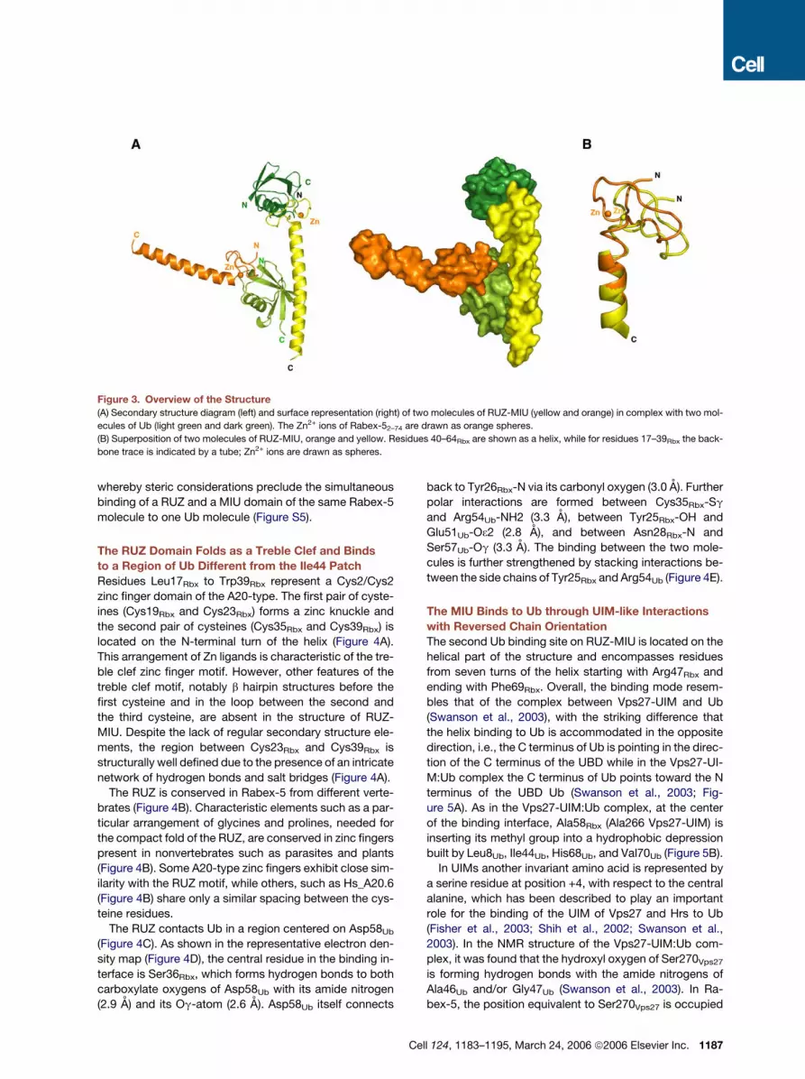
Figure 3. Overview of the Structure
(A) Secondary structure diagram (left) and surface representation (right) of two molecules of RUZ-MIU (yellow and orange) in complex with two mol-
ecules of Ub (light green and dark green). The Zn2+ ions of Rabex-52–74 are drawn as orange spheres.
(B) Superposition of two molecules of RUZ-MIU, orange and yellow. Residues 40–64Rbx are shown as a helix, while for residues 17–39Rbx the back-
bone trace is indicated by a tube; Zn2+ ions are drawn as spheres.
whereby steric considerations preclude the simultaneous
binding of a RUZ and a MIU domain of the same Rabex-5
molecule to one Ub molecule (Figure S5).
The RUZ Domain Folds as a Treble Clef and Binds
to a Region of Ub Different from the Ile44 Patch
Residues Leu17Rbx to Trp39Rbx represent a Cys2/Cys2
zinc finger domain of the A20-type. The first pair of cyste-
ines (Cys19Rbx and Cys23Rbx) forms a zinc knuckle and
the second pair of cysteines (Cys35Rbx and Cys39Rbx) is
located on the N-terminal turn of the helix (Figure 4A).
This arrangement of Zn ligands is characteristic of the tre-
ble clef zinc finger motif. However, other features of the
treble clef motif, notably b hairpin structures before the
first cysteine and in the loop between the second and
the third cysteine, are absent in the structure of RUZ-
MIU. Despite the lack of regular secondary structure ele-
ments, the region between Cys23Rbx and Cys39Rbx is
structurally well defined due to the presence of an intricate
network of hydrogen bonds and salt bridges (Figure 4A).
The RUZ is conserved in Rabex-5 from different verte-
brates (Figure 4B). Characteristic elements such as a par-
ticular arrangement of glycines and prolines, needed for
the compact fold of the RUZ, are conserved in zinc fingers
present in nonvertebrates such as parasites and plants
(Figure 4B). Some A20-type zinc fingers exhibit close sim-
ilarity with the RUZ motif, while others, such as Hs_A20.6
(Figure 4B) share only a similar spacing between the cys-
teine residues.
The RUZ contacts Ub in a region centered on Asp58Ub
(Figure 4C). As shown in the representative electron den-
sity map (Figure 4D), the central residue in the binding in-
terface is Ser36Rbx, which forms hydrogen bonds to both
carboxylate oxygens of Asp58Ub with its amide nitrogen
(2.9 A) and its Og-atom (2.6 A). Asp58Ub itself connects
C
back to Tyr26Rbx-N via its carbonyl oxygen (3.0 A). Further
polar interactions are formed between Cys35Rbx-Sg
and Arg54Ub-NH2 (3.3 A), between Tyr25Rbx-OH and
Glu51Ub-O32 (2.8 A), and between Asn28Rbx-N and
Ser57Ub-Og (3.3 A). The binding between the two mole-
cules is further strengthened by stacking interactions be-
tween the side chains of Tyr25Rbx and Arg54Ub (Figure 4E).
The MIU Binds to Ub through UIM-like Interactions
with Reversed Chain Orientation
The second Ub binding site on RUZ-MIU is located on the
helical part of the structure and encompasses residues
from seven turns of the helix starting with Arg47Rbx and
ending with Phe69Rbx. Overall, the binding mode resem-
bles that of the complex between Vps27-UIM and Ub
(Swanson et al., 2003), with the striking difference that
the helix binding to Ub is accommodated in the opposite
direction, i.e., the C terminus of Ub is pointing in the direc-
tion of the C terminus of the UBD while in the Vps27-UI-
M:Ub complex the C terminus of Ub points toward the N
terminus of the UBD Ub (Swanson et al., 2003; Fig-
ure 5A). As in the Vps27-UIM:Ub complex, at the center
of the binding interface, Ala58Rbx (Ala266 Vps27-UIM) is
inserting its methyl group into a hydrophobic depression
built by Leu8Ub, Ile44Ub, His68Ub, and Val70Ub (Figure 5B).
In UIMs another invariant amino acid is represented by
a serine residue at position +4, with respect to the central
alanine, which has been described to play an important
role for the binding of the UIM of Vps27 and Hrs to Ub
(Fisher et al., 2003; Shih et al., 2002; Swanson et al.,
2003). In the NMR structure of the Vps27-UIM:Ub com-
plex, it was found that the hydroxyl oxygen of Ser270Vps27
is forming hydrogen bonds with the amide nitrogens of
Ala46Ub and/or Gly47Ub (Swanson et al., 2003). In Ra-
bex-5, the position equivalent to Ser270Vps27 is occupied
ell 124, 1183–1195, March 24, 2006 ª2006 Elsevier Inc. 1187

Figure 4. The RUZ Binds to an Ub Surface Centered on Asp58
(A) Stereoview of the treble clef zinc finger domain of Rabex-5. Residues from 18 to 41 are shown in ball-and-sticks; hydrogen bonds are depicted as
black dashed lines.
(B) Alignment of representative ZnF_A20 domains. GenBank accession numbers are shown on the right. Numbers on top refer to human Rabex-5. The
multiple ZnF_A20 domains in the A20 protein are indicated by their numbers. Abbreviations are as in Figure S3.
(C) Schematic representation of the binding between RUZ (yellow) and Ub (green surface). The locations of Ile44Ub and Asp58Ub are indicated on the
surface of Ub in magenta and blue, respectively.
(D) 2Fobs� 1Fcalc difference electron density contoured at the 1s level around residue Asp58Ub (green) and residue Ser36Rbx (yellow). Hydrogen bonds
are indicated by dashed black lines.
(E) Hydrogen bonds and salt-bridge interactions between Rabex-5 (yellow) and Ub (green).
by Asp54Rbx whose carboxylate group is positioned such
that both oxygens can simultaneously engage in separate
hydrogen bonds with the amide nitrogens of Ala46Ub and
Gly47Ub (Figures 5C and 5D) leading to a tighter binding.
Shih et al. (2002) showed that mutation of Ser270 into
Asp in Vps27 reduced 5- to 6-fold the Ub binding ability
of the Vps27-UIM. An explanation for this apparent con-
tradiction came by the superimposition of the structures
(Figure 5D). The amide group of Gln50Rbx, which in the Ra-
bex5-MIU:Ub complex fixates the Asp54Rbx-carboxylate
in an optimal position via a salt bridge, is not present in
Vps27-UIM:Ub complex, where Ser274Vps27 substitutes
for Gln50Rbx.
The third region of importance in both UIM and MIU
binding is a group of glutamates C-terminal (MIU) or N-ter-
minal (UIM) of the central Ala. In both motifs, one of the
glutamates forms polar interactions with Arg42Ub and/or
Arg72Ub. In the MIU-Ub complex, the carboxylate group
of Glu65Rbx forms not only a salt bridge to Arg42Ub but
1188 Cell 124, 1183–1195, March 24, 2006 ª2006 Elsevier Inc.
also accepts a hydrogen bond from the amide nitrogen
of Leu73Ub. The latter interaction, together with a hydro-
phobic interaction between Phe69Rbx and the side chain
of Leu73Ub tacks the C terminus of Ub up to Leu73Ub to
Rabex-5 (Figure 5C).
N-terminal of Asp54Rbx, two more residues of Rabex-5
are involved in specific contacts to Ub and extend the
binding surface. The amide moiety of Gln50Rbx donates
a hydrogen bond to the carbonyl oxygen of Thr66Ub, and
the guanidinium group of Arg47Rbx forms a hydrogen
bond with the carbonyl oxygen of Lys63Ub (Figure 5C).
Thus, in total, the resulting binding region for the MIU in-
volves seven turns of the a helix.
Validation of the Two Surfaces of Interaction
between Rabex-5 and Ub
The structure of the RUZ-MIU:Ub complex reveals that the
two UBDs are able to interact with distinct surfaces on Ub,
centered on Asp58Ub and Ile44Ub, respectively. We tested

Figure 5. The MIU:Ub Complex Resembles the UIM:Ub Complex
(A) Structures of the Rabex-5-MIU:Ub (yellow and green) and the Vps27-UIM:Ub (red and green) complexes.
(B) 2Fobs � 1Fcalc difference electron density contoured at the 1s level around residue Ala58Rbx (yellow) and residues Ile44Ub and Val70Ub (green).
(C) Interactions between Ub (green) and MIU (yellow). Hydrogen bonds and salt bridges are indicated by dashed black lines.
(D) Ub binding residues of the MIU of Rabex-5 (yellow) and of the UIM of Vps27 (red). Models of the RZF-MIU:Ub complex and the Vps27-UIM:Ub
complex (Swanson et al., 2003) were superimposed based on residues 5–70 of the Ub molecules. Side chains involved in interaction between MIU
(yellow)/UIM (red) and Ub are shown in stick representation.
two Ub mutants, Ub-I44A and Ub-D58A, for binding to Ra-
bex-5. The I44A mutation strongly reduced the binding of
Ub to the MIU but not to the RUZ, while the D58A mutation
had the complementary effect (Figure 6A). Next, we ana-
lyzed mutants in either the RUZ or the MIU. In the context
of the isolated RUZ (aa 2–49), we mutagenized Tyr25 to
Phe to prevent the formation of the hydrogen bond be-
tween the Tyr25Rbx-OH and Glu51Ub. We also employed
the already-described mutant, A58G, in the context of
the isolated MIU (aa 48–74). Both mutants displayed
strongly reduced binding to monomeric Ub (Figure 6B).
Next, we characterized the interaction of the Rabex-5
UBDs with monomeric Ub, by isothermal titration calorim-
etry (ITC). The dissociation constants were 12.0 mM for the
RUZ:Ub interaction and 28.7 mM for the MIU:Ub one (Fig-
C
ures 6C and S6). The reactions were exothermic, with a
stoichiometry of 1:1.
The RUZ-MIU fragment displayed an apparent KD for in-
teraction with Ub of 6 mM, comparable to those of the iso-
lated domains. This confirms that the two UBDs of Rabex-
5 are oriented in a way that precludes their simultaneous
binding to a single molecule of Ub, a condition under which
a KD in the low nanomolar range would be predicted. Fi-
nally, the introduction of the A58G mutation in the RUZ-
MIU resulted in a 3-fold reduction in the affinity; con-
versely, the Y25F mutation did not significantly affected
the KD of the RUZ-MIU:Ub interaction, likely because of re-
sidual stacking interactions (Figures 6C and S6).
Of note, the ITC data suggest a 1:1 stoichiometry for
the RUZ-MIU:Ub complex (Figure 6C), a finding not
ell 124, 1183–1195, March 24, 2006 ª2006 Elsevier Inc. 1189

Figure 6. Validation of the Two Surfaces of Interaction between Rabex-5 and Ub
(A) Ub wt, Ub-I44A, and Ub-D58A were used in an in vitro pull-down assay with the indicated GST constructs of Rabex-5. Samples were resolved by
Tricine-PAGE and IB as indicated.
(B) GST-RUZ (aa 2–49), GST-MIU (aa 48–74), and their indicated mutants were used in an in vitro pull-down assay with wt Ub. Detection was as
in (A).
(C) Determination of binding affinities for Rabex-5 with Ub by ITC. Thermodynamic parameters for the interaction of the UBDs of Rabex-5 with Ub
were derived from experiments performed as described in Supplemental Experimental Procedures. KB, binding constant; KD, dissociation con-
stant; DHobs, observed binding enthalpy; DS, binding entropy; DGobs, observed Gibbs’ free energy; N, binding stoichiometry.
(D) Stoichiometry of the RUZ-MIU:Ub complex. One hundred mM purified RUZ-MIU was incubated (1 hr, 20ºC) with either Ub-I44A (100 mM) or Ub-
I44A/Ub-D58A (100 mM each), followed by fractionation on a Superdex75 column (Amersham). Fractions from 10.3 to 14.5 ml were separated by
Tricine-PAGE and stained with Coomassie (the Ub-D58A variant is retarded because of a 15 residues N-terminal tail).
1190 Cell 124, 1183–1195, March 24, 2006 ª2006 Elsevier Inc.

immediately reconcilable with the structural data. To clar-
ify this point, we analyzed, by size exclusion chromatogra-
phy, whether Ub-I44A and Ub-D58A, which are respec-
tively impaired in their binding to MIU or RUZ, can
simultaneously bind to RUZ-MIU. We observed a peak
shift in the elution profile of the RUZ-MIU incubated with
equimolar amounts of Ub-I44A and Ub-D58A, as com-
pared to that of the RUZ-MIU:Ub-I44A complex (Fig-
ure 6D). This indicates that Ub can enter a 2:1 complex
with RUZ-MIU in solution. The apparent 1:1 stoichiometry,
detected by ITC, is most likely a networking effect, due to
the presence of two binding sites on Ub for RUZ-MIU (see
Figure S7 for details and explanations).
The finding that there are two binding surfaces on Ub
might have interesting biological implications, if a single
Ub moiety were able to contact two different Ub recep-
tors. To investigate this possibility, we performed a binding
assay in which GST-MIU (aa 48–74), immobilized onto glu-
tathione-Sepharose beads (GSH), was first incubated with
Ub for 1 hr at room temperature and then with purified
RUZ (aa 2–49). As shown in Figure 6E, the RUZ domain
was efficiently retained on the beads, entering a trimeric
complex with the MIU:Ub complex. Thus, Ub can simulta-
neously interact with two different partners, raising the
possibility that in vivo it can bridge together different Ub
receptors.
DISCUSSION
In this study, we report the characterization of two UBDs,
contained in Rabex-5. These findings not only add to our
understanding of the Ub:UBDs interaction but also harbor
a number of interesting biological and functional implica-
tions. While this manuscript was undergoing review, two
papers reported the Ub binding properties (Mattera
et al., 2006) and the determination of the structure (Lee
et al., 2006) of the N terminus of Rabex-5. The conforma-
tion of the molecules, and the interaction surfaces be-
tween the N terminus of Rabex-5 and Ub, in this paper
and in that from Lee et al. [2006] are in agreement.
The MIU:Ub Interaction
The MIU can be roughly equaled to an inverted UIM. At the
level of primary sequence, the MIU did not show signifi-
cant similarity to the UIM, while it could be aligned rather
precisely to a UIM consensus in a reversed orientation
(Figure 1B). In addition, the HMM profile of the MIU did
not recognize any UIM in the database (Figure S3) and
vice-versa (S.C., unpublished data). Accordingly, the
MIU binds to Ub in a manner similar to the UIM but with
the helix running in the opposite direction (this paper;
Lee et al., 2006). In the UIM, the insertion of a methyl group
provided by the highly conserved alanine into the hydro-
phobic depression created on the Ub-surface by Leu8,
Ile44, His68, and Val70 identifies an essential contact
(Swanson et al., 2003). The MIU uses the same residue,
Ala58Rbx, to implement this central interaction, and in-
deed, the mutation of this residue to Gly strongly reduced
the binding to Ub.
The MIU:Ub interaction displays higher affinity (KD
�29 mM) with respect to that between UIMs and Ub (KDs
�100–500 mM; Hicke et al., 2005). This is mirrored by struc-
tural results that revealed the enforcement of individual
contacts and the increase in the number of interactions be-
tween MIU and Ub. For example, Asp54Rbx functionally
corresponds to Ser270 of the Vps27-UIM. In contrast to
the single hydroxyl oxygen of serine, the carboxylate group
of Asp54Rbx has the capacity to engage simultaneously in
two hydrogen bonds instead of one, thus gaining binding
energy. N-terminal of Ala58Rbx, the MIU binding interface
extends further with interactions mediated by Gln50Rbx
and Arg47Rbx resulting in a binding region that involves
seven turns of a helix and is significantly larger than the
binding region of Vps27-UIM, which involves four turns of
a helix. To provide all the interaction partners, the helix of
the MIU shows a significant bend in the complex (Fig-
ure 5A), which is not seen in the UIM helices (Swanson
et al., 2003). Given the size of the MIU-Ub binding interface
and its asymmetry, it is unlikely that few sequence alter-
ations could result in a reversal in the orientation of binding,
as observed for the SUMO binding motifs of PIASX (Song
et al., 2005) and RanBP2 (Reverter and Lima, 2005).
Another common feature of UIM and MIU is their ability
to drive coupled monoubiquitination (Hicke et al., 2005;
Figure 1E). Although the molecular mechanisms of this
phenomenon are still obscure, the ability of the UIM to
bind to Ub seems to be a necessary condition (Hicke
et al., 2005). It should be mentioned that in the study by
Mattera et al. (2006), a role for the Zinc finger domain of
Rabex-5 in coupled monoubiquitination was described.
Under our conditions of analysis, we did not evidence
a major contribution of this domain (Figure 1E).
It is also interesting that in UIM-dependent coupled
monoubiquitination, the ubiquitination event seems to oc-
cur at Lys residues N-terminal of the UIM (Miller et al.,
2004). This has been put in relationship with the antiparal-
lel binding of Ub to the UIM. In the case of the MIU:Ub
interaction, the two chains are in the same orientation,
raising the possibility that MIU-mediated coupled mono-
ubiquitination occurs C-terminal of the UBD. Indeed, we
have evidence that this is the case for Rabex-5 (L.P. and
S.P., unpublished data).
Finally, we identified a family of MIU-containing proteins
and provided evidence that some of them, including Myo-
sin 6, can bind to Ub. Future work will address whether
all MIU-containing proteins are endowed with similar
(E) Five micromolar GST-MIU or GST, immobilized onto GSH beads, was incubated with 20 mM of Ub and then 5 mM of purified RUZ (aa 2–49,
cleaved from the GST moiety) was added to the complex. Detection was performed as in (A). Comparable loading of GST-MIU is shown by Pon-
ceau staining (bottom panel). The presence of RUZ on beads was detected by IB with the anti-Rabex-5 antibody G32, which recognizes the first
49 aa of Rabex-5 (see Supplemental Experimental Procedures).
Cell 124, 1183–1195, March 24, 2006 ª2006 Elsevier Inc. 1191

properties and whether they are also subjected to coupled
monoubiquitination. In the case of Myosin 6, a series of ev-
idence (reviewed in Frank et al. [2004]) supports a role in
membrane trafficking, an area in which the involvement
of the Ub network is paramount. Thus, it will be of interest
to determine how the Ub binding properties of Myosin 6
regulate its function.
The RUZ:Ub Interaction
The second Ub binding motif in the N-terminal region of
Rabex-5 is a Cys2/Cys2 zinc finger of the A20 type. An-
other Ub binding Zinc finger, the NZF, has been structur-
ally characterized (Alam et al., 2004; Wang et al., 2003).
However, RUZ and NZF display different folds and modes
of interaction with Ub (Figure S8). RUZ binds Ub on a novel
surface centered on Asp58Ub (this paper; Lee et al., 2006).
This surface does not overlap either with the ‘‘canonical’’
I44-centered binding surface or with the ‘‘shifted’’ surface
(Bienko et al., 2005) responsible for interaction with UBM
(Figure S9).
An obvious question is whether all A20-type zinc fingers
display Ub binding properties. An alignment of some of
these domains (Figure 4B) revealed that the fourth zinc fin-
ger domain of the A20 protein, Znf4, displays the closest
homology to the RUZ of Rabex-5. In particular, the first
aromatic residue after the first pair of cysteines is a tyro-
sine, both in RUZ and in Znf4 and not a phenylalanine as
in many other A20-type zinc fingers. Of note, we show
that a Y to F mutation of that position impairs Ub binding.
Moreover, the equivalent of Ser36Rbx in the Znf4 is a thre-
onine, which is capable of forming the same interactions
as in the RUZ:Ub complex. Thus, the Znf4 of A20 might
be endowed with Ub binding properties.
We note that the A20 protein displays both deubiquiti-
nating and Ub ligase (E3) abilities (Wertz et al., 2004).
The Znf4 was identified as the critical determinant for the
E3 ligase activity (Wertz et al., 2004). In addition, Mattera
et al. (2006) and Lee et al. (2006) provided evidence com-
patible with the possibility that the RUZ domain of Rabex-
5 might be endowed with a similar activity. E3 ligases can
either possess intrinsic catalytic activity (HECT-type E3s),
or function as adaptors between E2 enzymes and sub-
strates (RING-type E3s; Pickart, 2001). In this latter case,
actual catalysis is executed by the E2. RUZ domains are
unlikely to possess intrinsic catalytic activity and therefore
are candidates to be E3 ligases of a RING-like type. Our
present results show that the RUZ domain binds to Ub.
To reconcile all these observations, one might postulate
that RUZ-containing proteins are recruited to ubiquiti-
nated E2 enzymes, thus allowing bridging with hypotheti-
cal substrates bound to other regions of the same RUZ-
containing proteins. In support of this, Lee et al. (2006)
have provided evidence for a Ub-mediated interaction be-
tween the RUZ and UbcH5C. Such a scenario could be
speculatively extended to various other Ub receptors.
Biological Implications
The Ile44-based surface on Ub surface is involved in liter-
ally hundreds of interactions with Ub receptors. Further
1192 Cell 124, 1183–1195, March 24, 2006 ª2006 Elsevier Inc.
work will be needed to establish whether the surface cen-
tered on Asp58 (this paper; Lee et al., 2006) is similarly pro-
miscuous or involved in more ‘‘specialized’’ functions.
Whatever the case, the fact that a single Ub moiety can si-
multaneously engage two different partners (Figure 6E)
adds a new dimension to the complexity of the Ub-based
network and provides interesting outlooks. For example,
ubiquitination is critical for intracellular trafficking of mem-
brane bound proteins (Hicke and Dunn, 2003). It was sug-
gested that Ub receptors act serially on monoubiquitinated
cargoes, during their transport from the cell surface to the
lysosome (Hicke et al., 2005). However, all UBDs de-
scribed so far engage the Ile44Ub-based pocket, making
such a ‘‘hand-off’’ mechanism difficult to conceptualize.
One possibility is that Ub receptors are organized in a hier-
archical cascade with increased affinity for Ub. However,
our results also open the possibility that Ub receptors
with specificity for noncanonical surfaces of Ub are inter-
calated, in the cascade, to ‘‘canonical’’ Ub receptors,
thus facilitating the hand-off of the ubiquitinated cargo.
Whether Rabex-5 has such a function remains to be estab-
lished. In addition, the dual binding ability of Ub might allow
recruitment of regulating enzymes, at specific points of the
cascade, without compromising the sequential delivery of
the ubiquitinated cargo: a possible mechanism well fitting
the known regulatory properties of Rabex-5.
In this context, the physical association between
Rabex-5 and the EGFR, reported here, identifies an impor-
tant target of the networking ability conferred upon
Rabex-5 by its UBDs. By drawing an analogy with the
well-characterized EGFR-Sos1-Ras system, we would
like to propose that the interaction with ubiquitinated
EGFRs allows relocalization of Rabex-5 to the plasma
membrane or to early endosomes (Figure 2C), thereby al-
lowing activation of Rab5. Another Vps9 domain-contain-
ing protein, RIN1, has been proposed for this role (Tall
et al., 2001), which can be directly recruited to the EGFR
through SH2:phosphotyrosine interactions (Barbieri
et al., 2003). Rabex-5 and RIN1 could work redundantly;
however, it is interesting that they bind to differently post-
translationally modified EGFRs, ubiquitinated and phos-
phorylated, respectively. Recent evidence has suggested
that the EGFR might follow different internalization routes,
as a function of its ubiquitination status (Sigismund et al.,
2005). Thus it is tempting to speculate that differential
mechanisms of Rab5 activation by EGFR have evolved
in the control (and possibly in the coordination) of different
endocytic pathways.
We finally note that, during evolution, Rabex-5 loses
a UBD (the CUE) but acquires two novel Ub-interacting
surfaces. This is reminiscent of what happens to the
UIM-containing protein eps15, which in mammals dis-
plays a UIM, while in yeast (Ede1p) displays a UBA do-
main. This suggests positive pressure on functions rather
than on structures and, at the same time, probably the
‘‘need’’ to evolve more complex modes of operation for
the Ub-based network. This might, in turn, reflect the chal-
lenge of adapting an originally simple signaling system

to the increasing complexity of the endocytic membrane
system throughout evolution.
EXPERIMENTAL PROCEDURES
Reagents and Constructs
Antibodies were: anti-Ub (P4D1, Santa Cruz Biotechnology), anti-
FLAG and anti-FLAG affinity gel (M2, Sigma), anti-HA (Babco), anti-Ra-
bex-5 monoclonal (G32; see Supplemental Experimental Procedures).
Polyubiquitin Lys48- and Lys63-linked chains (2–7 Ub moieties) and
bovine ubiquitin, employed in the crystallization studies, were from
Boston Biochem. All constructs were engineered by either site-di-
rected mutagenesis or recombinant PCR (see Supplemental Experi-
mental Procedures), and sequence verified.
Biochemical Studies
Transfections were performed using Lipofectamin (Invitrogen). Immu-
noprecipitation and immunoblotting (Fazioli et al., 1992) and immuno-
fluorescence and internalization assays were performed as described
(Sigismund et al., 2005).
GST fusion proteins were expressed and purified as described
(Sessa et al., 2005). Cleavage from GST was, as appropriate, with
PreScission Protease (Amersham Bioscience, 10 unit/mg of substrate,
16 hr, 4ºC), or with biotinylated thrombin (Thrombin Cleavage Capture
Kit, Novagen, 1 unit/mg of substrate, 16 hr, RT).
For crystallization studies, the cleaved RUZ-MIU fragment (aa 2–74)
was concentrated and loaded on a Superdex75 column (Amersham
Biosciences). Fractions containing RUZ-MIU were pooled and incu-
bated for 2 hr at RT with bovine Ub (1:1 molar ratio), followed by concen-
tration and chromatography on a Superdex75 column. Peak fractions
were concentrated with Vivaspin concentrators to 20 mg/ml. Usually,
1–2 mg of pure complex was obtained per liter of bacterial culture.
Details of the procedures used for pull-down experiments and ITC
assays are in Supplemental Experimental Procedures.
Crystallization and Structure Determination
Crystallization experiments were performed with the sitting drop vapor
diffusion technique at 20ºC. To screen for initial crystallization condi-
tions, 100 nl of protein solution, at a concentration of 20 mg/ml, was
mixed with 100 nl reservoir solution using a Cartesian Honeybee liquid
handler (Genomic Solutions) and equilibrated against 150 ml reservoir
solution. Rod-shaped hexagonal crystals (form I) grew with a reservoir
consisting of 0.2 M Li2SO4, 0.1 M MES (pH 6.5), and 25% PEG 3350,
while plate-like crystals (form II) grew with a reservoir buffer containing
0.2 M ammonium acetate, 0.1 M NaCitrate (pH 6.5), and 25% PEG
4000. For data collection, crystals were gradually transferred to
a cryo-buffer (reservoir buffer supplemented with 20% glycerol) and
flash-cooled in liquid N2. All data used in structure solution and refine-
ments were collected on beamlines ID29 and BM14 at ESRF (Greno-
ble, France). Data collection statistics are shown in Table S1. For
both crystal forms, all data were collected from one crystal. Data
were integrated and scaled with HKL2000 (Otwinowski and Minor,
1997) and merged with XPREP (Bruker AXS).
Initial phases were derived using the combination of SHELXC,
SHELXD, and SHELXE (Sheldrick, 2002) with default parameters
from the HKL2MAP graphical user interface. For crystal form I, model
building was initiated with a helical fragment placed into the electron
density by the HelixBuild module of ArpWarp (Morris et al., 2002).
For crystal form II, a first model containing 438 residues was automat-
ically built into the electron density by ArpWarp. Subsequent cycles of
model building and refinement were performed with Xfit (McRee, 1999)
and CNS (Brunger et al., 1998). In the final stages of refinement,
REFMAC5 (Murshudov et al., 1999) was used with a TLS description
of B values, whereby the rigid groups were identified by automatic
analysis of difference distance matrices (Schneider, 2002).
The structures were analyzed with Xfit, ESCET, and PROCHECK
(Laskowski et al., 1993). Figures were made with PYMOL (http://
pymol.sourceforge.net).
Supplemental Data
Supplemental Data include nine figures, three tables, and Supplemen-
tal Experimental Procedures and can be found with this article online
at http://www.cell.com/cgi/content/full/124/6/1183/DC1/.
ACKNOWLEDGMENTS
We thank L. Hicke and S. Giordano for reagents; E. Cavallaro, V. Ce-
catiello, and M. Garre for technical assistance; A. Mattevi for providing
X-ray facilities; C. Tarricone and R. Steiner for help in data collection;
the staff of beamlines ID29 and BM14 at ESRF; and the Monoclonal
Service at IFOM. This work was supported by grants from Associa-
zione Italiana per la Ricerca sul Cancro (S.P., P.P.D.F., T.R.S., A.M.);
Association for International Cancer Research (S.P.); European Com-
munity (VI Framework), Ministero della Salute, MIUR, Fondazione
Monzino (P.P.D.F.).
Received: December 7, 2005
Revised: February 6, 2006
Accepted: February 14, 2006
Published online: February 16, 2006
REFERENCES
Alam, S.L., Sun, J., Payne, M., Welch, B.D., Blake, B.K., Davis, D.R.,
Meyer, H.H., Emr, S.D., and Sundquist, W.I. (2004). Ubiquitin interac-
tions of NZF zinc fingers. EMBO J. 23, 1411–1421.
Barbieri, M.A., Kong, C., Chen, P.I., Horazdovsky, B.F., and Stahl, P.D.
(2003). The SRC homology 2 domain of Rin1 mediates its binding to
the epidermal growth factor receptor and regulates receptor endocy-
tosis. J. Biol. Chem. 278, 32027–32036.
Bienko, M., Green, C.M., Crosetto, N., Rudolf, F., Zapart, G., Coull, B.,
Kannouche, P., Wider, G., Peter, M., Lehmann, A.R., et al. (2005).
Ubiquitin-binding domains in Y-family polymerases regulate transle-
sion synthesis. Science 310, 1821–1824.
Brunger, A.T., Adams, P.D., Clore, G.M., DeLano, W.L., Gros, P.,
Grosse-Kunstleve, R.W., Jiang, J.S., Kuszewski, J., Nilges, M., Pannu,
N.S., et al. (1998). Crystallography & NMR system: a new software
suite for macromolecular structure determination. Acta Crystallogr. D
Biol. Crystallogr. 54, 905–921.
Chen, Z.J. (2005). Ubiquitin signalling in the NF-kappaB pathway. Nat.
Cell Biol. 7, 758–765.
Davies, B.A., Topp, J.D., Sfeir, A.J., Katzmann, D.J., Carney, D.S., Tall,
G.G., Friedberg, A.S., Deng, L., Chen, Z., and Horazdovsky, B.F.
(2003). Vps9p CUE domain ubiquitin binding is required for efficient
endocytic protein traffic. J. Biol. Chem. 278, 19826–19833.
Di Fiore, P.P., Polo, S., and Hofmann, K. (2003). When ubiquitin meets
ubiquitin receptors: a signalling connection. Nat. Rev. Mol. Cell Biol. 4,
491–497.
Donaldson, K.M., Yin, H., Gekakis, N., Supek, F., and Joazeiro, C.A.
(2003). Ubiquitin signals protein trafficking via interaction with a novel
ubiquitin binding domain in the membrane fusion regulator, Vps9p.
Curr. Biol. 13, 258–262.
Fazioli, F., Bottaro, D.P., Minichiello, L., Auricchio, A., Wong, W.T., Se-
gatto, O., and Di Fiore, P.P. (1992). Identification and biochemical
characterization of novel putative substrates for the epidermal growth
factor receptor kinase. J. Biol. Chem. 267, 5155–5161.
Fisher, R.D., Wang, B., Alam, S.L., Higginson, D.S., Robinson, H.,
Sundquist, W.I., and Hill, C.P. (2003). Structure and ubiquitin binding
of the ubiquitin-interacting motif. J. Biol. Chem. 278, 28976–28984.
Cell 124, 1183–1195, March 24, 2006 ª2006 Elsevier Inc. 1193

Frank, D.J., Noguchi, T., and Miller, K.G. (2004). Myosin VI: a structural
role in actin organization important for protein and organelle localiza-
tion and trafficking. Curr. Opin. Cell Biol. 16, 189–194.
Hama, H., Tall, G.G., and Horazdovsky, B.F. (1999). Vps9p is a guanine
nucleotide exchange factor involved in vesicle-mediated vacuolar pro-
tein transport. J. Biol. Chem. 274, 15284–15291.
Hicke, L., and Dunn, R. (2003). Regulation of membrane protein trans-
port by ubiquitin and ubiquitin-binding proteins. Annu. Rev. Cell Dev.
Biol. 19, 141–172.
Hicke, L., Schubert, H.L., and Hill, C.P. (2005). Ubiquitin-binding do-
mains. Nat. Rev. Mol. Cell Biol. 6, 610–621.
Hofmann, K., and Falquet, L. (2001). A ubiquitin-interacting motif con-
served in components of the proteasomal and lysosomal protein deg-
radation systems. Trends Biochem. Sci. 26, 347–350.
Horiuchi, H., Lippe, R., McBride, H.M., Rubino, M., Woodman, P.,
Stenmark, H., Rybin, V., Wilm, M., Ashman, K., Mann, M., and Zerial,
M. (1997). A novel Rab5 GDP/GTP exchange factor complexed to Ra-
baptin-5 links nucleotide exchange to effector recruitment and func-
tion. Cell 90, 1149–1159.
Kang, R.S., Daniels, C.M., Francis, S.A., Shih, S.C., Salerno, W.J.,
Hicke, L., and Radhakrishnan, I. (2003). Solution structure of a CUE-
ubiquitin complex reveals a conserved mode of ubiquitin binding.
Cell 113, 621–630.
Karplus, K., Barrett, C., and Hughey, R. (1998). Hidden Markov models
for detecting remote protein homologies. Bioinformatics 14, 846–856.
Kawasaki, M., Shiba, T., Shiba, Y., Yamaguchi, Y., Matsugaki, N., Igar-
ashi, N., Suzuki, M., Kato, R., Kato, K., Nakayama, K., and Wakatsuki,
S. (2005). Molecular mechanism of ubiquitin recognition by GGA3 GAT
domain. Genes Cells 10, 639–654.
Krishna, S.S., Majumdar, I., and Grishin, N.V. (2003). Structural classi-
fication of zinc fingers: survey and summary. Nucleic Acids Res. 31,
532–550.
Laskowski, R.A., MacArthur, M.W., Moss, D.S., and Thornton, J.M.
(1993). PROCHECK: a program to check the stereochemical quality
of protein structures. J. Appl. Crystallogr. 26, 283–291.
Lee, S., Tsai, Y.C., Mattera, R., Smith, W.J., Kostelansky, M.S., Weiss-
man, A.M., Bonifacino, J.S., and Hurley, J.H. (2006). Structural basis
for ubiquitin recognition and autoubiquitination by Rabex-5. Nat.
Struct. Mol. Biol., in press. Published online February 5, 2006. 10.
1038/nsmb1064.
Mattera, R., Tsai, Y.C., Weissman, A.M., and Bonifacino, J.S. (2006).
The Rab5 guanine nucleotide exchange factor Rabex-5 binds ubiquitin
and functions as a ubiquitin ligase through an atypical UIM and a zinc
finger domain. J. Biol. Chem., in press. Published online January 5,
2006. 10.1074/jbc.M509939200.
McRee, D.E. (1999). XtalView/Xfit—a versatile program for manipulat-
ing atomic coordinates and electron density. J. Struct. Biol. 125, 156–
165.
Miller, S.L., Malotky, E., and O’Bryan, J.P. (2004). Analysis of the role
of ubiquitin-interacting motifs in ubiquitin binding and ubiquitylation.
J. Biol. Chem. 279, 33528–33537.
Morris, R.J., Perrakis, A., and Lamzin, V.S. (2002). ARP/wARP’s
model-building algorithms. I. The main chain. Acta Crystallogr. D
Biol. Crystallogr. D58, 968–975.
Murshudov, G.N., Lebedev, A., Vagin, A.A., Wilson, K.S., and Dodson,
E.J. (1999). Efficient anisotropic refinement of Macromolecular struc-
tures using FFT. Acta Crystallogr. D Biol. Crystallogr. D55, 247–255.
Ohno, A., Jee, J., Fujiwara, K., Tenno, T., Goda, N., Tochio, H., Ko-
bayashi, H., Hiroaki, H., and Shirakawa, M. (2005). Structure of the
UBA domain of Dsk2p in complex with ubiquitin molecular determi-
nants for ubiquitin recognition. Structure (Camb.) 13, 521–532.
1194 Cell 124, 1183–1195, March 24, 2006 ª2006 Elsevier Inc.
Opipari, A.W., Jr., Boguski, M.S., and Dixit, V.M. (1990). The A20 cDNA
induced by tumor necrosis factor alpha encodes a novel type of zinc
finger protein. J. Biol. Chem. 265, 14705–14708.
Otwinowski, Z., and Minor, W. (1997). Processing of X-ray diffraction
data collected in oscillation mode. In Methods in Enzymology. Macro-
molecular Crystallography, Part A, C.W. Carter, Jr., and R.M. Sweet,
eds. (San Diego, CA: Academic Press.), pp. 307–326.
Pickart, C.M. (2001). Ubiquitin enters the new millennium. Mol. Cell 8,
499–504.
Polo, S., Confalonieri, S., Salcini, A.E., and Di Fiore, P.P. (2003). EH
and UIM: endocytosis and more. Sci. STKE 2003, re17.
Pornillos, O., Alam, S.L., Rich, R.L., Myszka, D.G., Davis, D.R., and
Sundquist, W.I. (2002). Structure and functional interactions of the
Tsg101 UEV domain. EMBO J. 21, 2397–2406.
Prag, G., Misra, S., Jones, E.A., Ghirlando, R., Davies, B.A., Horazdov-
sky, B.F., and Hurley, J.H. (2003). Mechanism of ubiquitin recognition
by the CUE domain of Vps9p. Cell 113, 609–620.
Prag, G., Lee, S., Mattera, R., Arighi, C.N., Beach, B.M., Bonifacino,
J.S., and Hurley, J.H. (2005). Structural mechanism for ubiquitinated-
cargo recognition by the Golgi-localized, gamma-ear-containing,
ADP-ribosylation-factor-binding proteins. Proc. Natl. Acad. Sci. USA
102, 2334–2339.
Raasi, S., Orlov, I., Fleming, K.G., and Pickart, C.M. (2004). Binding
of polyubiquitin chains to ubiquitin-associated (UBA) domains of
HHR23A. J. Mol. Biol. 341, 1367–1379.
Reverter, D., and Lima, C.D. (2005). Insights into E3 ligase activity re-
vealed by a SUMO-RanGAP1-Ubc9-Nup358 complex. Nature 435,
687–692.
Schneider, T.R. (2002). A genetic algorithm for the identification of con-
formationally invariant regions in protein molecules. Acta Crystallogr. D
Biol. Crystallogr. D58, 195–208.
Sessa, F., Mapelli, M., Ciferri, C., Tarricone, C., Areces, L.B.,
Schneider, T.R., Stukenberg, P.T., and Musacchio, A. (2005). Mecha-
nism of Aurora B activation by INCENP and inhibition by hesperadin.
Mol. Cell 18, 379–391.
Sheldrick, G.M. (2002). Macromolecular phasing with SHELXE. Z. Kris-
tallogr. 217, 644–650.
Shih, S.C., Katzmann, D.J., Schnell, J.D., Sutanto, M., Emr, S.D., and
Hicke, L. (2002). Epsins and Vps27p/Hrs contain ubiquitin-binding do-
mains that function in receptor endocytosis. Nat. Cell Biol. 4, 389–393.
Shih, S.C., Prag, G., Francis, S.A., Sutanto, M.A., Hurley, J.H., and
Hicke, L. (2003). A ubiquitin-binding motif required for intramolecular
monoubiquitylation, the CUE domain. EMBO J. 22, 1273–1281.
Sigismund, S., Woelk, T., Puri, C., Maspero, E., Tacchetti, C., Transi-
dico, P., Di Fiore, P.P., and Polo, S. (2005). Clathrin-independent endo-
cytosis of ubiquitinated cargos. Proc. Natl. Acad. Sci. USA 102, 2760–
2765.
Song, J., Zhang, Z., Hu, W., and Chen, Y. (2005). Small ubiquitin-like
modifier (SUMO) recognition of a SUMO binding motif: a reversal of
the bound orientation. J. Biol. Chem. 280, 40122–40129.
Sun, L., and Chen, Z.J. (2004). The novel functions of ubiquitination in
signaling. Curr. Opin. Cell Biol. 16, 119–126.
Sundquist, W.I., Schubert, H.L., Kelly, B.N., Hill, G.C., Holton, J.M.,
and Hill, C.P. (2004). Ubiquitin recognition by the human TSG101 pro-
tein. Mol. Cell 13, 783–789.
Swanson, K.A., Kang, R.S., Stamenova, S.D., Hicke, L., and Radhak-
rishnan, I. (2003). Solution structure of Vps27 UIM-ubiquitin complex
important for endosomal sorting and receptor downregulation.
EMBO J. 22, 4597–4606.
Tall, G.G., Barbieri, M.A., Stahl, P.D., and Horazdovsky, B.F. (2001).
Ras-activated endocytosis is mediated by the Rab5 guanine nucleo-
tide exchange activity of RIN1. Dev. Cell 1, 73–82.

Varadan, R., Assfalg, M., Haririnia, A., Raasi, S., Pickart, C., and Fush-
man, D. (2004). Solution conformation of Lys63-linked di-ubiquitin
chain provides clues to functional diversity of polyubiquitin signaling.
J. Biol. Chem. 279, 7055–7063.
Wang, B., Alam, S.L., Meyer, H.H., Payne, M., Stemmler, T.L., Davis,
D.R., and Sundquist, W.I. (2003). Structure and ubiquitin interactions
of the conserved zinc finger domain of Npl4. J. Biol. Chem. 278,
20225–20234.
Wertz, I.E., O’Rourke, K.M., Zhou, H., Eby, M., Aravind, L., Seshagiri,
S., Wu, P., Wiesmann, C., Baker, R., Boone, D.L., et al. (2004). De-
ubiquitination and ubiquitin ligase domains of A20 downregulate NF-
kappaB signalling. Nature 430, 694–699.
Yokouchi, M., Kondo, T., Houghton, A., Bartkiewicz, M., Horne, W.C.,
Zhang, H., Yoshimura, A., and Baron, R. (1999). Ligand-induced ubiqui-
tination of the epidermal growth factor receptor involves the interaction
of the c-Cbl RING finger and UbcH7. J. Biol. Chem. 274, 31707–31712.
Accession Numbers
The coordinates and structure factors have been deposited in the
Protein Data Bank (accession codes 2C7M and 2C7N).
Cell 124, 1183–1195, March 24, 2006 ª2006 Elsevier Inc. 1195


