Embed Size (px)
Citation preview

Lys6-modified Ubiquitin Inhibits Ubiquitin-dependent ProteinDegradation*
Fu Shang‡,§, Gejing Deng‡, Qing Liu‡, Weimin Guo‡, Arthur L. Haas¶, Bernat Crosas∥,Daniel Finley∥, and Allen Taylor‡‡Laboratory for Nutrition and Vision Research, Jean Mayer United States Department of AgricultureHuman Nutrition Research Center on Aging, Tufts University, Boston, Massachusetts 02111
¶Department of Biochemistry and Molecular Biology, Louisiana State University School of Medicine,New Orleans, Louisiana 70112
∥Department of Cell Biology, Harvard Medical School, Boston, Massachusetts 02138
AbstractUbiquitin plays essential roles in various cellular processes; therefore, it is of keen interest to studythe structure-function relationship of ubiquitin itself. We investigated the modification of Lys6 ofubiquitin and its physiological consequences. Mass spectrometry-based peptide mapping and N-terminal sequencing demonstrated that, of the 7 Lys residues in ubiquitin, Lys6 was the most readilylabeled with sulfosuccinimidobiotin. Lys6-biotinylated ubiquitin was incorporated into highmolecular mass ubiquitin conjugates as efficiently as unmodified ubiquitin. However, Lys6-biotinylated ubiquitin inhibited ubiquitin-dependent proteolysis, as conjugates formed with Lys6-biotinylated ubiquitin were resistant to proteasomal degradation. Ubiquitins with a mutation ofLys6 had similar phenotypes as Lys6-biotinylated ubiquitin. Lys6 mutant ubiquitins (K6A, K6R, andK6W) also inhibited ATP-dependent proteolysis and caused accumulation of ubiquitin conjugates.Conjugates formed with K6W mutant ubiquitin were also resistant to proteasomal degradation. Thedominant-negative effect of Lys6-modified ubiquitin was further demonstrated in intact cells.Overexpression of K6W mutant ubiquitin resulted in accumulation of intra-cellular ubiquitinconjugates, stabilization of typical substrates for ubiquitin-dependent proteolysis, and enhancedsusceptibility to oxidative stress. Taken together, these results show that Lys6-modified ubiquitin isa potent and specific inhibitor of ubiquitin-mediated protein degradation.
The ubiquitin/proteasome pathway (UPP)1 is involved in regulating the cell cycle, signaltransduction, differentiation, stress response, and DNA repair. Most of these functions aremediated by the conditional turnover of regulatory and abnormal proteins in eukaryotic cells(1-4). In this essential pathway, the conserved ubiquitin acts as a degradation signal uponcovalent linkage of multiple ubiquitins to cellular substrates. This is accomplished through theformation of an isopeptide bond between the C terminus of ubiquitin (Gly76) and an internallysine residue of the substrate. Formation of the ubiquitin-substrate conjugates is usuallyfollowed by additional rounds of conjugation of more ubiquitins to the initial adduct, also via
*This work was supported in part by National Institutes of Health Grants EY11717 (to F. S.), EY13250 (to A. T.), and GM34009 (to A.L. H.) and by the United States Department of Agriculture under Agreement 58-1950-9-001. The costs of publication of this article weredefrayed in part by the payment of page charges. This article must therefore be hereby marked “advertisement” in accordance with 18U.S.C. Section 1734 solely to indicate this fact.Correspondence to: Fu Shang.§To whom correspondence should be addressed: Lab. for Nutrition and Vision Research, Jean Mayer USDA HNRCA, Tufts University,711 Washington St., Boston, MA 02111. Tel.: 617-556-3158; Fax: 617-556-3344; E-mail: [email protected] abbreviations used are: UPP, ubiquitin/proteasome pathway; sulfo-NHS-biotin, sulfosuccinimidobiotin; GFP, green fluorescent protein; HPLC, high pressure liquid chromatography; RPE, retinal pigment epithelial; GST, glutathione S-transferase; E1, ubiquitin-activating enzyme; E2, ubiquitin carrier protein; E3, ubiquitin-protein isopeptide ligase; UbG76V, G76V mutant ubiquitin.
NIH Public AccessAuthor ManuscriptJ Biol Chem. Author manuscript; available in PMC 2006 February 27.
Published in final edited form as:J Biol Chem. 2005 May 27; 280(21): 20365–20374.
NIH
-PA Author Manuscript
NIH
-PA Author Manuscript
NIH
-PA Author Manuscript

isopeptide bonds, leading to the assembly of long polyubiquitin chains. Although polyubiquitinchains formed via Lys6 (5), Lys11 (6), Lys29 (7), and Lys63 (7-10) are detected in cell-freesystems and/or in whole cells and are competent for 26 S proteasome-dependent degradation(6,11), Lys48-linked polyubiquitin chains represent the predominant signal for targetingsubstrates to the 26 S proteasome (12-14). Polyubiquitin chains linked through lysine residuesother than Lys48 have functions principally independent of targeting proteins for degradation,such as DNA repair and signal transduction (8-11).
The 26 S proteasome specifically degrades ubiquitin-conjugated proteins, and it is assembledfrom a 20 S catalytic core and two 19 S regulatory complexes (also called PA700) that cap theentry to the cylindrical catalytic core (15-18). It is the 19 S regulatory complex that recruitsubiquitin-protein conjugates to the 26 S proteasome and translocates the ubiquitinated proteinsinto the catalytic cavity. It has been demonstrated that S5a (also called MBP1 and Mcb1p), theATPase S6[H11032] subunit of the 19 S regulatory complex, and Rad23, a proteasome-associated protein, can each bind polyubiquitin chains (6,19-23). The 19 S regulatory complexalso has deubiquitinating activity, which removes ubiquitin from the substrate prior to orcoordinated with degradation of the substrate proteins. Such deubiquitination may serve bothin degradation and in proofreading or editing functions (24-26).
Biotinylated ubiquitin has been used as a probe to study the function of the UPP (27). However,biotinylated ubiquitin has not been characterized in detail, and the potential kinetic artifacts ofbiotinylated ubiquitin have not been assessed. In most cases, Lys residues are modified bybiotinylating reagents such as sulfosuccinimidobiotin (sulfo-NHS-biotin). Some of the 7 Lysresidues in ubiquitin are essential for formation of polyubiquitin chains. Modification of 1 ormore of the critical Lys residues may abolish the ability of ubiquitin to form polyubiquitinchains. Modifications of some Lys residues may also alter the surface property of ubiquitinchains and may interfere with the interaction between ubiquitin conjugates and the proteasome.Previous studies indicated that Lys6 is the most susceptible residue to modification by variouschemicals, such as p-nitrophenyl acetate (28), aspirin (29), acetic anhydride (30), and Oregongreen succinimidyl ester (31). This preference for modification could be due to the differentialamine surface accessibility (29) or to the catalytic microenvironment. The latter is most likelywith respect to Lys6 because acetylation of Lys6 depends upon its interaction with His68 (30).In the three-dimensional structure of ubiquitin, His68 is located in the vicinity of Lys6 (32,33). The interaction between Lys6 and His68 provides a catalytic microenvironment for themodification of Lys6 (30). Given the similarity between biotinylation and the chemicalmodifications mentioned above, Lys6 could also be the dominant site of biotinylation.
Individual Lys-to-Arg mutations are often used to test the role of particular Lys residues inubiquitin chain formation. However, the physiological consequences of Lys-to-Arg mutationsmay be due to factors unrelated to whether that particular Lys is used for chain synthesis.Disruption of a face on ubiquitin necessary for recognition by enzymes involved in ubiquitinconjugation or degradation could be one of the factors. A detailed characterization ofbiotinylated ubiquitin will allow us to determine the role of a particular Lys residue in ubiquitinchain formation and in maintaining the surface property for proper interaction with enzymesinvolved in ubiquitination, deubiquitination, and degradation of ubiquitin conjugates.
In this study, we ascertained which Lys residues are modified by sulfo-NHS-biotin.Subsequently, we determined the ability of various biotinylated ubiquitins to form ubiquitinconjugates and to support ubiquitin-dependent degradation. The results show that 1) Lys6 wasthe most readily labeled with sulfo-NHS-biotin; 2) Lys6-biotinylated ubiquitin was used asefficiently as unmodified ubiquitin to form high molecular mass ubiquitin conjugates; 3)Lys6-biotinylated ubiquitin inhibited ubiquitin-dependent degradation because conjugatesformed with Lys6-biotinylated ubiquitin were less susceptible to degradation by the 26 S
Shang et al. Page 2
J Biol Chem. Author manuscript; available in PMC 2006 February 27.
NIH
-PA Author Manuscript
NIH
-PA Author Manuscript
NIH
-PA Author Manuscript

proteasome; 4) K6W mutant ubiquitin had effects similar to those of Lys6-biotinylatedubiquitin; and 5) intracellular expression of K6W mutant ubiquitin resulted in stabilization ofsubstrates for the UPP and enhanced susceptibility to oxidative stress. The data indicate thatLys6-modified ubiquitin is a specific inhibitor of the UPP and a valuable reagent for studyingfunctions of ubiquitin-dependent cellular processes.
EXPERIMENTAL PROCEDURESMaterials—Tris, acrylamide, N,N′-methylenebisacrylamide, N,N,N′,N′-tetramethylenediamine, 2-mercaptoethanol, SDS, glycine, and protein molecular massstandards were purchased from Bio-Rad. Sulfo-NHS-biotin and chemiluminescence detectionSuperSignal kits were purchased from Pierce. Ubiquitin, bovine α-lactalbumin, dithiothreitol,MgCl2,Me2SO, ATP, creatine phosphate, and creatine phosphokinase were purchased fromSigma. 125I-Protein A and Na125I were obtained from PerkinElmer Life Sciences. Anti-ubiquitin antibody was produced in New Zealand White rabbits. Antibodies to p21WAF1 andgreen fluorescent protein (GFP) were purchased from Santa Cruz Biotechnology, Inc. (SantaCruz, CA). Ubiquitin aldehyde was purchased from Boston Biochem (Cambridge, MA).Sequencing grade trypsin and Lys-C were purchased from Roche Diagnostics. Cell culturemedium and Lipofectamine 2000 were purchased from Invitrogen.
Labeling of Ubiquitin with Biotin and Purification of Biotin-labeled Ubiquitin—Ubiquitin wasdissolved in phosphate-buffered saline at a concentration of 10 mg/ml at pH 7.4. 100 mM sulfo-NHS-biotin stock solution was made in Me2SO. To obtain ubiquitins with various levels ofbiotin attached, an appropriate volume of sulfo-NHS-biotin stock solution was mixed with theubiquitin solution to achieve molar ratios of sulfo-NHS-biotin to ubiquitin of 2, 3.5, and 7. Thereaction was carried out on ice for 2 h with occasional shaking and was terminated by additionof lysine at a final concentration of 20 mM. The biotinylated ubiquitins were purified byreversed-phase HPLC (HP 1100) on a Vydac C18 column (4.6 × 250 mm, 5-μm particle size,90-Å pore size) using water/acetonitrile linear gradients containing 0.05% trifluoroacetic acidin both mobile phases. The elution gradient started at 20% acetonitrile and increased to 70%acetonitrile in a 40-min period. Elution was monitored at 254 and 280 nm. Peaks were collectedindividually. The masses of ubiquitin in each fraction were determined by a matrix-assistedlaser desorption ionization time-of-flight mass spectrometer (Voyager-DE Biospectrometryworkstation, PerSeptive Biosystems) or by an in-line electrospray ionization ion-trap massspectrometer (Esquire-LC, Bruker Daltonik GmbH).
Mapping the Sites of Biotinylation—Each fraction of biotinylated ubiquitin was lyophilizedand redissolved in 100 mM Tris-HCl (pH 8.0) containing 1 mM EDTA at a concentration of1 mg/ml. Twenty μg of biotinylated ubiquitin was digested with 2 μg of sequencing grade Lys-C or trypsin at 32 °C for 16 h. The resulting peptides were separated by HPLC (HP 1100) ona Vydac C18 column (1 × 50 mm, 5-μm particle size, 300-Å pore size) with gradients from 0to 60% acetonitrile over 40 min. The masses of the separated peptides were determined withthe Esquire-LC in-line electrospray ionization ion-trap mass spectrometer. The sites ofbiotinylation were determined by comparing the observed peptide masses with the calculatedmasses of all peptides that could be derived from ubiquitin after digestion with Lys-C or trypsin.The sites of biotinylation were also confirmed by N-terminal sequencing of biotinylatedubiquitins. To do this, each of the purified biotinylated ubiquitins was sequenced for 34 cyclesusing a protein sequencer (Ap-plied Biosystems).
Generation of Ubiquitin Mutants—K6A and K6R mutant ubiquitins were generated by PCR-based site-directed mutagenesis and expressed in Escherichia coli as described previously(34). K6W mutant ubiquitin was generated by PCR amplification of mouse polyubiquitincDNA (35) with forward primer 5′-CGCCACCATGCAGATCTTCGTGTGGACC-3′ and
Shang et al. Page 3
J Biol Chem. Author manuscript; available in PMC 2006 February 27.
NIH
-PA Author Manuscript
NIH
-PA Author Manuscript
NIH
-PA Author Manuscript

reverse primer 5′-TTAGCCACCTCTCAGGCAAGGACC-3′. The PCR products werepurified and inserted into the pAdTrack-CMV vector (36) for expression in mammalian cellsor subcloned into the pET15b vector (Novagen) for expression in E. coli. The sequence of themutant ubiquitin gene was confirmed by sequencing the entire coding region. Recombinantadenoviruses were generated using the AdEasy adenoviral vector system (36). To study theeffect of K6W mutant ubiquitin in HEK293 cells, wild-type and K6W mutant ubiquitinconstructs were introduced into 293 cells using Lipofectamine according to the manufacturer'sinstructions. To study the effect of K6W mutant ubiquitin in human lens epithelial cells, thecells were infected with control recombinant adenovirus (no ubiquitin) and with recombinantadenovirus encoding K6W mutant ubiquitin.
Detection of Ubiquitin Conjugates—Levels of endogenous ubiquitin conjugates weredetermined by Western blotting as described previously (37). Briefly, proteins were resolvedby SDS-PAGE on 12% gels and transferred to nitrocellulose. The blots were probed withantibody to ubiquitin. The specifically bound antibody was detected with 125I-protein A,followed by autoradiography or by chemiluminescence detection using the SuperSignal kit.
De Novo Ubiquitin Conjugation—The ability of biotinylated ubiqui-tins to supportubiquitination was determined in fraction II of rabbit reticulocytes (38) or in the supernatant(S100) of retinal pigment epithelial (RPE) cells. Fraction II of reticulocytes and RPE cell lysatewere prepared as described previously (38,39). The conjugation activity was determined usingendogenous enzymes and exogenous 125I-labeled bovine α-lactalbumin or bovine transducin-α as substrate. Briefly, the assay was conducted in a final volume of 25 μl containing (finalconcentrations) 12 mg/ml RPE cell supernatant or 10 mg/ml fraction II, 50 mM Tris (pH 7.6),2 mM ATP, 1 mM dithiothreitol, 5 mM MgCl2, 4 μM 125I-labeled substrate, and 8 μM wild-type or mutant ubiquitin. The mixture was incubated at 37 °C for 20 min, and the reaction wasstopped by addition of 25 μl of 2× SDS-PAGE loading buffer. After boiling at 100 °C for 3min, aliquots of the mixture were resolved by SDS-PAGE. The ubiquitin conjugates formedde novo were visualized by autoradiography.
Ubiquitin-dependent Degradation of Model Substrates—The ability of differentially modifiedubiquitins to support ATP-dependent degradation was determined in RPE cell lysatesusing 125I-labeled α-crystallin or transducin-α as substrate. Briefly, the assay was conductedin a final volume of 25 μl containing (final concentrations) 12 mg/ml RPE cell supernatant, 8μM wild-type or mutant ubiquitin, 50 mM Tris (pH 7.6), 2 mM ATP, 1 mM dithiothreitol, 5mM MgCl2, 4 mM creatine phosphate, 2 μg/ml creatine phosphokinase, and 2—4 μM 125I-labeled substrate. The same conditions were used to measure ATP-independent degradationexcept that ATP, creatine phosphate, and creatine phosphokinase were replaced with 30 mM2-deoxyglucose. The reaction was carried out at 37 °C for 2 h and then stopped by addition of200 μl of cold 1% (w/v) bovine serum albumin solution and 50 μl of 100% (w/v) trichloroaceticacid. The mixture was centrifuged at 14,000 × g for 10 min after standing on ice for 10 min,and the radioactivity in the supernatants and pellets was counted. The degradation rates areexpressed as percentages of the 125I-labeled substrates that became trichloroacetic acid-soluble(40).
Proteasome Binding Assay—Bovine α-lactalbumin was first labeled with 125I and used assubstrate for ubiquitin conjugation. Ubiquitin conjugates of 125I-labeled α-lactalbumin wereformed in proteasome-free fraction II of rabbit reticulocytes upon addition of wild-type,Lys6-biotinylated, K6W, or L8A mutant ubiquitin. To obtain proteasome-free fraction II, rabbitreticulocyte fraction II was prepared as described (41), and the proteasome was removed fromfraction II by centrifugation at 100,000 × g for 5 h (42). The ubiquitin conjugates of 125I-labeledα-lactalbumin were isolated chromatographically using a DE52 column. Proteasome fromrabbit reticulocyte was resolved by SDS-PAGE and transferred to polyvinylidene difluoride
Shang et al. Page 4
J Biol Chem. Author manuscript; available in PMC 2006 February 27.
NIH
-PA Author Manuscript
NIH
-PA Author Manuscript
NIH
-PA Author Manuscript

membranes. After denaturation and renaturation procedures (19,20), the membranes wereprobed with the respective ubiquitin conjugates of 125I-labeled α-lactalbumin. Toquantitatively test the capability of binding to S5a, glutathione S-transferase (GST)-S5a fusionprotein was immobilized on glutathione-agarose as described (43) and incubated overnightwith ubiquitinated 125I-labeled α-lactalbumin at 4 °C. After five washes with 50 mM Tris, 150mM NaCl, and 0.2% Tween 20, bound ubiquitin conjugates, together with GST-S5a, wereeluted with 10 mM glutathione and quantified using a γ-counter. GST-glutathione-agarose wasused as negative control to subtract nonspecific binding.
Sensitivity to Oxidative Stress Assays—Human lens epithelial cells at ∼50% confluence wereinfected with recombinant adenovirus encoding K6W mutant ubiquitin or with controladenovirus for 48 h. The cells were then exposed to ∼20 μM H2O2 constantly generated byaddition of glucose oxidase to the medium (0.1 unit/dish) as described (4). After exposure toH2O2 for 8 h, cell viability was determined by 3-(4,5-dimethylthiazol-2-yl)-2,5-diphenyltetrazolium bromide assay using re-agents from Promega. Cells detached from theplate were collected and stained with trypan blue, and the stained and unstained cells were thencounted using a hemocytometer. Cells adhering to the dishes were collected with trypsin/EDTAand counted to determine the total number of cells in each dish. (>99% of the adherent cellswere viable.) Susceptibility to oxidative stress was indicated by a decrease in cell viability andby an increase in the number of cells stained with trypan blue (dead cells) after exposure toH2O2.
Sensitivity to Canavanine—K6R mutant ubiquitin was constructed and expressed in yeast asdescribed (8). In brief, wild-type or K6R mutant ubiquitin under the control of the CUP1promoter (16) was transformed into a yeast strain (SUB328) in which the genes encodingubiquitin-ribosomal protein fusions (UBI1 to UBI3) and polyubiquitin (UBI4) had been deleted(44). The yeast cells were cultured in the presence or absence of 1 μM canavanine, an aminoacid analog.
RESULTSLys6 of Ubiquitin Is the Most Readily Labeled with Sulfo-NHS-biotin—To characterizerelations between the stoichiometry of sulfo-NHS-biotin and the extent and location ofubiquitin modification, we isolated biotinylated ubiquitins that were formed using differentamounts of sulfo-NHS-biotin (Fig. 1A). There are 7 Lys residues in a ubiquitin molecule(Lys6, Lys11, Lys27, Lys29, Lys33, Lys48, and Lys63). Together with the N-terminal aminegroup, there are eight possible positions that can be labeled with sulfo-NHS-biotin. When thesulfo-NHSbiotin/ubiquitin ratio was 2, >95% of the ubiquitin was modified, and the majorityof the ubiquitin was monobiotinylated (Fig. 1B). When the sulfo-NHS-biotin/ubiquitin ratiowas increased to 3.5, 100% of the ubiquitin was modified by at least one biotin moiety (Fig.1C). The modified ubiquitins were separated by HPLC, and their masses were determined usingan in-line mass spectrometer. The peak that eluted at ∼22.5 min had a mass of 8565.0 Da,which corresponds to unmodified ubiquitin. The peak that eluted at ∼23.5 min had a mass of8790.3 Da, which corresponds to monobiotinylated ubiquitin. The peaks that eluted at ∼24.5and 26 min, respectively, had an identical mass of 9016.3 Da, corresponding to dibiotinylatedubiquitin. The peak that eluted at ∼27 min had a mass of 9242.8 Da, corresponding totribiotinylated ubiquitin.
There are eight positions where a single biotin might attach and 28 or 56 combinations ofpossible di- or tribiotinylated ubiquitins, respectively. To determine which Lys residues werelabeled with biotin, the differentially labeled ubiquitins were digested with endopeptidase Lys-C, which cleaves peptide bonds after Lys residues. The resulting peptides were separated byHPLC, and the masses were determined as described above. Except for the Ala28—Lys29 and
Shang et al. Page 5
J Biol Chem. Author manuscript; available in PMC 2006 February 27.
NIH
-PA Author Manuscript
NIH
-PA Author Manuscript
NIH
-PA Author Manuscript

Ile30—Lys33 peptides, all peptides were recovered stoichiometrically during peptide mapping.Comparison of the masses of peptides from monobiotinylated ubiquitin and from unmodifiedubiquitin showed that, upon biotinylation, only the mass of the Met1—Lys11 peptide increasedby the mass of the biotin adduct (227 atomic mass units) (Table I). Thus, the biotin inmonobiotinylated ubiquitin is exclusively located between Met1 and Lys11. One of the biotinsin both dibiotinylated and tribiotinylated ubiquitins is also located within this fragment. Edmandegradation analysis of this fragment revealed that only Lys6 was labeled and that Met1 andLys11 were free of modification. The other biotin in dibiotinylated ubiquitin that eluted at 26min was located in the Gln49—Lys63 fragment (Table I). Thus, this dibiotinylated ubiquitinwas labeled at Lys6 and Lys63.
One of the biotins in dibiotinylated ubiquitin that eluted at ∼24.5 min was located at Glu34—Lys63 (Table I). This dibiotinylated ubiquitin had a different retention time compared withLys6/Lys63-dibiotinylated ubiquitin, suggesting that it was labeled at Lys6 and Lys48. Todefinitively determine this labeling site, this dibiotinylated ubiquitin was digested with trypsin,which cleaves peptide bonds after Lys and Arg residues. Peptide mapping showed that onebiotin was located in the Met1—Lys11 fragment and that the other was located at the Leu43—Arg54 fragment (Table II), confirming that this ubiquitin was labeled at Lys6 and Lys48.
Peptides containing Lys29 and Lys33 were not detected by peptide mapping because of poorretention of these small fragments on the HPLC column. To determine whether these residueswere labeled with biotin, biotinylated ubiquitins were sequenced by Edman degradation for 34cycles, which revealed that ∼10% of the dibiotinylated ubiquitin in the peak that eluted at∼24.5 min was labeled at Lys6 and Lys33. No modification at Lys11, Lys27, or Lys29 wasobserved unless the Sulfo-NHS-biotin/ubiquitin ratio was >7 (data not shown). Peptidemapping and Edman degradation analysis showed that biotins were at Lys6, Lys48, andLys63 in tribiotinylated ubiquitin (Table I). Taken together, these data indicate that the orderof susceptibility of amino groups in ubiquitin to modification by sulfo-NHS-biotin is Lys6 >>Lys63 > Lys48 >> Lys33 >> Lys11, Lys27, and Lys29.
Biotinylated Ubiquitin Is Efficiently Used by the Ubiquitin Conjugation System to FormUbiquitin Conjugates de Novo—The differentially biotinylated ubiquitins were isolated byHPLC, and their abilities to form ubiquitin conjugates were tested using reticulocyte fractionII. Fraction II is largely free of ubiquitin, but contains E1 and most E2 and E3 enzymes.Therefore, it allowed us to evaluate the utilization of biotinylated ubiquitin by the endogenouscellular ubiquitin conjugation machinery rather than by a single combination of E1, E2, andE3. To determine the relative extent of ubiquitination of endogenous substrates withbiotinylated ubiquitin, 2 μg of the differentially biotinylated ubiquitins was added to each 25-μl assay, and the levels of ubiquitin conjugates were determined by Western blotting. Adramatic increase in high molecular mass ubiquitin conjugates was observed when wild-typeubiquitin was added to the assay (Fig. 2A, compare lanes 1 and 2), confirming that fraction IIis largely devoid of ubiquitin. Addition of biotinylated ubiquitin to the assay resulted in acomparable increase in ubiquitin conjugates (Fig. 2A, compare lane 2 with lanes 3—6),indicating that these biotinylated ubiquitins were efficiently incorporated into high molecularmass ubiquitin conjugates. Because it is thought that Lys48 and Lys63 are usually used to formthe inter-ubiquitin linkages that allow for polyubiquitin chain assembly, it was surprising tofind that even when Lys6 and Lys48 or Lys6 and Lys63 were blocked (Lys6/Lys48- and Lys6/Lys63-dibiotinylated ubiquitins, respectively), these biotinylated ubiquitins were capable offorming high molecular mass ubiquitin conjugates in this system. These data indicate that, inaddition to Lys48 and Lys63, other Lys residues in ubiquitin are capable of forming highmolecular mass conjugates, particularly when Lys48 or Lys63 is not available. The specificlinkage of polyubiquitin chains may also be substrate-dependent because both substratespecificity and polyubiquitin chain linkage are governed by combinations of specific E2 and
Shang et al. Page 6
J Biol Chem. Author manuscript; available in PMC 2006 February 27.
NIH
-PA Author Manuscript
NIH
-PA Author Manuscript
NIH
-PA Author Manuscript

E3 enzymes (45). As shown in Fig. 3A, Lys6/Lys48-dibiotinylated ubiquitin could not formhigh molecular mass conjugates with transducin-α. Under the steady-state conditions used inthis assay, the levels of ubiquitin conjugates reflect the balance between conjugate formationand degradation/deconjugation. Therefore, although the levels of ubiquitin conjugates appearto be very similar for each of the modified ubiquitins, the rates of conjugation and degradation/deconjugation may vary for the differentially modified ubiquitins.
To further determine whether Lys6-biotinylated ubiquitin was used as efficiently as unmodifiedubiquitin by ubiquitin conjugation enzymes, we determined the abilities of Lys6-biotinylatedubiquitin to compete with 125I-labeled wild-type ubiquitin in the formation of high molecularmass ubiquitin conjugates. Fig. 2B (compare lane 1 with lanes 2—6 and lane 7 with lanes 8—12) shows that unmodified and Lys6-biotinylated ubiquitins were capable of competingwith 125I-labeled ubiquitin as indicated by the progressively reduced levels of 125I-labeledubiquitin conjugates in the presence of increasing levels of wild-type or Lys6-modifiedubiquitin, respectively. Lys6-biotinylated ubiquitin competed with 125I-labeled ubiquitinalmost as efficiently as unmodified ubiquitin (Fig. 2, B, compare lanes 1—6 with lanes 7—12; and C). In a reciprocal experiment, we determined the competition of wild-type ubiquitinwith Lys6-biotinylated ubiquitin (Fig. 2D). As shown by the progressive decrease in the levelsof biotinylated ubiquitin conjugates, which were detected by horseradish peroxidase-conjugated avidin, wild-type ubiquitin competed with Lys6-biotinylated ubiquitin in a dose-dependent manner. These data demonstrate that unmodified and Lys6-biotinylated ubiquitinsare equally used by the ubiquitin conjugation system.
Lys6-modified Ubiquitin Inhibits ATP-dependent Degradation—To further characterize theability of biotinylated ubiquitin to support ATP-dependent degradation, proteolysis ofexogenous 125I-labeled substrates was tested in RPE cell supernatants. These RPE cellpreparations have limited amounts of free ubiquitin (39). Endogenous ubiquitin supportedmodest ATP-dependent degradation (22%) (Table III). Addition of wild-type ubiquitin to theRPE cell supernatant increased the ATP-dependent degradation by ∼100% (Table III). Incontrast, addition of Lys6-biotinylated ubiquitin had the opposite effect, inhibiting the ATP-dependent degradation by ∼45%. The dominant-negative effect of biotinylated ubiquitinappears not to be due to steric effects because K6A mutant ubiquitin, which has no steric bulk,inhibited degradation to a similar extent (Table III). Similarly, K6W mutant ubiquitin alsoinhibited ATP-dependent degradation. Lys6/Lys48- and Lys6/Lys63-dibiotinylated ubiquitinsand Lys6/Lys48/Lys63-tribiotinylated ubiquitin also inhibited proteolysis in the RPE cellsupernatant (Table III).
The dominant-negative effect of Lys6-modified ubiquitin was not limited to RPE cells.Addition of Lys6-biotinylated or K6W mutant ubiquitin to rabbit reticulocyte lysate, which hassufficient endogenous ubiquitin, also inhibited the ATP-dependent degradation of αA-crystallin by 60—70%, whereas addition of wild-type ubiquitin had little effect (Table IV).
Lys6-modified Ubiquitin Causes Accumulation of High Molecular Mass Ubiquitin Conjugates—To determine the mechanism of the inhibitory effect of Lys6-modified ubiquitin on ATP-dependent degradation, we studied the stability of ubiquitinated substrates. Transducin and α-lactalbumin were labeled with 125I and used as substrates for conjugation assays in the RPEcell supernatant. Fig. 3A (compare lanes 1 and 2) shows that a slight increase in the levels ofubiquitin conjugates was observed when additional wild-type ubiquitin was included in theassay. When equivalent amounts of Lys6-modified ubiquitin were used in the conjugationassays, significant amounts of high molecular mass ubiquitin conjugates were observed (Fig.3A, compare lanes 2 and 3). Similar results were obtained with Lys6/Lys63-dibiotinylatedubiquitin as with Lys6-monobiotinylated ubiquitin (Fig. 3A, compare lane 2 with lanes 3 and5). In contrast, the level of high molecular mass conjugates of transducin was much lower when
Shang et al. Page 7
J Biol Chem. Author manuscript; available in PMC 2006 February 27.
NIH
-PA Author Manuscript
NIH
-PA Author Manuscript
NIH
-PA Author Manuscript

Lys6/Lys48-dibiotinylated or Lys6/Lys48/Lys63-tribiotinylated ubiquitin was used (Fig. 3A,lanes 4 and 6), indicating that high molecular mass conjugates of transducin are linked mainlyvia the Lys48 isopeptide bond. The higher levels of ubiquitin conjugates observed in assayscontaining Lys6-monobiotinylated and Lys6/Lys63-dibiotinylated ubiquitins may result fromenhanced conjugate formation or diminished degradation/deconjugation of these conjugates.However, the latter is more likely because Lys6-modified ubiquitin competed with 125I-labeledubiquitin similarly compared with unmodified ubiquitin for conjugate formation (Fig. 2, B andC), but it inhibited proteolysis (Tables III and IV). The increased levels of high molecular massubiquitin conjugates in these assays suggest that Lys6 is required for degradation and/ordeconjugation of ubiquitin conjugates and that blocking Lys6 by biotinylation diminishes thedegradation or deconjugation process. This hypothesis is supported by the observation that anincrease in high molecular mass conjugates was also detected when K6A, K6R, or K6W mutantubiquitin was used in place of Lys6-modified ubiquitin (Fig. 3, B, compare lane 2 with lanes3—5; and C, compare lanes 2 and 3).
Conjugates Formed with Lys6-modified Ubiquitin Are Resistant to Degradation by theProteasome, but Are Not Resistant to Isopeptidases—The data in Figs. 2 and 3 and Tables IIIand IV suggest that, although Lys6-modified ubiquitin is conjugation-competent, it does notsupport degradation, perhaps due to an inability to degrade conjugates formed with modifiedor mutant ubiquitin. To directly test the susceptibility of conjugates formed with Lys6-modifiedubiquitin to proteasomal degradation, 125I-labeled α-lactalbumin was ubiquitinated in protea-some-free fraction II of rabbit reticulocyte using wild-type, Lys6-biotinylated, or mutantubiquitin. The conjugates were then separated from non-ubiquitinated 125I-labeled α-lactalbumin by ion-exchange chromatography and subjected to degradation by thereconstituted 26 S proteasome. The profiles of the isolated conjugates formed with eachmodified or mutant ubiquitin are shown in Fig. 4.A Although the profiles of conjugates formedwith Lys6-modified or mutant ubiquitin were not identical to those formed with wild-typeubiquitin, they were similar. The rates of proteasome-dependent degradation of conjugatesformed with Lys6-modified or mutant ubiquitin were ∼80% lower than those formed with wild-type ubiquitin (Fig. 4B). It has been reported that mutation of the hydrophobic patch (Leu8,Ile44, and Val70) also results in accumulation of ubiquitin conjugates due to resistance toproteasomal degradation (46,47). We compared the susceptibilities of conjugates formed withLys6-modified ubiquitin with those formed with L8A mutant ubiquitin. The data show thatconjugates formed with L8A mutant ubiquitin were even less susceptible to proteasomaldegradation than those formed with Lys6-modified or K6W mutant ubiquitin. The proteasome-dependent degradation of conjugates formed with L8A mutant ubiquitin was only ∼10% ofthose formed with wild-type ubiquitin (Fig. 5B). Although conjugates formed with L8A mutantubiquitin were more resistant to proteasomal degradation than those formed with Lys6-modified ubiquitin (Fig. 5B), L8A mutant ubiquitin was less potent than Lys6-biotinylated orK6W mutant ubiquitin in inhibiting ATP-dependent degradation of α-crystallin in rabbitreticulocyte lysate (Table IV). This may be due to reduced incorporation of L8A mutantubiquitin into polyubiquitin conjugates in this system.
To determine whether altered susceptibility to isopeptidases also plays a role in theaccumulation of conjugates formed with Lys6-modified ubiquitin, we determined the stabilityof these conjugates in proteasome-free fraction II. Wild-type and Lys6-biotinylated ubiquitinswere labeled with 125I, and ubiquitin conjugates were formed in proteasome-free fraction II.The stability of the 125I-labeled ubiquitin conjugates was determined in the presence of a 20-fold excess of unlabeled wild-type ubiquitin. As shown in Fig. 4C, the levels of conjugatesformed with wild-type ubiquitin (lanes 1—3) or Lys6-modified ubiquitin (lanes 4—6)decreased rapidly during the chase period. There were no significant differences in the ratesof deubiquitination (Fig. 4C, compare lanes 1—3 with lanes 4—6). If isopeptidases wereinhibited by ubiquitin aldehyde, ubiquitin conjugates were stable during the chase period (Fig.
Shang et al. Page 8
J Biol Chem. Author manuscript; available in PMC 2006 February 27.
NIH
-PA Author Manuscript
NIH
-PA Author Manuscript
NIH
-PA Author Manuscript
4C, lanes 7 and 8). In addition, several low molecular mass ubiquitin conjugates were observedin the presence of ubiquitin aldehyde (Fig. 4C, lane 8). These data indicate that conjugatesformed with Lys6-modified ubiquitin are not resistant to deubiquitination by isopeptidases.
Conjugates Formed with Lys6-modified Ubiquitin Bind the 26 S Proteasome with ReducedAvidity—To determine the molecular mechanisms that underlie the resistance to proteasomaldegradation of conjugates formed with Lys6-modified ubiquitin, we determined the capabilityof these conjugates to interact with the proteasome. To do this, ubiquitinated 125I-labeled α-lactalbumin was formed and isolated as described above. Proteasome from rabbit reticulocytewas resolved by SDS-PAGE and transferred to polyvinylidene difluoride membranes. Afterdenaturation and renaturation procedures (19,20), the membranes were probed with therespective ubiquitin conjugates of 125I-labeled α-lactalbumin. Conjugates formed with wild-type ubiquitin bound a proteasome subunit with an apparent molecular mass of 53 kDa on themembrane (Fig. 5A, lane 1). Binding to this or any other proteasome subunit was not observedwhen Lys6-biotinylated ubiquitin was used to form the conjugates (Fig. 5B, lane 2). The 53-kDa proteasome subunit that bound the ubiquitin conjugates was recognized by antibody toS5a (Fig. 5A, lane 3). Because only S5a bound ubiquitin conjugates under these conditions,we further quantitatively compared the binding to recombinant S5a. Fig. 5B shows thatconjugates formed with Lys6-biotinylated ubiquitin bound immobilized S5a ∼80% less thanthose formed with wild-type ubiquitin. Conjugates formed with K6W mutant ubiquitin alsobound S5a substantially less than those formed with wild-type ubiquitin (Fig. 5B). We alsocompared the proteasome-binding capabilities of conjugates formed with Lys6-modifiedubiquitins with those of conjugates formed with L8A mutant ubiquitin, a ubiquitin mutantdemonstrated to impair proteasome binding (46-48). As shown in Fig. 5B, conjugates formedwith Lys6-modified ubiquitin bound S5a a little stronger than those formed with L8A mutantubiquitin.
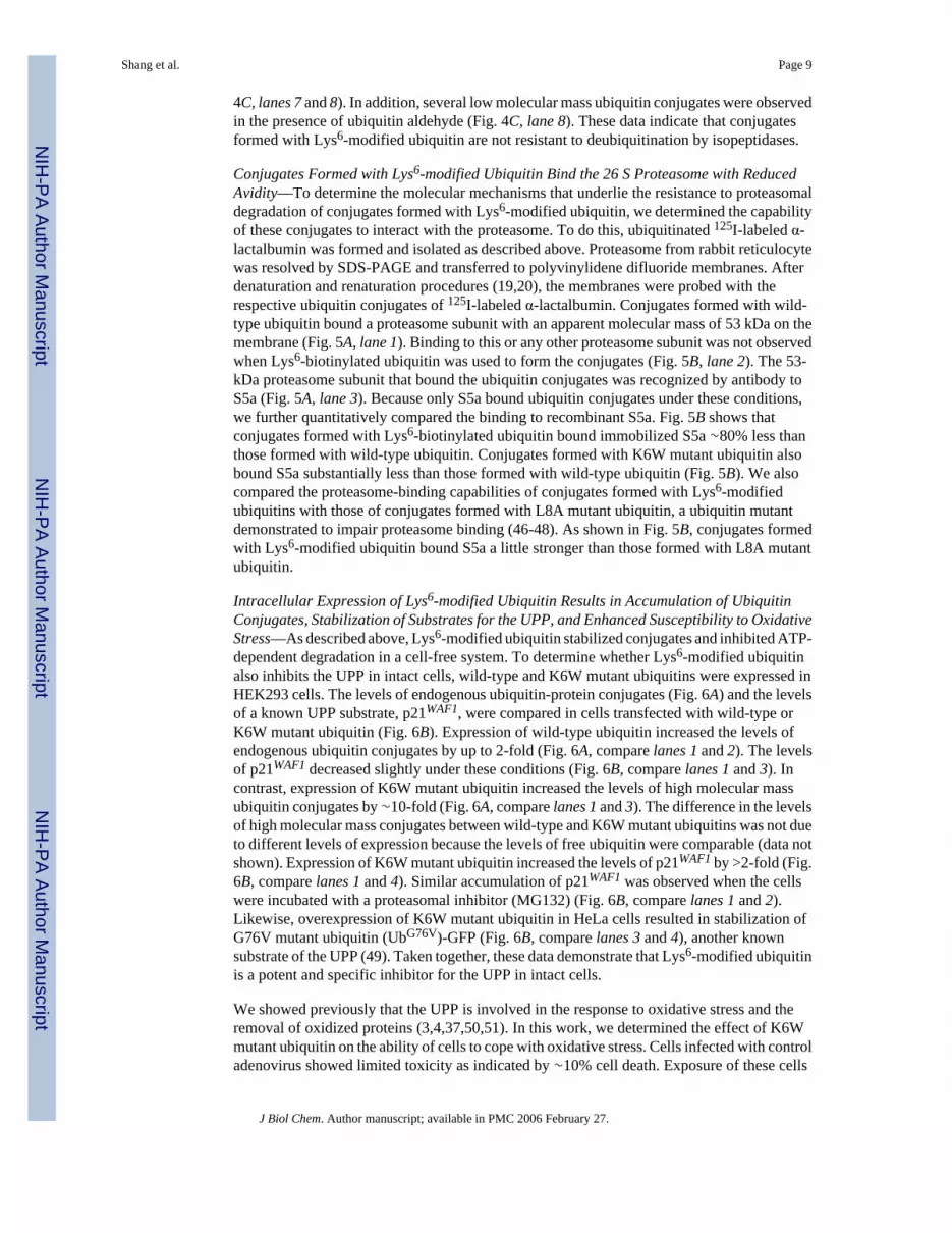
Intracellular Expression of Lys6-modified Ubiquitin Results in Accumulation of UbiquitinConjugates, Stabilization of Substrates for the UPP, and Enhanced Susceptibility to OxidativeStress—As described above, Lys6-modified ubiquitin stabilized conjugates and inhibited ATP-dependent degradation in a cell-free system. To determine whether Lys6-modified ubiquitinalso inhibits the UPP in intact cells, wild-type and K6W mutant ubiquitins were expressed inHEK293 cells. The levels of endogenous ubiquitin-protein conjugates (Fig. 6A) and the levelsof a known UPP substrate, p21WAF1, were compared in cells transfected with wild-type orK6W mutant ubiquitin (Fig. 6B). Expression of wild-type ubiquitin increased the levels ofendogenous ubiquitin conjugates by up to 2-fold (Fig. 6A, compare lanes 1 and 2). The levelsof p21WAF1 decreased slightly under these conditions (Fig. 6B, compare lanes 1 and 3). Incontrast, expression of K6W mutant ubiquitin increased the levels of high molecular massubiquitin conjugates by ∼10-fold (Fig. 6A, compare lanes 1 and 3). The difference in the levelsof high molecular mass conjugates between wild-type and K6W mutant ubiquitins was not dueto different levels of expression because the levels of free ubiquitin were comparable (data notshown). Expression of K6W mutant ubiquitin increased the levels of p21WAF1 by >2-fold (Fig.6B, compare lanes 1 and 4). Similar accumulation of p21WAF1 was observed when the cellswere incubated with a proteasomal inhibitor (MG132) (Fig. 6B, compare lanes 1 and 2).Likewise, overexpression of K6W mutant ubiquitin in HeLa cells resulted in stabilization ofG76V mutant ubiquitin (UbG76V)-GFP (Fig. 6B, compare lanes 3 and 4), another knownsubstrate of the UPP (49). Taken together, these data demonstrate that Lys6-modified ubiquitinis a potent and specific inhibitor for the UPP in intact cells.
We showed previously that the UPP is involved in the response to oxidative stress and theremoval of oxidized proteins (3,4,37,50,51). In this work, we determined the effect of K6Wmutant ubiquitin on the ability of cells to cope with oxidative stress. Cells infected with controladenovirus showed limited toxicity as indicated by ∼10% cell death. Exposure of these cells
Shang et al. Page 9
J Biol Chem. Author manuscript; available in PMC 2006 February 27.
NIH
-PA Author Manuscript
NIH
-PA Author Manuscript
NIH
-PA Author Manuscript

to 20 μM H2O2 for 8 h did not significantly alter cells viability or the percentage of dead cells(Fig. 7, A and B), indicating that these cells could withstand this modest level of oxidativestress and that infection with control adenovirus had no effect on the susceptibility of thesecells to oxidative stress. In contrast, cells infected with the same amount of K6W mutantubiquitin-encoding adenovirus resulted in a slight decrease in cell viability compared with cellsinfected with control adenovirus (Fig. 7, A and B). The cytotoxicity of K6W mutant ubiquitinmay be associated with the inhibition of ubiquitin-dependent proteolysis because proteasomalinhibition also resulted in cytotoxicity in these cells (data not shown). Furthermore, exposureof the cells expressing K6W mutant ubiquitin to 20 μM H2O2 for 8 h dramatically decreasedcell viability and increased cell death by ∼80% as compared with those not treated withH2O2 (Fig. 7, B). Likewise, yeast cells expressing K6R mutant ubiquitin as the sole source ofubiquitin grew similarly to yeast cells expressing wild-type ubiquitin under normal conditions.However, in the presence of canavanine, yeast cells expressing K6R mutant ubiquitin grewsignificantly slower than yeast cells expressing wild-type ubiquitin (Fig. 7C), although wild-type and K6R mutant ubiquitins were expressed at similar levels (data not shown). Theenhanced susceptibility of Lys6 mutant ubiquitin-expressing cells to stresses may be due toimpaired proteasomal degradation of damaged proteins or canavanine-containing proteins. Ithas been shown that some oxidized and canavanine-containing proteins are degraded by theUPP. However, the effects of expression of Lys6 mutant ubiquitin on cell viability may alsobe related to the abrogation of Lys6-linked ubiquitin chains (52).
DISCUSSIONThe major observations of this work include the following. 1) Lys6 was the most readilychemically modified of the 7 Lys residues in human ubiquitin. 2) Lys6-modified ubiquitin wasincorporated into ubiquitin conjugates as efficiently as wild-type ubiquitin. 3) Mutation orchemical modification of Lys6 of ubiquitin reduced its ability to stimulate ATP-dependentprotein degradation. 4) Conjugates formed with Lys6-modified ubiquitin were resistant todegradation by the proteasome, as the conjugates formed with Lys6-modified ubiquitin boundto the proteasome with reduced avidity. 5) Expression of K6W mutant ubiquitin in mammaliancells also inhibited ubiquitin-dependent proteolysis. 6) Impairment of ubiquitin function bymodification or mutation of Lys6 compromised cell viability and survival, particularly understress conditions.
The results show that Lys6-modified ubiquitin is a potent and specific inhibitor of the UPP.Lys6-modified ubiquitin in cell-free assays significantly and specifically inhibited ATP-dependent proteolysis (Table III). Overexpression of K6W mutant ubiquitin in cultured cellsresulted in accumulation of high molecular mass ubiquitin conjugates and stabilization ofp21WAF1 and UbG76V-GFP (Fig. 6), typical substrates for the ubiquitin-proteasome pathway.In human lens epithelial cells, expression of K6W mutant ubiquitin enhanced the susceptibilityof cells to oxidative stress (Fig. 7). The modest cytotoxicity of K6W mutant ubiquitin undernon-stress conditions may be due to the insufficient expression of the mutant ubiquitin, whichonly partially inhibited the UPP. Under these experimental conditions, only ∼60% of the cellsexpressed the mutant ubiquitin. It is likely that only the cells expressing high levels of themutant ubiquitin showed cytotoxicity.
The mechanism of the inhibitory effects of Lys6-modified ubiquitin appears to be related tothe attenuated avidity of the interaction between ubiquitin conjugates and the 26 S protea-some(Fig. 5). It was well established that the hydrophobic patch formed by Leu8, Ile44, and Val70
on the surface of ubiquitin is critical for docking polyubiquitinated substrates to the 26 Sproteasome (46-48), and conjugates formed with L8A mutant ubiquitin were resistant toproteasomal degradation (Fig. 4B). Incorporation of Lys6-modified ubiquitin intopolyubiquitin chains (formed with wild-type and Lys6-modified ubiquitins) may disrupt the
Shang et al. Page 10
J Biol Chem. Author manuscript; available in PMC 2006 February 27.
NIH
-PA Author Manuscript
NIH
-PA Author Manuscript
NIH
-PA Author Manuscript

arrangement of signals in polyubiquitin chains that are required for proteasomal recognition.This hypothesis is consistent with previous results showing that incorporation of one L8Amutant ubiquitin into a tetramer composed of three other wild-type ubiquitins dramaticallyreduced the binding affinity of the heterotetramer for the proteasome (46).
It is intriguing to discover that conjugates formed with L8A mutant ubiquitin were moreresistant to proteasomal degradation than those formed with K6W mutant ubiquitin (Fig. 4B),but that L8A mutant ubiquitin was not as potent as K6W mutant ubiquitin in inhibiting ATP-dependent degradation in reticulocyte lysate (Table IV). Preliminary data indicate that L8Amutant ubiquitin was not used as efficiently as wild-type ubiquitin in fraction II of reticulocytes(data not shown). The hydrophobic patch formed by Leu8, Ile44, and Val70 on the ubiquitinsurface that is recognized by the 19 S complex of the proteasome may also be recognized byother ubiquitin-binding proteins such as E1. It has been reported that this hydrophobic patchis also required for monoubiquitin-mediated processes such as endocytosis (53). Lys6 ofubiquitin is important for proteasome-dependent degradation, but it may not be essential forother ubiquitin-dependent processes. This may explain why mutations of residues related tothe hydrophobic patch are lethal, whereas yeast cells expressing K6A mutant ubiquitin cansurvive under normal conditions (53).
The 26 S proteasome (rather than the 20 S proteasome) is involved in the degradation ofubiquitinated substrates. However, none of the commercially available proteasomal inhibitors,including MG132, lactacystin, and epoxomicin, allow distinction of degradation via the 26 Sproteasome versus the 20 S proteasome (54). Thus, stabilization of a protein by theseproteasomal inhibitors does not prove that the protein is degraded in the UPP because the 20S proteasome degrades proteins in a ubiquitin-independent manner (54). Our demonstrationthat Lys6-modified ubiquitin is a potent and specific inhibitor of the UPP provides a usefulreagent for studying the function of this pathway. Together with the ability to express K6Wmutant ubiquitin at high levels in intact cells, Lys6-modified ubiquitin can be used to distinguishubiquitin-dependent from ubiquitin-independent protein degradation. The accumulation ofubiquitin conjugates by Lys6-modified ubiquitin will also facilitate the isolation andidentification of ubiquitinated proteins in the cells.
Our observation that, of the 7 Lys residues, Lys6 is the most readily modified with sulfo-NHS-biotin in the ubiquitin molecule (Tables I and II) is consistent with prior observations thatLys6 is the most readily modified by p-nitrophenyl acetate (28), aspirin (29), acetic anhydride(30), and Oregon green succinimidyl ester (31). Acetylation and biotinylation of Lys residuesshare the same mechanism as other nonenzymatic modifications such as nonenzymaticglycosylation (glycation). This result suggests that Lys6 of ubiquitin may also be moresusceptible to glycation or modification by 4-hydroxy-2-nonenal, a lipid peroxidation product.Thus, it is reasonable to expect that other types of modifications of ubiquitin Lys6 will alsointerfere with proteasomal degradation and result in the accumulation of ubiquitin conjugates.This might explain why ubiquitin conjugates are accumulated in response to oxidative stressand upon aging (3,37,50,55-58). Accumulation of ubiquitin conjugates in the brain is acharacteristic of age-related diseases such as Alzheimer and Parkinson and cataracts (59,60).Further studies regarding relationships between Lys6 modification and accumulation ofubiquitin per se or ubiquitin conjugates will reveal new roles for ubiquitin in health and disease.
Acknowledgments
We thank Dr. Rob Layfield for providing GSTS5a and Mark Siegal and Matthew Gallagher for help in the preparationof this manuscript.
Shang et al. Page 11
J Biol Chem. Author manuscript; available in PMC 2006 February 27.
NIH
-PA Author Manuscript
NIH
-PA Author Manuscript
NIH
-PA Author Manuscript

REFERENCES1. Hershko A, Ciechanover A. Annu. Rev. Biochem 1998;67:425–479. [PubMed: 9759494]2. Ciechanover A, Orian A, Schwartz AL. J. Cell. Biochem. Suppl 2000;34:40–51. [PubMed: 10762014]3. Shang F, Gong X, Taylor A. J. Biol. Chem 1997;272:23086–23093. [PubMed: 9287309]4. Shang F, Nowell TR Jr. Taylor A. Exp. Eye Res 2001;73:229–238. [PubMed: 11446773]5. Haas A, Reback PM, Pratt G, Rechsteiner M. J. Biol. Chem 1990;265:21664–21669. [PubMed:
2174883]6. Baboshina OV, Haas AL. J. Biol. Chem 1996;271:2823–2831. [PubMed: 8576261]7. Arnason T, Ellison MJ. Mol. Cell. Biol 1994;14:7876–7883. [PubMed: 7969127]8. Spence J, Sadis S, Haas AL, Finley D. Mol. Cell. Biol 1995;15:1265–1273. [PubMed: 7862120]9. Wang C, Deng L, Hong M, Akkaraju GR, Inoue J, Chen ZJ. Nature 2001;412:346–351. [PubMed:
11460167]10. Deng L, Wang C, Spencer E, Yang L, Braun A, You J, Slaughter C, Pickart C, Chen ZJ. Cell
2000;103:351–361. [PubMed: 11057907]11. Hofmann RM, Pickart CM. J. Biol. Chem 2001;276:27936–27943. [PubMed: 11369780]12. Chau V, Tobias JW, Bachmair A, Marriott D, Ecker DJ, Gonda DK, Varshavsky A. Science
1989;243:1576–1583. [PubMed: 2538923]13. Gregori L, Poosch MS, Cousins G, Chau V. J. Biol. Chem 1990;265:8354–8357. [PubMed: 2160452]14. Pickart CM. FASEB J 1997;11:1055–1066. [PubMed: 9367341]15. Hoffman L, Pratt G, Rechsteiner M. J. Biol. Chem 1992;267:22362–22368. [PubMed: 1331052]16. DeMartino GN, Moomaw CR, Zagnitko OP, Proske RJ, Chu-Ping M, Afendis SJ, Swaffield JC,
Slaughter CA. J. Biol. Chem 1994;269:20878–20884. [PubMed: 8063704]17. Lowe J, Stock D, Jap B, Zwickl P, Baumeister W, Huber R. Science 1995;268:533–539. [PubMed:
7725097]18. Groll M, Ditzel L, Lowe J, Stock D, Bochtler M, Bartunik HD, Huber R. Nature 1997;386:463–471.
[PubMed: 9087403]19. Deveraux Q, Ustrell V, Pickart C, Rechsteiner M. J. Biol. Chem 1994;269:7059–7061. [PubMed:
8125911]20. van Nocker S, Deveraux Q, Rechsteiner M, Vierstra RD. Proc. Natl. Acad. Sci. U. S. A 1996;93:856–
860. [PubMed: 8570648]21. Elsasser S, Gali RR, Schwickart M, Larsen CN, Leggett DS, Muller B, Feng MT, Tubing F, Dittmar
GA, Finley D. Nat. Cell Biol 2002;4:725–730. [PubMed: 12198498]22. Elsasser S, Chandler-Militello D, Muller B, Hanna J, Finley D. J. Biol. Chem 2004;279:26817–26822.
[PubMed: 15117949]23. Verma R, Oania R, Graumann J, Deshaies RJ. Cell 2004;118:99–110. [PubMed: 15242647]24. Lam YA, Xu W, DeMartino GN, Cohen RE. Nature 1997;385:737–740. [PubMed: 9034192]25. Lam YA, DeMartino GN, Pickart CM, Cohen RE. J. Biol. Chem 1997;272:28438–28446. [PubMed:
9353303]26. Yao T, Cohen RE. Nature 2002;419:403–407. [PubMed: 12353037]27. Corsi D, Galluzzi L, Crinelli R, Magnani M. J. Biol. Chem 1995;270:8928–8935. [PubMed: 7721801]28. Jabusch JR, Deutsch HF. Arch. Biochem. Biophys 1985;238:170–177. [PubMed: 2984995]29. Macdonald JM, LeBlanc DA, Haas AL, London RE. Biochem. Pharmacol 1999;57:1233–1244.
[PubMed: 10230767]30. Macdonald JM, Haas AL, London RE. J. Biol. Chem 2000;275:31908–31913. [PubMed: 10906321]31. Wee KE, Lai Z, Auger KR, Ma J, Horiuchi KY, Dowling RL, Dougherty CS, Corman JI, Wynn R,
Copeland RA. J. Protein Chem 2000;19:489–498. [PubMed: 11195973]32. Vijay-Kumar S, Bugg CE, Cook WJ. J. Mol. Biol 1987;194:531–544. [PubMed: 3041007]33. Vijay-Kumar S, Bugg CE, Wilkinson KD, Vierstra RD, Hatfield PM, Cook WJ. J. Biol. Chem
1987;262:6396–6399. [PubMed: 3032965]34. Burch TJ, Haas AL. Biochemistry 1994;33:7300–7308. [PubMed: 8003494]
Shang et al. Page 12
J Biol Chem. Author manuscript; available in PMC 2006 February 27.
NIH
-PA Author Manuscript
NIH
-PA Author Manuscript
NIH
-PA Author Manuscript

35. Finch JS, Bonham K, Krieg P, Bowden GT. Nucleic Acids Res 1990;18:1907–1907. [PubMed:2159627]
36. He TC, Zhou S, da Costa LT, Yu J, Kinzler KW, Vogelstein B. Proc. Natl. Acad. Sci. U. S. A1998;95:2509–2514. [PubMed: 9482916]
37. Shang F, Taylor A. Biochem. J 1995;307:297–303. [PubMed: 7717989]38. Ciechanover A, Elias S, Heller H, Hershko A. J. Biol. Chem 1982;257:2537–2542. [PubMed:
6277904]39. Obin M, Nowell T, Taylor A. Curr. Eye Res 1995;14:751–760. [PubMed: 8529413]40. Shang F, Huang L, Taylor A. Curr. Eye Res 1994;13:423–431. [PubMed: 7924406]41. Huang LL, Jahngen-Hodge J, Taylor A. Biochim. Biophys. Acta 1993;1175:181–187. [PubMed:
8380340]42. Gaczynska M, Goldberg AL, Tanaka K, Hendil KB, Rock KR. J. Biol. Chem 1996;271:17275–17280.
[PubMed: 8663318]43. Layfield R, Tooth D, Landon M, Dawson S, Mayer J, Alban A. Proteomics 2001;1:773–777.
[PubMed: 11677784]44. Finley D, Sadis S, Monia BP, Boucher P, Ecker DJ, Crooke ST, Chau V. Mol. Cell. Biol
1994;14:5501–5509. [PubMed: 8035826]45. Pickart CM, Fushman D. Curr. Opin. Chem. Biol 2004;8:610–616. [PubMed: 15556404]46. Beal R, Deveraux Q, Xia G, Rechsteiner M, Pickart C. Proc. Natl. Acad. Sci. U. S. A 1996;93:861–
866. [PubMed: 8570649]47. Beal RE, Toscano-Cantaffa D, Young P, Rechsteiner M, Pickart CM. Biochemistry 1998;37:2925–
2934. [PubMed: 9485444]48. Lam YA, Lawson TG, Velayutham M, Zweier JL, Pickart CM. Nature 2002;416:763–767. [PubMed:
11961560]49. Dantuma NP, Lindsten K, Glas R, Jellne M, Masucci MG. Nat. Biotechnol 2000;18:538–543.
[PubMed: 10802622]50. Shang F, Gong X, Palmer HJ, Nowell TR, Taylor A. Exp. Eye Res 1997;64:21–30. [PubMed:
9093017]51. Dudek EJ, Shang F, Taylor A. Free Radic. Biol. Med 2001;31:651–658. [PubMed: 11522450]52. Morris JR, Solomon E. Hum. Mol. Genet 2004;13:807–817. [PubMed: 14976165]53. Sloper-Mould KE, Jemc JC, Pickart CM, Hicke L. J. Biol. Chem 2001;276:30483–30489. [PubMed:
11399765]54. Grune T, Reinheckel T, Davies KJ. FASEB J 1997;11:526–534. [PubMed: 9212076]55. Ivy GO, Kitani K, Ihara Y. Brain Res 1989;498:360–365. [PubMed: 2477115]56. Kudo T, Iqbal K, Ravid R, Swaab DF, Grundke-Iqbal I. Brain Res 1994;639:1–7. [PubMed: 8180825]57. Scrofano MM, Shang F, Nowell TR Jr. Gong X, Smith DE, Kelliher M, Dunning J, Mura CV, Taylor
A. Mech. Ageing Dev 1998;105:273–290. [PubMed: 9862235]58. Scrofano MM, Shang F, Nowell TR Jr. Gong X, Smith DE, Kelliher M, Dunning J, Mura CV, Taylor
A. Mech. Ageing Dev 1998;101:277–296. [PubMed: 9622231]59. Jahngen-Hodge J, Cyr D, Laxman E, Taylor A. Exp. Eye Res 1992;55:897–902. [PubMed: 1336733]60. Jahngen JH, Lipman RD, Eisenhauer DA, Jahngen EG, Taylor A. Arch. Biochem. Biophys
1990;276:32–37. [PubMed: 2153364]
Shang et al. Page 13
J Biol Chem. Author manuscript; available in PMC 2006 February 27.
NIH
-PA Author Manuscript
NIH
-PA Author Manuscript
NIH
-PA Author Manuscript

FIG. 1.Characterization of biotin-labeled ubiquitin. Sulfo-NHS-biotin modifies primary aminegroups on proteins to form amide bonds (A). Ubiquitin was labeled in PBS (pH 7.5) withsulfoNHS-biotin at molar ratios of 2 (B) and 3.5 (C). The labeled ubiquitin was separated byHPLC, and elution was monitored at 280 nm (B and C). Peaks were collected, and the molecularmass of each peak was determined by mass spectrometry. The peak that eluted at ∼22.6 minhad a mass of 8565.0 Da, which corresponds to unmodified ubiquitin (Ub). The peak that elutedat ∼23.5 min had a mass of 8790.3 Da, which corresponds to monobiotinylated ubiquitin((Bio)1Ub). The peaks that eluted at ∼24.5 and 26 min had an identical mass of 9016.3 Da,which corresponds to dibiotinylated ubiquitin ((Bio)2Ub). The peak that eluted at ∼27 min hada mass of 9242.8 Da, which corresponds to tribiotinylated ubiquitin ((Bio)3Ub). mAU, milli-absorbance units.
Shang et al. Page 14
J Biol Chem. Author manuscript; available in PMC 2006 February 27.
NIH
-PA Author Manuscript
NIH
-PA Author Manuscript
NIH
-PA Author Manuscript

FIG. 2.Biotin-labeled ubiquitin is efficiently used by the ubiquitin conjugation system to formubiquitin conjugates. A, 2 μg of wild-type ubiquitin (wt Ub) or purified Lys6-biotinylatedubiquitin (K6-Bio Ub) was added to fraction II of rabbit reticulocytes in the presence of 2 mMATP in a 25-μl assay. After incubation at 37 °C for 30 min, the reaction was terminated byaddition of 25 μl of 2× SDS gel loading buffer. The mixture was resolved by SDS-PAGE andtransferred to a nitrocellulose membrane. The levels of ubiquitin conjugates (Ub-conj.) weredetected by Western blot analysis using anti-ubiquitin antibodies. K6K48-dibio Ub, Lys6/Lys48-dibiotinylated ubiquitin; K6K63-dibio Ub, Lys6/Lys63-dibiotinylated ubiquitin;K6K48K63-tribio Ub, Lys6/Lys48/Lys63-tribiotinylated ubiquitin. B, 0—4 μg of unmodifiedubiquitin (lanes 1—6) or Lys6-biotinylated ubiquitin (lanes 7—12) was added to theconjugation assays, which contained 0.5 μgof 125I-labeled wild-type ubiquitin. Ubiquitinconjugation was performed as described for A, and ubiquitin conjugates were monitored byautoradiography. C, the results in B were quantitated by densitometry. D, 0—4 μg of wild-typeubiquitin was added to the conjugation assays, which contained 0.5 μg of Lys6-biotinylatedubiquitin. Biotinylated ubiquitin conjugates were determined with horseradish peroxidase-conjugated avidin using a chemiluminescence detection kit.
Shang et al. Page 15
J Biol Chem. Author manuscript; available in PMC 2006 February 27.
NIH
-PA Author Manuscript
NIH
-PA Author Manuscript
NIH
-PA Author Manuscript

FIG. 3.Lys6-modified ubiquitin causes accumulation of high molecular mass ubiquitinconjugates. Transducin (A) and α-lactalbumin (B and C) were labeled with 125I and used assubstrates for conjugation assays in the RPE cell supernatant. Each 25-μl assay contained 2μg of native or modified ubiquitin (Ub) as indicated. The reaction mixture was resolved bySDS-PAGE, and the levels of ubiquitinated substrates were determined by autoradiography.In C, K6W mutant ubiquitin was His6-tagged; therefore, the masses of the mono-, di, andtriubiquitinated α-lactalbumin conjugates formed with this mutant ubiquitin were a little higherthan those formed with wild-type ubiquitin (wt Ub). K6-Bio Ub, Lys6-biotinylated ubiquitin;K6K48-Dibio Ub, Lys6/Lys48-dibiotinylated ubiquitin; K6K63-Dibio Ub, Lys6/Lys63-dibiotinylated ubiquitin; K6K48K63 Tribio, Lys6/Lys48/Lys63-tribiotinylated ubiquitin.
Shang et al. Page 16
J Biol Chem. Author manuscript; available in PMC 2006 February 27.
NIH
-PA Author Manuscript
NIH
-PA Author Manuscript
NIH
-PA Author Manuscript

FIG. 4.Conjugates formed with Lys6-modified ubiquitin are relatively resistant to proteasomaldegradation, but are not resistant to isopeptidases. Ubiquitin conjugates were formed withwild-type, Lys6-biotinylated, or K6W or L8A mutant ubiquitin in proteasome-free fraction IIof reticulocytes using 125I-labeled α-lactalbumin as substrate. The ubiquitinated 125I-labeledα-lactalbumin was separated from non-ubiquitinated 125I-labeled α-lactalbumin by ion-exchange chromatography, and its susceptibility to purified proteasome was determined. A,profiles of ubiquitinated α-lactalbumin (Ub-lact) formed with ubiquitin variants. Lane 1, wild-type ubiquitin (wt); lane 2, Lys6-biotinylated ubiquitin (K6-Bio); lane 3, K6W mutantubiquitin; and lane 4, L8A mutant ubiquitin. B, proteasomal degradation ofubiquitinated 125I-labeled α-lactalbumin in the RPE cell supernatant. Ubiquitin conjugatesof 125I-labeled α-lactalbumin were incubated with the reconstituted 26 S proteasome(composed of 0.25 μg of 20 S and 0.5 μg of 19 S) in the presence of ATP. Degradation rateswere determined as the percentage of the substrates that became trichloroacetic acid-solubleafter incubation with proteasome. wt Ub, wild-type ubiquitin; K6-Bio Ub, Lys6-biotinylatedubiquitin; K6W Ub, K6W mutant ubiquitin; L8A Ub, L8A mutant ubiquitin. C, deubiquitinationassay. Wild-type (lanes 1—3) or Lys6-modified (lanes 4—6) ubiquitin was labeled with 125I,and ubiquitin conjugates (Ub-conj.) were formed in proteasome-free fraction II of rabbitreticulocyte. Deubiquitination by isopeptidases of the 125I-labeled ubiquitin conjugates wasdetermined in the presence of a 20-fold excess of unlabeled wild-type ubiquitin conjugates. Inlanes 7 and 8, ubiquitin aldehyde (Ubal) was added to inhibit isopeptidases.
Shang et al. Page 17
J Biol Chem. Author manuscript; available in PMC 2006 February 27.
NIH
-PA Author Manuscript
NIH
-PA Author Manuscript
NIH
-PA Author Manuscript

FIG. 5.Conjugates formed with Lys6-modified ubiquitin bind S5a of the proteasome withreduced avidity. Ubiquitin conjugates of α-lactalbumin were formed with wild-type, Lys6-biotinylated, or K6W or L8A mutant ubiquitin and isolated as described in the legend to Fig.4. A, proteasome isolated from rabbit reticulocytes was resolved by SDS-PAGE and transferredto polyvinylidene difluoride membranes. After denaturation and renaturation procedures, themembranes were incubated overnight with conjugates of 125I-labeled α-lactalbumin formedwith wild-type or Lys6-biotinylated ubiquitin. After removal of nonspecifically boundradioactivity, the membranes were exposed to x-ray film, and the specific binding wasvisualized by autoradiography. Lane 1, conjugates formed with wild-type ubiquitin (wt UB);lane 2, conjugates formed with Lys6-biotinylated ubiquitin (K6-Bio Ub); lane 3, Western blotanalysis (immunoblotting (IB)) of the 26 S proteasome using antibody to S5a. B, shown arethe results from S5a binding assay. Equal amounts of ubiquitinated 125I-labeled α-lactalbumin(4 × 10⁴ cpm) formed with wild-type, Lys6-biotinylated, or K6W or L8A mutant ubiquitinwere incubated with GST-S5a, which was immobilized overnight on glutathione-agarose beadswith constant shaking. After extensive washing, ubiquitin conjugates were eluted from thebeads with glutathione and quantified using a γ-counter. The data presented are the relativebinding capabilities, where the binding capability of conjugates formed with wild-typeubiquitin is designated as 100%.
Shang et al. Page 18
J Biol Chem. Author manuscript; available in PMC 2006 February 27.
NIH
-PA Author Manuscript
NIH
-PA Author Manuscript
NIH
-PA Author Manuscript
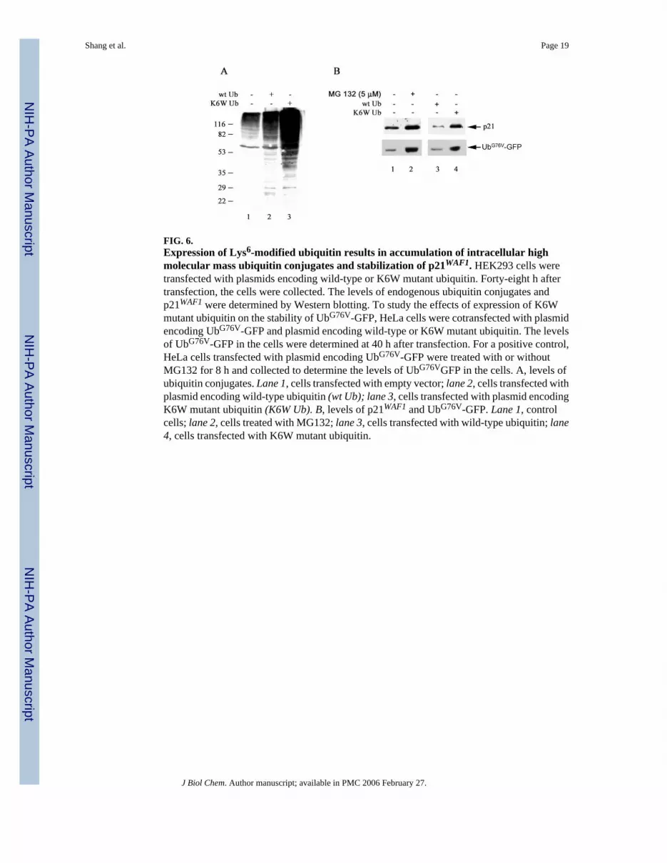
FIG. 6.Expression of Lys6-modified ubiquitin results in accumulation of intracellular highmolecular mass ubiquitin conjugates and stabilization of p21WAF1. HEK293 cells weretransfected with plasmids encoding wild-type or K6W mutant ubiquitin. Forty-eight h aftertransfection, the cells were collected. The levels of endogenous ubiquitin conjugates andp21WAF1 were determined by Western blotting. To study the effects of expression of K6Wmutant ubiquitin on the stability of UbG76V-GFP, HeLa cells were cotransfected with plasmidencoding UbG76V-GFP and plasmid encoding wild-type or K6W mutant ubiquitin. The levelsof UbG76V-GFP in the cells were determined at 40 h after transfection. For a positive control,HeLa cells transfected with plasmid encoding UbG76V-GFP were treated with or withoutMG132 for 8 h and collected to determine the levels of UbG76VGFP in the cells. A, levels ofubiquitin conjugates. Lane 1, cells transfected with empty vector; lane 2, cells transfected withplasmid encoding wild-type ubiquitin (wt Ub); lane 3, cells transfected with plasmid encodingK6W mutant ubiquitin (K6W Ub). B, levels of p21WAF1 and UbG76V-GFP. Lane 1, controlcells; lane 2, cells treated with MG132; lane 3, cells transfected with wild-type ubiquitin; lane4, cells transfected with K6W mutant ubiquitin.
Shang et al. Page 19
J Biol Chem. Author manuscript; available in PMC 2006 February 27.
NIH
-PA Author Manuscript
NIH
-PA Author Manuscript
NIH
-PA Author Manuscript

FIG. 7.Expression of Lys6 mutant ubiquitin increases the susceptibility of cells to stress. Humanlens epithelial cells at ∼50% confluence were infected with recombinant adenovirus encodingK6W mutant ubiquitin or with control adenovirus for 48 h. The cells were then exposed to∼20 μM H2O2 constantly generated by addition of glucose oxidase to the medium for 8 h. A,cell viability was determined by 3-(4,5-dimethylthiazol-2-yl)-5-(3-carboxymethoxyphenyl)-2-(4-sulfophenyl)-2H-tetrazolium bromide assay. B, the percentage of dead cells that werestained by trypan blue was determined. The data represent the means ± S.D. of sixmeasurements. C, yeast cells expressing wild-type (wt) or K6R mutant ubiquitin were culturedin the absence or presence of 1 μM canavanine.
Shang et al. Page 20
J Biol Chem. Author manuscript; available in PMC 2006 February 27.
NIH
-PA Author Manuscript
NIH
-PA Author Manuscript
NIH
-PA Author Manuscript

NIH
-PA Author Manuscript
NIH
-PA Author Manuscript
NIH
-PA Author Manuscript
Shang et al. Page 21
TABLE IBiotinylation sites of biotin-labeled ubiquitin as determined by peptide mass mapping after endopeptidase Lys-C digestion. Biotin-labeled ubiquitin was purified by reversed-phase HPLC and digested by endopeptidase Lys-C. The resulting peptides were separated by reversed-phase HPLC, and their masses were determined using anin-line coupled mass spectrometer. In monobiotinylated ubiquitin ((Bio)1Ub), the biotin was exclusively attachedto residues between Met1 and Lys11. In dibiotinylated ubiquitin ((Bio)2Ub), one biotin was located betweenMet1 and Lys11, and the other biotin was located between Glu34 and Lys63 (peak eluting at 24.5 min) or Gln49
and Lys63 (peak eluting at 26 min). In tribiotinylated ubiquitin ((Bio)3Ub), one biotin was located betweenMet1 and Lys11, and the other two biotins were located between Glu34 and Lys63. The mass for the biotin moietyis 227 atomic mass units. WT Ub, wild-type ubiquitin.
WT Ub Peak at 23.5 min,(Bio)1Ub
Peak at 24.5 min,(Bio)2Ub
Peak at 26 min,(Bio)2Ub
Peak at 27 min,(Bio)3Ub
Masses Positions Masses Positions Masses Positions Masses Positions Masses Positions
Da Da Da Da Da
764.4 Met1-Lys6 1490.0
Met1-Lys11
biotin1490.2
Met1-Lys11
biotin1490.2
Met1-Lys11
biotin1490.0
Met1-Lys11
biotin518.3 Thr7-
Lys11
1786.7 Thr12-Lys27 1786.5 Thr12-
Lys27 1786.2 Thr12-Lys27 1786.7 Thr12-
Lys27 1786.2 Thr12-Lys27
1667.9 Glu34-Lys48 1668.2 Glu34-
Lys48 3773.2Glu34-Lys63
biotin1668.0 Glu34-
Lys48 3999.5Glu34-Lys63
dibiotin
1778.9 Gln49-Lys63 1779.2 Gln49-
Lys63 2006.3Gln49-Lys63
biotin1449.8 Glu64-
Gly76 1450.0 Glu64-Gly76 1449.5 Glu64-
Gly76 1450.0 Glu64-Gly76 1449.5 Glu64-
Gly76
J Biol Chem. Author manuscript; available in PMC 2006 February 27.

NIH
-PA Author Manuscript
NIH
-PA Author Manuscript
NIH
-PA Author Manuscript
Shang et al. Page 22
TABLE IIBiotinylation sites of dibiotin-labeled ubiquitin determined by peptide mass mapping after trypsin digestion.Unmodified and dibiotinylated ((Bio)2Ub; peak eluting at 24.5 min) ubiquitins were digested by trypsin. Theresulting peptides were separated by reversed-phase HPLC, and their masses were determined using an in-linecoupled mass spectrometer. One biotin was located between Met1 and Lys11, and the other biotin of thisdibiotinylated ubiquitin was located between Leu43 and Arg54. WT Ub, wild-type ubiquitin.
WT Ub Peak at 24.5 min, (Bio)2Ub
Masses Positions Masses Positions
Da Da764.2 Met1-Lys6 1490.0 Met1-Lys11 biotin518.2 Thr7-Lys11
1786.7 Thr12-Lys27 1786.5 Thr12-Lys27
1038 Glu34-Arg42 1038.2 Glu34-Arg42
643.4 Leu43-Lys48 1572.2 Leu43-Arg54 biotin716.8 Gln49-Arg54
1080.3 Thr55-Lys63 1080.5 Thr55-Lys63
1066.4 Glu64-Arg72 1066.7 Glu64-Arg72
J Biol Chem. Author manuscript; available in PMC 2006 February 27.
NIH
-PA Author Manuscript
NIH
-PA Author Manuscript
NIH
-PA Author Manuscript
Shang et al. Page 23
TABLE IIILys6-modified ubiquitin inhibits ATP-dependent degradation of transducin in the RPE cell lysate. The ability ofdifferentially labeled ubiquitin (Ub) and K6A or K6W mutant ubiquitin to support ATP-dependent degradationwas determined in supernatants of RPE cells using 125I-labeled transducin as substrate. The final concentrationof the added ubiquitin was 8 μM in a 25-μl assay. The degradation rates are expressed as percentages of 125I-labeled substrate degraded into acid-soluble fragments in a 2-h period. WT Ub, wild-type ubiquitin.
ATP-dependent degradation Stimulation
% %Without addition of Ub 22 ± 2.2 0WT Ub 44 ± 3.4 100Lys6-biotinylated Ub 12 ± 1.9 -45K6A Ub 13 ± 1.2 -41K6W Ub 10 ± 2.5 -54Lys6/Lys48-dibiotinylated Ub 18 ± 1.1 -18Lys6/Lys63-dibiotinylated Ub 19 ± 1.8 -14
J Biol Chem. Author manuscript; available in PMC 2006 February 27.

NIH
-PA Author Manuscript
NIH
-PA Author Manuscript
NIH
-PA Author Manuscript
Shang et al. Page 24
TABLE IVK6W mutant ubiquitin inhibits ATP-dependent degradation of α-crystallin in reticulocyte lysate. The effects ofwild-type (WT), Lys6-biotinylated, or K6W or L8A mutant ubiquitin (Ub) on ATP-dependent degradation ofαA-crystallin were determined in supernatants of rabbit reticulocyte lysate. The final concentration of the addedubiquitin was 8 μM in a 25-μl assay. The degradation rates are expressed as percentages of 125I-labeled αA-crystallin degraded into acid-soluble fragments in a 90-min period.
ATP-dependentdegradation Stimulation
% %Without addition of Ub 13.4 ± 2.5 0WT Ub 12.9 ± 3.1 -3Lys6-biotinylated Ub 5.4 ± 2.1 -60K6W Ub 4.1 ± 1.8 -69L8A Ub 7.7 ± 1.4 -42
J Biol Chem. Author manuscript; available in PMC 2006 February 27.


