Embed Size (px)
Citation preview
Victor C.Hou, Robert Lersch, Sherry L.Gee,Julie L.Ponthier, Annie J.Lo, Michael Wu1,Chris W.Turck2, Mark Koury3,Adrian R.Krainer4, Akila Mayeda5 andJohn G.Conboy6
Lawrence Berkeley National Laboratory, Life Sciences Divisionand 1Department of Molecular and Cellular Biology, University ofCalifornia at Berkeley, Berkeley, CA 94720, 2University of California,San Francisco, HHMI, Department of Medicine and CardiovascularResearch Institute, San Francisco, CA 94143, 3Department ofMedicine, Vanderbilt University, Veterans Affairs Medical Centers,Nashville, TN 37232, 4Cold Spring Harbor Laboratory, Cold SpringHarbor, NY 11724 and 5University of Miami School of Medicine,Department of Biochemistry and Molecular Biology, Miami,FL 33136, USA
6Corresponding authore-mail: [email protected]
A physiologically important alternative pre-mRNAsplicing switch, involving activation of protein 4.1Rexon 16 (E16) splicing, is required for the establish-ment of proper mechanical integrity of the erythro-cyte membrane during erythropoiesis. Here weidentify a conserved exonic splicing silencer element(CE16) in E16 that interacts with hnRNP A/B proteinsand plays a role in repression of E16 splicing duringearly erythropoiesis. Experiments with model pre-mRNAs showed that CE16 can repress splicing ofupstream introns, and that mutagenesis or replace-ment of CE16 can relieve this inhibition. An af®nityselection assay with biotinylated CE16 RNA demon-strated speci®c binding of hnRNP A/B proteins.Depletion of hnRNP A/B proteins from nuclearextract signi®cantly increased E16 inclusion, whilerepletion with recombinant hnRNP A/B restored E16silencing. Most importantly, differentiating mouseerythroblasts exhibited a stage-speci®c activation ofthe E16 splicing switch in concert with a dramatic andspeci®c down-regulation of hnRNP A/B proteinexpression. These ®ndings demonstrate that naturaldevelopmental changes in hnRNP A/B proteins caneffect physiologically important switches in pre-mRNA splicing.Keywords: alternative splicing/exonic splicing silencer/hnRNP A and B/protein 4.1R
Introduction
Alternative splicing of pre-mRNAs from a single genefacilitates expression of multiple protein isoforms that canhave different functional characteristics (reviewed inChabot, 1996; Smith and Valcarcel, 2000). Recent analy-sis of the Human Genome Project predicts that themajority of human genes undergo some form of alternative
splicing (Lander et al., 2001), indicating that this is amajor mechanism for regulating gene expression. Thesealternative splicing events are often developmentallyregulated and/or exhibit tissue-speci®c variations. One ofthe major challenges in the ®eld is to de®ne the molecularmechanisms whereby splicing factors with splicingenhancer or silencer activity help regulate alternativepre-mRNA splicing in the appropriate developmentalpatterns.
In many cases, the primary sequence of an alternativeexon plays a dual role: not only does the sequence performan obvious protein-coding function, but it also often servesas binding sites for speci®c splicing factor proteins thatregulate post-transcriptional processing. Alternativelyspliced exons commonly are associated with both posi-tively and negatively cis-acting elements that determinethe usage of the (typically weak) ¯anking splice sites. Agreat deal of work over the past several years has beendevoted to identi®cation of exonic splicing enhancer(ESE) elements in both alternative exons (reviewed inReed, 1996; Hertel et al., 1997; Wang and Manley, 1997)and constitutive exons (Mayeda et al., 1999; Schaal andManiatis, 1999).
Many alternative exons also possess negatively actingsequences termed exonic splicing silencer (ESS) elementsthat can antagonize splicing enhancer function and preventexon inclusion until the appropriate developmental stage.Recently, a number of alternative exons have been shownto contain ESS elements (Graham et al., 1992; Amendtet al., 1995; Del Gatto and Breathnach, 1995; Staffa et al.,1997; Konig et al., 1998; Si et al., 1998; Caputi et al.,1999; Kan and Green, 1999; Mayeda et al., 1999). Amongthe trans-acting factors that appear to be capable of actingthrough silencer elements to mediate a repressive effectare snRNP complexes (Nelson and Green, 1990; Siebelet al., 1992; Kan and Green, 1999) and members ofthe hnRNP family such as the A/B proteins or hnRNP I/polypyrimidine tract binding protein (PTB) (Ashiya andGrabowski, 1997; Chan and Black, 1997; Caputi et al.,1999; Del Gatto-Konczak et al., 1999; Matter et al., 2000;Bilodeau et al., 2001; Tange et al., 2001; Zhu et al., 2001).
The cytoskeletal protein 4.1R gene exhibits severaldevelopmentally regulated alternative splicing events inerythroid, epithelial and muscle cell types (Conboy, 1999).In particular, a regulated splicing switch involving exon 16(E16) during erythroid differentiation plays a criticalphysiological role in establishing the appropriate red cellmembrane material properties. E16 is skipped in earlyerythroid progenitors but included ef®ciently in matureerythroblasts (Chasis et al., 1993), leading to synthesis of4.1R protein isoforms that bind spectrin and actin andassemble stably into the membrane skeleton (Horne et al.,1993; Discher et al., 1995; Schischmanoff et al., 1995).E16 also encodes part of a nuclear localization signal and
Decrease in hnRNP A/B expression duringerythropoiesis mediates a pre-mRNA splicing switch
The EMBO Journal Vol. 21 No. 22 pp. 6195±6204, 2002
Published by Oxford University Press 6195
is thus an important determinant of subcellular localizationin certain nucleated cell types (Luque et al., 1998; Gascardet al., 1999).
We have developed a heterologous system in whichthe alternative splicing of E16 can be reconstituted inthe context of a three-exon model pre-mRNA, and thesequence determinants for ef®cient inclusion can beexplored (Gee et al., 2000). These studies yieldedevidence for an ordered splicing model in which theintron downstream of E16 is removed preferentially priorto excision of the upstream intron. Coordinated interactionof regulatory elements in a speci®c spatial and temporalmanner is postulated for proper regulation of E16 inclu-sion/exclusion during erythrocyte development. One ofthese key regulatory elements is the suboptimal 5¢ splicesite (5¢ss) adjacent to E16 (Gee et al., 2000), whichprevents constitutive splicing of the exon. Recent parallel
studies have indicated the involvement of additionalregulatory elements in exon 16 itself as well as the¯anking intron sequences (Deguillien et al., 2001). In thisreport, we characterize an exonic RNA element that isinvolved in E16 splicing regulation, a phylogeneticallyconserved silencer element in E16 that functions throughthe binding of the hnRNP A/B proteins to repress E16inclusion. Importantly, down-regulation of hnRNP A/Bprotein expression temporally correlates with the acti-vation of E16 splicing during erythropoiesis, suggestingthat this is the functional switch for activation of E16expression in the 4.1R gene during erythropoiesis.
Results
Alternative splicing of 4.1R E16 is conserved among threevertebrate orders (Winardi et al., 1995; Conboy, 1999).
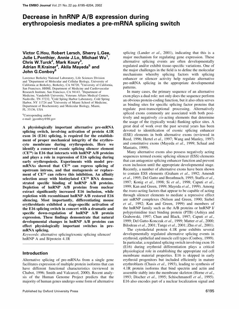
Fig. 1. Conservation of 4.1R E16 and ¯anking intron sequences. Conserved sequence elements are boxed. M, mouse; h, human; f, frog; c, chicken;b, bovine; introns are in lower case and exons in upper case.
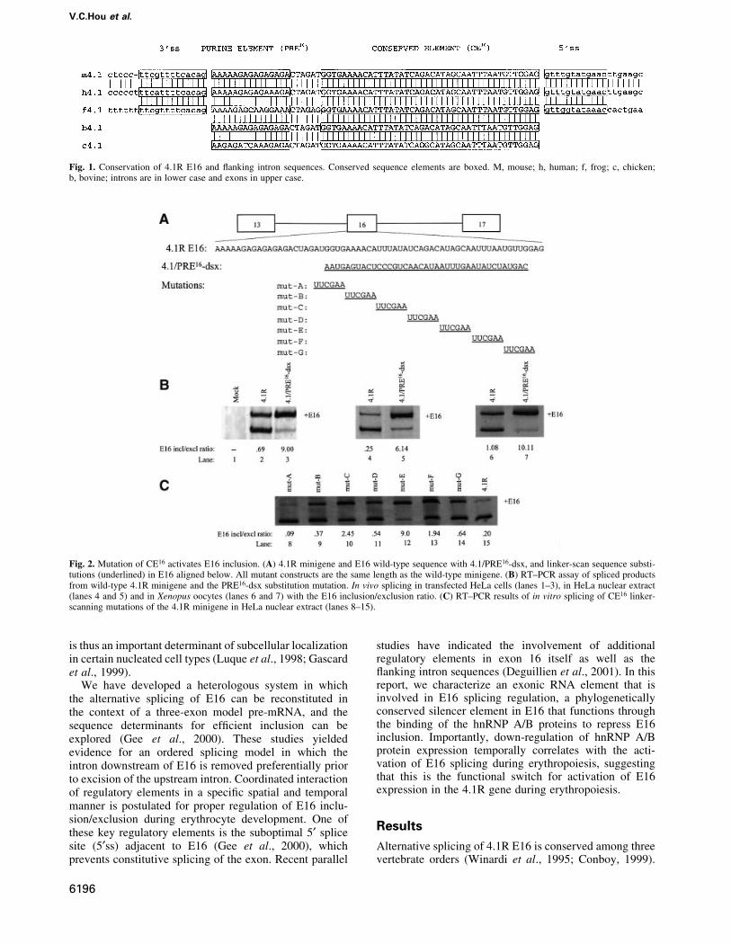
Fig. 2. Mutation of CE16 activates E16 inclusion. (A) 4.1R minigene and E16 wild-type sequence with 4.1/PRE16-dsx, and linker-scan sequence substi-tutions (underlined) in E16 aligned below. All mutant constructs are the same length as the wild-type minigene. (B) RT±PCR assay of spliced productsfrom wild-type 4.1R minigene and the PRE16-dsx substitution mutation. In vivo splicing in transfected HeLa cells (lanes 1±3), in HeLa nuclear extract(lanes 4 and 5) and in Xenopus oocytes (lanes 6 and 7) with the E16 inclusion/exclusion ratio. (C) RT±PCR results of in vitro splicing of CE16 linker-scanning mutations of the 4.1R minigene in HeLa nuclear extract (lanes 8±15).
V.C.Hou et al.
6196
With the assumption that splicing regulatory sequencesmight be phylogenetically conserved, we compared thenucleotide sequences around E16 in the mammalian(human, mouse and bovine), amphibian (frog) and avian(chicken) genes (Figure 1). Several candidate regulatoryelements were identi®ed, including a conserved 3¢ss, aweak 5¢ss (Gee et al., 2000), a purine-rich element withinthe 5¢ portion of E16 (PRE16) and a highly conservedelement (CE16) of ~40 nucleotides spanning most of theremainder of the exon. As shown below, some of theseelements appear to play a cooperative role in regulating theordered splicing of the introns ¯anking E16.
We previously reported that the 5¢ss of 4.1R E16 in themouse gene was suboptimal, and that substitution of aconsensus 5¢ss sequence resulted in greatly increased E16inclusion (Gee et al., 2000). Here we show (Figure 1) thatthe 5¢ss in the mouse, human and frog genes is divergedsimilarly from consensus by virtue of having pyrimidinenucleotides at the +3 (frog) or +3 and +4 positions (humanand mouse) of the intron. Therefore, a weak 5¢ss is aconsistent feature of E16 in several species.
Identi®cation of an exonic splicingsilencer elementTwo distinct domains are evident within E16: a 15nucleotide purine-rich element (PRE16), whose primarysequence varies among species but whose purine-richnature is conserved; and a 42 nucleotide element (CE16) inwhich the primary sequence is extraordinarily conserved
(Figure 1). Indeed, the sequences are invariant in four ofthe ®ve species, with a single transition present in thechicken exon. This strict sequence conservation suggeststhat constraints in addition to coding for protein mustgovern the evolution of this sequence. Functional experi-ments shown below support the hypothesis that the CE16
domain of E16 functions as an ESS.As an initial attempt to determine whether CE16
contains splicing regulatory elements, a substitutionmutation was made in the 4.1R minigene (Gee et al.,2000), in which most of CE16 was replaced with an equallength of dsx exon 4 sequence to generate construct 4.1/PRE-dsx (Figure 2A). This dsx sequence was demon-strated in previous studies to lack active enhancer orsilencer elements (Watakabe et al., 1993; Lynch andManiatis, 1995). Splicing of the modi®ed 4.1R pre-mRNAwas tested in three different splicing assays. Transfectionof HeLa cells showed that the level of E16 inclusion in thewild-type minigene (Figure 2B, lane 2) was greatlyenhanced in the dsx substitution mutant (lane 3). Sincethe substituted region of dsx exon 4 lacks enhanceractivity, the improved inclusion of E16 is most probablydue to the loss of an ESS in CE16. Similar results wereobtained in in vitro splicing assays and in microinjectedXenopus oocytes (compare lanes 4 and 5 and lanes 6 and 7,respectively), suggesting that the regulatory machinery hasbeen evolutionarily conserved, and that the process isreproduced accurately in cell-free systems amenable tomore detailed analysis.
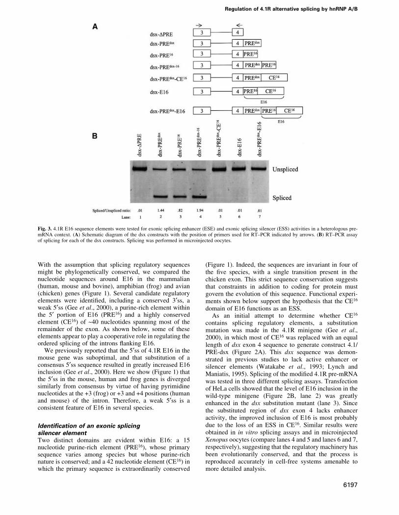
Fig. 3. 4.1R E16 sequence elements were tested for exonic splicing enhancer (ESE) and exonic splicing silencer (ESS) activities in a heterologous pre-mRNA context. (A) Schematic diagram of the dsx constructs with the position of primers used for RT±PCR indicated by arrows. (B) RT±PCR assayof splicing for each of the dsx constructs. Splicing was performed in microinjected oocytes.
Regulation of 4.1R alternative splicing by hnRNP A/B
6197
To de®ne boundaries of the ESS element further, linker-scanning mutagenesis of CE16 was performed in thecontext of the 4.1R minigene. Seven mutants weregenerated, each containing three to ®ve nucleotide substi-tutions (Figure 2A). In vitro splicing reactions in HeLa cellnuclear extract revealed that one of these mutants (mutE)almost completely destroyed silencer activity, resulting invery ef®cient inclusion of exon 16 (lane 12). Notably, thismutation altered a sequence motif (UAG) that is charac-teristic of binding sites for the splicing silencer proteinhnRNP A1 (Del Gatto et al., 1996), and its disruptionquantitatively mimicked the effect of complete substitu-tion of CE16 sequences observed in pre-mRNA substratePRE16-dsx. Together, these observations demonstrate thatthe region altered in mutE comprises a critical part of thesilencer element. However, the strong evolutionary con-servation of CE16 and the ®nding that other mutations inthis region partially disrupt silencer activity (Figure 2C,lanes 5±12) suggest that additional sequences affect thefunction of the putative A1-binding site.
The next experiments tested whether CE16 silenceractivity can function in a heterologous context, in dsx-based pre-mRNAs used previously to demonstrate splicingenhancer activity (Figure 3A; Watakabe et al., 1993;Lynch and Maniatis, 1995). dsx DPRE, an enhancerlessconstruct from which the endogenous enhancer elementshave been deleted (Lynch and Maniatis, 1995), has a weak3¢ss and splices poorly unless provided with a splicingenhancer such as the endogenous dsx E4 purine-richelement (dsx-PREdsx) (Figure 3B, lanes 1 and 2). Whentested in parallel under identical conditions, the PRE16
element of E16 also signi®cantly enhanced splicing of dsxpre-mRNA (lane 3). A double enhancer PREdsx-16 (lane 4)had even higher levels of splicing. Importantly, juxtapos-ition of CE16 to the enhancer(s) in all three of these testconstructs led to complete repression of splicing activity(lanes 5±7). The repressive activity of CE16 can thus
counteract at least two distinct enhancers, and can functionup to 143 nucleotides from an upstream 3¢ss (the distancein pre-mRNA dsx-PREdsx-E16). Similar results wereobtained in two different assay systems employingmicroinjected oocytes (Figure 3) and HeLa cell nuclearextract (not shown), indicating that the CE16 silencer canfunction in different cell types and species. Moreover,recent studies have shown that a partial silencing effect isobserved when this E16 region is inserted into aheterologous b-globin exon (Deguillien et al., 2001).
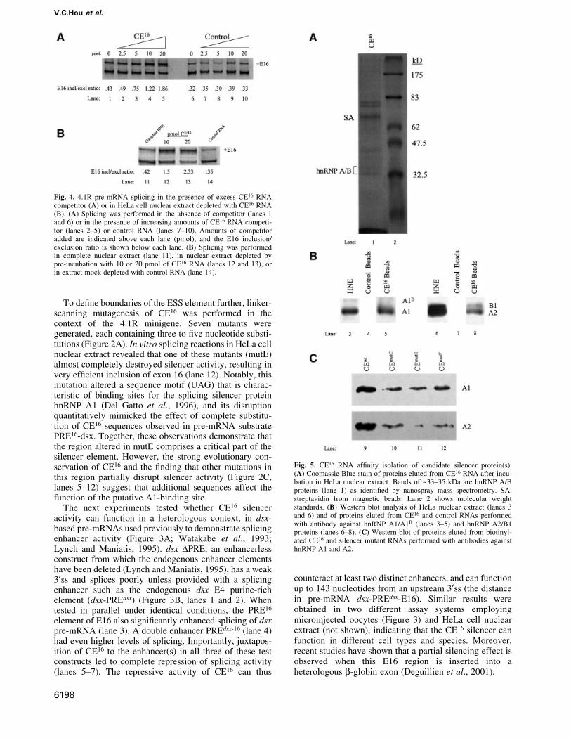
Fig. 4. 4.1R pre-mRNA splicing in the presence of excess CE16 RNAcompetitor (A) or in HeLa cell nuclear extract depleted with CE16 RNA(B). (A) Splicing was performed in the absence of competitor (lanes 1and 6) or in the presence of increasing amounts of CE16 RNA competi-tor (lanes 2±5) or control RNA (lanes 7±10). Amounts of competitoradded are indicated above each lane (pmol), and the E16 inclusion/exclusion ratio is shown below each lane. (B) Splicing was performedin complete nuclear extract (lane 11), in nuclear extract depleted bypre-incubation with 10 or 20 pmol of CE16 RNA (lanes 12 and 13), orin extract mock depleted with control RNA (lane 14).
Fig. 5. CE16 RNA af®nity isolation of candidate silencer protein(s).(A) Coomassie Blue stain of proteins eluted from CE16 RNA after incu-bation in HeLa nuclear extract. Bands of ~33±35 kDa are hnRNP A/Bproteins (lane 1) as identi®ed by nanospray mass spectrometry. SA,streptavidin from magnetic beads. Lane 2 shows molecular weightstandards. (B) Western blot analysis of HeLa nuclear extract (lanes 3and 6) and of proteins eluted from CE16 and control RNAs performedwith antibody against hnRNP A1/A1B (lanes 3±5) and hnRNP A2/B1proteins (lanes 6±8). (C) Western blot of proteins eluted from biotinyl-ated CE16 and silencer mutant RNAs performed with antibodies againsthnRNP A1 and A2.
V.C.Hou et al.
6198
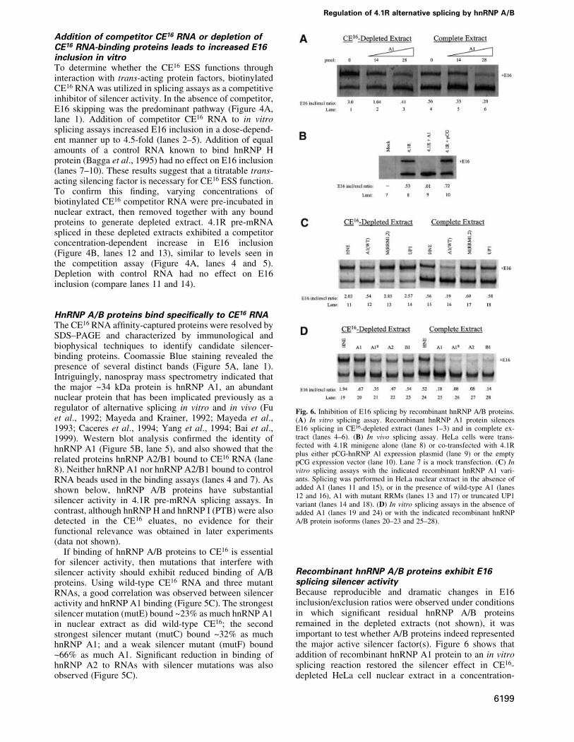
Addition of competitor CE16 RNA or depletion ofCE16 RNA-binding proteins leads to increased E16inclusion in vitroTo determine whether the CE16 ESS functions throughinteraction with trans-acting protein factors, biotinylatedCE16 RNA was utilized in splicing assays as a competitiveinhibitor of silencer activity. In the absence of competitor,E16 skipping was the predominant pathway (Figure 4A,lane 1). Addition of competitor CE16 RNA to in vitrosplicing assays increased E16 inclusion in a dose-depend-ent manner up to 4.5-fold (lanes 2±5). Addition of equalamounts of a control RNA known to bind hnRNP Hprotein (Bagga et al., 1995) had no effect on E16 inclusion(lanes 7±10). These results suggest that a titratable trans-acting silencing factor is necessary for CE16 ESS function.To con®rm this ®nding, varying concentrations ofbiotinylated CE16 competitor RNA were pre-incubated innuclear extract, then removed together with any boundproteins to generate depleted extract. 4.1R pre-mRNAspliced in these depleted extracts exhibited a competitorconcentration-dependent increase in E16 inclusion(Figure 4B, lanes 12 and 13), similar to levels seen inthe competition assay (Figure 4A, lanes 4 and 5).Depletion with control RNA had no effect on E16inclusion (compare lanes 11 and 14).
HnRNP A/B proteins bind speci®cally to CE16 RNAThe CE16 RNA af®nity-captured proteins were resolved bySDS±PAGE and characterized by immunological andbiophysical techniques to identify candidate silencer-binding proteins. Coomassie Blue staining revealed thepresence of several distinct bands (Figure 5A, lane 1).Intriguingly, nanospray mass spectrometry indicated thatthe major ~34 kDa protein is hnRNP A1, an abundantnuclear protein that has been implicated previously as aregulator of alternative splicing in vitro and in vivo (Fuet al., 1992; Mayeda and Krainer, 1992; Mayeda et al.,1993; Caceres et al., 1994; Yang et al., 1994; Bai et al.,1999). Western blot analysis con®rmed the identity ofhnRNP A1 (Figure 5B, lane 5), and also showed that therelated proteins hnRNP A2/B1 bound to CE16 RNA (lane8). Neither hnRNP A1 nor hnRNP A2/B1 bound to controlRNA beads used in the binding assays (lanes 4 and 7). Asshown below, hnRNP A/B proteins have substantialsilencer activity in 4.1R pre-mRNA splicing assays. Incontrast, although hnRNP H and hnRNP I (PTB) were alsodetected in the CE16 eluates, no evidence for theirfunctional relevance was obtained in later experiments(data not shown).
If binding of hnRNP A/B proteins to CE16 is essentialfor silencer activity, then mutations that interfere withsilencer activity should exhibit reduced binding of A/Bproteins. Using wild-type CE16 RNA and three mutantRNAs, a good correlation was observed between silenceractivity and hnRNP A1 binding (Figure 5C). The strongestsilencer mutation (mutE) bound ~23% as much hnRNP A1in nuclear extract as did wild-type CE16; the secondstrongest silencer mutant (mutC) bound ~32% as muchhnRNP A1; and a weak silencer mutant (mutF) bound~66% as much A1. Signi®cant reduction in binding ofhnRNP A2 to RNAs with silencer mutations was alsoobserved (Figure 5C).
Recombinant hnRNP A/B proteins exhibit E16splicing silencer activityBecause reproducible and dramatic changes in E16inclusion/exclusion ratios were observed under conditionsin which signi®cant residual hnRNP A/B proteinsremained in the depleted extracts (not shown), it wasimportant to test whether A/B proteins indeed representedthe major active silencer factor(s). Figure 6 shows thataddition of recombinant hnRNP A1 protein to an in vitrosplicing reaction restored the silencer effect in CE16-depleted HeLa cell nuclear extract in a concentration-
Fig. 6. Inhibition of E16 splicing by recombinant hnRNP A/B proteins.(A) In vitro splicing assay. Recombinant hnRNP A1 protein silencesE16 splicing in CE16-depleted extract (lanes 1±3) and in complete ex-tract (lanes 4±6). (B) In vivo splicing assay. HeLa cells were trans-fected with 4.1R minigene alone (lane 8) or co-transfected with 4.1Rplus either pCG-hnRNP Al expression plasmid (lane 9) or the emptypCG expression vector (lane 10). Lane 7 is a mock transfection. (C) Invitro splicing assays with the indicated recombinant hnRNP A1 vari-ants. Splicing was performed in HeLa nuclear extract in the absence ofadded A1 (lanes 11 and 15), or in the presence of wild-type A1 (lanes12 and 16), A1 with mutant RRMs (lanes 13 and 17) or truncated UP1variant (lanes 14 and 18). (D) In vitro splicing assays in the absence ofadded A1 (lanes 19 and 24) or with the indicated recombinant hnRNPA/B protein isoforms (lanes 20±23 and 25±28).
Regulation of 4.1R alternative splicing by hnRNP A/B
6199
dependent manner, reducing the levels of E16 inclusion(Figure 6A, lanes 1±3). These add-back experiments wereperformed with amounts of recombinant A/B proteinsnearly identical to those employed in studies of the HIV tatexon 2 silencer, and are within the range of endogenoushnRNP A1 protein estimated to be present in HeLa cellnuclear extracts (Caputi et al., 1999). Addition of hnRNPA1 protein to complete extract reduced E16 inclusion toeven lower levels (lanes 4±6). These results suggest thatE16 inclusion is very sensitive to the levels of the hnRNPA/B family of proteins. Control experiments showed thatsubstrates with a mutated CE16 domain and reduced A1binding were much less sensitive to added A/B proteins(not shown).
To validate the silencing effect of hnRNP A1 proteinsobserved in vitro, a comparable experiment was performedusing transfection to alter hnRNP A1 levels in intact cells.Overexpression of hnRNP A1 in transfected HeLa cellssigni®cantly decreased inclusion of E16 in a co-transfected 4.1R minigene (compare lanes 8 and 9).Control experiments showed that co-transfection with theempty expression vector had little effect on E16 inclusion(lane 10).
To determine which domains of hnRNP A1 are requiredfor silencer activity, variant hnRNP A1 proteins (Mayedaet al., 1994) were tested in splicing assays. In contrast tothe strong silencer activity of wild-type hnRNP A1(Figure 6C, lanes 12 and 16), hnRNP A1 protein withmutated RNA recognition motifs (RRMs) had no silenceractivity (compare lanes 12 and 13 with lanes 16 and 17).The C-terminal truncation variant UP1, which lacks theglycine-rich protein±protein interaction domain of hnRNPA1 but has wild-type RRMs, was also inactive for silenceractivity (lanes 14 and 18). These results indicate that bothRNA±protein and protein±protein interactions are requiredfor the splicing silencer activity of hnRNP A1.
Silencer activity among other members of the hnRNPA/B family of proteins was also investigated. In agreementwith results reported for splicing of exon 2 in humanimmunode®ciency virus (HIV) tat pre-mRNA (Caputiet al., 1999; Bilodeau et al., 2001), the alternativelyspliced isoform of hnRNP A1, hnRNP A1B, effectivelysilenced protein 4.1R E16 inclusion (lanes 21 and 26).Similarly, hnRNP A2 and its alternatively spliced isoformhnRNP B1 exhibited strong silencing activity in parallelassays (lanes 22 and 27, and lanes 23 and 28, respectively)compared with the control splicing reactions (lanes 19and 24).
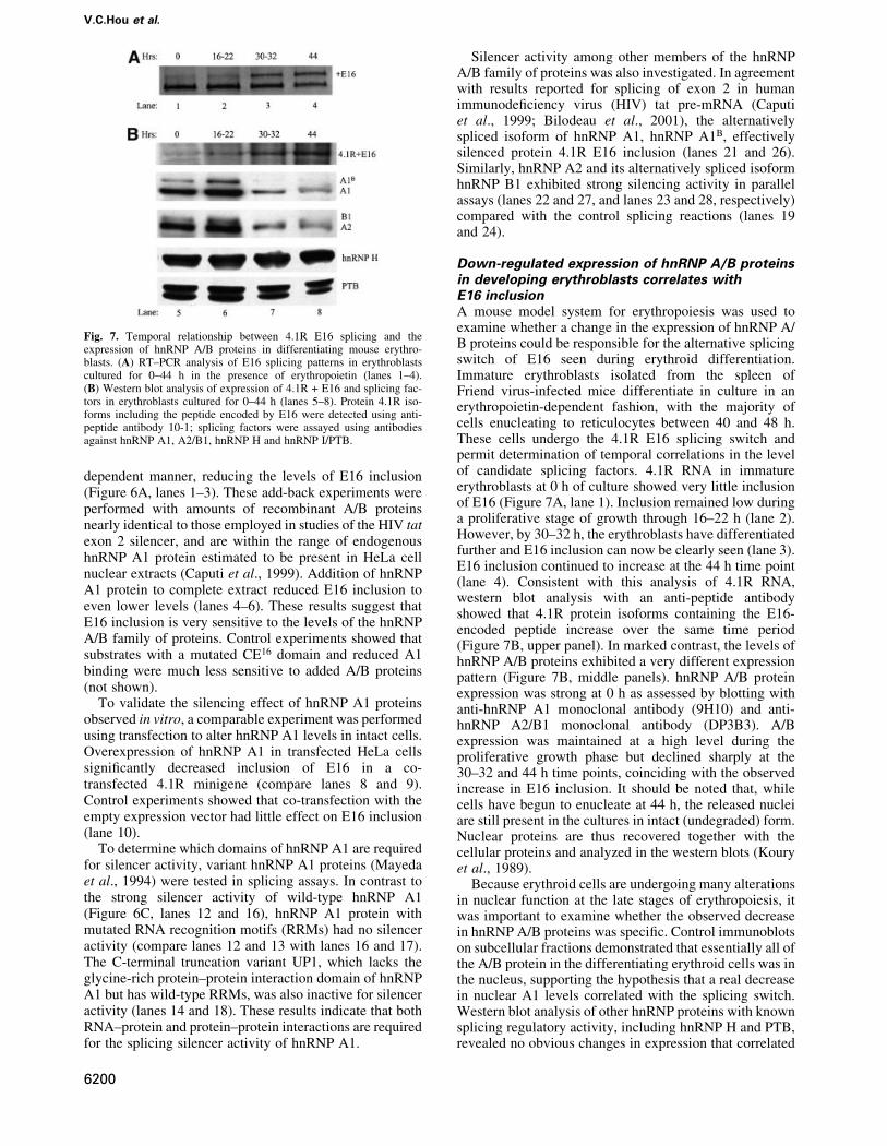
Down-regulated expression of hnRNP A/B proteinsin developing erythroblasts correlates withE16 inclusionA mouse model system for erythropoiesis was used toexamine whether a change in the expression of hnRNP A/B proteins could be responsible for the alternative splicingswitch of E16 seen during erythroid differentiation.Immature erythroblasts isolated from the spleen ofFriend virus-infected mice differentiate in culture in anerythropoietin-dependent fashion, with the majority ofcells enucleating to reticulocytes between 40 and 48 h.These cells undergo the 4.1R E16 splicing switch andpermit determination of temporal correlations in the levelof candidate splicing factors. 4.1R RNA in immatureerythroblasts at 0 h of culture showed very little inclusionof E16 (Figure 7A, lane 1). Inclusion remained low duringa proliferative stage of growth through 16±22 h (lane 2).However, by 30±32 h, the erythroblasts have differentiatedfurther and E16 inclusion can now be clearly seen (lane 3).E16 inclusion continued to increase at the 44 h time point(lane 4). Consistent with this analysis of 4.1R RNA,western blot analysis with an anti-peptide antibodyshowed that 4.1R protein isoforms containing the E16-encoded peptide increase over the same time period(Figure 7B, upper panel). In marked contrast, the levels ofhnRNP A/B proteins exhibited a very different expressionpattern (Figure 7B, middle panels). hnRNP A/B proteinexpression was strong at 0 h as assessed by blotting withanti-hnRNP A1 monoclonal antibody (9H10) and anti-hnRNP A2/B1 monoclonal antibody (DP3B3). A/Bexpression was maintained at a high level during theproliferative growth phase but declined sharply at the30±32 and 44 h time points, coinciding with the observedincrease in E16 inclusion. It should be noted that, whilecells have begun to enucleate at 44 h, the released nucleiare still present in the cultures in intact (undegraded) form.Nuclear proteins are thus recovered together with thecellular proteins and analyzed in the western blots (Kouryet al., 1989).
Because erythroid cells are undergoing many alterationsin nuclear function at the late stages of erythropoiesis, itwas important to examine whether the observed decreasein hnRNP A/B proteins was speci®c. Control immunoblotson subcellular fractions demonstrated that essentially all ofthe A/B protein in the differentiating erythroid cells was inthe nucleus, supporting the hypothesis that a real decreasein nuclear A1 levels correlated with the splicing switch.Western blot analysis of other hnRNP proteins with knownsplicing regulatory activity, including hnRNP H and PTB,revealed no obvious changes in expression that correlated
Fig. 7. Temporal relationship between 4.1R E16 splicing and theexpression of hnRNP A/B proteins in differentiating mouse erythro-blasts. (A) RT±PCR analysis of E16 splicing patterns in erythroblastscultured for 0±44 h in the presence of erythropoietin (lanes 1±4).(B) Western blot analysis of expression of 4.1R + E16 and splicing fac-tors in erythroblasts cultured for 0±44 h (lanes 5±8). Protein 4.1R iso-forms including the peptide encoded by E16 were detected using anti-peptide antibody 10-1; splicing factors were assayed using antibodiesagainst hnRNP A1, A2/B1, hnRNP H and hnRNP I/PTB.
V.C.Hou et al.
6200
temporally with the E16 splicing switch (Figure 7B, lowerpanels). Together, these data implicate the modulatedexpression of the hnRNP A/B proteins as the develop-mental switch responsible for activation of exon 16inclusion during erythropoiesis.
Discussion
The present study provides insight into the molecularmechanism responsible for a pre-mRNA alternative spli-cing switch that operates during erythroid differentiationto induce physiologically important changes in the struc-ture and function of protein 4.1R. Activation of E16inclusion is critical for establishment of normal membranemechanical stability during erythropoiesis, as red cellmembranes assembled with protein 4.1R isoforms pos-sessing a complete spectrin±actin-binding domain (trans-lated from mRNAs including E16) exhibit considerableresistance to shear forces. In contrast, membranes assem-bled with 4.1R bearing a truncated spectrin±actin-bindingdomain (translated from mRNAs excluding E16) areunstable in shear assays, and may result in hemolyticanemia due to increased fragmentation in the circulation(Takakuwa et al., 1986; Conboy et al., 1991b; Discheret al., 1993; Horne et al., 1993).
Analysis of 4.1R pre-mRNA splicing supports a modelin which exon 16 splicing is regulated primarily bymodulation of hnRNP A/B protein levels in differentiatingerythroid cells. In early erythroid progenitors, high levelsof hnRNP A/B proteins are present, leading to repressionof exon 16 splicing. Then, as the cells differentiate intomature erythroblasts, a dramatic decrease in levels ofhnRNP A/B proteins leads to derepression of splicing andactivation of exon 16 inclusion in mature 4.1R mRNA.This model is supported by in vitro and in vivo datashowing that hnRNP A/B proteins bind to a conservedsplicing silencer element in exon 16, that the ef®ciency ofexon 16 inclusion in functional splicing assays is correl-ated directly with the levels of hnRNP A/B proteins andthat a speci®c decrease in hnRNP A/B proteins correlateswith the activation of exon 16 inclusion in differentiatingerythroid cells. These ®ndings are consistent with previousstudies demonstrating that hnRNP A/B proteins canfunction as ESS-responsive negative regulators of splicing(Caputi et al., 1999; Del Gatto-Konczak et al., 1999;Matter et al., 2000; Bilodeau et al., 2001; Zhu et al., 2001),and that changes in the relative amounts of hnRNP A/Bproteins and SR proteins can alter either the alternativesplice site choice or the inclusion/exclusion ratio ofselected alternative exons (Fu et al., 1992; Mayeda andKrainer, 1992; Mayeda et al., 1993; Caceres et al., 1994;Yang et al., 1994; Bai et al., 1999; Blanchette and Chabot,1999). The most novel ®nding in the current study is thedemonstration that a natural developmental change in thelevel of endogenous hnRNP A/B proteins in differentiatingerythroid cells can mediate an important pre-mRNAsplicing switch.
Proper regulation of exon 16 ordered splicing, in whichdownstream splicing precedes excision of the upstreamintron (Gee et al., 2000), requires mechanisms to preventinappropriate splicing of the ¯anking introns. We proposethat the CE16±hnRNP A/B interaction plays an importantrole in regulating both upstream and downstream intron
splicing. This is demonstrated most easily in the case ofupstream splicing: in vitro experiments with model 4.1Rpre-mRNAs (Gee et al., 2000) and heterologous dsx pre-mRNAs (Figure 3) indicate that upstream splicing isinef®cient in the presence of intact CE16. In vivo, thisactivity may be critical to prevent premature splicing ofthe upstream intron in nascent 4.1R pre-mRNA transcripts,because disruption of the normal `downstream ®rst'ordered splicing would lead to inappropriate activationof a cryptic splice site and to the generation of aberrantmRNAs (Gee et al., 2000). With regard to splicing of thedownstream intron, we have shown previously that thisstep is inef®cient in the absence of a strong exon 16 3¢ss(Gee et al., 2000). Therefore, inhibition of 3¢ss function byhnRNP A/B bound at the splicing silencer may indirectlyblock activation of the downstream 5¢ss, perhaps byinterference with critical exon-bridging interactions(Berget, 1995).
The ®nding that CE16 is essentially identical amongmammalian, avian and amphibian species clearly indicatesthat this region of exon 16 is critical to proper 4.1R geneexpression. At least two speci®c functions can now beascribed to this domain. First, CE16 is important for proteinfunction, because it encodes a protein domain that isessential for interaction of 4.1R protein with spectrin andactin in the erythroid membrane skeleton (Horne et al.,1993; Discher et al., 1995). Secondly, CE16 plays a keyrole in 4.1R pre-mRNA processing by negatively regulat-ing E16 splicing in cells where it may be important toprevent expression of an intact spectrin±actin-bindingdomain. A similar dual constraint on exon sequenceevolution has been proposed earlier in the case of exonicsplicing enhancer elements (Liu et al., 1998; Schaal andManiatis, 1999).
Future studies will focus on identi®cation of themechanism by which hnRNP A/B protein(s) binding tothe CE16 silencer can block recognition and/or splicing ofE16 by the nuclear spliceosomal machinery, and what rolethese interactions play in the ordered splicing of introns¯anking E16 (Gee et al., 2000). Most probably, properregulation requires additional interactions among hnRNPA/B proteins (Ding et al., 1999) or between hnRNP A/Bproteins and other splicing factors, acting in a concertedfashion to orchestrate the developmental switch in E16splicing during erythropoiesis. In particular, it will beimportant to clarify the possible function of SR proteins inE16 splicing, the role of enhancer activity in PRE16 andpotential interaction(s) between PRE16 and CE16.Moreover, important cis-acting elements are likely to belocated not only in E16, but also in the ¯anking intronsequences (Gee et al., 1998; Deguillien et al., 2001).Further characterization of these elements and theircognate interacting proteins will be necessary to gain abetter understanding of the mechanism(s) by which E16splicing is regulated during erythropoiesis.
Materials and methods
Construction of plasmids4.1R minigene substitution mutation, 4.1/PRE16-dsx and 4.1/PRE16-dsx(pcDNA) for transfection. Splice overlap extension PCR was used toreplace the E16 silencer element with an equal length of Drosophila dsxexon 4 sequence containing no known splicing enhancer or silencer
Regulation of 4.1R alternative splicing by hnRNP A/B
6201
activity. A 5¢ fragment containing 4.1R exon 13, the upstream intron andpart of exon 16 fused to dsx sequences was ampli®ed usingoligonucleotides T7, 5¢-TAATACGACTCACTATAGG-3¢ and CE-DSX3¢, 5¢-GTCATAGATATTCAAATTATGTTGACGGGAGTACTCATT-ATCTAGTCTCTCTCTCTTTTT-3¢. The 3¢ half of the constructcontaining dsx sequences linked to the last two nucleotides of exon 16,plus the downstream intron and exon 17, was ampli®ed using primersCE-DSX 5¢, 5¢-AATGAGTACTCCCGTCAACATAATTTGAATATCT-ATGACGAGGTTTGTATGAACTTGAAG-3¢, and E17.1, 5¢-GCG-AATTCCCGGGATTCAGT-3¢. A mixture of the two fragments,containing an overlap of 39 nucleotides (underlined), was ampli®edwith primers T7 and E17.1 to create the full minigene (4.1/PRE16-dsx)with a substituted silencer region.
4.1/PRE16-dsx(pcDNA) was made by amplifying the above minigenewith primers E13, 5¢-AGCCATTGCTCAGAGTCAGG-3¢, and E17.1using Pfu polymerase (Stratagene) to make a blunt-ended insert. After T7polynucleotide kinase treatment, the insert was ligated into the plasmidpcDNA 3.1 (Invitrogen) that had been digested with BamHI and EcoRVand the ends made blunt with mung bean nuclease.
dsx plasmid constructs dsx-PRE16, dsx-E16, dsx-PREdsx-CE16, dsx-PREdsx-16
and dsx-PREdsx-E16. A series of modi®ed dsx constructs containingelements PRE16 or CE16 was generated by annealing complementaryoligonucleotides into the ClaI site of plasmid pdsxT7 (Lynch andManiatis, 1995). For dsx-PREdsx-16, dsx-PREdsx-CE16 and dsx-PREdsx-E16,the same annealed oligonucleotides were made blunt with mung beannuclease and then subcloned into pCSC-PU (Lynch and Maniatis, 1995)linearized with SmaI. The following oligonucleotides were used: PRE16
sense, 5¢-CGAAAAAGAGAGAGAGA-3¢; PRE16 antisense, 5¢-CGT-CTCTCTCTCTTTTT-3¢; CE16 sense, 5¢- CGGTGAAAACATTTATA-TCAGACATAGCAATTTAATGTTGGAG-3¢; CE16 antisense, 5¢-CGC-TCCAACATTAAATTGCTATGTCTGATATAAATGTTTTCAC-3¢; E16sense, 5¢-CGAAAAAGAGAGAGAGACTAGATGGTGAAAACATTT-ATATCAGACATAGCAATTTAATGTTGGAG-3¢; and E16 antisense,5¢-CGCTCCAACATTAAATTGCTATGTCTGATATAAATGTTTTC-ACCATCTAGTCTCTCTCTCTTTTT-3¢.
4.1R CE16 linker-scanning mutants. A BstBI restriction site (TTCGAA)was engineered into the 4.1R minigene at 6 bp increments across CE16
using the QuikChange kit described above. A representative oligonucleo-tide primer pair used to generate the 4.1R mut-A mutation is given.Mutations mutB through mutG used the same methodology and primerdesign outlined. A1, 5¢-TTCACAGAAAAAGAGAGAGAGACTAG-TTCGAAAAAACATTTATATCAGACATAGCAAT-3¢; and A2, 5¢-ATTGCTATGTCTGATATAAATGTTTTTTCGAACTAGTCTCTCT-CTCTTTTTCTGTGAA-3¢.
Genomic sequencing of Xenopus 4.1R E16A Xenopus genomic library (lFIX II, Stratagene) was screened byhybridization to radiolabeled Xenopus 4.1R cDNA. A 5 kb DNA fragmenthybridizing to E16 was subcloned and ¯anking introns were sequencedusing Xenopus-speci®c primers located within E16. Mouse and humangenomic sequences at the intron/exon ¯anking E16 (Huang et al., 1993;Baklouti et al., 1997) and avian E16 sequence (Yew et al., 1987) havebeen published previously.
Synthesis of pre-mRNAs and microinjection into oocytesSynthesis of capped RNA transcripts was done using the mMESSAGEmMACHINE (Ambion, Inc., Austin, TX) in vitro transcription kitaccording to the manufacturer's protocols. 4.1R and b-globin plasmidswere linearized and transcribed as described previously (Gee et al., 2000).dsx constructs were linearized with BamHI and transcribed using the T7promoter kit. Transcripts were puri®ed using RNeasy (Qiagen, Valencia,CA) columns and microinjected into oocytes as described previously(Gee et al., 2000).
In vitro splicing assaysHeLa cell nuclear extract was prepared as described (Mayeda andKrainer, 1999b). The 25 ml splicing reactions, containing 6.25 fmol ofRNA substrate in 40% HeLa cell nuclear extract, 3.2 mM MgCl2, 1 mMATP, 20 mM creatine phosphate, 3.1% polyvinyl alcohol and 40 U ofRNasin (Promega Corp., Madison, WI) were incubated 2 h at 30°C(Mayeda and Krainer, 1999a). All in vitro splicing experiments wereperformed at least three times.
Competitor splicing assays were performed by adding a 46 nucleotide5¢-biotinylated RNA (Dharmacon Research, Inc., Lafayette, CO)containing the conserved element of E16 (CE16) plus the two ¯anking
nucleotides on each end with 2¢O-methyl group modi®cations. Abiotinylated RNA containing a high-af®nity hnRNP H-binding site, 5¢-AAGGGGGAGGUGUGGGUC-3¢ (Bagga et al., 1995), was used as acontrol. Depleted nuclear extract was generated by pre-incubating thesplicing mixture with up to 28 pmol of biotinylated RNA for 30 min at4°C, then removing RNA and bound proteins by a 15 min capture step at4°C with 150 ml of streptavidin MagneSphere paramagnetic particles(Promega) pre-washed in buffer D [20 mM HEPES±KOH pH 8, 100 mMKCl, 0.2 mM EDTA, 20% (v/v) glycerol, 0.5 mM phenylmethylsulfonyl¯uoride (PMSF), 1 mM dithiothreitol (DTT)]. Recombinant hnRNP A1,A1B, A2, B1 and mutants A1-M(RRM1,2) and UP1 proteins wereexpressed in Escherichia coli and puri®ed as described previously(Mayeda and Krainer, 1992; Mayeda et al., 1994).
Transient transfection of HeLa cellsHeLa cells were grown to ~80% con¯uency in six-well plates andtransfected using lipofectamine (Gibco-BRL, Gaithersburg, MD) accord-ing to the manufacturer's protocol. In brief, 1 mg of each plasmid DNAwas used in 10 ml of lipofectamine and transfected for 18 h beforeremoval and addition of fresh media. Total RNA was then isolated after48 h of incubation. The plasmid pCG-hnRNP A1 was used foroverexpression of hnRNP A1 as described previously (Caceres et al.,1994). Control co-transfection experiments were performed using theempty expression vector pCG.
RT±PCR analysis of spliced pre-mRNAAnalysis of splicing reactions was carried out as described previously(Gee et al., 2000) using DNA primers in protein 4.1R exons 13 (forward)and 17 (reverse). Twenty-®ve cycles of PCR were used routinely toanalyze the products of the splicing reactions; this condition was in thelinear range of ampli®cation since similar exon inclusion/exclusion ratioswere obtained even after 35 cycles. As described previously (Gee et al.,2000), duplicate splicing reactions exhibited very little intra-experimentalvariability when processed in parallel under identical conditions.Although inter-experimental variability in absolute splicing ef®ciencieswas observed, relative splicing ef®ciencies among various pre-mRNAsremained extremely consistent. Moreover, the relative splicingef®ciencies of various pre-mRNA substrates was very similar in splicingassays with microinjected oocytes or transfected HeLa cells. Together,these results strongly support the validity of the RT±PCR results forin vitro splicing assays.
Image and densitometry measurements were done using the IS 1000digital imaging system and software (Alpha Innotech Corp., San Leandro,CA). All labeled bands in the ®gures have been con®rmed by sequencing.DNA primers used for analysis of dsx RNAs were located in exon 3(sense) and exon 4 (antisense), as follows: E3, 5¢-GGAGCTGATG-CCACTCATGTATG-3¢; and E4, 5¢-GCTCACCCCCGTCATAGATA-TTC-3¢. The E4 primer was used for both the RT±PCRs.
Erythroblast cell procurement and cultureErythroid cells obtained from the spleens of mice infected with theanemia-inducing strain of Friend erythroleukemia virus were isolated andcultured as described previously (Koury et al., 1984; Sawyer et al., 1987).Cells at t = 0 h are mainly proerythroblasts, which then differentiate over~48 h into late-stage erythroblasts and enucleated reticulocytes. TotalRNA was isolated from cell pellets using RNeasy columns, and proteinsprepared by lysing cells in 53 protein sample buffer (Conboy et al.,1991a) at a concentration of 20 000 cells/ml. Western blot analysis wasdone as described previously (Conboy et al., 1991a). Protein from 2 3 106
cells per sample was loaded for 4.1R + E16 (anti-SAB antibody)detection, and 5 3 105 cells per sample for hnRNP A/B (9H10 andDP3B3 monoclonal antibodies, respectively). Control blots wereperformed using anti-PTB and anti-hnRNP H antibodies (kindly providedby Doug Black).
Nanospray mass spectrometryBands of interest were excised and subjected to in-gel tryptic digestion(Hellman et al., 1995). Tryptic peptides were extracted from the gelpieces and cleaned using a gel-loader pipet tip ®lled with 100 nl ofPOROS C18 resin (PE Biosytems, Foster City, CA). The peptide mixturewas eluted into a nanospray glass capillary (PROTANA, Odense,Denmark) using 500 nl of 60% methanol/5% formic acid, then peptidesolutions were infused into an LCQ Iontrap mass spectrometer(FinniganMat, San Jose, CA) at a ¯ow rate of 10 nl/min. Individualpeptide ions were isolated and subjected to MS/MS analysis. Theacquired MS/MS spectra were then subjected to protein and DNAdatabase searches using the SEQUEST program (Eng et al., 1994).
V.C.Hou et al.
6202
Acknowledgements
We thank G.Dreyfuss for kindly providing the monoclonal antibodies9H10 and DP3B3, D.Black for antibodies to hnRNP H and PTB, andT.Maniatis for providing the pdsxT7 and pCSC-PU plasmids. This workwas supported by NIH grant HL45182 to J.G.C., by the Director, Of®ce ofBiological and Environmental Research, US Department of Energy undercontract DE-AC03-76SF00098, and by a Merit Review Award from theDepartment of Veterans Affairs to M.J.K. A.M. is a research member ofthe Sylvester Comprehensive Cancer Center and supported by the FloridaBiomedical Research Program Grant (BM031) from the FDH. A.R.K.acknowledges support from National Cancer Institute grant CA13106.
References
Amendt,B.A., Si,Z.H. and Stoltzfus,C.M. (1995) Presence of exonsplicing silencers within human immunode®ciency virus type 1 tatexon 2 and tat±rev exon 3: evidence for inhibition mediated bycellular factors. Mol. Cell. Biol., 15, 4606±4615.
Ashiya,M. and Grabowski,P.J. (1997) A neuron-speci®c splicing switchmediated by an array of pre-mRNA repressor sites: evidence of aregulatory role for the polypyrimidine tract binding protein and abrain-speci®c PTB counterpart. RNA, 3, 996±1015.
Bagga,P.S., Ford,L.P., Chen,F. and Wilusz,J. (1995) The G-richauxiliary downstream element has distinct sequence and positionrequirements and mediates ef®cient 3¢ end pre-mRNA processingthrough a trans-acting factor. Nucleic Acids Res., 23, 1625±1631.
Bai,Y., Lee,D., Yu,T. and Chasin,L.A. (1999) Control of 3¢ splice sitechoice in vivo by ASF/SF2 and hnRNP A1. Nucleic Acids Res., 27,1126±1134.
Baklouti,F., Huang,S.C., Vulliamy,T.J., Delaunay,J. and Benz,E.J.,Jr(1997) Organization of the human protein 4.1 genomic locus: newinsights into the tissue-speci®c alternative splicing of the pre-mRNA.Genomics, 39, 289±302.
Berget,S.M. (1995) Exon recognition in vertebrate splicing. J. Biol.Chem., 270, 2411±2414.
Bilodeau,P.S., Domsic,J.K., Mayeda,A., Krainer,A.R. and Stoltzfus,C.M.(2001) RNA splicing at human immunode®ciency virus type 1 3¢splice site A2 is regulated by binding of hnRNP A/B proteins to anexonic splicing silencer element. J. Virol., 75, 8487±8497.
Blanchette,M. and Chabot,B. (1999) Modulation of exon skipping byhigh-af®nity hnRNP A1-binding sites and by intron elements thatrepress splice site utilization. EMBO J., 18, 1939±1952.
Caceres,J.F., Stamm,S., Helfman,D.M. and Krainer,A.R. (1994)Regulation of alternative splicing in vivo by overexpression ofantagonistic splicing factors. Science, 265, 1706±1709.
Caputi,M., Mayeda,A., Krainer,A.R. and Zahler,A.M. (1999) hnRNP A/B proteins are required for inhibition of HIV-1 pre-mRNA splicing.EMBO J., 18, 4060±4067.
Chabot,B. (1996) Directing alternative splicing: cast and scenarios.Trends Genet., 12, 472±478.
Chan,R.C. and Black,D.L. (1997) The polypyrimidine tract bindingprotein binds upstream of neural cell-speci®c c-src exon N1 to repressthe splicing of the intron downstream. Mol. Cell. Biol., 17, 4667±4676.
Chasis,J.A., Coulombel,L., Conboy,J., McGee,S., Andrews,K.,Kan,Y.W. and Mohandas,N. (1993) Differentiation-associatedswitches in protein 4.1 expression. Synthesis of multiple structuralisoforms during normal human hematopoiesis. J. Clin. Invest., 91,329±338.
Conboy,J. (1999) The role of alternative pre-mRNA splicing inregulating the structure and function of skeletal protein 4.1. Proc.Soc. Exp. Biol. Med., 220, 73±78.
Conboy,J.G., Chan,J., Chasis,J.A., Kan,Y.W. and Mohandas,N. (1991a)Tissue- and development-speci®c alternative RNA splicing regulatesexpression of multiple isoforms of erythroid membrane protein 4.1.J. Biol. Chem., 266, 8273±8280.
Conboy,J.G., Shitamoto,R., Parra,M., Winardi,R., Kabra,A., Smith,J. andMohandas,N. (1991b) Hereditary elliptocytosis due to both qualitativeand quantitative defects in membrane skeletal protein 4.1. Blood, 78,2438±2443.
Deguillien,M., Huang,S.C., Moriniere,M., Dreumont,N., Benz,E.J.,Jrand Baklouti,F. (2001) Multiple cis elements regulate an alternativesplicing event at 4.1R pre-mRNA during erythroid differentiation.Blood, 98, 3809±3816.
Del Gatto,F. and Breathnach,R. (1995) Exon and intron sequences,
respectively, repress and activate splicing of a ®broblast growth factorreceptor 2 alternative exon. Mol. Cell. Biol., 15, 4825±4834.
Del Gatto,F., Gesnel,M.C. and Breathnach,R. (1996) The exon sequenceTAGG can inhibit splicing. Nucleic Acids Res., 24, 2017±2021.
Del Gatto-Konczak,F., Olive,M., Gesnel,M.C. and Breathnach,R. (1999)hnRNP A1 recruited to an exon in vivo can function as an exonsplicing silencer. Mol. Cell. Biol., 19, 251±260.
Ding,J., Hayashi,M.K., Zhang,Y., Manche,L., Krainer,A.R. and Xu,R.M.(1999) Crystal structure of the two-RRM domain of hnRNP A1 (UP1)complexed with single-stranded telomeric DNA. Genes Dev., 13,1102±1115.
Discher,D., Parra,M., Conboy,J.G. and Mohandas,N. (1993)Mechanochemistry of the alternatively spliced spectrin±actin bindingdomain in membrane skeletal protein 4.1. J. Biol. Chem., 268,7186±7195.
Discher,D.E., Winardi,R., Schischmanoff,P.O., Parra,M., Conboy,J.G.and Mohandas,N. (1995) Mechanochemistry of protein 4.1'sspectrin±actin-binding domain: ternary complex interactions,membrane binding, network integration, structural strengthening.J. Cell Biol., 130, 897±907.
Eng,Y., McCormack,A.L. and Yates,J.R.,III (1994) An approach tocorrelate tandem mass spectral data of peptides with amino acidsequences in a protein database. J. Am. Soc. Mass Spectrom., 5,976±989.
Fu,X.D., Mayeda,A., Maniatis,T. and Krainer,A.R. (1992) Generalsplicing factors SF2 and SC35 have equivalent activities in vitroand both affect alternative 5¢ and 3¢ splice site selection. Proc. NatlAcad. Sci. USA, 89, 11224±11228.
Gascard,P., Nunomura,W., Lee,G., Walensky,L., Krauss,S.W.,Takakuwa,Y., Chasis,J.A., Mohandas,M. and Conboy,J.G. (1999)Deciphering the nuclear import pathway for the cytoskeletal protein4.1R. Mol. Biol. Cell, 10, 1783±1798.
Gee,S.L., Parra,M., Willig,T., Hou,V.C., Chan,N., Wu,M. andConboy,J.G. (1998) Alternative splicing of protein 4.1 exon 16 isregulated in part by downstream intron elements. Blood, 92, Suppl. 1,5a.
Gee,S.L., Aoyagi,K., Lersch,R., Hou,V., Wu,M. and Conboy,J.G. (2000)Alternative splicing of protein 4.1R exon 16: ordered excision of¯anking introns ensures proper splice site choice. Blood, 95, 692±699.
Graham,I.R., Hamshere,M. and Eperon,I.C. (1992) Alternative splicingof a human a-tropomyosin muscle-speci®c exon: identi®cation ofdetermining sequences. Mol. Cell. Biol., 12, 3872±3882.
Hellman,U., Wernstedt,C., Gonez,J. and Heldin,C.H. (1995)Improvement of an in-gel digestion procedure for themicropreparation of internal protein fragments for amino acidsequencing. Anal. Biochem., 224, 451±455.
Hertel,K.J., Lynch,K.W. and Maniatis,T. (1997) Common themes in thefunction of transcription and splicing enhancers. Curr. Opin. CellBiol., 9, 350±357.
Horne,W.C., Huang,S.C., Becker,P.S., Tang,T.K. and Benz,E.J.J. (1993)Tissue-speci®c alternative splicing of protein 4.1 inserts an exonnecessary for formation of the ternary complex with erythrocytespectrin and F-actin. Blood, 82, 2558±2563.
Huang,J.-P., Tang,C.-J., Kou,G.-H., Marchesi,T., Benz,E.J.,Jr andTang,T.K. (1993) Genomic structure of the locus encoding protein4.1. Structural basis for complex combinational patterns of tissue-speci®c alternative RNA splicing. J. Biol. Chem., 268, 3758±3766.
Kan,J.L. and Green,M.R. (1999) Pre-mRNA splicing of IgM exons M1and M2 is directed by a juxtaposed splicing enhancer and inhibitor.Genes Dev., 13, 462±471.
Konig,H., Ponta,H. and Herrlich,P. (1998) Coupling of signaltransduction to alternative pre-mRNA splicing by a composite spliceregulator. EMBO J., 17, 2904±2913.
Koury,M.J., Sawyer,S.T. and Bondurant,M.C. (1984) Splenicerythroblasts in anemia-inducing Friend disease: a source of cellsfor studies of erythropoietin-mediated differentiation. J. Cell. Physiol.,121, 526±532.
Koury,S.T., Koury,M.J. and Bondurant,M.C. (1989) Cytoskeletaldistribution and function during the maturation and enucleation ofmammalian erythroblasts. J. Cell Biol., 109, 3005±3013.
Lander,E.S. et al. (2001) Initial sequencing and analysis of the humangenome. Nature, 409, 860±921.
Liu,H.X., Zhang,M. and Krainer,A.R. (1998) Identi®cation of functionalexonic splicing enhancer motifs recognized by individual SR proteins.Genes Dev., 12, 1998±2012.
Luque,C.M., Lallena,M.J., Alonso,M.A. and Correas,I. (1998) An
Regulation of 4.1R alternative splicing by hnRNP A/B
6203
alternative domain determines nuclear localization in multifunctionalprotein 4.1. J. Biol. Chem., 273, 11643±11649.
Lynch,K.W. and Maniatis,T. (1995) Synergistic interactions betweentwo distinct elements of a regulated splicing enhancer. Genes Dev., 9,284±293.
Matter,N., Marx,M., Weg-Remers,S., Ponta,H., Herrlich,P. and Konig,H.(2000) Heterogeneous ribonucleoprotein A1 is part of an exon-speci®csplice-silencing complex controlled by oncogenic signaling pathways.J. Biol. Chem., 275, 35353±35360.
Mayeda,A. and Krainer,A.R. (1992) Regulation of alternative pre-mRNA splicing by hnRNP A1 and splicing factor SF2. Cell, 68,365±375.
Mayeda,A. and Krainer,A.R. (1999a) Mammalian in vitro splicingassays. Methods Mol. Biol., 118, 315±321.
Mayeda,A. and Krainer,A.R. (1999b) Preparation of HeLa cell nuclearand cytosolic S100 extracts for in vitro splicing. Methods Mol. Biol.,118, 309±314.
Mayeda,A., Helfman,D.M. and Krainer,A.R. (1993) Modulation of exonskipping and inclusion by heterogeneous nuclear ribonucleoprotein A1and pre-mRNA splicing factor SF2/ASF [published erratum appears inMol. Cell. Biol. (1993) 13, 4458]. Mol. Cell. Biol., 13, 2993±3001.
Mayeda,A., Munroe,S.H., Caceres,J.F. and Krainer,A.R. (1994) Functionof conserved domains of hnRNP A1 and other hnRNP A/B proteins.EMBO J., 13, 5483±5495.
Mayeda,A., Screaton,G.R., Chandler,S.D., Fu,X.D. and Krainer,A.R.(1999) Substrate speci®cities of SR proteins in constitutive splicingare determined by their RNA recognition motifs and composite pre-mRNA exonic elements. Mol. Cell. Biol., 19, 1853±1863.
Nelson,K.K. and Green,M.R. (1990) Mechanism for cryptic splice siteactivation during pre-mRNA splicing. Proc. Natl Acad. Sci. USA, 87,6253±6257.
Reed,R. (1996) Initial splice-site recognition and pairing during pre-mRNA splicing. Curr. Opin. Genet. Dev., 6, 215±220.
Sawyer,S.T., Koury,M.J. and Bondurant,M.C. (1987) Large-scaleprocurement of erythropoietin-responsive erythroid cells: assay forbiological activity of erythropoietin. Methods Enzymol., 147,340±352.
Schaal,T.D. and Maniatis,T. (1999) Multiple distinct splicing enhancersin the protein-coding sequences of a constitutively spliced pre-mRNA.Mol. Cell. Biol., 19, 261±273.
Schischmanoff,P.O., Winardi,R., Discher,D.E., Parra,M.K., Bicknese,S.E., Witkowska,H.E., Conboy,J.G. and Mohandas,N. (1995) De®ningthe minimal domain of protein 4.1 involved in spectrin±actin binding.J. Biol. Chem., 270, 21243±21250.
Si,Z.H., Rauch,D. and Stoltzfus,C.M. (1998) The exon splicing silencerin human immunode®ciency virus type 1 Tat exon 3 is bipartite andacts early in spliceosome assembly. Mol. Cell. Biol., 18, 5404±5413.
Siebel,C.W., Fresco,L.D. and Rio,D.C. (1992) The mechanism ofsomatic inhibition of Drosophila P-element pre-mRNA splicing:multiprotein complexes at an exon pseudo-5¢ splice site control U1snRNP binding. Genes Dev., 6, 1386±1401.
Smith,C.W. and Valcarcel,J. (2000) Alternative pre-mRNA splicing: thelogic of combinatorial control. Trends Biochem. Sci., 25, 381±388.
Staffa,A., Acheson,N.H. and Cochrane,A. (1997) Novel exonic elementsthat modulate splicing of the human ®bronectin EDA exon. J. Biol.Chem., 272, 33394±33401.
Takakuwa,Y., Tchernia,G., Rossi,M., Benabadji,M. and Mohandas,N.(1986) Restoration of normal membrane stability to unstable protein4.1-de®cient membranes by incorporation of puri®ed protein 4.1.J. Clin. Invest., 78, 80±85.
Tange,T.O., Damgaard,C.K., Guth,S., Valcarcel,J. and Kjems,J. (2001)The hnRNP A1 protein regulates HIV-1 tat splicing via a novel intronsilencer element. EMBO J., 20, 5748±5758.
Wang,J. and Manley,J.L. (1997) Regulation of pre-mRNA splicing inmetazoa. Curr. Opin. Genet. Dev., 7, 205±211.
Watakabe,A., Tanaka,K. and Shimura,Y. (1993) The role of exonsequences in splice site selection. Genes Dev., 7, 407±418.
Winardi,R., Discher,D., Kelley,C., Zon,L., Mays,K., Mohandas,N. andConboy,J.G. (1995) Evolutionarily conserved alternative pre-mRNAsplicing regulates structure and function of the spectrin±actin bindingdomain of erythroid protein 4.1. Blood, 86, 4315±4322.
Yang,X., Bani,M.R., Lu,S.J., Rowan,S., Ben-David,Y. and Chabot,B.(1994) The A1 and A1B proteins of heterogeneous nuclearribonucleoparticles modulate 5¢ splice site selection in vivo. Proc.Natl Acad. Sci. USA, 91, 6924±6928.
Yew,N.S., Choi,H.R., Gallarda,J.L. and Engel,J.D. (1987) Expression of
cytoskeletal protein 4.1 during avian erythroid cellular maturation.Proc. Natl Acad. Sci. USA, 84, 1035±1039.
Zhu,J., Mayeda,A. and Krainer,A.R. (2001) Exon identity establishedthrough differential antagonism between exonic splicing silencer-bound hnRNP A1 and enhancer-bound SR proteins. Mol. Cell, 8,1351±1361.
Received April 15, 2002; revised September 4, 2002;accepted September 30, 2002
V.C.Hou et al.
6204


