Embed Size (px)
Citation preview
MOLECULAR AND CELLULAR BIOLOGY,0270-7306/98/$04.0010
Nov. 1998, p. 6224–6237 Vol. 18, No. 11
Copyright © 1998, American Society for Microbiology. All Rights Reserved.
Ectopic Expression of cdc2/cdc28 Kinase Subunit Homo sapiens 1Uncouples Cyclin B Metabolism from the Mitotic
Spindle Cell Cycle CheckpointMARY L. HIXON, ANA I. FLORES, MARK W. WAGNER, AND ANTONIO GUALBERTO*
Department of Physiology & Biophysics and Ireland Cancer Center, School of Medicine,Case Western Reserve University, Cleveland, Ohio 44106
Received 27 March 1998/Returned for modification 15 May 1998/Accepted 4 August 1998
Primary human fibroblasts arrest growth in response to the inhibition of mitosis by mitotic spindle-depoly-merizing drugs. We show that the mechanism of mitotic arrest is transient and implicates a decrease in the ex-pression of cdc2/cdc28 kinase subunit Homo sapiens 1 (CKsHs1) and a delay in the metabolism of cyclin B.Primary human fibroblasts infected with a retroviral vector that drives the expression of a mutant p53 proteinfailed to downregulate CKsHs1 expression, degraded cyclin B despite the absence of chromosomal segregation,and underwent DNA endoreduplication. In addition, ectopic expression of CKsHs1 interfered with the controlof cyclin B metabolism by the mitotic spindle cell cycle checkpoint and resulted in a higher tendency to undergoDNA endoreduplication. These results demonstrate that an altered regulation of CKsHs1 and cyclin B in cellsthat carry mutant p53 undermines the mitotic spindle cell cycle checkpoint and facilitates the development ofaneuploidy. These data may contribute to the understanding of the origin of heteroploidy in mutant p53 cells.
The onset of each phase of the cell cycle depends on thecompletion of the previous phase. These events are coordi-nated by cell cycle checkpoints (19). Cell cycle checkpoints arepathways that ensure the timely progression of the cell cycle,and they play a crucial role in the maintenance of genomicintegrity by coordinating the cell cycle regulatory machinerywith DNA repair and cell death pathways. At mitosis, themitotic spindle cell cycle checkpoint prevents the onset ofanaphase, the actual segregation of chromosomes, if the integ-rity of the mitotic spindle is compromised. Recent data havesuggested a role for the p53 tumor suppressor gene product inthe control of cell ploidy. Fibroblasts isolated from individualswith Li-Fraumeni syndrome have a marked tendency to be-come heteroploid in culture (5). These individuals are bornwith heterozygous mutations in the p53 tumor suppressor gene(39, 57). Heteroploidy is also commonly found in p53 null cellsin culture (27) and in vivo (13) in p53 knockout mice. Intrigu-ingly, overexpression of a mutant p53 protein on a p53 nullbackground accelerated the appearance of polyploidy in a my-elomonocytic cell line (50). Also, the expression of mutant p53proteins in human colon carcinoma cells and murine cell linescauses karyotypic abnormalities, including an increase in ploi-dy levels during growth in culture (1). We have seen that Li-Fraumeni syndrome fibroblasts that carry heterozygous struc-tural p53 mutant proteins progress to polyploidy when in-cubated in the presence of mitotic spindle inhibitors (22).However, normal human fibroblasts, p53 null Li-Fraumeni fibro-blasts, and normal human fibroblasts infected with a retrovirusthat expresses the human papillomavirus 16 E6, which binds toand promotes the degradation of p53, arrest growth whenincubated in the presence of mitotic inhibitors (22). Progres-sion to polyploidy in E6-expressing human fibroblasts, how-ever, has been reported by others (16). In agreement with ourprevious results, Lanni and Jacks have recently reported that
p53 null mouse fibroblasts have a normal mitotic spindle check-point (35). However, these fibroblasts may progress to poly-ploidy due to the inactivation of a p53-dependent postmitoticcheckpoint (35). In addition, inactivation of wild-type p53 bythe overexpression of a truncated (C terminus) p53 protein ina murine prolymphocytic cell line led to polyploidy (41).
The cell cycle G2/M transition and progression through mi-tosis is driven by the kinase activity of a complex referred to asmaturation- or M-phase-promoting factor (MPF). This com-plex consists of a catalytic subunit (34-kDa cyclin-dependentkinase, cdc2), a regulatory subunit (cyclin B proteins), andassociated proteins (47). Entry into mitosis requires MPF ac-tivation, a process that depends upon an increase in cyclin Bexpression and the dephosphorylation of cdc2. Progressionthrough mitosis and cytokinesis requires the subsequent inac-tivation of MPF, which depends in part on cyclin B degrada-tion. Experiments with yeast indicate that the mitotic spindlecell cycle checkpoint feeds into the cell cycle regulatory ma-chinery at mitosis by a pathway that delays the degradation ofcyclin B and maintains cdc2 kinase activity (3). Thus, cyclin Bis degraded and MPF is inactivated only after certain aspects ofmitosis related to spindle assembly and disassembly are prop-erly completed. The activity of the p34cdc2-cyclin B complex isthought to be regulated by its interaction with other proteins(47). We have focused our attention on two low-molecular-weight proteins known as cdc2/cdc28 kinase subunit Homosapiens, CKsHs1 and CKsHs2 (53). These proteins were pre-viously identified as the human homologs of the small cdc28-and cdc2-associated proteins of Saccharomyces cerevisiae,Cks1, and Schizosaccharomyces pombe, Suc1 (53). CKsHs1and CKsHs2 bind the cyclin B-cdc2 complex (53). In S. pombe,inactivation of suc1 causes cells to arrest in mitosis with highlevels of Cdc13 (the S. pombe cyclin B homolog) and high MPFkinase activity (4, 42). Thus, suc1 inactivation mimics the ef-fects of the mitotic spindle cell cycle checkpoint on cyclin Blevels and MPF activity. It has been proposed that Cks familymembers may play a critical role in the mitotic spindle cellcycle checkpoint (54).
We show that primary human fibroblasts arrest growth in
* Corresponding author. Mailing address: Physiology & BiophysicsSOM E553, CWRU, 10900 Euclid Ave., Cleveland, OH 44106-4970.Phone: (216) 368-3487. Fax: (216) 368-3952. E-mail: [email protected].
6224
on March 2, 2015 by guest
http://mcb.asm
.org/D
ownloaded from
response to the inhibition of mitosis by mitotic spindle-depoly-merizing drugs. This growth arrest was transient and was ac-companied by a delay in the metabolism of cyclin B and atransient decrease in the expression of CKsHs1. Inactivation ofp53 by the expression of the human papillomavirus 16 proteinE6 did not affect the effect of a mitotic inhibitor on cyclin Bmetabolism. By contrast, primary human fibroblasts expressinga dominant mutant p53 protein responded very differently tomitotic inhibition: they failed to downregulate CKsHs1 expres-sion, degraded cyclin B despite the absence of chromosomalsegregation, and underwent DNA endoreduplication. Theseresults show that in human cells, mutant p53 proteins abrogatethe mitotic spindle cell cycle checkpoint by interfering with theregulation of CKsHs1 expression and cyclin B metabolism atmitosis. Consistent with this conclusion, we further show thatectopic expression of CKsHs1 abrogates the control of cyclin Bmetabolism by the mitotic spindle cell cycle checkpoint path-way and facilitates the development of polyploidy. These datademonstrate that CKsHs1 plays a key role in the control ofmitosis in human cells and that altered expression of CKsHs1in human cells carrying mutant p53 proteins contributes to thedevelopment of aneuploidy.
MATERIALS AND METHODS
Plasmids and cell culture. The expression vectors pBabe CKsHs1 and pBabeCKsHs2 were created by subcloning CKsHs1 and CKsHs2 cDNA fragments intothe retroviral vector pBabe, which contains a puromycin selectable marker (42a).CKsHs1 and CKsHs2 cDNA fragments were cloned by reverse transcriptionPCR (RT-PCR) with primers 59-AGAGCGATCATGTCGCACAAACAA-39and 59-TCATTTCTTTGGTTTCTTGGGTAG-39 (CKsHs1) and primers 59-ACGAGGATGGCCCACAAGCAGATCTACTAC-39 and 59-TTTTTGTTGATCTTTTGGAAGAGG-39 (CKsHs2) and 1 mg of poly(A) mRNA isolated fromneonatal human foreskin fibroblasts (NHF). CKsHs cDNA sequences were thensubcloned into the pBabe snaB1 site. Plasmid construction was verified by DNAsequencing. The retroviral expression vector pBabe p53 143A was a gift from J.Jacobberger, Case Western Reserve University (Cleveland, Ohio), and was gen-erated by the subcloning of a p53 mutant 143A cDNA fragment formed byBamHI digestion of plasmid CMV-p53 143A into the pBabe BamHI site (33).The murine sarcoma virus long terminal repeat-based p53 expression vectorswere a gift from C. Finlay, GlaxoWellcome (Research Triangle Park, N.C.) (21).The retroviral vectors LXSN and LXSN-E6 were a gift from D. A. Galloway,Fred Hutchinson Cancer Research Center (Seattle, Wash.) (26). The CMV-bcl2plasmid was a gift from M. W. Mayo and A. S. Baldwin, University of NorthCarolina Lineberger Comprehensive Cancer Center (Chapel Hill, N.C.).
NHF, C2C12, and 10T1/2 cells were incubated in Dulbecco’s modified Eagle’smedium (DMEM) plus 10% dialyzed fetal bovine serum (FBS; GIBCO) sup-plemented with penicillin (10 U/ml) and streptomycin (10 U/ml). NHF were agift from C. Muro-Cacho, University of South Florida (Tampa, Fla.). C2C12 and10T1/2 cells were a gift from K. Guo, Rhone Poulenc Rorer (Philadelphia, Pa.).NHF were incubated in Polybrene-supplemented medium obtained from PA317cells infected with the retroviral vector pBabe, pBabe-p53 143A, or pBabe-CKsHs1. Selection was carried out by incubation in media supplemented with 3mg of puromycin (Sigma) per ml. NHF pBabe/LXSN, NHF pBabe/LXSN-E6,and NHF pBabe 143A/LXSN-E6 cells were generated by coinfection of primaryNHF with the corresponding pBabe- and LXSN-based vectors followed by dou-ble selection in puromycin (3 mg/ml)- and G418 (400 mg/ml)-supplementedmedia. C2C12-pBabe and C2C12-pBabe 143A cells were generated by transfec-tion of pBabe or pBabe p53 143A followed by selection in puromycin-supple-mented medium. C2C12-bcl2/pBabe and C2C12-bcl2/pBabe p53 143A cells weregenerated by cotransfection of C2C12 cells with CMVneo-bcl2 and pBabe orpBabe p53 143A and double selection in puromycin (3 mg/ml)- and G418 (500mg/ml)-supplemented media. Control 10T1/2 cells (10T1/2 pBabe) and 10T1/2cells overexpressing CKsHs1 or CKsHs2 were generated by transfection of10T1/2 cells with the respective pBabe-based vector followed by selection inpuromycin-supplemented medium. Cell populations were analyzed at passages 1to 3 after drug selection.
Analysis of mitotic spindle cell cycle checkpoint status. Cells were analyzed bya modification of the technique described previously (22). When both total DNAcontent and newly synthesized DNA were determined, the cells were labeledwith 10 mM bromodeoxyuridine (BrdU) for 4 h, trypsinized, counted, and fixedwith 70% ethanol. Fixed cells were centrifuged and treated with 0.08% pepsin forthe preparation of nuclei. The nuclear pellet was resuspended in 100 ml of a 1:5dilution of anti-BrdU fluorescein isothiocyanate-conjugated antibody (BectonDickinson), incubated for 30 min, washed, stained with 50 mg of propidiumiodide (Aldrich Chemical Co.) per ml, and analyzed by flow cytometry for cellcycle distribution of the DNA content. For sorting, cells were stained with
Hoechst 33342 (Sigma) at a final concentration of 2.0 mg/ml in culture mediumat 37°C for 1 h. Flow cytometry was carried out with a Coulter Elite ESP flowcytometer and analyzed with CellQuest software (Becton Dickinson).
Immunoprecipitations and Western analysis. Antibodies were purchased fromSanta Cruz Biotech, except for anti-b-actin, which was from Sigma. In immuno-precipitation studies, 2 3 107 human fibroblasts were washed twice for 10 min in10 ml of methionine-free DMEM and incubated for 4 h at 37°C in 5 ml of newmedium supplemented with 2.5 mCi of [35S]methionine (1,175 Ci/mmol; NEN).The cells were collected by centrifugation, lysed in 1 ml of immunoprecipitationbuffer (phosphate-buffered saline containing 1% Triton X-100, 0.1% sodiumdodecyl sulfate [SDS], 1 mM sodium orthovanadate, 1 mM dithiothreitol, and1 mM phenylmethylsulfonyl fluoride), and subjected to centrifugation at 1,500 3g for 10 min. Cell lysates were incubated in the presence of 1 mg of the indicatedantibody at 4°C for 4 h and then subjected to incubation for 1 h with proteinA/G-agarose (Santa Cruz). Immunoprecipitates were collected by centrifugationat 5,000 3 g for 10 min, washed twice with 250 ml of immunoprecipitation buffer,and resuspended in SDS-polyacrylamide gel electrophoresis (PAGE) samplebuffer, boiled for 5 min, and subjected to SDS-PAGE (15% polyacrylamide).Gels were exposed to a PhosphorImager screen and analyzed with ImageQuantsoftware (Molecular Dynamics).
For Western analysis, cells were harvested, lysed in 1 ml of lysis buffer (phos-phate-buffered saline containing 1% Triton X-100, 0.1% SDS, 1 mM dithiothre-itol, and 1 mM phenylmethylsulfonyl fluoride), and subjected to centrifugation at1,500 3 g for 5 min. Equal amounts of proteins were assayed under each set ofconditions as determined by the Bradford protein assay (Bio-Rad). SDS-PAGEwas carried out at a 30-mA constant current in a 15% polyacrylamide gel (Bio-Rad). Proteins were transferred to Immobilon-P membranes (Millipore) andprobed as recommended by the manufacturer. Anti-p53 (DO-1), anti-bcl2,anti-CKsHs1, and anti-b-actin antibodies were used (unless otherwise indicated)at dilutions of 1:500, 1:1,000, 1:100 and 1:10,000, respectively, in phosphate-buffered saline–5% dry milk. The membranes were hybridized overnight at 4°C.For detection, the membranes were incubated for 1 h in a 1:10,000 or 1:5,000dilution of horseradish peroxidase-linked anti-mouse or anti-rabbit immunoglob-ulin G, respectively (Santa Cruz). Horseradish peroxidase luminescence reac-tions were carried out with the ECL kit (Amersham). The membranes wereexposed to Hyperfilm (Kodak), and protein bands were detected by autoradiog-raphy. Low-exposure autoradiographs were scanned with an LKB densitometerto determine peak areas.
Northern analysis. RNA was isolated from human fibroblasts, C2C12 cells, or10T1/2 cells with Trizol reagent (GIBCO). For Northern analysis, 30 mg of totalRNA was resolved in a 1.3% agarose–formaldehyde gel, visualized with ethidiumbromide, transferred to nitrocellulose filters (Amersham), fixed by UV cross-linking, and baked at 80°C for 1 h. For hybridizations, 106 cpm of a random-primed 32P-labeled (Boehringer Mannheim) CKsHs1 or glyceraldehyde-3-phos-phate dehydrogenase (GAPDH) cDNA fragment per ml was used as a probe.Hybridizations were carried out as described previously (52). The membraneswere exposed to Hyperfilm (Kodak), and RNA bands were detected by autora-diography.
RESULTS
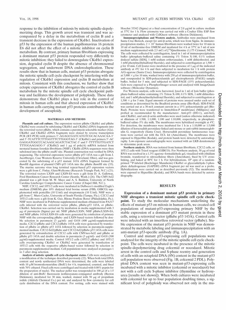
Expression of a dominant mutant p53 protein in primaryNHF abrogates a transient mitotic spindle cell cycle check-point. To study the molecular mechanisms underlining theeffects of mutant p53 on mitosis in human cells, we created cellpopulations of mutant-p53-expressing primary NHF by thestable expression of a dominant p53 mutant protein in thesecells, using a retroviral vector (pBabe p53 143A). Control cellswere infected with an insertless retroviral vector (pBabe). Sta-ble expression of the mutant p53 protein in NHF was demon-strated by metabolic labeling and immunoprecipitation with ananti-mutant p53-specific antibody (Fig. 1A).
Control and mutant p53-expressing cell populations wereanalyzed for the integrity of the mitotic spindle cell cycle check-point. The cells were incubated in the presence of the mitoticspindle-depolymerizing drug colcemid or nocodazol. Mitoticarrest in the control cells and S-phase reentry and generationof cells with an octaploid DNA (8N) content in the mutant-p53cell population were observed (Fig. 1B, colcemid 2 PDL). Poly-ploid DNA content was seen in mutant-p53-expressing cellsincubated with a mitotic inhibitor (colcemid or nocodazol) butnot with a cell cycle S-phase inhibitor (thymidine or hydroxy-urea [results not shown]). When both cultures were incubatedwith colcemid for up to four population doubling times, a sig-nificant level of polyploidy was observed not only in the mu-
VOL. 18, 1998 MUTANT p53 ALTERS MITOSIS VIA CKsHs1 6225
on March 2, 2015 by guest
http://mcb.asm
.org/D
ownloaded from
tant-p53-expressing cells but also in the control cells (Fig. 1B,NHF pBabe, colcemid 4 PDL, 12% of 8N cells). Nevertheless,the proportion of cells with a polyploid DNA content wasalways higher in the mutant-p53-expressing cell population(Fig. 1B, NHF pBabe-p53 143A, colcemid 4 PDL, 28% of 8Ncells and 4% of 16N cells). The presence of polyploidy in thesecells was confirmed by counting chromosomes in metaphasespreads (results not shown). These results extend our previousobservations with mutant-p53-expressing fibroblasts (22) andsuggest that the activation of the mitotic spindle cell cyclecheckpoint in human cells does not result in permanent growtharrest but in transient delay of mitosis. We hypothesized thatthe cell population carrying a mutant p53 protein accumulatedpolyploid cells at a higher rate than normal cells did becausemutant p53 proteins abrogate a transient mechanism of mitoticarrest.
The results shown in Fig. 1 are consistent with an alternativemechanism. These cells were obtained from biopsy specimensof human skin. Therefore, they are not clonal in origin, and sopolyploidy could be originated by the growth of a subpopula-tion of cells with an inactive mitotic spindle cell cycle check-point, namely, mitotic checkpoint-negative cells. In this sce-nario, the effect of mutant p53 could be but an increase in thefraction of mitotic checkpoint-negative cells rather than theabrogation of the mitotic spindle checkpoint. This hypothesiswas discarded on the basis of experiments in which NHF ar-rested by colcemid at the G2/M boundary (mitotic checkpoint-positive cells) were sorted, grown, and then incubated for fourpopulation doubling times in the presence of colcemid. Again,the generation of polyploid cells was observed (data not shown).These experiments support the conclusion that NHF arrestgrowth in response to an anomalous chromosomal segregationbut that this growth arrest is transient and may be abrogated bythe expression of a dominant mutant p53 protein.
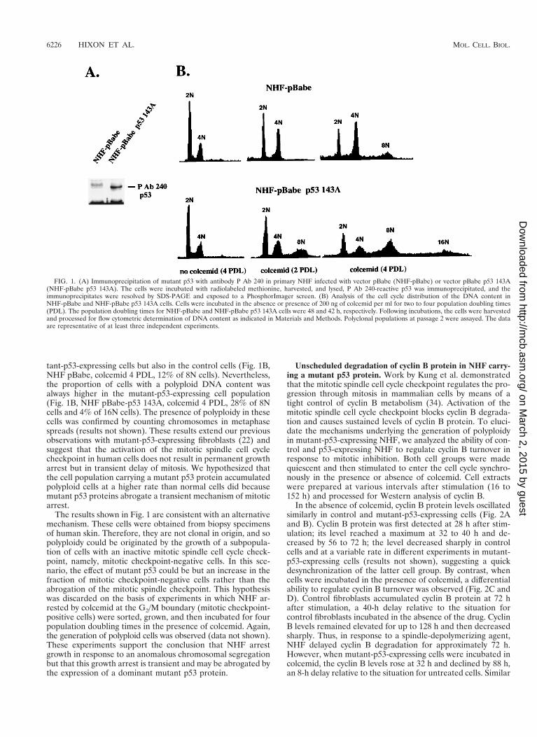
Unscheduled degradation of cyclin B protein in NHF carry-ing a mutant p53 protein. Work by Kung et al. demonstratedthat the mitotic spindle cell cycle checkpoint regulates the pro-gression through mitosis in mammalian cells by means of atight control of cyclin B metabolism (34). Activation of themitotic spindle cell cycle checkpoint blocks cyclin B degrada-tion and causes sustained levels of cyclin B protein. To eluci-date the mechanisms underlying the generation of polyploidyin mutant-p53-expressing NHF, we analyzed the ability of con-trol and p53-expressing NHF to regulate cyclin B turnover inresponse to mitotic inhibition. Both cell groups were madequiescent and then stimulated to enter the cell cycle synchro-nously in the presence or absence of colcemid. Cell extractswere prepared at various intervals after stimulation (16 to152 h) and processed for Western analysis of cyclin B.
In the absence of colcemid, cyclin B protein levels oscillatedsimilarly in control and mutant-p53-expressing cells (Fig. 2Aand B). Cyclin B protein was first detected at 28 h after stim-ulation; its level reached a maximum at 32 to 40 h and de-creased by 56 to 72 h; the level decreased sharply in controlcells and at a variable rate in different experiments in mutant-p53-expressing cells (results not shown), suggesting a quickdesynchronization of the latter cell group. By contrast, whencells were incubated in the presence of colcemid, a differentialability to regulate cyclin B turnover was observed (Fig. 2C andD). Control fibroblasts accumulated cyclin B protein at 72 hafter stimulation, a 40-h delay relative to the situation forcontrol fibroblasts incubated in the absence of the drug. CyclinB levels remained elevated for up to 128 h and then decreasedsharply. Thus, in response to a spindle-depolymerizing agent,NHF delayed cyclin B degradation for approximately 72 h.However, when mutant-p53-expressing cells were incubated incolcemid, the cyclin B levels rose at 32 h and declined by 88 h,an 8-h delay relative to the situation for untreated cells. Similar
FIG. 1. (A) Immunoprecipitation of mutant p53 with antibody P Ab 240 in primary NHF infected with vector pBabe (NHF-pBabe) or vector pBabe p53 143A(NHF-pBabe p53 143A). The cells were incubated with radiolabeled methionine, harvested, and lysed, P Ab 240-reactive p53 was immunoprecipitated, and theimmunoprecipitates were resolved by SDS-PAGE and exposed to a PhosphorImager screen. (B) Analysis of the cell cycle distribution of the DNA content inNHF-pBabe and NHF-pBabe p53 143A cells. Cells were incubated in the absence or presence of 200 ng of colcemid per ml for two to four population doubling times(PDL). The population doubling times for NHF-pBabe and NHF-pBabe p53 143A cells were 48 and 42 h, respectively. Following incubations, the cells were harvestedand processed for flow cytometric determination of DNA content as indicated in Materials and Methods. Polyclonal populations at passage 2 were assayed. The dataare representative of at least three independent experiments.
6226 HIXON ET AL. MOL. CELL. BIOL.
on March 2, 2015 by guest
http://mcb.asm
.org/D
ownloaded from
results were obtained with another two human cell populationsinfected with the pBabe or pBabe 143A vectors. These resultsshow that mutant-p53-expressing fibroblasts fail to regulatecyclin B levels in response to mitotic spindle depolymerization.These results underscore the role played by the control cyclinB metabolism at the mitotic spindle checkpoint in human cells.Moreover, since cyclin B metabolism eventually proceeded incontrol cells, these experiments supported the conclusion thatthe mitotic spindle checkpoint arrest is transient. The acceler-ated cyclin B degradation in mutant-p53-expressing NHF wasaccompanied by S-phase reentry as determined by flow cytom-etry of DNA content (results not shown). Mutant p53 proteinlevels did not change during the mitotic progression (resultsnot shown).
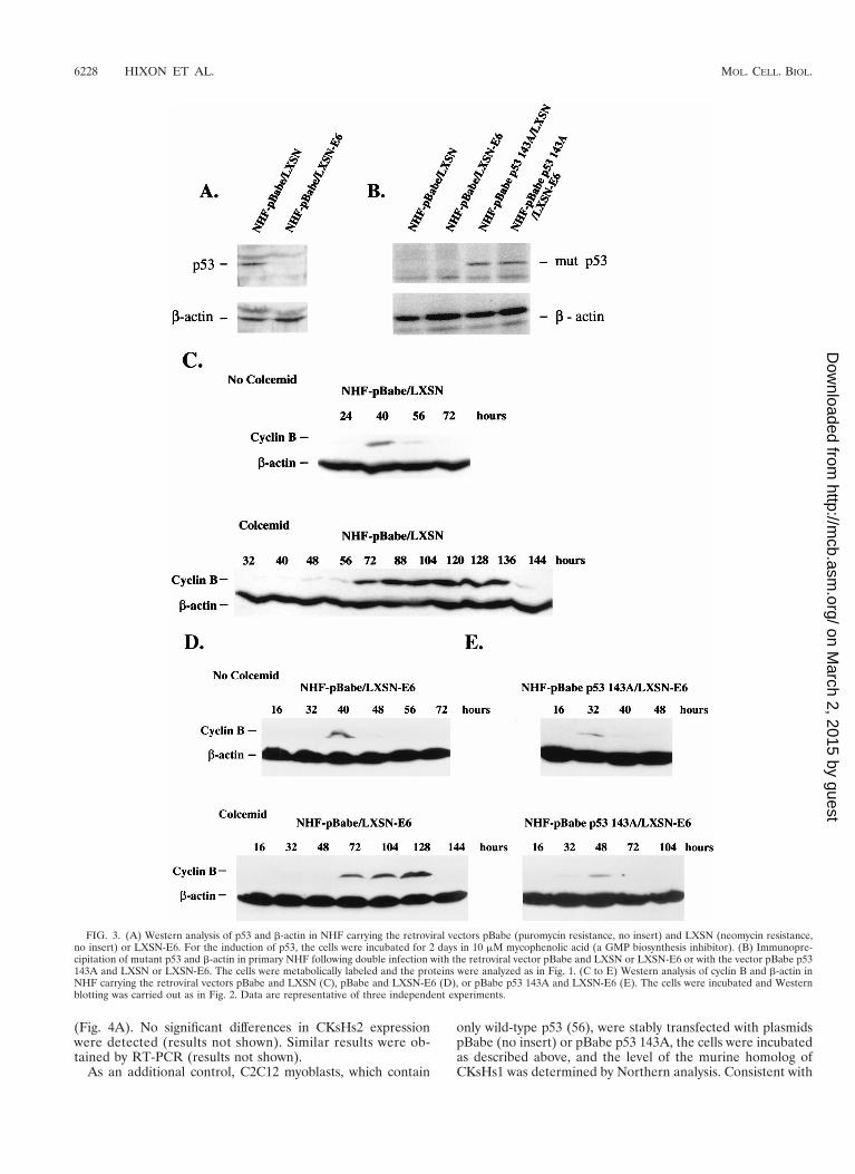
NHF expressing the human papillomavirus type 16 (HPV16)E6 oncoprotein regulate cyclin B metabolism in response tothe inhibition of mitosis. Mutant p53 proteins may exert dom-inant negative effects on wild-type p53 functions. Alternatively,mutant p53 proteins may exhibit properties that are not orig-inated by the inactivation of wild-type p53 (gain-of-function)(36). Thus, expression of a mutant p53 protein in NHF mayinterfere with putative wild-type p53-dependent or -inde-pendent mechanisms of checkpoint control at mitosis. To dis-tinguish between these two possibilities, we created a func-tional knockout of wild-type p53 in primary NHF by theexpression of the human papillomavirus type 16 E6 oncopro-tein in these cells with the retroviral vector LXSN (26). Severalexperimental controls were introduced in these series of ex-periments. Primary NHF were coinfected with LXSN (controlempty vector) and pBabe (control empty vector) or withLXSN-E6 and pBabe p53 143A. Cells infected with LXSN-E6were also coinfected with pBabe. The status of p53 in these cellpopulations is shown in Fig. 3A and B. HPV16 E6 promotedselectively the degradation of wild-type but not mutant p53.Therefore, NHF pBabe p53 143A/LXSN-E6 showed a dra-matic decrease in wild-type p53 levels but expressed similarlevels of mutant p53 protein to those found in the NHF pBabep53 143A/LXSN population. Primary NHF coinfected withboth control retroviral vectors demonstrated an 80-h delay in
cyclin B metabolism in response to colcemid (Fig. 3C). Wethen compared the ability of the NHF pBabe/LXSN-E6 andNHF pBabe p53 143A/LXSN-E6 to regulate cyclin B metab-olism in response to the mitotic inhibitor. E6-expressing NHFdelayed cyclin B metabolism in response to colcemid in asimilar way to the control population, i.e., by approximately80 h (Fig. 3C and D). However, the cell population coexpressingE6 and p53 143A showed a much shorter mitotic pause, i.e.,16 h, in response to the mitotic inhibitor (Fig. 3E), similar towhat we previously observed in mutant-p53-expressing cells(Fig. 2B and C). A second E6-expressing NHF population alsodisplayed a prolonged delay in cyclin B metabolism when in-cubated in the presence of colcemid (results not shown). Theseexperiments suggest that expression of a mutant p53 protein,but not wild-type p53 inactivation, abrogates the ability ofNHF to regulate the metabolism of cyclin B protein in re-sponse to an anomalous chromosomal segregation (see Dis-cussion). This hypothesis is in agreement with recent data byLanni and Jacks that shows similar mitotic delays in normaland p53 null mouse embryo fibroblasts challenged by a mitoticinhibitor (35). These results do not contradict the fact that p53inactivation may facilitate an increase in the fraction ofpolyploid cells in a given cell population by a different mech-anism, such as the abrogation of a postmitotic p53-dependentcell death response (35, 41).
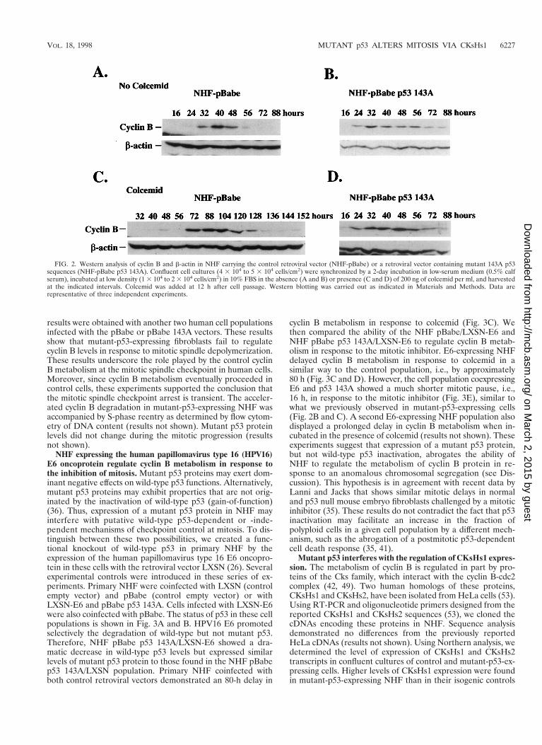
Mutant p53 interferes with the regulation of CKsHs1 expres-sion. The metabolism of cyclin B is regulated in part by pro-teins of the Cks family, which interact with the cyclin B-cdc2complex (42, 49). Two human homologs of these proteins,CKsHs1 and CKsHs2, have been isolated from HeLa cells (53).Using RT-PCR and oligonucleotide primers designed from thereported CKsHs1 and CKsHs2 sequences (53), we cloned thecDNAs encoding these proteins in NHF. Sequence analysisdemonstrated no differences from the previously reportedHeLa cDNAs (results not shown). Using Northern analysis, wedetermined the level of expression of CKsHs1 and CKsHs2transcripts in confluent cultures of control and mutant-p53-ex-pressing cells. Higher levels of CKsHs1 expression were foundin mutant-p53-expressing NHF than in their isogenic controls
FIG. 2. Western analysis of cyclin B and b-actin in NHF carrying the control retroviral vector (NHF-pBabe) or a retroviral vector containing mutant 143A p53sequences (NHF-pBabe p53 143A). Confluent cell cultures (4 3 104 to 5 3 104 cells/cm2) were synchronized by a 2-day incubation in low-serum medium (0.5% calfserum), incubated at low density (1 3 104 to 2 3 104 cells/cm2) in 10% FBS in the absence (A and B) or presence (C and D) of 200 ng of colcemid per ml, and harvestedat the indicated intervals. Colcemid was added at 12 h after cell passage. Western blotting was carried out as indicated in Materials and Methods. Data arerepresentative of three independent experiments.
VOL. 18, 1998 MUTANT p53 ALTERS MITOSIS VIA CKsHs1 6227
on March 2, 2015 by guest
http://mcb.asm
.org/D
ownloaded from
(Fig. 4A). No significant differences in CKsHs2 expressionwere detected (results not shown). Similar results were ob-tained by RT-PCR (results not shown).
As an additional control, C2C12 myoblasts, which contain
only wild-type p53 (56), were stably transfected with plasmidspBabe (no insert) or pBabe p53 143A, the cells were incubatedas described above, and the level of the murine homolog ofCKsHs1 was determined by Northern analysis. Consistent with
FIG. 3. (A) Western analysis of p53 and b-actin in NHF carrying the retroviral vectors pBabe (puromycin resistance, no insert) and LXSN (neomycin resistance,no insert) or LXSN-E6. For the induction of p53, the cells were incubated for 2 days in 10 mM mycophenolic acid (a GMP biosynthesis inhibitor). (B) Immunopre-cipitation of mutant p53 and b-actin in primary NHF following double infection with the retroviral vector pBabe and LXSN or LXSN-E6 or with the vector pBabe p53143A and LXSN or LXSN-E6. The cells were metabolically labeled and the proteins were analyzed as in Fig. 1. (C to E) Western analysis of cyclin B and b-actin inNHF carrying the retroviral vectors pBabe and LXSN (C), pBabe and LXSN-E6 (D), or pBabe p53 143A and LXSN-E6 (E). The cells were incubated and Westernblotting was carried out as in Fig. 2. Data are representative of three independent experiments.
6228 HIXON ET AL. MOL. CELL. BIOL.
on March 2, 2015 by guest
http://mcb.asm
.org/D
ownloaded from
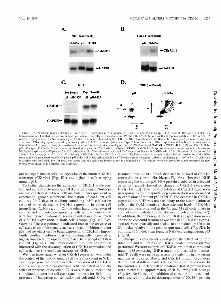
our findings in human cells, the expression of the murine CKsHs1transcript (CKsMm1 [Fig. 4B]) was higher in cells carryingmutant p53.
To further characterize the expression of CKsHs1 in the con-trol and mutant-p53-expressing NHF, we performed Northernanalysis of CKsHs1 in these cells incubated under quiescent orexponential growth conditions. Incubation of confluent cellcultures for 2 days in medium containing 0.5% calf serumresulted in no detectable CKsHs1 expression in either cellgroup (Fig. 4C, No Serum). On the other hand, incubation ofcontrol and mutant-p53-expressing cells at low density andwith high concentrations of serum resulted in similar levelsof CKsHs1 expression in both cells groups (Fig. 4C, Exp.Growth). These experiments indicated that in the absence ofcell cycle checkpoint signals, such as contact inhibition, mutantp53 had no effect on the basal expression of CKsHs1. Impor-tantly, confluent cultures of p53-expressing cells incubated inhigh-serum medium rapidly progressed to a polyploid DNAcontent (Fig. 4D). Thus, expression of a mutant p53 proteininterfered with the downregulation of CKsHs1 expression andcell cycle arrest in confluent cultures of NHF.
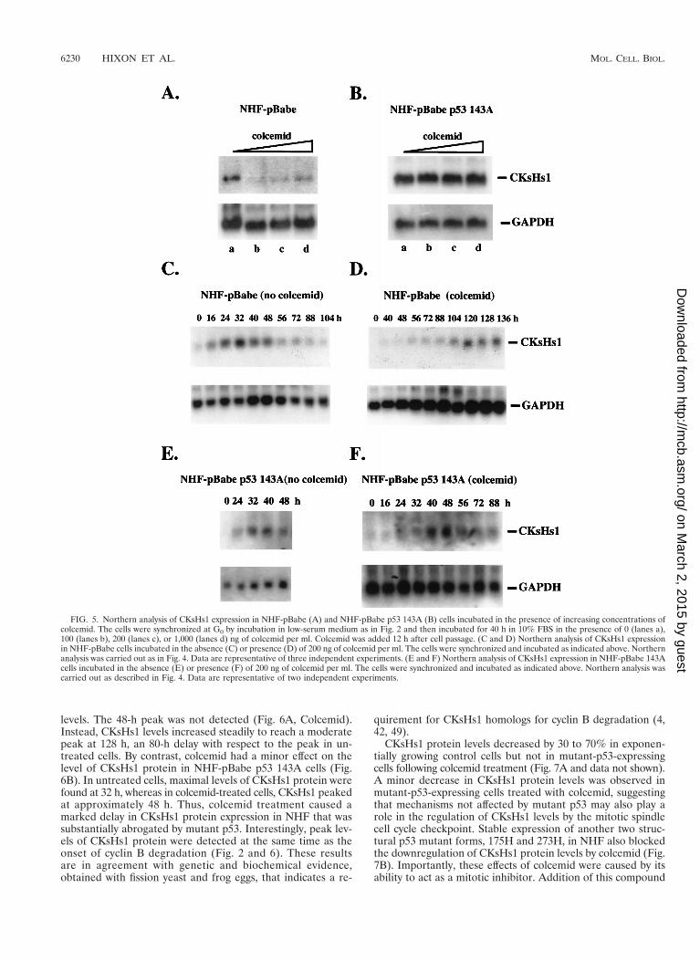
We then investigated whether CKsHs1 expression was underthe control of the mitotic spindle cell cycle checkpoint in NHF.For this purpose, we determined the expression of CKsHs1 incontrol and mutant-p53-expressing cells incubated in the ab-sence or presence of colcemid. Cells were made quiescent andstimulated to enter the cell cycle synchronously for 40 h in thepresence of increasing concentrations of colcemid. Colcemid
treatment resulted in a drastic decrease in the level of CKsHs1expression in control fibroblasts (Fig. 5A). However, NHFexpressing the mutant p53 143A protein incubated in colcemidat up to 1 mg/ml showed no change in CKsHs1 expressionlevels (Fig. 5B). Thus, downregulation of CKsHs1 expressionin response to mitotic spindle depolymerization was abrogatedby expression of mutant p53 in NHF. The decrease in CKsHs1expression in NHF was not secondary to the accumulation ofcells at the G2/M boundary, since maximal levels of CKsHs1expression were observed at the G2 and M cell cycle phase incontrol cells incubated in the absence of colcemid (Fig. 5C).In addition, the downregulation of CKsHs1 expression in re-sponse to colcemid treatment was transient. CKsHs1 expres-sion recovered partially and peaked at approximately 120 h, an80-h delay relative to the peak in untreated cells (Fig. 5D). Incontrast, a 16-h delay was found in NHF expressing mutant p53(Fig. 5E).
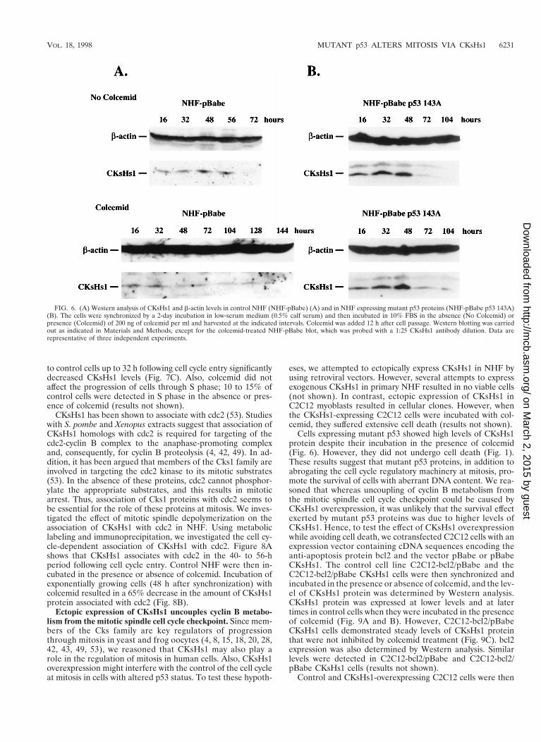
Subsequent experiments investigated the effect of mitoticinhibition and mutant p53 on CKsHs1 protein expression. Weperformed Western analysis of CKsHs1 protein in control andmutant-p53-expressing NHF treated or not treated with colce-mid. The cells were made quiescent by incubation in low-serummedium as indicated above, and CKsHs1 protein levels weredetermined at different intervals following cell cycle entry. Inthe control NHF-pBabe cell population, CKsHs1 protein levelswere maximal at approximately 48 h following cell passage(Fig. 6A, No Colcemid). Addition of colcemid to the cell cul-ture resulted in a drastic downregulation of CKsHs1 protein
FIG. 4. (A) Northern analysis of CKsHs1 and GAPDH expression in NHF-pBabe (pB), NHF-pBabe p53 143A (pB-143A), and HT1080 cells. HT1080 is afibrosarcoma cell line that carries two mutated p53 alleles. The cells were incubated in DMEM with 10% FBS until confluent (approximately 4 3 104 to 5 3 104
cells/cm2) and processed for Northern analysis. A CKsHs1 sequence obtained by RT-PCR from NHF was subcloned into Bluescribe (Stratagene), sequenced, and usedas a probe. RNA integrity was verified by reprobing with a GAPDH sequence (American Type Culture Collection). Other experimental details were as indicated inMaterials and Methods. (B) Northern analysis of the expression of a murine homolog of CKsHs1 (CKsMm1) and GAPDH in C2C12-pBabe (pB) and C2C12-pBabep53 143A (pB-143A) cells. The cells were incubated as in panel A. (C) Northern analysis of CKsHs1 and GAPDH expression in quiescent or exponentially growingNHF-pBabe (pB) and NHF-pBabe p53 143A (pB-143A) cells. The cells were incubated for 2 days at confluence in DMEM with 0.5% calf serum (No Serum) or for2 days at low density (1 3 104 to 2 3 104 cells/cm2) in DMEM with 10% FBS (Exp. Growth). (D) Flow-cytometric analysis of the cell cycle distribution of the DNAcontent in NHF-pBabe (pB) and NHF-pBabe p53 143A (pB-143A) cells at confluence. The cells were incubated for 2 days at confluence (4 3 104 to 5 3 104 cells/cm2)in DMEM with 10% FBS, 100 mM BrdU was added, and the cells were incubated for an additional 4 h. The cultures were harvested, fixed, and processed for flowcytometry as indicated in Materials and Methods.
VOL. 18, 1998 MUTANT p53 ALTERS MITOSIS VIA CKsHs1 6229
on March 2, 2015 by guest
http://mcb.asm
.org/D
ownloaded from
levels. The 48-h peak was not detected (Fig. 6A, Colcemid).Instead, CKsHs1 levels increased steadily to reach a moderatepeak at 128 h, an 80-h delay with respect to the peak in un-treated cells. By contrast, colcemid had a minor effect on thelevel of CKsHs1 protein in NHF-pBabe p53 143A cells (Fig.6B). In untreated cells, maximal levels of CKsHs1 protein werefound at 32 h, whereas in colcemid-treated cells, CKsHs1 peakedat approximately 48 h. Thus, colcemid treatment caused amarked delay in CKsHs1 protein expression in NHF that wassubstantially abrogated by mutant p53. Interestingly, peak lev-els of CKsHs1 protein were detected at the same time as theonset of cyclin B degradation (Fig. 2 and 6). These resultsare in agreement with genetic and biochemical evidence,obtained with fission yeast and frog eggs, that indicates a re-
quirement for CKsHs1 homologs for cyclin B degradation (4,42, 49).
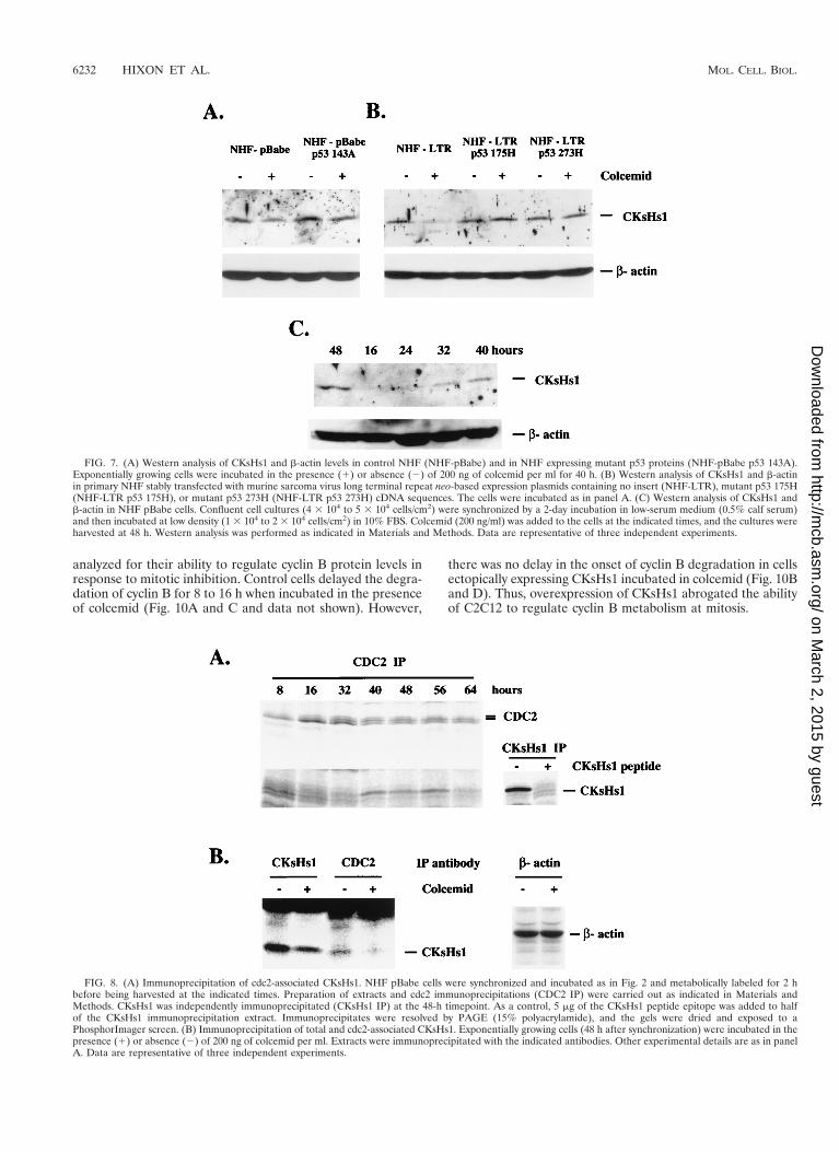
CKsHs1 protein levels decreased by 30 to 70% in exponen-tially growing control cells but not in mutant-p53-expressingcells following colcemid treatment (Fig. 7A and data not shown).A minor decrease in CKsHs1 protein levels was observed inmutant-p53-expressing cells treated with colcemid, suggestingthat mechanisms not affected by mutant p53 may also play arole in the regulation of CKsHs1 levels by the mitotic spindlecell cycle checkpoint. Stable expression of another two struc-tural p53 mutant forms, 175H and 273H, in NHF also blockedthe downregulation of CKsHs1 protein levels by colcemid (Fig.7B). Importantly, these effects of colcemid were caused by itsability to act as a mitotic inhibitor. Addition of this compound
FIG. 5. Northern analysis of CKsHs1 expression in NHF-pBabe (A) and NHF-pBabe p53 143A (B) cells incubated in the presence of increasing concentrations ofcolcemid. The cells were synchronized at G0 by incubation in low-serum medium as in Fig. 2 and then incubated for 40 h in 10% FBS in the presence of 0 (lanes a),100 (lanes b), 200 (lanes c), or 1,000 (lanes d) ng of colcemid per ml. Colcemid was added 12 h after cell passage. (C and D) Northern analysis of CKsHs1 expressionin NHF-pBabe cells incubated in the absence (C) or presence (D) of 200 ng of colcemid per ml. The cells were synchronized and incubated as indicated above. Northernanalysis was carried out as in Fig. 4. Data are representative of three independent experiments. (E and F) Northern analysis of CKsHs1 expression in NHF-pBabe 143Acells incubated in the absence (E) or presence (F) of 200 ng of colcemid per ml. The cells were synchronized and incubated as indicated above. Northern analysis wascarried out as described in Fig. 4. Data are representative of two independent experiments.
6230 HIXON ET AL. MOL. CELL. BIOL.
on March 2, 2015 by guest
http://mcb.asm
.org/D
ownloaded from
to control cells up to 32 h following cell cycle entry significantlydecreased CKsHs1 levels (Fig. 7C). Also, colcemid did notaffect the progression of cells through S phase; 10 to 15% ofcontrol cells were detected in S phase in the absence or pres-ence of colcemid (results not shown).
CKsHs1 has been shown to associate with cdc2 (53). Studieswith S. pombe and Xenopus extracts suggest that association ofCKsHs1 homologs with cdc2 is required for targeting of thecdc2-cyclin B complex to the anaphase-promoting complexand, consequently, for cyclin B proteolysis (4, 42, 49). In ad-dition, it has been argued that members of the Cks1 family areinvolved in targeting the cdc2 kinase to its mitotic substrates(53). In the absence of these proteins, cdc2 cannot phosphor-ylate the appropriate substrates, and this results in mitoticarrest. Thus, association of Cks1 proteins with cdc2 seems tobe essential for the role of these proteins at mitosis. We inves-tigated the effect of mitotic spindle depolymerization on theassociation of CKsHs1 with cdc2 in NHF. Using metaboliclabeling and immunoprecipitation, we investigated the cell cy-cle-dependent association of CKsHs1 with cdc2. Figure 8Ashows that CKsHs1 associates with cdc2 in the 40- to 56-hperiod following cell cycle entry. Control NHF were then in-cubated in the presence or absence of colcemid. Incubation ofexponentially growing cells (48 h after synchronization) withcolcemid resulted in a 65% decrease in the amount of CKsHs1protein associated with cdc2 (Fig. 8B).
Ectopic expression of CKsHs1 uncouples cyclin B metabo-lism from the mitotic spindle cell cycle checkpoint. Since mem-bers of the Cks family are key regulators of progressionthrough mitosis in yeast and frog oocytes (4, 8, 15, 18, 20, 28,42, 43, 49, 53), we reasoned that CKsHs1 may also play arole in the regulation of mitosis in human cells. Also, CKsHs1overexpression might interfere with the control of the cell cycleat mitosis in cells with altered p53 status. To test these hypoth-
eses, we attempted to ectopically express CKsHs1 in NHF byusing retroviral vectors. However, several attempts to expressexogenous CKsHs1 in primary NHF resulted in no viable cells(not shown). In contrast, ectopic expression of CKsHs1 inC2C12 myoblasts resulted in cellular clones. However, whenthe CKsHs1-expressing C2C12 cells were incubated with col-cemid, they suffered extensive cell death (results not shown).
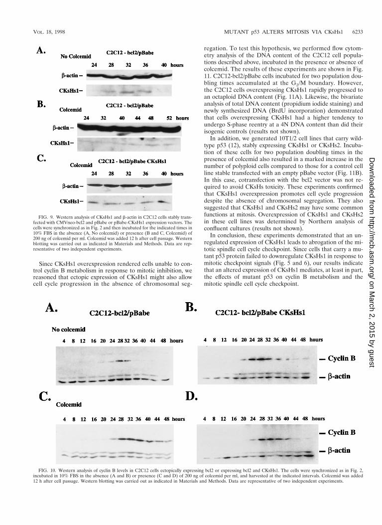
Cells expressing mutant p53 showed high levels of CKsHs1protein despite their incubation in the presence of colcemid(Fig. 6). However, they did not undergo cell death (Fig. 1).These results suggest that mutant p53 proteins, in addition toabrogating the cell cycle regulatory machinery at mitosis, pro-mote the survival of cells with aberrant DNA content. We rea-soned that whereas uncoupling of cyclin B metabolism fromthe mitotic spindle cell cycle checkpoint could be caused byCKsHs1 overexpression, it was unlikely that the survival effectexerted by mutant p53 proteins was due to higher levels ofCKsHs1. Hence, to test the effect of CKsHs1 overexpressionwhile avoiding cell death, we cotransfected C2C12 cells with anexpression vector containing cDNA sequences encoding theanti-apoptosis protein bcl2 and the vector pBabe or pBabeCKsHs1. The control cell line C2C12-bcl2/pBabe and theC2C12-bcl2/pBabe CKsHs1 cells were then synchronized andincubated in the presence or absence of colcemid, and the lev-el of CKsHs1 protein was determined by Western analysis.CKsHs1 protein was expressed at lower levels and at latertimes in control cells when they were incubated in the presenceof colcemid (Fig. 9A and B). However, C2C12-bcl2/pBabeCKsHs1 cells demonstrated steady levels of CKsHs1 proteinthat were not inhibited by colcemid treatment (Fig. 9C). bcl2expression was also determined by Western analysis. Similarlevels were detected in C2C12-bcl2/pBabe and C2C12-bcl2/pBabe CKsHs1 cells (results not shown).
Control and CKsHs1-overexpressing C2C12 cells were then
FIG. 6. (A) Western analysis of CKsHs1 and b-actin levels in control NHF (NHF-pBabe) (A) and in NHF expressing mutant p53 proteins (NHF-pBabe p53 143A)(B). The cells were synchronized by a 2-day incubation in low-serum medium (0.5% calf serum) and then incubated in 10% FBS in the absence (No Colcemid) orpresence (Colcemid) of 200 ng of colcemid per ml and harvested at the indicated intervals. Colcemid was added 12 h after cell passage. Western blotting was carriedout as indicated in Materials and Methods, except for the colcemid-treated NHF-pBabe blot, which was probed with a 1:25 CKsHs1 antibody dilution. Data arerepresentative of three independent experiments.
VOL. 18, 1998 MUTANT p53 ALTERS MITOSIS VIA CKsHs1 6231
on March 2, 2015 by guest
http://mcb.asm
.org/D
ownloaded from
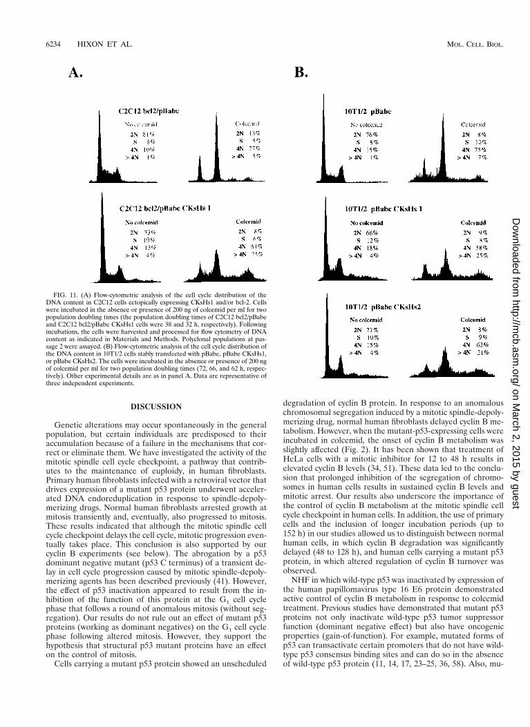
analyzed for their ability to regulate cyclin B protein levels inresponse to mitotic inhibition. Control cells delayed the degra-dation of cyclin B for 8 to 16 h when incubated in the presenceof colcemid (Fig. 10A and C and data not shown). However,
there was no delay in the onset of cyclin B degradation in cellsectopically expressing CKsHs1 incubated in colcemid (Fig. 10Band D). Thus, overexpression of CKsHs1 abrogated the abilityof C2C12 to regulate cyclin B metabolism at mitosis.
FIG. 7. (A) Western analysis of CKsHs1 and b-actin levels in control NHF (NHF-pBabe) and in NHF expressing mutant p53 proteins (NHF-pBabe p53 143A).Exponentially growing cells were incubated in the presence (1) or absence (2) of 200 ng of colcemid per ml for 40 h. (B) Western analysis of CKsHs1 and b-actinin primary NHF stably transfected with murine sarcoma virus long terminal repeat neo-based expression plasmids containing no insert (NHF-LTR), mutant p53 175H(NHF-LTR p53 175H), or mutant p53 273H (NHF-LTR p53 273H) cDNA sequences. The cells were incubated as in panel A. (C) Western analysis of CKsHs1 andb-actin in NHF pBabe cells. Confluent cell cultures (4 3 104 to 5 3 104 cells/cm2) were synchronized by a 2-day incubation in low-serum medium (0.5% calf serum)and then incubated at low density (1 3 104 to 2 3 104 cells/cm2) in 10% FBS. Colcemid (200 ng/ml) was added to the cells at the indicated times, and the cultures wereharvested at 48 h. Western analysis was performed as indicated in Materials and Methods. Data are representative of three independent experiments.
FIG. 8. (A) Immunoprecipitation of cdc2-associated CKsHs1. NHF pBabe cells were synchronized and incubated as in Fig. 2 and metabolically labeled for 2 hbefore being harvested at the indicated times. Preparation of extracts and cdc2 immunoprecipitations (CDC2 IP) were carried out as indicated in Materials andMethods. CKsHs1 was independently immunoprecipitated (CKsHs1 IP) at the 48-h timepoint. As a control, 5 mg of the CKsHs1 peptide epitope was added to halfof the CKsHs1 immunoprecipitation extract. Immunoprecipitates were resolved by PAGE (15% polyacrylamide), and the gels were dried and exposed to aPhosphorImager screen. (B) Immunoprecipitation of total and cdc2-associated CKsHs1. Exponentially growing cells (48 h after synchronization) were incubated in thepresence (1) or absence (2) of 200 ng of colcemid per ml. Extracts were immunoprecipitated with the indicated antibodies. Other experimental details are as in panelA. Data are representative of three independent experiments.
6232 HIXON ET AL. MOL. CELL. BIOL.
on March 2, 2015 by guest
http://mcb.asm
.org/D
ownloaded from
Since CKsHs1 overexpression rendered cells unable to con-trol cyclin B metabolism in response to mitotic inhibition, wereasoned that ectopic expression of CKsHs1 might also allowcell cycle progression in the absence of chromosomal seg-
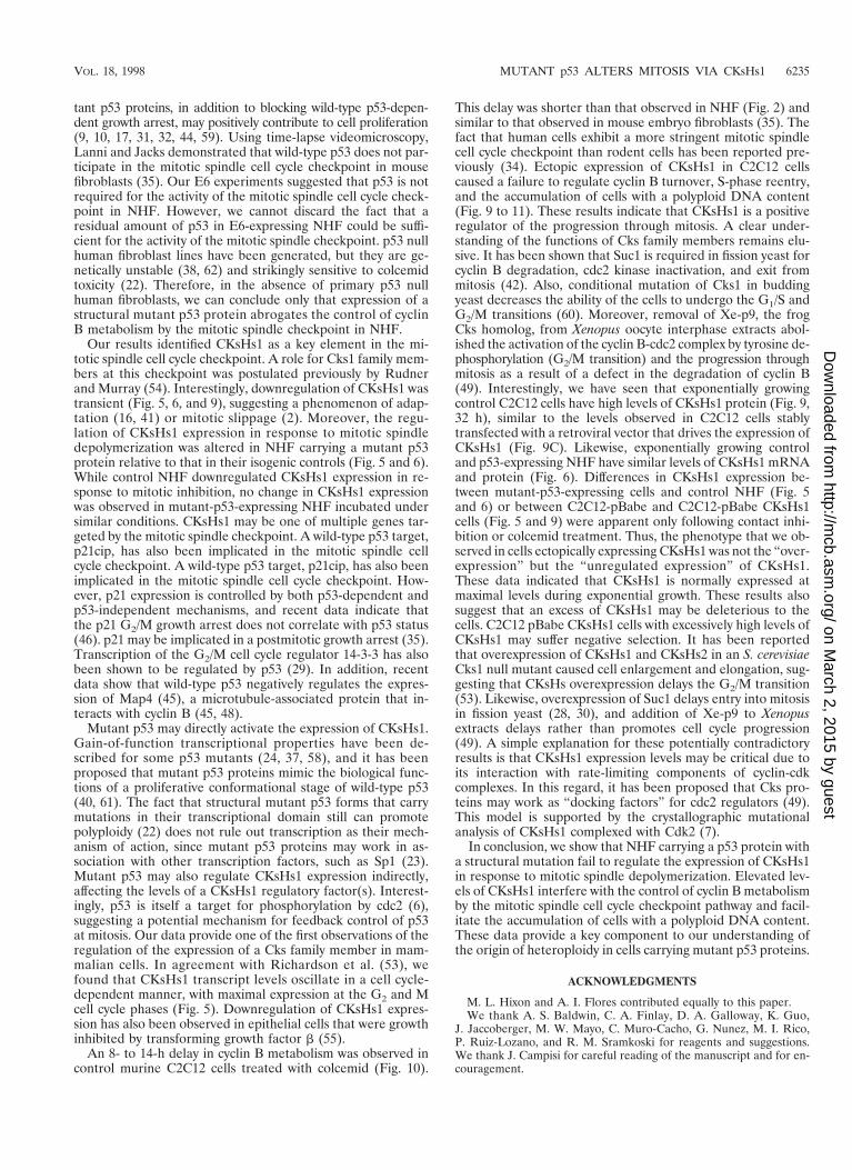
regation. To test this hypothesis, we performed flow cytom-etry analysis of the DNA content of the C2C12 cell popula-tions described above, incubated in the presence or absence ofcolcemid. The results of these experiments are shown in Fig.11. C2C12-bcl2/pBabe cells incubated for two population dou-bling times accumulated at the G2/M boundary. However,the C2C12 cells overexpressing CKsHs1 rapidly progressed toan octaploid DNA content (Fig. 11A). Likewise, the bivariateanalysis of total DNA content (propidium iodide staining) andnewly synthesized DNA (BrdU incorporation) demonstratedthat cells overexpressing CKsHs1 had a higher tendency toundergo S-phase reentry at a 4N DNA content than did theirisogenic controls (results not shown).
In addition, we generated 10T1/2 cell lines that carry wild-type p53 (12), stably expressing CKsHs1 or CKsHs2. Incuba-tion of these cells for two population doubling times in thepresence of colcemid also resulted in a marked increase in thenumber of polyploid cells compared to those for a control cellline stable transfected with an empty pBabe vector (Fig. 11B).In this case, cotransfection with the bcl2 vector was not re-quired to avoid CKsHs toxicity. These experiments confirmedthat CKsHs1 overexpression promotes cell cycle progressiondespite the absence of chromosomal segregation. They alsosuggested that CKsHs1 and CKsHs2 may have some commonfunctions at mitosis. Overexpression of CKsHs1 and CKsHs2in these cell lines was determined by Northern analysis ofconfluent cultures (results not shown).
In conclusion, these experiments demonstrated that an un-regulated expression of CKsHs1 leads to abrogation of the mi-totic spindle cell cycle checkpoint. Since cells that carry a mu-tant p53 protein failed to downregulate CKsHs1 in response tomitotic checkpoint signals (Fig. 5 and 6), our results indicatethat an altered expression of CKsHs1 mediates, at least in part,the effects of mutant p53 on cyclin B metabolism and themitotic spindle cell cycle checkpoint.
FIG. 10. Western analysis of cyclin B levels in C2C12 cells ectopically expressing bcl2 or expressing bcl2 and CKsHs1. The cells were synchronized as in Fig. 2,incubated in 10% FBS in the absence (A and B) or presence (C and D) of 200 ng of colcemid per ml, and harvested at the indicated intervals. Colcemid was added12 h after cell passage. Western blotting was carried out as indicated in Materials and Methods. Data are representative of two independent experiments.
FIG. 9. Western analysis of CKsHs1 and b-actin in C2C12 cells stably trans-fected with CMVneo-bcl2 and pBabe or pBabe-CKsHs1 expression vectors. Thecells were synchronized as in Fig. 2 and then incubated for the indicated times in10% FBS in the absence (A, No colcemid) or presence (B and C, Colcemid) of200 ng of colcemid per ml. Colcemid was added 12 h after cell passage. Westernblotting was carried out as indicated in Materials and Methods. Data are rep-resentative of two independent experiments.
VOL. 18, 1998 MUTANT p53 ALTERS MITOSIS VIA CKsHs1 6233
on March 2, 2015 by guest
http://mcb.asm
.org/D
ownloaded from
DISCUSSION
Genetic alterations may occur spontaneously in the generalpopulation, but certain individuals are predisposed to theiraccumulation because of a failure in the mechanisms that cor-rect or eliminate them. We have investigated the activity of themitotic spindle cell cycle checkpoint, a pathway that contrib-utes to the maintenance of euploidy, in human fibroblasts.Primary human fibroblasts infected with a retroviral vector thatdrives expression of a mutant p53 protein underwent acceler-ated DNA endoreduplication in response to spindle-depoly-merizing drugs. Normal human fibroblasts arrested growth atmitosis transiently and, eventually, also progressed to mitosis.These results indicated that although the mitotic spindle cellcycle checkpoint delays the cell cycle, mitotic progression even-tually takes place. This conclusion is also supported by ourcyclin B experiments (see below). The abrogation by a p53dominant negative mutant (p53 C terminus) of a transient de-lay in cell cycle progression caused by mitotic spindle-depoly-merizing agents has been described previously (41). However,the effect of p53 inactivation appeared to result from the in-hibition of the function of this protein at the G1 cell cyclephase that follows a round of anomalous mitosis (without seg-regation). Our results do not rule out an effect of mutant p53proteins (working as dominant negatives) on the G1 cell cyclephase following altered mitosis. However, they support thehypothesis that structural p53 mutant proteins have an effecton the control of mitosis.
Cells carrying a mutant p53 protein showed an unscheduled
degradation of cyclin B protein. In response to an anomalouschromosomal segregation induced by a mitotic spindle-depoly-merizing drug, normal human fibroblasts delayed cyclin B me-tabolism. However, when the mutant-p53-expressing cells wereincubated in colcemid, the onset of cyclin B metabolism wasslightly affected (Fig. 2). It has been shown that treatment ofHeLa cells with a mitotic inhibitor for 12 to 48 h results inelevated cyclin B levels (34, 51). These data led to the conclu-sion that prolonged inhibition of the segregation of chromo-somes in human cells results in sustained cyclin B levels andmitotic arrest. Our results also underscore the importance ofthe control of cyclin B metabolism at the mitotic spindle cellcycle checkpoint in human cells. In addition, the use of primarycells and the inclusion of longer incubation periods (up to152 h) in our studies allowed us to distinguish between normalhuman cells, in which cyclin B degradation was significantlydelayed (48 to 128 h), and human cells carrying a mutant p53protein, in which altered regulation of cyclin B turnover wasobserved.
NHF in which wild-type p53 was inactivated by expression ofthe human papillomavirus type 16 E6 protein demonstratedactive control of cyclin B metabolism in response to colcemidtreatment. Previous studies have demonstrated that mutant p53proteins not only inactivate wild-type p53 tumor suppressorfunction (dominant negative effect) but also have oncogenicproperties (gain-of-function). For example, mutated forms ofp53 can transactivate certain promoters that do not have wild-type p53 consensus binding sites and can do so in the absenceof wild-type p53 protein (11, 14, 17, 23–25, 36, 58). Also, mu-
FIG. 11. (A) Flow-cytometric analysis of the cell cycle distribution of theDNA content in C2C12 cells ectopically expressing CKsHs1 and/or bcl-2. Cellswere incubated in the absence or presence of 200 ng of colcemid per ml for twopopulation doubling times (the population doubling times of C2C12 bcl2/pBabeand C2C12 bcl2/pBabe CKsHs1 cells were 38 and 32 h, respectively). Followingincubations, the cells were harvested and processed for flow cytometry of DNAcontent as indicated in Materials and Methods. Polyclonal populations at pas-sage 2 were assayed. (B) Flow-cytometric analysis of the cell cycle distribution ofthe DNA content in 10T1/2 cells stably transfected with pBabe, pBabe CKsHs1,or pBabe CKsHs2. The cells were incubated in the absence or presence of 200 ngof colcemid per ml for two population doubling times (72, 66, and 62 h, respec-tively). Other experimental details are as in panel A. Data are representative ofthree independent experiments.
6234 HIXON ET AL. MOL. CELL. BIOL.
on March 2, 2015 by guest
http://mcb.asm
.org/D
ownloaded from
tant p53 proteins, in addition to blocking wild-type p53-depen-dent growth arrest, may positively contribute to cell proliferation(9, 10, 17, 31, 32, 44, 59). Using time-lapse videomicroscopy,Lanni and Jacks demonstrated that wild-type p53 does not par-ticipate in the mitotic spindle cell cycle checkpoint in mousefibroblasts (35). Our E6 experiments suggested that p53 is notrequired for the activity of the mitotic spindle cell cycle check-point in NHF. However, we cannot discard the fact that aresidual amount of p53 in E6-expressing NHF could be suffi-cient for the activity of the mitotic spindle checkpoint. p53 nullhuman fibroblast lines have been generated, but they are ge-netically unstable (38, 62) and strikingly sensitive to colcemidtoxicity (22). Therefore, in the absence of primary p53 nullhuman fibroblasts, we can conclude only that expression of astructural mutant p53 protein abrogates the control of cyclinB metabolism by the mitotic spindle checkpoint in NHF.
Our results identified CKsHs1 as a key element in the mi-totic spindle cell cycle checkpoint. A role for Cks1 family mem-bers at this checkpoint was postulated previously by Rudnerand Murray (54). Interestingly, downregulation of CKsHs1 wastransient (Fig. 5, 6, and 9), suggesting a phenomenon of adap-tation (16, 41) or mitotic slippage (2). Moreover, the regu-lation of CKsHs1 expression in response to mitotic spindledepolymerization was altered in NHF carrying a mutant p53protein relative to that in their isogenic controls (Fig. 5 and 6).While control NHF downregulated CKsHs1 expression in re-sponse to mitotic inhibition, no change in CKsHs1 expressionwas observed in mutant-p53-expressing NHF incubated undersimilar conditions. CKsHs1 may be one of multiple genes tar-geted by the mitotic spindle checkpoint. A wild-type p53 target,p21cip, has also been implicated in the mitotic spindle cellcycle checkpoint. A wild-type p53 target, p21cip, has also beenimplicated in the mitotic spindle cell cycle checkpoint. How-ever, p21 expression is controlled by both p53-dependent andp53-independent mechanisms, and recent data indicate thatthe p21 G2/M growth arrest does not correlate with p53 status(46). p21 may be implicated in a postmitotic growth arrest (35).Transcription of the G2/M cell cycle regulator 14-3-3 has alsobeen shown to be regulated by p53 (29). In addition, recentdata show that wild-type p53 negatively regulates the expres-sion of Map4 (45), a microtubule-associated protein that in-teracts with cyclin B (45, 48).
Mutant p53 may directly activate the expression of CKsHs1.Gain-of-function transcriptional properties have been de-scribed for some p53 mutants (24, 37, 58), and it has beenproposed that mutant p53 proteins mimic the biological func-tions of a proliferative conformational stage of wild-type p53(40, 61). The fact that structural mutant p53 forms that carrymutations in their transcriptional domain still can promotepolyploidy (22) does not rule out transcription as their mech-anism of action, since mutant p53 proteins may work in as-sociation with other transcription factors, such as Sp1 (23).Mutant p53 may also regulate CKsHs1 expression indirectly,affecting the levels of a CKsHs1 regulatory factor(s). Interest-ingly, p53 is itself a target for phosphorylation by cdc2 (6),suggesting a potential mechanism for feedback control of p53at mitosis. Our data provide one of the first observations of theregulation of the expression of a Cks family member in mam-malian cells. In agreement with Richardson et al. (53), wefound that CKsHs1 transcript levels oscillate in a cell cycle-dependent manner, with maximal expression at the G2 and Mcell cycle phases (Fig. 5). Downregulation of CKsHs1 expres-sion has also been observed in epithelial cells that were growthinhibited by transforming growth factor b (55).
An 8- to 14-h delay in cyclin B metabolism was observed incontrol murine C2C12 cells treated with colcemid (Fig. 10).
This delay was shorter than that observed in NHF (Fig. 2) andsimilar to that observed in mouse embryo fibroblasts (35). Thefact that human cells exhibit a more stringent mitotic spindlecell cycle checkpoint than rodent cells has been reported pre-viously (34). Ectopic expression of CKsHs1 in C2C12 cellscaused a failure to regulate cyclin B turnover, S-phase reentry,and the accumulation of cells with a polyploid DNA content(Fig. 9 to 11). These results indicate that CKsHs1 is a positiveregulator of the progression through mitosis. A clear under-standing of the functions of Cks family members remains elu-sive. It has been shown that Suc1 is required in fission yeast forcyclin B degradation, cdc2 kinase inactivation, and exit frommitosis (42). Also, conditional mutation of Cks1 in buddingyeast decreases the ability of the cells to undergo the G1/S andG2/M transitions (60). Moreover, removal of Xe-p9, the frogCks homolog, from Xenopus oocyte interphase extracts abol-ished the activation of the cyclin B-cdc2 complex by tyrosine de-phosphorylation (G2/M transition) and the progression throughmitosis as a result of a defect in the degradation of cyclin B(49). Interestingly, we have seen that exponentially growingcontrol C2C12 cells have high levels of CKsHs1 protein (Fig. 9,32 h), similar to the levels observed in C2C12 cells stablytransfected with a retroviral vector that drives the expression ofCKsHs1 (Fig. 9C). Likewise, exponentially growing controland p53-expressing NHF have similar levels of CKsHs1 mRNAand protein (Fig. 6). Differences in CKsHs1 expression be-tween mutant-p53-expressing cells and control NHF (Fig. 5and 6) or between C2C12-pBabe and C2C12-pBabe CKsHs1cells (Fig. 5 and 9) were apparent only following contact inhi-bition or colcemid treatment. Thus, the phenotype that we ob-served in cells ectopically expressing CKsHs1 was not the “over-expression” but the “unregulated expression” of CKsHs1.These data indicated that CKsHs1 is normally expressed atmaximal levels during exponential growth. These results alsosuggest that an excess of CKsHs1 may be deleterious to thecells. C2C12 pBabe CKsHs1 cells with excessively high levels ofCKsHs1 may suffer negative selection. It has been reportedthat overexpression of CKsHs1 and CKsHs2 in an S. cerevisiaeCks1 null mutant caused cell enlargement and elongation, sug-gesting that CKsHs overexpression delays the G2/M transition(53). Likewise, overexpression of Suc1 delays entry into mitosisin fission yeast (28, 30), and addition of Xe-p9 to Xenopusextracts delays rather than promotes cell cycle progression(49). A simple explanation for these potentially contradictoryresults is that CKsHs1 expression levels may be critical due toits interaction with rate-limiting components of cyclin-cdkcomplexes. In this regard, it has been proposed that Cks pro-teins may work as “docking factors” for cdc2 regulators (49).This model is supported by the crystallographic mutationalanalysis of CKsHs1 complexed with Cdk2 (7).
In conclusion, we show that NHF carrying a p53 protein witha structural mutation fail to regulate the expression of CKsHs1in response to mitotic spindle depolymerization. Elevated lev-els of CKsHs1 interfere with the control of cyclin B metabolismby the mitotic spindle cell cycle checkpoint pathway and facil-itate the accumulation of cells with a polyploid DNA content.These data provide a key component to our understanding ofthe origin of heteroploidy in cells carrying mutant p53 proteins.
ACKNOWLEDGMENTS
M. L. Hixon and A. I. Flores contributed equally to this paper.We thank A. S. Baldwin, C. A. Finlay, D. A. Galloway, K. Guo,
J. Jaccoberger, M. W. Mayo, C. Muro-Cacho, G. Nunez, M. I. Rico,P. Ruiz-Lozano, and R. M. Sramkoski for reagents and suggestions.We thank J. Campisi for careful reading of the manuscript and for en-couragement.
VOL. 18, 1998 MUTANT p53 ALTERS MITOSIS VIA CKsHs1 6235
on March 2, 2015 by guest
http://mcb.asm
.org/D
ownloaded from
This work was supported in part by grants NIH AR39750, ACSIRG91022, AHA 9750205N, and CWRU RIG to A.G.
ADDENDUM IN PROOF
While the manuscript was under review, D. Patra and W. G.Dunphy (Genes Dev. 12:2549–2559, 1998) reported that thefrog homolog of CKsHs1 is required for the hyperphosphory-lation of cdc27 by the cdc2-cyclin B complex and the activationof the cyclin B destruction machinery. These results indicatethat Cks proteins regulate substrate recognition by cdc2-cy-clinB.
REFERENCES1. Agapova, L. S., G. V. Ilyinskaya, N. A. Turovets, A. V. Ivanov, P. M. Chu-
makov, and B. P. Kopnin. 1996. Chromosome changes caused by alterationsof p53 expression. Mutat. Res. 354:129–138.
2. Andreassen, P. R., and R. L. Margolis. 1994. Microtubule dependency ofp34cdc2 inactivation and mitotic exit in mammalian cells. J. Cell Biol. 127:789–802.
3. Basi, G., and G. Draetta. 1995. The cdc2 kinase: structure, activation, and itsrole at mitosis in vertebrate cells, p. 106–134. In C. Hutchison and D. M.Glover (ed.), Cell cycle control, vol. 10. Oxford University Press, Oxford,United Kingdom.
4. Basi, G., and G. Draetta. 1995. p13suc1 of Schizosaccharomyces pomberegulates two distinct forms of the mitotic cdc2 kinase. Mol. Cell. Biol. 15:2028–2036.
5. Bischoff, F. Z., S. O. Yim, S. Pathak, G. Grant, M. J. Siciliano, B. C.Giovanella, L. C. Strong, and M. A. Tainsky. 1990. Spontaneous abnormal-ities in normal fibroblasts from patients with Li-Fraumeni cancer syndrome:aneuploidy and immortalization. Cancer Res. 50:7979–7984.
6. Bischoff, J. R., P. N. Friedman, D. R. Marshak, C. Prives, and D. Beach.1990. Human p53 is phosphorylated by p60-cdc2 and cyclin B-cdc2. Proc.Natl. Acad. Sci. USA 87:4766–4770.
7. Bourne, Y., M. H. Watson, M. J. Hickey, W. Holmes, W. Rocque, S. I. Reed,and J. A. Tainer. 1996. Crystal structure and mutational analysis of thehuman CDK2 kinase complex with cell cycle-regulatory protein CksHs1. Cell84:863–874.
8. Brizuela, L., G. Draetta, and D. Beach. 1987. p13suc1 acts in the fission yeastcell division cycle as a component of the p34cdc2 protein kinase. EMBO J.6:3507–3514.
9. Chen, P. L., Y. M. Chen, R. Bookstein, and W. H. Lee. 1990. Geneticmechanisms of tumor suppression by the human p53 gene. Science 250:1576–1580.
10. Chen, Y., P. L. Chen, and W. H. Lee. 1994. Hot-spot p53 mutants interactspecifically with two cellular proteins during progression of the cell cycle.Mol. Cell. Biol. 14:6764–6772.
11. Chin, K. V., K. Ueda, I. Pastan, and M. M. Gottesman. 1992. Modulation ofactivity of the promoter of the human MDR1 gene by Ras and p53. Science255:459–462.
12. Coleman, W. B., J. W. Grisham, and G. J. Smith. 1994. Morphologic trans-formation of the C3H 10T1/2 cell line is accompanied by altered expressionof the p53 tumor suppressor gene. Carcinogenesis 15:145–152.
13. Cross, S. M., C. A. Sanchez, C. A. Morgan, M. K. Schimke, S. Ramel, R. L.Idzerda, W. H. Raskind, and B. J. Reid. 1995. A p53-dependent mousespindle checkpoint. Science 267:1353–1356.
14. Deb, S., C. T. Jackson, M. A. Subler, and D. W. Martin. 1992. Modulation ofcellular and viral promoters by mutant human p53 proteins found in tumorcells. J. Virol. 66:6164–6170.
15. De Veylder, L., G. Segers, N. Glab, P. Casteels, M. Van Montagu, and D.Inze. 1997. The Arabidopsis Cks1At protein binds the cyclin-dependentkinases cdc2aAt and cdc2bAt. FEBS Lett. 412:446–452.
16. Di Leonardo, A., S. H. Khan, S. P. Linke, V. Greco, G. Seidita, and G. M.Wahl. 1997. DNA rereplication in the presence of mitotic spindle inhibitorsin human and mouse fibroblasts lacking either p53 or pRb function. CancerRes. 57:1013–1019.
17. Dittmer, D., S. Pati, G. Zambetti, S. Chu, A. K. Teresky, M. Moore, C.Finlay, and A. J. Levine. 1993. Gain of function mutations in p53. Nat.Genet. 4:42–46.
18. Draetta, G., L. Brizuela, J. Potashkin, and D. Beach. 1987. Identification ofp34 and p13, human homologs of the cell cycle regulators of fission yeastencoded by cdc21 and suc11. Cell 50:319–325.
19. Elledge, S. J. 1996. Cell cycle checkpoints: preventing an identity crisis.Science 274:1664–1672.
20. Endicott, J. A., and P. Nurse. 1995. The cell cycle and suc1: from structureto function? Structure 3:321–325.
21. Finlay, C. A., P. W. Hinds, T. H. Tan, D. Eliyahu, M. Oren, and A. J. Levine.1988. Activating mutations for transformation by p53 produce a gene prod-uct that forms an hsc70-p53 complex with an altered half-life. Mol. Cell. Biol.8:531–539.
22. Gualberto, A., K. Aldape, K. Kozakiewicz, and T. D. Tlsty. 1998. An onco-genic form of p53 confers a dominant, gain-of-function phenotype thatdisrupts spindle checkpoint control. Proc. Natl. Acad. Sci. USA 95:5166–5171.
23. Gualberto, A., and A. S. Baldwin, Jr. 1995. p53 and Sp1 interact and coop-erate in the tumor necrosis factor-induced transcriptional activation of theHIV-1 long terminal repeat. J. Biol. Chem. 270:19680–19683.
24. Gualberto, A., M. L. Hixon, T. S. Finco, N. D. Perkins, G. J. Nabel, and A. S.Baldwin, Jr. 1995. A proliferative p53-responsive element mediates tumornecrosis factor alpha induction of the human immunodeficiency virus type 1long terminal repeat. Mol. Cell. Biol. 15:3450–3459.
25. Gualberto, A., G. Marquez, M. Carballo, G. L. Youngblood, S. W. R. Hunt,A. S. Baldwin, and F. Sobrino. 1998. p53 transactivation of the HIV-1 longterminal repeat is blocked by PD 144795, a calcineurin-inhibitor with anti-HIV properties. J. Biol. Chem. 273:7088–7093.
26. Halbert, C. L., G. W. Demers, and D. A. Galloway. 1992. The E6 and E7genes of human papillomavirus type 6 have weak immortalizing activity inhuman epithelial cells. J. Virol. 66:2125–2134.
27. Harper, J. W., G. R. Adami, N. Wei, K. Keyomarsi, and S. J. Elledge. 1993.The p21 Cdk-interacting protein Cip1 is a potent inhibitor of G1 cyclin-dependent kinases. Cell 75:805–816.
28. Hayles, J., D. Beach, B. Durkacz, and P. Nurse. 1986. The fission yeast cellcycle control gene cdc2: isolation of a sequence suc1 that suppresses cdc2mutant function. Mol. Gen. Genet. 202:291–293.
29. Hermeking, H., C. Lengauer, K. Polyak, T.-C. He, L. Zhang, S. Thiagal-ingam, K. W. Kinzler, and B. Vogelstein. 1998. 14-3-3s is a p53-regulatedinhibitor of G2/M progression. Mol. Cell 1:3–11.
30. Hindley, J., G. Phear, M. Stein, and D. Beach. 1987. Suc11 encodes apredicted 13-kilodalton protein that is essential for cell viability and is di-rectly involved in the division cycle of Schizosaccharomyces pombe. Mol. Cell.Biol. 7:504–511.
31. Hsiao, M., J. Low, E. Dorn, D. Ku, P. Pattengale, J. Yeargin, and M. Haas.1994. Gain-of-function mutations of the p53 gene induce lymphohematopoi-etic metastatic potential and tissue invasiveness. Am. J. Pathol. 145:702–714.
32. Iwamoto, K. S., T. Mizuno, T. Ito, N. Tsuyama, S. Kyoizumi, and T. Seyama.1996. Gain-of-function p53 mutations enhance alteration of the T-cell re-ceptor following X-irradiation, independently of the cell cycle and cell sur-vival. Cancer Res. 56:3862–3865.
33. Kern, S. E., J. A. Pietenpol, S. Thiagalingam, A. Seymour, K. W. Kinzler, andB. Vogelstein. 1992. Oncogenic forms of p53 inhibit p53-regulated geneexpression. Science 256:827–830.
34. Kung, A. L., S. W. Sherwood, and R. T. Schimke. 1990. Cell line-specificdifferences in the control of cell cycle progression in the absence of mitosis.Proc. Natl. Acad. Sci. USA 87:9553–9557.
35. Lanni, J. S., and T. Jacks. 1998. Characterization of the p53-dependentpostmitotic checkpoint following spindle disruption. Mol. Cell. Biol. 18:1055–1064.
36. Levine, A. J., M. C. Wu, A. Chang, A. Silver, E. F. Attiyeh, J. Lin, and C. B.Epstein. 1995. The spectrum of mutations at the p53 locus. Evidence fortissue-specific mutagenesis, selection of mutant alleles, and a “gain of func-tion” phenotype. Ann. N. Y. Acad. Sci. 768:111–128.
37. Lin, J., A. K. Teresky, and A. J. Levine. 1995. Two critical hydrophobicamino acids in the N-terminal domain of the p53 protein are required for thegain of function phenotypes of human p53 mutants. Oncogene 10:2387–2390.
38. Livingstone, L. R., A. White, J. Sprouse, E. Livanos, T. Jacks, and T. D.Tlsty. 1992. Altered cell cycle arrest and gene amplification potential accom-pany loss of wild-type p53. Cell 70:923–935.
39. Malkin, D., F. P. Li, L. C. Strong, J. F. Fraumeni, Jr., C. E. Nelson, D. H.Kim, J. Kassel, M. A. Gryka, F. Z. Bischoff, M. A. Tainsky, et al. 1990. Germline p53 mutations in a familial syndrome of breast cancer, sarcomas, andother neoplasms. Science 250:1233–1238.
40. Milner, J., and J. V. Watson. 1990. Addition of fresh medium induces cellcycle and conformation changes in p53, a tumour suppressor protein. On-cogene 5:1683–1690.
41. Minn, A. J., L. H. Boise, and C. B. Thompson. 1996. Expression of Bcl-xLand loss of p53 can cooperate to overcome a cell cycle checkpoint induced bymitotic spindle damage. Genes Dev. 10:2621–2631.
42. Moreno, S., J. Hayles, and P. Nurse. 1989. Regulation of p34cdc2 proteinkinase during mitosis. Cell 58:361–372.
42a.Morgenstern, J. P., and H. Land. 1990. Advanced mammalian gene transfer:high titer retroviral vectors with multiple drug selection markers and com-plementary helper-free packaging cell line. Nucleic Acids Res. 18:3587–3596.
43. Mottram, J. C., and K. M. Grant. 1996. Leishmania mexicana p12cks1, ahomologue of fission yeast p13suc1, associates with a stage-regulated histoneH1 kinase. Biochem. J. 316:833–839.
44. Muller, B. F., D. Paulsen, and W. Deppert. 1996. Specific binding of MAR/SAR DNA-elements by mutant p53. Oncogene 12:1941–1952.
45. Murphy, M., A. Hinman, and A. J. Levine. 1996. Wild-type p53 negativelyregulates the expression of a microtubule-associated protein. Genes Dev. 10:2971–2980.
46. Niculescu, A. B., III, X. Chen, M. Smeets, L. Hengst, C. Prives, and S. I.
6236 HIXON ET AL. MOL. CELL. BIOL.
on March 2, 2015 by guest
http://mcb.asm
.org/D
ownloaded from
Reed. 1998. Effects of p21cip1/Waf1 at both the G1/S and the G2/M cell cycletransitions: pRb is a critical determinant in blocking DNA replication and inpreventing endoreduplication. Mol. Cell. Biol. 18:629–643.
47. Norbury, C., and P. Nurse. 1992. Animal cell cycles and their control. Annu.Rev. Biochem. 61:441–470.
48. Ookata, K., S. Hisanaga, E. Okumura, and T. Kishimoto. 1993. Associationof p34cdc2/cyclin B complex with microtubules in starfish oocytes. J. Cell Sci.105:873–881.
49. Patra, D., and W. G. Dunphy. 1996. Xe-p9, a Xenopus Suc1/Cks homolog,has multiple essential roles in cell cycle control. Genes Dev. 10:1503–1515.
50. Peled, A., D. Schwartz, N. B. Elkind, R. Wolkowicz, R. Li, and V. Rotter.1996. The role of p53 in the induction of polyploidity of myelomonocyticleukemic M1/2 cells. Oncogene 13:1677–1685.
51. Pines, J., and T. Hunter. 1989. Isolation of a human cyclin cDNA: evidencefor cyclin mRNA and protein regulation in the cell cycle and for interactionwith p34cdc2. Cell 58:833–846.
52. Reed, S. I., J. Ferguson, and J. C. Groppe. 1982. Preliminary characterizationof the transcriptional and translational products of the Saccharomyces cer-evisiae cell division cycle gene CDC28. Mol. Cell. Biol. 2:412–425.
53. Richardson, H. E., C. S. Stueland, J. Thomas, P. Russell, and S. I. Reed.1990. Human cDNAs encoding homologs of the small p34cdc28/cdc2-asso-ciated protein of Saccharomyces cerevisiae and Schizosaccharomycespombe. Genes Dev. 4:1332–1344.
54. Rudner, A. D., and A. W. Murray. 1996. The spindle assembly checkpoint.Curr. Opin. Cell Biol. 8:773–780.
55. Simon, K. E., H. H. Cha, and G. L. Firestone. 1995. Transforming growthfactor beta down-regulation of CKShs1 transcripts in growth-inhibited epi-thelial cells. Cell Growth Differ. 6:1261–1269.
56. Soddu, S., G. Blandino, R. Scardigli, S. Coen, A. Marchetti, M. G. Rizzo, G.Bossi, L. Cimino, M. Crescenzi, and A. Sacchi. 1996. Interference with p53protein inhibits hematopoietic and muscle differentiation. J. Cell Biol. 134:193–204.
57. Srivastava, S., Z. Q. Zou, K. Pirollo, W. Blattner, and E. H. Chang. 1990.Germ-line transmission of a mutated p53 gene in a cancer-prone family withLi-Fraumeni syndrome. Nature 348:747–749.
58. Subler, M. A., D. W. Martin, and S. Deb. 1994. Activation of the humanimmunodeficiency virus type 1 long terminal repeat by transforming mutantsof human p53. J. Virol. 68:103–110.
59. Sun, Y., K. Nakamura, E. Wendel, and N. Colburn. 1993. Progression towardtumor cell phenotype is enhanced by overexpression of a mutant p53 tumor-suppressor gene isolated from nasopharyngeal carcinoma. Proc. Natl. Acad.Sci. USA 90:2827–2831.
60. Tang, Y., and S. I. Reed. 1993. The Cdk-associated protein Cks1 functionsboth in G1 and G2 in Saccharomyces cerevisiae. Genes Dev. 7:822–832.
61. Ullrich, S. J., C. W. Anderson, W. E. Mercer, and E. Appella. 1992. The p53tumor suppressor protein, a modulator of cell proliferation. J. Biol. Chem.267:15259–15262.
62. Yin, Y., M. A. Tainsky, F. Z. Bischoff, L. C. Strong, and G. M. Wahl. 1992.Wild-type p53 restores cell cycle control and inhibits gene amplification incells with mutant p53 alleles. Cell 70:937–948.
VOL. 18, 1998 MUTANT p53 ALTERS MITOSIS VIA CKsHs1 6237
on March 2, 2015 by guest
http://mcb.asm
.org/D
ownloaded from