Embed Size (px)
Citation preview
Global Expression of Cell Surface Proteins in EmbryonicStem CellsBin Gu1, Jiarong Zhang1, Wei Wang1, Lijuan Mo1, Yang Zhou1, Liangbiao Chen2, Yusen Liu3, Ming
Zhang1*
1 The Institute of Cell Biology and Genetics, College of Life Sciences, Zhejiang University, Hangzhou, China, 2 The Institute of Genetics and Developmental Biology,
Chinese Academic of Sciences, Beijing, China, 3 Center for Perinatal Research, The Research Institute at Nationwide Children’s Hospital, Department of Pediatrics, The Ohio
State University College of Medicine, Columbus, Ohio, United States of America
Abstract
Background: Recent studies have shown that embryonic stem (ES) cells globally express most genes in the genome at themRNA level; however, it is unclear whether this global expression is propagated to the protein level. Cell surface proteinscould perform critical functions in ES cells, so determining whether ES cells globally express cell surface proteins would havesignificant implications for ES cell biology.
Methods and Principal Findings: The surface proteins of mouse ES cells were purified by biotin labeling and subjected toproteomics analysis. About 1000 transmembrane or secreted cell surface proteins were identified. These proteins covered alarge variety if functional categories including signal transduction, adhesion and transporting. More over, mES cellspromiscuously expressed a wide variety of tissue specific surface proteins. And many surface proteins were expressedheterogeneously on mES cells. We also find that human ES cells express a wide variety of tissue specific surface proteins.
Conclusions/Significance: Our results indicate that global gene expression is not simply a result of leaky gene expression,which could be attributed to the loose chromatin structure of ES cells; it is also propagated to the functional level. ES cellsmay use diverse surface proteins to receive signals from the diverse extracellular stimuli that initiate differentiation.Moreover, the promiscuous expression of tissue specific surface proteins illuminate new insights into the strategies of cellsurface marker screening.
Citation: Gu B, Zhang J, Wang W, Mo L, Zhou Y, et al. (2010) Global Expression of Cell Surface Proteins in Embryonic Stem Cells. PLoS ONE 5(12): e15795.doi:10.1371/journal.pone.0015795
Editor: Robert Feil, Institute of Molecular Genetics, CNRS, France
Received August 22, 2010; Accepted November 29, 2010; Published December 29, 2010
Copyright: � 2010 Gu et al. This is an open-access article distributed under the terms of the Creative Commons Attribution License, which permits unrestricteduse, distribution, and reproduction in any medium, provided the original author and source are credited.
Funding: This work was supported by the Ministry of Science and Technology of China and the National Key Scientific Research Program of China (2007947804,http://www.973.gov.cn). The funders had no role in study design, data collection and analysis, decision to publish, or preparation of the manuscript.
Competing Interests: The authors have declared that no competing interests exist.
* E-mail: [email protected]
Introduction
Embryonic stem (ES) cells are pluripotent stem cells from early
embryos [1,2]. It has been proposed that the maintenance of their
self- renewal capacity depends on the sustained expression of ES-
specific genes like Oct4 and Nanog and the suppressed expression
of differentiation-associated genes [3,4,5]. However, recent studies
have shown that ES cells possess a loose chromatin structure
[6,7,8], and most genes in the genome of ES cells are associated
with activating epigenetic modifications and are expressed at low
levels as transcripts [9,10]. Moreover, Nishikawa et al. and our
group have shown that the core regulator Aire, which promotes
the promiscuous expression of tissue-specific genes in the thymus,
is expressed in ES cells and induced pluripotent stem cell(iPS) cells
[11,12]. With these findings, the phenomenon that ES cells
globally express genes on the mRNA level seems to be well
established. However, whether this global expression is just leaky
transcription (as a consequence of loose chromatin), or has an
actual functional significance, is an issue of debate. Proteins are the
functional entities of genes, so determining whether ES cells
globally express genes at the protein level would help to resolve the
debate and elucidate the biological significance of global gene
expression.
Embryonic stem cells depend on specific extracellular signals,
like LIF signaling, and metabolites, like threonine, to maintain
their self-renewal capacity [13,14]. ES cells also depend on
extracellular signals to initiate their differentiation [15]. Cell
surface proteins mediate the interaction of ES cells with
extracellular factors, making them an important functional group
in ES cells. Moreover, cell surface proteins are candidates for use
as specific markers in screening [16]. Therefore, exploring the
pattern of cell surface protein expression on ES cells is important
for understanding the mechanisms of ES cell self-renewal and
differentiation and can help to establish strategies for surface
marker discovery.
Proteomics technologies allow for the large-scale scanning of
proteins. However, because a significant fraction of cell surface
proteins are transmembrane and have a relatively low abundance
and solubility [17], differential extraction is required to reduce the
abundance range and the complexity of the samples to acquire good
quality results. Cell surface labeling and affinity purification is a
standard method to selectively extract cell surface proteins [18].
PLoS ONE | www.plosone.org 1 December 2010 | Volume 5 | Issue 12 | e15795
In this study, we labeled the surface proteins of mouse ES (mES)
cells with membrane-impermeable biotins and then purified the
proteins by streptavidin affinity purification. The purified proteins
were analyzed by LC-MS/MS, and 991 cell surface proteins were
identified. Bioinformatics studies showed that mES cells expressed a
large variety of cell surface proteins with a broad range of functions
and tissue distributions. The results were further confirmed by
several biochemical methods. Moreover, we showed that hES cells
also expressed a variety of tissue-specific surface proteins. Our
results demonstrate that the global gene expression in ES cells is
propagated to the protein level, which may have a functional
significance. Moreover, we propose that new strategies should be
implemented to screen for specific surface markers of ES cells.
Results
Proteomics analysis of cell surface proteins on mES cellsTo explore the expression pattern of ES cell surface proteins, we
extracted mES cell surface proteins by biotin labeling and
performed protein identification by LC-MS/MS. Before labeling,
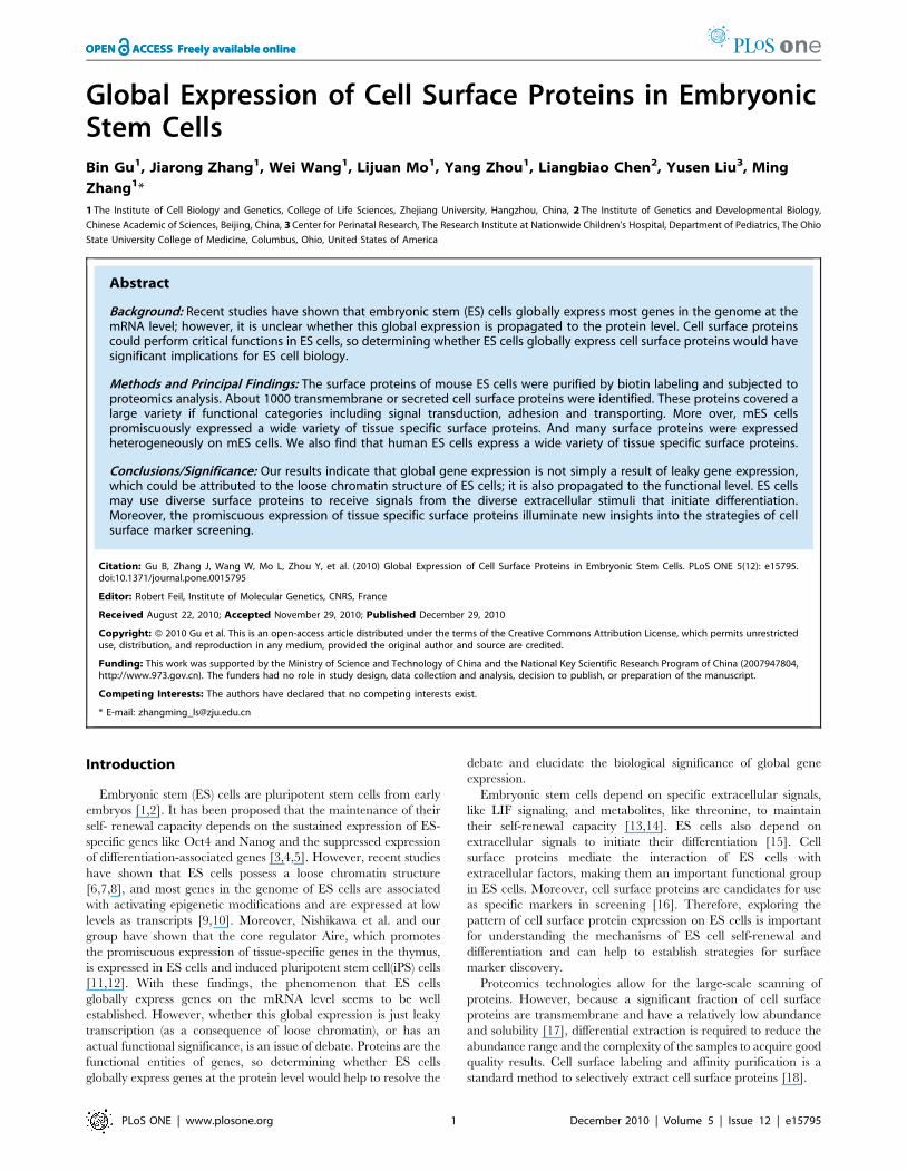
the quality of the mES cells was evaluated. As shown in Figure 1A,
the mES cells used in this study grew with typical colony
Figure 1. Labeling of mES cell surface proteins. A. Undifferentiated state of mES cells used to purify cell surface proteins. Left panel.Morphology of mES cells used in this study. The undifferentiated mES cells grew as compact colonies. Middle panel. mES cells expressed Alkalinephospatase (ALP). Right panel, Immunocytochemistry staining showed that mES cells expressed Oct4. The bars in left and middle panel represented100 um while the bar in the right panel represented 20 um. B. Flow cytometry showed that most mES cells used in this study expressed the ESspecific surface marker SSEA-1. FITC-streptavidin staining showed that most biotins were labeled on the cell surface.doi:10.1371/journal.pone.0015795.g001
Global Expression of Surface Proteins in ES Cells
PLoS ONE | www.plosone.org 2 December 2010 | Volume 5 | Issue 12 | e15795
morphology and homogeneously expressed alkaline phosphatase
(ALP) and Oct4. Quantitative analysis by flow cytometry showed
that more than 97% of the cells were positive for SSEA-1. These
data demonstrate that most mES cells used in this study were
undifferentiated. The surface proteins of the mES cells were then
labeled with membrane-impermeable biotin reagents and the
labeling efficiency was monitored by streptavidin-FITC staining.
As shown in Figure 1C, most of the cells were labeled with biotin
on the cell surface, although some intracellular labeling could be
observed, which could be explained by the staining of apoptotic
cells that are common in mES populations.
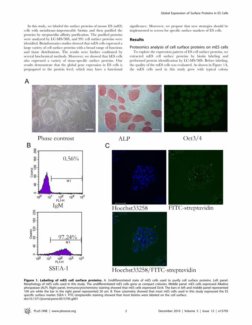
The biotin-labeled proteins were resolved by SDS-PAGE and
analyzed by LC-MS/MS. A total of 3468 proteins were identified.
The transmembrane structure and signal peptides were predicted
using SOSUI software[19]. Proteins annotated as ‘membrane’ in
gene ontology or those predicted to contain transmembrane
domains or signal peptides were annotated as general membrane
proteins. Of the identified proteins, 1699 were annotated as general
membrane proteins, of which 778 were integral membrane proteins
with a transmembrane domain or a lipid anchorage, 213 were
secreted proteins and 698 were membrane-associated proteins
(Figure 2A). Therefore, about half of the identified proteins were
general membrane proteins, which is consistent with other reports
that used the same methods. We selected the integral membrane
proteins and secreted proteins that adhere to the cell surface as cell
surface proteins and performed further analysis (Table S1). We first
evaluated the expression of 350 randomly selected surface proteins
by RT-PCR, and 274 of them were confirmed to be expressed on
mES cells. Therefore, our results should be at least 75% accurate.
We performed a gene ontology analysis according to the Molecular
Function annotations. As shown in Figure 2B, the cell surface
proteins of mES cells perform a wide variety of molecular functions.
Moreover, each functional category included many functional
surface proteins. For example, many different signaling receptors
from different pathways were identified (discussed below). Diverse
adhesion molecules, including types of cadherins/protocadherins,
cell adhesion molecules, and integrins were identified in this study.
Various transporting proteins were also identified, including 50
types of channel proteins and 66 types of transporter proteins.
Among these were 12 types of ABC-type ATPases from five
different families. Diverse extracellular matrix proteins were also
identified. Moreover, 180 uncharacterized cell surface proteins were
identified, which could serve as candidates for surface marker
screening.
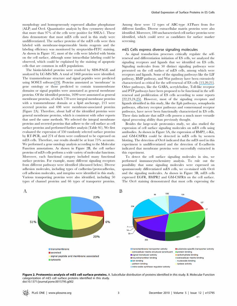
mES Cells express diverse signaling moleculesAs signal transduction processes critically regulate the self-
renewal and differentiation initiation of ES cells, we analyzed the
signaling receptors and ligands that we identified on ES cells.
Signaling molecules from 50 distinct signaling pathways were
presented on the cell surface of mES cells, among which were
receptors and ligands. Some of the signaling pathways like the LIF
pathway, BMP pathway, and Wnt pathway have been extensively
characterized as critical for the self-renewal of ES cells [13,20,21].
Other pathways, like the GABA, acetylcholine, Toll-like receptor
and PTP pathways have been proposed to be functional in the self-
renewal and proliferation of ES cells according to some reports
[22,23,24,25]. However, most of the signaling receptors and
ligands identified in this study, like the Eph pathways, semaphorin
pathways, olfactory receptor pathways and vomeronasal receptor
pathways, have never been functionally characterized in ES cells.
These data indicate that mES cells possess a much more versatile
signal processing ability than previously thought.
Besides the large-scale proteomics study, we also studied the
expression of cell surface signaling molecules on mES cells using
antibodies. As shown in Figure 3A, the expression of BMP2, c-Kit,
and GM-CSFRa could be detected in mES cells by western
blotting. The detection of Oct4 indicated that the mES used in this
experiment is undifferentiated and the detection of E-cadherin
indicated that membrane proteins were successfully extracted in
this experiment.
To detect the cell surface signaling molecules in situ, we
performed immunocytochemistry analysis. To rule out the
possibility that some signaling molecules were expressed on
spontaneously differentiated mES cells, we co-stained with Oct4
and the signaling molecules. As shown in Figure 3B, mES cells
expressed EGFR, BMPR2 and GM-CSFRa on the cell surface.
The Oct4 staining demonstrated that most cells expressing the
Figure 2. Proteomics analysis of mES cell surface proteins. A. Subcellular distribution of proteins identified in this study. B. Molecular Functioncategorization of mES cell surface proteins identified in this study.doi:10.1371/journal.pone.0015795.g002
Global Expression of Surface Proteins in ES Cells
PLoS ONE | www.plosone.org 3 December 2010 | Volume 5 | Issue 12 | e15795
signaling molecules were undifferentiated mES cells. Another
interesting phenomenon is that the staining of signaling molecules
on mES cells was heterogeneous, the fluorescence strength of
varied between Oct4 positive cells. To further confirm the
heterogeneous expression of cell surface signaling molecules in
mES populations, we performed a flow cytometry analysis. As
shown in Figure 3C, mES cells showed a heterogeneous expression
for BMPR2, GM-CSFRa and EGFR. Although the whole
fluorescent peak moved to the right, indicating positive staining.
Only a fraction of mES D3 cells strongly expressed BMPR2, GM-
CSFRa and EGFR (approximately 10% for BMPR2, 11% for
GM-CSFRa and 15% for EGFR). About 90% of the cells stained
positive for SSEA-1, indicating undifferentiated state. However,
even for SSEA-1, the fluorescent level varied widely. Therefore,
mES cells not only expressed signal molecules but also surface
markers heterogeneously. To rule out the possibility that the
heterogeneous expression was due to the in vitro culture features of
this specific cell line, we performed the same analysis on two other
ES cell lines established in our Lab, ZjuJ1 and ZjuJ2. As shown in
Figure 3C, both lines expressed BMPR2, GM-CSFRa and EGFR,
Figure 3. Cell surface signal molecules expressed on mES cells. A. Western blotting showed that mES cells expressed BMP2, c-Kit and GM-CSFRa along with mES specific marker Oct4 and mES surface protein E-cadherin. B. Immunocytochemistry staining showed that mES cells expressedBMPR2, EGFR and GM-CSFRa. Left panel, ICC staining of the cell surface signal molecules on mES cells. Middle panel, ICC staining of Oct4 on mES cells.Right panel, co-staining of the cell surface signal molecules and Oct4 on mES cells. The bar represented 100 um. C. Flow cytometry analysis showedthat mES cells heterogeneously expressed BMPR2, EGFR and GM-CSFRa. IgM, IgM control (For SSEA-1 staining). IgG, IgG control(For cell surface signalmolecules staining). D3, mES D3 cell line. ZJ1, mES ZJ1 cell line. ZJ2, mES ZJ2 cell line. D3-2, a single cell derived cell line derived from D3.doi:10.1371/journal.pone.0015795.g003
Global Expression of Surface Proteins in ES Cells
PLoS ONE | www.plosone.org 4 December 2010 | Volume 5 | Issue 12 | e15795
Global Expression of Surface Proteins in ES Cells
PLoS ONE | www.plosone.org 5 December 2010 | Volume 5 | Issue 12 | e15795
although the percentage of cells that strongly expressed them was
different from the D3 cell line. Previous reports have shown that
the heterogeneous expressions of some genes like Nanog, Rex1
and Stella in ES populations are subject to epigenetic regulation
and have equilibrium properties [26,27,28]. To determine
whether this also held true for cell surface signaling molecules,
we isolated single cell clonal cell lines from mES D3. We analyzed
four single cell clonal cell lines and got similar results, therefore
only one is shown as a representative. As shown in Figure 3C, the
cell lines established from single mES cells also heterogeneously
expressed BMPR2, GM-CSFRa and EGFR, while the percentage
of cells strongly expressing them was different from the parental
D3 cell line. These results indicated that mES cells heteroge-
neously expressed cell surface signaling molecules and argued for a
stochastic mechanism for the regulation of their expressions.
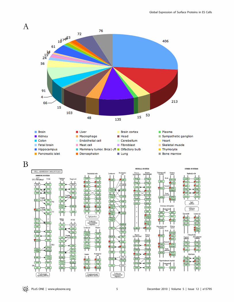
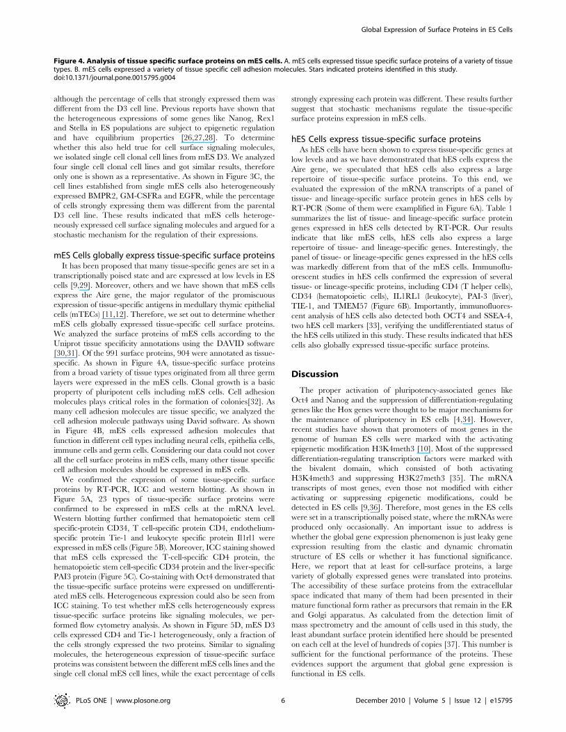
mES Cells globally express tissue-specific surface proteinsIt has been proposed that many tissue-specific genes are set in a
transcriptionally poised state and are expressed at low levels in ES
cells [9,29]. Moreover, others and we have shown that mES cells
express the Aire gene, the major regulator of the promiscuous
expression of tissue-specific antigens in medullary thymic epithelial
cells (mTECs) [11,12]. Therefore, we set out to determine whether
mES cells globally expressed tissue-specific cell surface proteins.
We analyzed the surface proteins of mES cells according to the
Uniprot tissue specificity annotations using the DAVID software
[30,31]. Of the 991 surface proteins, 904 were annotated as tissue-
specific. As shown in Figure 4A, tissue-specific surface proteins
from a broad variety of tissue types originated from all three germ
layers were expressed in the mES cells. Clonal growth is a basic
property of pluripotent cells including mES cells. Cell adhesion
molecules plays critical roles in the formation of colonies[32]. As
many cell adhesion molecules are tissue specific, we analyzed the
cell adhesion molecule pathways using David software. As shown
in Figure 4B, mES cells expressed adhesion molecules that
function in different cell types including neural cells, epithelia cells,
immune cells and germ cells. Considering our data could not cover
all the cell surface proteins in mES cells, many other tissue specific
cell adhesion molecules should be expressed in mES cells.
We confirmed the expression of some tissue-specific surface
proteins by RT-PCR, ICC and western blotting. As shown in
Figure 5A, 23 types of tissue-specific surface proteins were
confirmed to be expressed in mES cells at the mRNA level.
Western blotting further confirmed that hematopoietic stem cell
specific-protein CD34, T cell-specific protein CD4, endothelium-
specific protein Tie-1 and leukocyte specific protein Il1rl1 were
expressed in mES cells (Figure 5B). Moreover, ICC staining showed
that mES cells expressed the T-cell-specific CD4 protein, the
hematopoietic stem cell-specific CD34 protein and the liver-specific
PAI3 protein (Figure 5C). Co-staining with Oct4 demonstrated that
the tissue-specific surface proteins were expressed on undifferenti-
ated mES cells. Heterogeneous expression could also be seen from
ICC staining. To test whether mES cells heterogeneously express
tissue-specific surface proteins like signaling molecules, we per-
formed flow cytometry analysis. As shown in Figure 5D, mES D3
cells expressed CD4 and Tie-1 heterogeneously, only a fraction of
the cells strongly expressed the two proteins. Similar to signaling
molecules, the heterogeneous expression of tissue-specific surface
proteins was consistent between the different mES cells lines and the
single cell clonal mES cell lines, while the exact percentage of cells
strongly expressing each protein was different. These results further
suggest that stochastic mechanisms regulate the tissue-specific
surface proteins expression in mES cells.
hES Cells express tissue-specific surface proteinsAs hES cells have been shown to express tissue-specific genes at
low levels and as we have demonstrated that hES cells express the
Aire gene, we speculated that hES cells also express a large
repertoire of tissue-specific surface proteins. To this end, we
evaluated the expression of the mRNA transcripts of a panel of
tissue- and lineage-specific surface protein genes in hES cells by
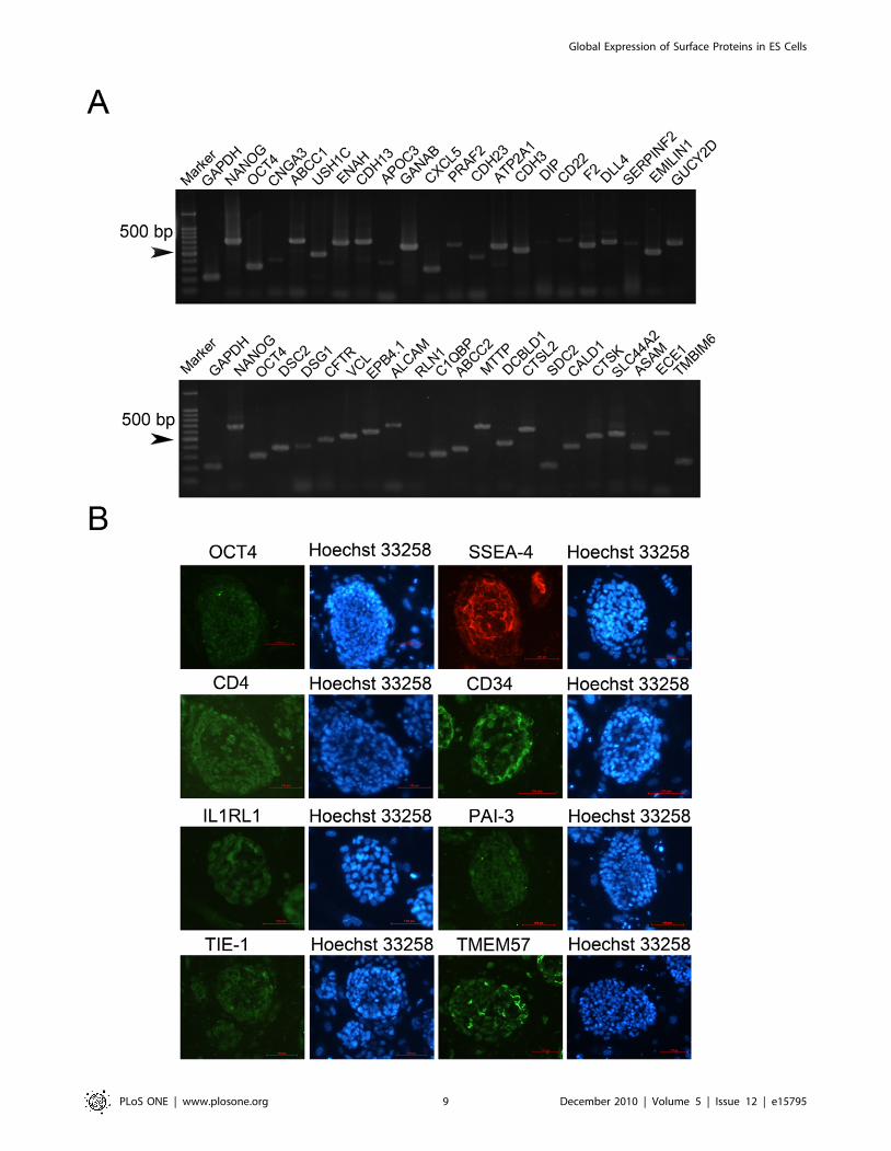
RT-PCR (Some of them were examplified in Figure 6A). Table 1
summarizes the list of tissue- and lineage-specific surface protein
genes expressed in hES cells detected by RT-PCR. Our results
indicate that like mES cells, hES cells also express a large
repertoire of tissue- and lineage-specific genes. Interestingly, the
panel of tissue- or lineage-specific genes expressed in the hES cells
was markedly different from that of the mES cells. Immunoflu-
orescent studies in hES cells confirmed the expression of several
tissue- or lineage-specific proteins, including CD4 (T helper cells),
CD34 (hematopoietic cells), IL1RL1 (leukocyte), PAI-3 (liver),
TIE-1, and TMEM57 (Figure 6B). Importantly, immunofluores-
cent analysis of hES cells also detected both OCT4 and SSEA-4,
two hES cell markers [33], verifying the undifferentiated status of
the hES cells utilized in this study. These results indicated that hES
cells also globally expressed tissue-specific surface proteins.
Discussion
The proper activation of pluripotency-associated genes like
Oct4 and Nanog and the suppression of differentiation-regulating
genes like the Hox genes were thought to be major mechanisms for
the maintenance of pluripotency in ES cells [4,34]. However,
recent studies have shown that promoters of most genes in the
genome of human ES cells were marked with the activating
epigenetic modification H3K4meth3 [10]. Most of the suppressed
differentiation-regulating transcription factors were marked with
the bivalent domain, which consisted of both activating
H3K4meth3 and suppressing H3K27meth3 [35]. The mRNA
transcripts of most genes, even those not modified with either
activating or suppressing epigenetic modifications, could be
detected in ES cells [9,36]. Therefore, most genes in the ES cells
were set in a transcriptionally poised state, where the mRNAs were
produced only occasionally. An important issue to address is
whether the global gene expression phenomenon is just leaky gene
expression resulting from the elastic and dynamic chromatin
structure of ES cells or whether it has functional significance.
Here, we report that at least for cell-surface proteins, a large
variety of globally expressed genes were translated into proteins.
The accessibility of these surface proteins from the extracellular
space indicated that many of them had been presented in their
mature functional form rather as precursors that remain in the ER
and Golgi apparatus. As calculated from the detection limit of
mass spectrometry and the amount of cells used in this study, the
least abundant surface protein identified here should be presented
on each cell at the level of hundreds of copies [37]. This number is
sufficient for the functional performance of the proteins. These
evidences support the argument that global gene expression is
functional in ES cells.
Figure 4. Analysis of tissue specific surface proteins on mES cells. A. mES cells expressed tissue specific surface proteins of a variety of tissuetypes. B. mES cells expressed a variety of tissue specific cell adhesion molecules. Stars indicated proteins identified in this study.doi:10.1371/journal.pone.0015795.g004
Global Expression of Surface Proteins in ES Cells
PLoS ONE | www.plosone.org 6 December 2010 | Volume 5 | Issue 12 | e15795
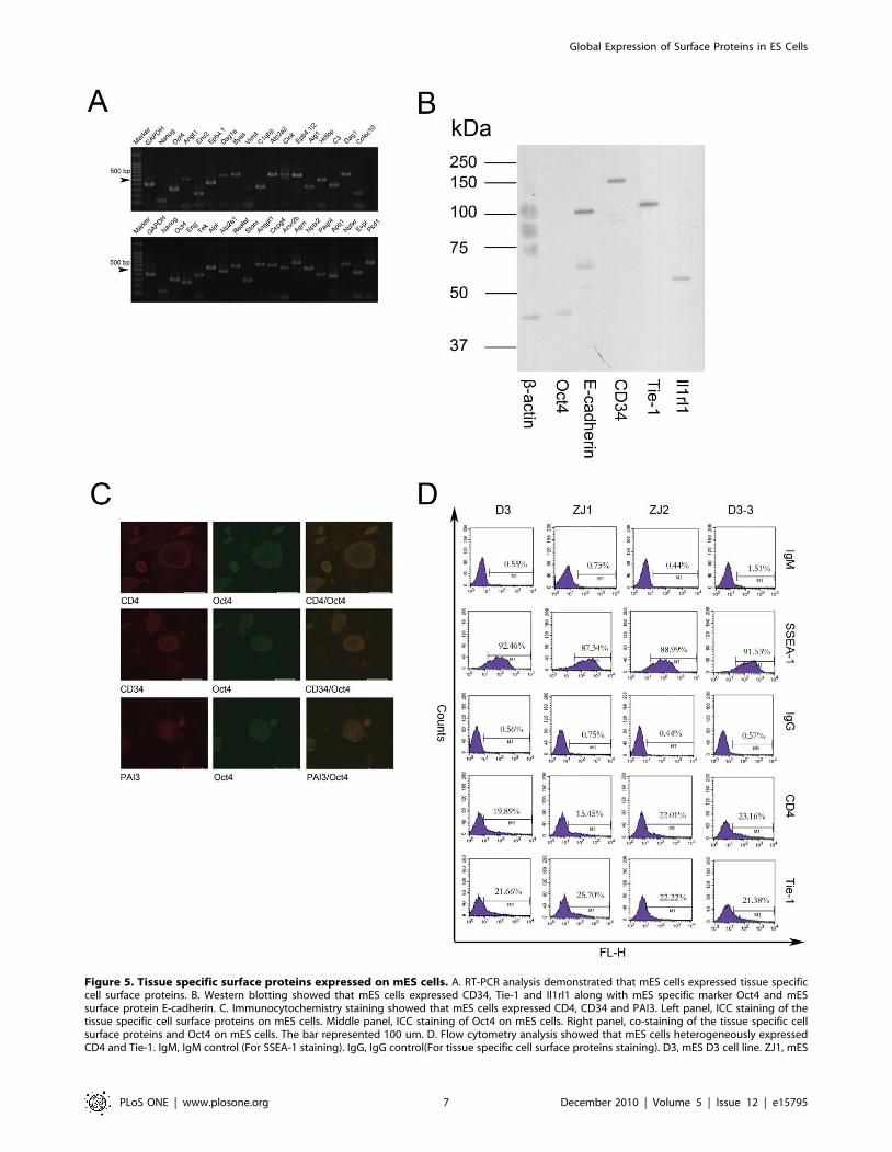
Figure 5. Tissue specific surface proteins expressed on mES cells. A. RT-PCR analysis demonstrated that mES cells expressed tissue specificcell surface proteins. B. Western blotting showed that mES cells expressed CD34, Tie-1 and Il1rl1 along with mES specific marker Oct4 and mESsurface protein E-cadherin. C. Immunocytochemistry staining showed that mES cells expressed CD4, CD34 and PAI3. Left panel, ICC staining of thetissue specific cell surface proteins on mES cells. Middle panel, ICC staining of Oct4 on mES cells. Right panel, co-staining of the tissue specific cellsurface proteins and Oct4 on mES cells. The bar represented 100 um. D. Flow cytometry analysis showed that mES cells heterogeneously expressedCD4 and Tie-1. IgM, IgM control (For SSEA-1 staining). IgG, IgG control(For tissue specific cell surface proteins staining). D3, mES D3 cell line. ZJ1, mES
Global Expression of Surface Proteins in ES Cells
PLoS ONE | www.plosone.org 7 December 2010 | Volume 5 | Issue 12 | e15795
ES cells are versatile signal transformersIn contrast to terminally differentiated cell types, which mount
restricted responses to various stimuli, ES cells mount essentially
infinite responses, differentiating to all cell types in the organism.
Two seemingly contradictory responses of ES cells to stimulus
were observed. First, ES cells respond differently to different
stimuli, to the same stimulus of different strengths and to different
combinations of stimuli. For example, Zansdtra et al. showed a
concentration-dependent effect of LIF on mES cells [38]. Second,
ES cells respond differently to the same stimulus. For example,
when cultured in suspension, ES cells in identical medium form
embryonic bodies, which consist of cells of the three germ layers.
The evidence we present here that ES cells globally express a large
diversity of surface signaling molecules in a heterogeneous manner
may partially explain these properties. The versatility of signaling
receptors could enable ES cells to transform diverse stimulus into
highly variable differentiation behaviors. In addition, the hetero-
geneity of the signal-accepting ability caused by the differential
expression of signaling molecules could enable ES cells to
transform the same stimulus to different differentiation behaviors.
The global and heterogeneous expression of signaling molecules
make ES cells versatile signal transformers, ensuring their plasticity
and pluripotency.
Implications of the population heterogeneity of ES cellsIt has been demonstrated that ES cells heterogeneously express
genes like Nanog, Rex-1, Stella and CD133 [26,27,28,39]. The
different subpopulations sorted according to these markers
possess different self-renewal abilities and differentiation poten-
tials. Moreover, a recent study has shown that a subpopulation of
undifferentiated mES cells that express the primitive endoderm
(PrEn)-specific gene Hex at very low level has early PrEn
properties and that their differentiation into PrEn was favored
[40]. Our results showed that undifferentiated mES cells actually
expressed a large repertoire of tissue-specific surface proteins at
low levels and the expression of many of them tended to be
heterogeneous. These results indicate that ES cells actually
consist of different subpopulations expressing different tissue-
specific proteins and have different differentiation tendencies.
Our results supported the idea that ES cells are a equilibrium
population that consist of subpopulations of different differenti-
ation potentials.
Implications for surface marker screening of ES cellsES cell surface markers are valuable tools for the characteriza-
tion, quality control and purification of ES cells. Extensive efforts
have been mounted for decades to discover the surface markers of
ES cells. Until now, the most widely used specific ES cell surface
markers (SSEA1, SSEA3, SSEA4, Tra-1-60, Tra-1-81) were all
glycan epitopes on glycoproteins or glycolipids [33,41]. Although
some proteins like CD9, HSPA8 and PODXL have been proposed
to be specific surface markers of ES cells, they are all only
relatively specific and only expressed in certain tissues, according
to data from the human protein atlas [16,33,42,43,44]. The
question remains whether or not there is a specific surface protein
marker that is exclusive to ES cells. Our results showed that mES
cells globally expressed a large repertoire of tissue-specific cell
surface proteins. It indicated that, at least for mES cells, an
exclusive, specific surface protein marker is not easy to identify.
The same thing may also hold true for hES cells because we have
shown that hES cells express tissue-specific surface proteins.
Therefore, new strategies should be employed to screen for surface
markers of ES cells. Hematopoietic stem cells (HSC) are another
type of stem cells that exhibit promiscuous surface protein
expression [45]. HSCs express genes specific to differentiated
hematopoietic cells and other cell types, like neural cells.
Researchers have determined quantitatively the combination of
markers, like the lineage markers Sca-1, c-Kit or SLAMs, to
identify HSCs [46,47,48,49]. We suggest that the same idea is
applicable to ES cells. The quantitative combination of a group of
surface proteins of different tissue specificities could identify an ES-
specific surface protein pattern. Therefore, perhaps efforts should
be shifted from screening for exclusive, specific markers to
determining the combinations.
In conclusion, we demonstrated that mES cells globally
expressed cell surface proteins of diverse functions and tissue
specificities. Our results support the idea that global gene
expression in ES cells is functional. Moreover, our results indicate
that ES cells are versatile in their signal reception and
transduction ability. Our results also have profound implications
for understanding the functional and population properties of ES
cells and they also indicate new strategies for surface marker
screening.
Materials and Methods
Ethics StatementWe do not require a Ethics Statement because we only used
mouse cell lines and commercially available human cell lines. No
animals or human samples were used. And the approval of a
named review board institution or ethics committee is not needed
for the same reason.
Cell lines and Cell cultureGamma irradiation inactivated mouse embryonic fibroblast
(MEF) feeder cells isolated from the embryos of ICR mice at
gestational day 13.5 were purchased from Invitogen (Invitrogen,
Carlsbad, CA). MEFs were thawed in DMEM supplemented with
10% fetal bovine serum(Invitrogen) at 37uC and plated at a density
of 46104 cells/cm2 for ES culture.
Mouse ES(mES) cell D3 line was purchased from ATCC
(Manassas, VA). ZJ1 and ZJ2 mouse ES cell lines were established
in our laboratory[50]. mES cells (D3, ZJ1 and ZJ2) were cultured
on gamma irradiation inactivated MEFs in DMEM supplemented
with 15% fetal bovine serum (Invitrogen) and 1000 ng/ml LIF
(Millipore, Billerica, MA) at 37uC in 5% CO2. The pluripotency of
the mES cells was routinely analyzed using ALP staining
Kit(Sigma), SSEA-1 staining and teratoma formation. In addition,
the karyotype was checked routinely.
Human embryonic stem cells H9 were purchased from WiCell
(Madison,WI) and cultured on gamma irradiation inactivated
MEFs in Knockout DMEM supplemented with 20% KOSR
(Invitrogen) and 1000 ng/ml bFGF (Millipore) at 37uC in 5%
CO2. The pluripotency of the hES cells was routinely analyzed
using ALP staining (Sigma), SSEA-4 staining and teratoma
formation. In addition, the karyotype was checked routinely.
ZJ1 cell line. ZJ2, mES ZJ2 cell line. D3-2, a single cell derived cell line derived from D3. (Same controls and SSEA-1 staining as in Figure 3 were usedsince the datas shown here were generated in the same experiment).doi:10.1371/journal.pone.0015795.g005
Global Expression of Surface Proteins in ES Cells
PLoS ONE | www.plosone.org 8 December 2010 | Volume 5 | Issue 12 | e15795
Global Expression of Surface Proteins in ES Cells
PLoS ONE | www.plosone.org 9 December 2010 | Volume 5 | Issue 12 | e15795
Cell surface labeling and affinity purificationThe mES D3 cells (56108) cultured gamma irradiation
inactivated MEFs were trypsinized to single cells and plated on
gelatin-coated 100 mm culture dishes. After 1 hour, most MEFs
adhered to the culture dish. The mES cells in suspension were
collected and biotin labeled.
For biotin labeling, the cells were incubated with 1 mg/ml
Sulfo-NHS-SS-Biotin (Pierce, Rockford, IL) in PBS for 30
minutes. Excess biotin was quenched using 10 mM Glycin for
10 minutes, and then the cells were washed three times with PBS.
Next, the cells were homogenized in ice-cold cell lysis buffer
(50 mM Tris-Cl, pH 7.4, 1% NP-40 substitute (Sigma), 150 mM
NaCl, 1 mM EDTA, 1 mM PMSF(Sigma)) using a Dounce
Homogenizer (30 strokes). The homogenate was put on ice for
1 hour with gentle vortexing to extract the membrane proteins.
After that, the homogenate was centrifuged at 12000 g for ten
minutes to remove the nuclei, unbroken cells and cell fragments.
The supernatant was mixed with streptavidin-coupled LATEX
(300 nm diameter) beads and vortexed at 4uC for 1 hour. The
LATEX beads were precipitated by centrifugation and washed
twice with 0.1 M Na2CO2 and once with 1 M KCl to remove the
contaminant proteins. After that, the disulfide bonds linking biotin
and the purified proteins were cleaved by 100 mM DTT(Sigma) to
elute the purified proteins. Approximately 100 mg of membrane
protein could be purified from 108 cells. The labeling efficiency
was monitored using FITC-streptavidin staining.
SDS-PAGE separationThe purified proteins were separated by SDS-PAGE using a
12.5% SDS-PAGE gel. After electrophoresis, the gels were stained
with Coomassie Blue R250(Sigma) and then dissected into 8 bands
for LC-MS/MS analysis.
Enzyme digestion, LC-MS/MS analysis and databasesearching
The enzyme digestion was performed as previously described
[51]. The peptides from each band were separated on a Paradigm
MS4N Nano/Capillary HS MDLC (Michrom Bioresources, Inc.
USA) using a 100 mm6150 mm C18 reverse phase column. The
LC separation was conducted with a linear gradient of 5–35%
buffer B for 50 min, followed by 35–90% buffer B for 10 minutes,
followed by 90% buffer B for 10 minutes (buffer A: 0.1% formic
acid in a 2% acetonitrile H2O solution; buffer B: 0.1% formic acid
in a 98% acetonitrile H2O solution) at a flow rate of 500 nl/min.
The separated peptides were then analyzed on a LTQ-MS
(Thermol, USA) coupled with a Michrome Advanced nanospray
apparatus (Microm). The peak list files generated by the Bioworks
software (Applied Biosystems, USA) using the default parameters
were searched against databases for protein identification using the
Sequest software. The searching parameters were: for 2 or 3 valent
ions, Xcorr $2; for 1 valent ion, Xcorr $1.5;,Deltacn $0.1; and
two nonredundant peptides identified on a unique protein.
Antibodies and immunocytochemistryThe following antibodies were used: Oct-4 (R&D MAB 1759),
SSEA-1 (R&D MAB2155), CD34 (HUABIO, Hangzhou, China),
c-Kit (HUABIO), EGFR (HUABIO), BMPR2 (HUABIO), E-
Cadherin (HUABIO 0407-25), BMP2 (HUABIO 0806-2), GM-
CSF Ra (HUABIO 0804-8), CD4 (HUABIO), TIE-1 (HUABIO
0804-11), PAI-3 (HUABIO), TMEM57(HUABIO), R-PE-conju-
gated goat anti-rabbit IgG (Proteintech Chicago, USA), and Alexa
488-conjugated goat anti-rabbit IgG (Invitrogen).
For double staining, the cells were fixed using 4% paraformal-
dehyde according to the standard protocol, blocked with blocking/
permeating buffer (PBS with 10% goat serum and 0.3% Triton-
X100) and then incubated with Rat anti-human Oct4 monoclonal
antibody overnight at 4uC. After wash, the cells were incubated with
a Alexa 488-conjugated Goat-anti Rat for 1 hour at 37uC. After
wash, the cells were incubated with rabbit polyclonal antibodies
against cell surface molecules for 1 hour at 37uC. After wash, the
cells were Alexa 555-conjugated Goat-anti Rabbit for 1 hour at
37uC and then observed under a LSM500 Confocal Microscope
(Zeiss, Germany). For single staining, the cells were fixed using 4%
paraformaldehyde according to the standard protocol, blocked with
blocking/permeating buffer (PBS with 10% goat serum and 0.3%
Triton-X100) and then incubated with primary antibodies for
1 hour at 37uC. After washing, the cells were incubated with a
Alexa 488-conjugated secondary antibodies for 1 hour at 37uC and
then observed under a fluorescent microscope(Nikon, Japan).
Biotin-labeled mES cells were fixed with 4% paraformaldehyde
overnight at 4uC and then stained with FITC-conjugated
streptavidin (Sigma) for 30 minutes to monitor the surface labeling.
RT-PCRRT-PCR was performed as previously described [12]. Total
RNA was extracted using the Trizol Reagent (Takara, Japan),
retro-transcribed and then PCR-amplified. The primers were
designed using the PRIMER PREMIER 5 software.
ALP stainingALP staining was performed with an ALP assay kit (Sigma).
Figure 6. hES cells expressed tissue specific cell surface proteins. A. RT-PCR analysis demonstrated that hES cells expressed tissue specific cellsurface proteins. B. Immunocytochemistry staining showed that mES cells expressed CD4, CD34, Il1Rl1, PAI-3, TIE-1 and TMEM57 along with hESspecific marker OCT4 and SSEA4.doi:10.1371/journal.pone.0015795.g006
Table 1. Tissue specific cell surface proteins expressed onhES cells.
Tissue TRA
Cardiovascular CDH13;CDH3;DLL4; BSG
Endothelia Amot; CD31; KDR; Tie-1;
Epithelia DCD;EVPL;EPN3;CFTR;DSC2;DSG1;ENAH;LIMA1;SDC4;ENAM;SLC44A2;ABCA4;CDH23; CNGA3
Gastrointestinal CA9;APOA1;APOC3;ABCC2;USH1C;PRAF2;MTTP;PTGER3
Hematopoietic&immune
ALCAM; CD4;CD22; CD34;EPB41;CD79B;FCER1G;NCKAP1L;
Kidney BBS1;CDH16;SLC12A1
Liver AGXT;SERPINF2;ABCB11;F2;C1QBP;HPX;PAI-3
Muscle ATP2A1;CACNB1;CALD1;CROT;DYSF;UTRN;VCL
Neural HTR1D;CEND1;CHRM2;CNTN4;GABRA5;GRIA4; TMEM57
Reproductive GANAB;AMIGO2;RLN1;CTSL2;CXCL5;CXCL6;CMTM1;TMBIM6;DNAJB13
Respiratory ABCC1;BMPER;DIP;DCBLD1;EMILIN1;EMCN;ECE1
doi:10.1371/journal.pone.0015795.t001
Global Expression of Surface Proteins in ES Cells
PLoS ONE | www.plosone.org 10 December 2010 | Volume 5 | Issue 12 | e15795
Flow cytometryThe mES cells were dissociated with 0.05 mM EDTA and then
washed with PBS and 3% FBS to remove the EDTA. The cells
were then incubated with a primary antibody for 1 hour on ice.
After thorough washes, the cells were incubated with fluorescent
secondary antibodies for 30 minutes on ice. The cells were then
washed with PBS and analyzed by BDLSR flow cytometry (BD
Biosciences, San Jose, CA).
Western blottingWestern blotting was carried out as described previously[52].
Briefly, ES cells were harvested in lysis buffer (50 mM Tris-HCl
pH 7.4, 1% NP-40, 1% SDS), and equal amounts of the protein
lysate were separated by electrophoresis on a 12.5% Laemmli
SDS-polyacrylamide gel. The proteins were then transferred onto
PVDF membranes. After incubation with primary and secondary
antibodies, the membranes were developed using an ECL kit
purchased from GE Healthcare Life Sciences (Piscataway, NJ).
Bioinformatics analysisThe subcellular localization of the proteins was annotated
according to the Swissprot annotation, the Sosui prediction
software and the literature. Proteins containing transmembrane
domains, secreted proteins and proteins annotated as cell surface
proteins by either Swissprot or the existing literature were all
considered cell surface proteins. A gene ontology (GO) analysis
was done using the DAVID software and database [30,31]. The
molecular pathways were analyzed according to the KEGG
pathway annotations. The tissue specificity of the surface proteins
was annotated according to Uniprot annotations.
Supporting Information
Table S1 The list of cell surface proteins identified inthis study.
(DOC)
Acknowledgments
We thank Dr. Kerry Nugent and Mr. Yixin Zhu from Microm Inc. for
their assistance in Mass Spectrometry analysis. The streptavidin conjugated
Latex was provided by Yuanji Lin. We thank Mr. Xiaoli Zhao for his help
in cell culture.
Author Contributions
Conceived and designed the experiments: BG LC YL MZ. Performed the
experiments: BG JZ WW LM YZ. Analyzed the data: BG JZ YL LC MZ.
Wrote the paper: BG YL LC MZ.
References
1. Evans MJ, Kaufman MH (1981) Establishment in culture of pluripotential cells
from mouse embryos. Nature 292: 154–156.
2. Thomson JA, Itskovitz-Eldor J, Shapiro SS, Waknitz MA, Swiergiel JJ, et al.
(1998) Embryonic stem cell lines derived from human blastocysts. Science 282:
1145–1147.
3. Chambers I, Silva J, Colby D, Nichols J, Nijmeijer B, et al. (2007) Nanog
safeguards pluripotency and mediates germline development. Nature 450:
1230–1234.
4. Lee TI, Jenner RG, Boyer LA, Guenther MG, Levine SS, et al. (2006) Control
of developmental regulators by Polycomb in human embryonic stem cells. Cell
125: 301–313.
5. Nichols J, Zevnik B, Anastassiadis K, Niwa H, Klewe-Nebenius D, et al. (1998)
Formation of pluripotent stem cells in the mammalian embryo depends on the
POU transcription factor Oct4. Cell 95: 379–391.
6. Azuara V, Perry P, Sauer S, Spivakov M, Jorgensen HF, et al. (2006) Chromatin
signatures of pluripotent cell lines. Nat Cell Biol 8: 532–538.
7. Meshorer E, Misteli T (2006) Chromatin in pluripotent embryonic stem cells
and differentiation. Nat Rev Mol Cell Biol 7: 540–546.
8. Meshorer E, Yellajoshula D, George E, Scambler PJ, Brown DT, et al. (2006)
Hyperdynamic plasticity of chromatin proteins in pluripotent embryonic stem
cells. Dev Cell 10: 105–116.
9. Efroni S, Duttagupta R, Cheng J, Dehghani H, Hoeppner DJ, et al. (2008)
Global transcription in pluripotent embryonic stem cells. Cell Stem Cell 2:
437–447.
10. Guenther MG, Levine SS, Boyer LA, Jaenisch R, Young RA (2007) A
chromatin landmark and transcription initiation at most promoters in human
cells. Cell 130: 77–88.
11. Nishikawa Y, Hirota F, Yano M, Kitajima H, Miyazaki J, et al. (2010) Biphasic
Aire expression in early embryos and in medullary thymic epithelial cells before
end-stage terminal differentiation. J Exp Med 207: 963–971.
12. Gu B, Zhang J, Chen Q, Tao B, Wang W, et al. (2010) Aire regulates the
expression of differentiation-associated genes and self-renewal of embryonic stem
cells. Biochem Biophys Res Commun 394: 418–423.
13. Davey RE, Onishi K, Mahdavi A, Zandstra PW (2007) LIF-mediated control of
embryonic stem cell self-renewal emerges due to an autoregulatory loop.
FASEB J 21: 2020–2032.
14. Wang J, Alexander P, Wu L, Hammer R, Cleaver O, et al. (2009) Dependence
of mouse embryonic stem cells on threonine catabolism. Science 325: 435–439.
15. Hansson M, Olesen DR, Peterslund JM, Engberg N, Kahn M, et al. (2009) A
late requirement for Wnt and FGF signaling during activin-induced formation of
foregut endoderm from mouse embryonic stem cells. Dev Biol 330: 286–304.
16. Choo AB, Tan HL, Ang SN, Fong WJ, Chin A, et al. (2008) Selection against
undifferentiated human embryonic stem cells by a cytotoxic antibody
recognizing podocalyxin-like protein-1. Stem Cells 26: 1454–1463.
17. Speers AE, Wu CC (2007) Proteomics of integral membrane proteins–theory
and application. Chem Rev 107: 3687–3714.
18. Zhao Y, Zhang W, Kho Y (2004) Proteomic analysis of integral plasma
membrane proteins. Anal Chem 76: 1817–1823.
19. Hirokawa T, Boon-Chieng S, Mitaku S (1998) SOSUI: classification and
secondary structure prediction system for membrane proteins. Bioinformatics
14: 378–379.
20. Hao J, Li T-G, Qi X, Zhao D-F, Zhao G-Q (2006) WNT/[beta]-catenin
pathway up-regulates Stat3 and converges on LIF to prevent differentiation of
mouse embryonic stem cells. Developmental Biology 290: 81–91.
21. Ying QL, Nichols J, Chambers I, Smith A (2003) BMP induction of Id proteins
suppresses differentiation and sustains embryonic stem cell self-renewal in
collaboration with STAT3. Cell 115: 281–292.
22. Kim M, Kim M, Heo J, Kim J, Han H (2008) Acetylcholine inhibits long-term
hypoxia-induced apoptosis by suppressing the oxidative stress-mediated MAPKs
activation as well as regulation of Bcl-2, c-IAPs, and caspase-3 in mouse
embryonic stem cells. Apoptosis 13: 295–304.
23. Schwirtlich M, Emri Z, Antal K, Mate Z, Katarova Z, et al. (2010) GABA(A)
and GABA(B) receptors of distinct properties affect oppositely the proliferation
of mouse embryonic stem cells through synergistic elevation of intracellular
Ca(2+). FASEB J 24: 1218–1228.
24. Soh BS, Song CM, Vallier L, Li P, Choong C, et al. (2007) Pleiotrophin
enhances clonal growth and long-term expansion of human embryonic stem
cells. Stem Cells 25: 3029–3037.
25. Taylor T, Kim YJ, Ou X, Derbigny W, Broxmeyer HE (2010) Toll Like
Receptor 2 Mediates Proliferation, Survival, NFkappaB Translocation, and
Cytokine mRNA Expression in LIF-Maintained Mouse Embryonic Stem Cells.
Stem Cells Dev 19: 1333–41.
26. Singh AM, Hamazaki T, Hankowski KE, Terada N (2007) A heterogeneous
expression pattern for Nanog in embryonic stem cells. Stem Cells 25: 2534–2542.
27. Toyooka Y, Shimosato D, Murakami K, Takahashi K, Niwa H (2008)
Identification and characterization of subpopulations in undifferentiated ES cell
culture. Development 135: 909–918.
28. Hayashi K, Lopes SM, Tang F, Surani MA (2008) Dynamic equilibrium and
heterogeneity of mouse pluripotent stem cells with distinct functional and
epigenetic states. Cell Stem Cell 3: 391–401.
29. Xu J, Watts JA, Pope SD, Gadue P, Kamps M, et al. (2009) Transcriptional
competence and the active marking of tissue-specific enhancers by defined
transcription factors in embryonic and induced pluripotent stem cells. Genes
Dev 23: 2824–2838.
30. Dennis G, Jr., Sherman BT, Hosack DA, Yang J, Gao W, et al. (2003) DAVID:
Database for Annotation, Visualization, and Integrated Discovery. Genome Biol
4: P3.
31. Huang da W, Sherman BT, Lempicki RA (2009) Systematic and integrative
analysis of large gene lists using DAVID bioinformatics resources. Nat Protoc 4:
44–57.
32. Spencer HL, Eastham AM, Merry CL, Southgate TD, Perez-Campo F, et al.
(2007) E-cadherin inhibits cell surface localization of the pro-migratory 5T4
oncofetal antigen in mouse embryonic stem cells. Mol Biol Cell 18: 2838–2851.
33. Adewumi O, Aflatoonian B, Ahrlund-Richter L, Amit M, Andrews PW, et al.
(2007) Characterization of human embryonic stem cell lines by the International
Stem Cell Initiative. Nat Biotechnol 25: 803–816.
Global Expression of Surface Proteins in ES Cells
PLoS ONE | www.plosone.org 11 December 2010 | Volume 5 | Issue 12 | e15795

34. Rao M (2004) Conserved and divergent paths that regulate self-renewal in
mouse and human embryonic stem cells. Dev Biol 275: 269–286.35. Bernstein BE, Mikkelsen TS, Xie X, Kamal M, Huebert DJ, et al. (2006) A
bivalent chromatin structure marks key developmental genes in embryonic stem
cells. Cell 125: 315–326.36. Pan G, Tian S, Nie J, Yang C, Ruotti V, et al. (2007) Whole-genome analysis of
histone H3 lysine 4 and lysine 27 methylation in human embryonic stem cells.Cell Stem Cell 1: 299–312.
37. Djuro Josic JGC (2007) Mammalian plasma membrane proteomics. Proteomics
7: 3010–3029.38. Zandstra PW, Le HV, Daley GQ, Griffith LG, Lauffenburger DA (2000)
Leukemia inhibitory factor (LIF) concentration modulates embryonic stem cellself-renewal and differentiation independently of proliferation. Biotechnol
Bioeng 69: 607–617.39. King FW, Ritner C, Liszewski W, Kwan HC, Pedersen A, et al. (2009)
Subpopulations of Human Embryonic Stem Cells with Distinct Tissue-Specific
Fates Can Be Selected from Pluripotent Cultures. Stem Cells Dev 18: 1441–50.40. Canham MA, Sharov AA, Ko MS, Brickman JM (2010) Functional
Heterogeneity of Embryonic Stem Cells Revealed through TranslationalAmplification of an Early Endodermal Transcript. PLoS Biol 8: e1000379.
41. Nagano K, Yoshida Y, Isobe T (2008) Cell surface biomarkers of embryonic
stem cells. Proteomics 8: 4025–4035.42. Son YS, Park JH, Kang YK, Park JS, Choi HS, et al. (2005) Heat shock 70-kDa
protein 8 isoform 1 is expressed on the surface of human embryonic stem cellsand downregulated upon differentiation. Stem Cells 23: 1502–1513.
43. Uhlen M, Bjorling E, Agaton C, Szigyarto CA-K, Amini B, et al. (2005) AHuman Protein Atlas for Normal and Cancer Tissues Based on Antibody
Proteomics. Mol Cell Proteomics 4: 1920–1932.
44. Berglund L, Bjorling E, Oksvold P, Fagerberg L, Asplund A, et al. (2008) A
genecentric Human Protein Atlas for expression profiles based on antibodies.
Mol Cell Proteomics 7: 2019–2027.
45. Akashi K, He X, Chen J, Iwasaki H, Niu C, et al. (2003) Transcriptional
accessibility for genes of multiple tissues and hematopoietic lineages is
hierarchically controlled during early hematopoiesis. Blood 101: 383–389.
46. Spangrude GJ, Heimfeld S, Weissman IL (1988) Purification and characteriza-
tion of mouse hematopoietic stem cells. Science 241: 58–62.
47. Uchida N, Weissman IL (1992) Searching for hematopoietic stem cells: evidence
that Thy-1.1lo Lin- Sca-1+ cells are the only stem cells in C57BL/Ka-Thy-1.1
bone marrow. J Exp Med 175: 175–184.
48. Spangrude GJ, Smith L, Uchida N, Ikuta K, Heimfeld S, et al. (1991) Mouse
hematopoietic stem cells. Blood 78: 1395–1402.
49. Kiel MJ, Yilmaz OH, Iwashita T, Terhorst C, Morrison SJ (2005) SLAM family
receptors distinguish hematopoietic stem and progenitor cells and reveal
endothelial niches for stem cells. Cell 121: 1109–1121.
50. Jiang ZY, Wu RR, Yuan YJ, Chen LB, Zhao XL, et al. (2005) Research on the
high efficient methodology of Establishment of 129S1/SvImJ murine embryonic
stem cells JOURNAL OF ZHEJIANG UNIVERSITY (SCIENCE EDITION)
32: 574–578.
51. Shevchenko A, Wilm M, Vorm O, Mann M (1996) Mass Spectrometric
Sequencing of Proteins from Silver-Stained Polyacrylamide Gels. Anal Chem
68: 850–858.
52. Huang Y, Gu B, Wu R, Zhang J, Li Y, et al. (2007) Development of a rabbit
monoclonal antibody group against Smads and immunocytochemical study of
human and mouse embryonic stem cells. Hybridoma (Larchmt) 26: 387–391.
Global Expression of Surface Proteins in ES Cells
PLoS ONE | www.plosone.org 12 December 2010 | Volume 5 | Issue 12 | e15795


