Embed Size (px)
Citation preview

Interactions of Al(III) with a neurofilament heptapeptide fragment:AcLysSerProValValGluGly
T. Kiss a,b,*, M. Kilyen b, A. Lakatos b, F. Evanics c, T. Kortvelyesi d,f, Gy. Dombi c,Zs. Majer e, M. Hollosi e
a Department of Inorganic and Analytical Chemistry, University of Szeged, PO Box 440, Szeged, Hungaryb Biocoordination Chemistry Research Group of the Hungarian Academy of Sciences, University of Szeged, PO Box 440, Szeged, Hungary
c Department of Pharmaceutical Analytical Chemistry, University of Szeged, PO Box 427, Szeged, Hungaryd Department of Biomedical Engineering, Boston University, Boston, MA 02215, USA
e Department of Organic Chemistry, Eotvos University, PO Box 32, Budapest, Hungaryf Department of Physical Chemistry, University of Szeged, PO Box 105, Szeged, Hungary
Received 14 August 2001; accepted 12 February 2002
Abstract
The solution state of the neurotoxic Al(III) in biological systems is discussed briefly, and the importance of the Al(III)�/peptide
and Al(III)�/protein interactions in the various neurodegeneration processes is emphasised and evaluated. The possible involvement
of Al(III) in the formation of Alzheimer’s disease marker senile plaques and neurofilament tangles is discussed in light of the
solution speciation of the Al(III)�/AcLysSerProValValGluGly system and the structural features of the complexes formed. # 2002
Elsevier Science B.V. All rights reserved.
Keywords: Al(III)-complexes; Speciation; NMR; Molecular dynamics calculations
Contents
Abstract . . . . . . . . . . . . . . . . . . . . . . . . . . . . . . . . . . . . . . . . . . . . . . . . . . . . . . . . . . . . . . . . . . . . . . . . . 227
1. Introduction . . . . . . . . . . . . . . . . . . . . . . . . . . . . . . . . . . . . . . . . . . . . . . . . . . . . . . . . . . . . . . . . . . . 228
2. Chemical forms of Al(III) in biological systems . . . . . . . . . . . . . . . . . . . . . . . . . . . . . . . . . . . . . . . . . . . . . . . . 228
3. Interactions of Al(III) with oligopeptides . . . . . . . . . . . . . . . . . . . . . . . . . . . . . . . . . . . . . . . . . . . . . . . . . . . . 229
4. The Al(III)�/AcLysSerProValValGluGly heptapeptide system . . . . . . . . . . . . . . . . . . . . . . . . . . . . . . . . . . . . . . . . 230
4.1 Experimental . . . . . . . . . . . . . . . . . . . . . . . . . . . . . . . . . . . . . . . . . . . . . . . . . . . . . . . . . . . . . . . 230
4.1.1 Materials . . . . . . . . . . . . . . . . . . . . . . . . . . . . . . . . . . . . . . . . . . . . . . . . . . . . . . . . . . . . . 230
4.1.2 Potentiometric measurements . . . . . . . . . . . . . . . . . . . . . . . . . . . . . . . . . . . . . . . . . . . . . . . . . 230
4.1.3 NMR spectroscopy . . . . . . . . . . . . . . . . . . . . . . . . . . . . . . . . . . . . . . . . . . . . . . . . . . . . . . . 230
4.1.4 Molecular modelling calculations . . . . . . . . . . . . . . . . . . . . . . . . . . . . . . . . . . . . . . . . . . . . . . . 231
4.1.5 Set-up of the MD simulation . . . . . . . . . . . . . . . . . . . . . . . . . . . . . . . . . . . . . . . . . . . . . . . . . 231
4.1.6 Trajectory analysis . . . . . . . . . . . . . . . . . . . . . . . . . . . . . . . . . . . . . . . . . . . . . . . . . . . . . . . 231
4.2 Results and discussion . . . . . . . . . . . . . . . . . . . . . . . . . . . . . . . . . . . . . . . . . . . . . . . . . . . . . . . . . . 231
4.2.1 Speciation . . . . . . . . . . . . . . . . . . . . . . . . . . . . . . . . . . . . . . . . . . . . . . . . . . . . . . . . . . . . 231
4.2.2 NMR features of the Al(III)-ligand system . . . . . . . . . . . . . . . . . . . . . . . . . . . . . . . . . . . . . . . . . . 232
4.2.3 Molecular dynamics calculations . . . . . . . . . . . . . . . . . . . . . . . . . . . . . . . . . . . . . . . . . . . . . . . 234
5. Conclusions . . . . . . . . . . . . . . . . . . . . . . . . . . . . . . . . . . . . . . . . . . . . . . . . . . . . . . . . . . . . . . . . . . . . 235
Acknowledgements . . . . . . . . . . . . . . . . . . . . . . . . . . . . . . . . . . . . . . . . . . . . . . . . . . . . . . . . . . . . . . . . . . . 236
References . . . . . . . . . . . . . . . . . . . . . . . . . . . . . . . . . . . . . . . . . . . . . . . . . . . . . . . . . . . . . . . . . . . . . . . . 236
* Corresponding author. Tel.: �/36-62-544-337; fax: �/36-62-420-505.
E-mail address: [email protected] (T. Kiss).
Coordination Chemistry Reviews 228 (2002) 227�/236
www.elsevier.com/locate/ccr
0010-8545/02/$ - see front matter # 2002 Elsevier Science B.V. All rights reserved.
PII: S 0 0 1 0 - 8 5 4 5 ( 0 2 ) 0 0 0 7 3 - 5

1. Introduction
Aluminium has been recognised to be a neurotoxic
element, but its aetiological role in Alzheimer’s disease
(AD) and other neurological disorders is rather con-
troversial [1]. There seems to be no single unifying
mechanism that can explain the wide variety of patho-
logical, neurochemical and behavioural consequences of
exposure to Al(III). It appears very likely, however, that
Al(III) can strongly alter normal cellular metabolic
pathways. As a strong Lewis acid and hard metal ion,
Al(III) may react with numerous enzyme- and non-
enzyme proteins, mostly through interactions with the
negatively charged oxygen donor side-chain functional
groups [2,3]. Although it has not been unambiguously
clarified, Al(III) is almost certainly present at elevated
concentration in the brain in many neurological dis-
orders. Al(III) may occur both inside and outside
neurons in the brain. There are several ways by which
enhanced Al(III) levels may influence the structural or
functional protein constituents of nerve cells. The
neurotoxicity of Al(III) may stem from its promotion
of the aggregation of phosphorylated neurofilament
subunits, its catalysis of phosphorylation, or its inhibi-
tion of the dephosphorylation of tau protein, the main
constituent of neurofibrillary tangles (NFTs). The other
main form of neuronal degradation is the formation of
senile plaques (SPs), which, like the tangles, are char-
acteristic markers of AD. These amyloid plaques were
found to accumulate Al(III). It is not clear, however,
whether these metal ion binders behave only as a sink of
metal ions, such as Al(III), or whether their formation is
also induced or accelerated by Al(III). A further, and
again rather unclear area is the possible effect of
chelator molecules on these neurological disorders.
Chelation is accepted therapy in cases involving Al(III)
overload and there is evidence that desferrioxamine
(DFO) and various pyridinone derivatives, for instance,
can reduce the Al(III) level in the brain. As concerns the
application of DFO in AD, there is a single paper from
MacLachlan et al. [4], which reports that the sustained
administration of DFO slowed down the progression of
AD-type dementia. Their work has been criticised from
many respects [5], but never repeated.In 1995 the Vancouver Workshop on Aluminium
Toxicity posed the following questions concerning the
problem of whether the controversy of the role of Al(III)
in AD can be resolved [6]:
1) Are there elevations of the concentration of Al in
the brains of AD patients?2) Is there a relationship between environmental
exposure to Al(III), particularly in the drinking
water, and an increased risk of AD?
3) Is treatment with DFO a potentially useful ther-
apeutic approach, and to what extent might bene-
ficial effects of DFO implicate Al(III) in the
aetiology of AD?
4) Are there similarities between experimental animal
studies and AD, particularly in the development ofabnormal forms of tau protein seen in NFTs?
5) Does Al(III) promote deposition of the b-amyloid
peptide in AD?
6) Does hyperaluminaemia associated with long-term
haemodialysis treatment induce neurofibrillary de-
generation?
Although these questions cannot be answered easily
and unambiguously, it is surprising how little has been
achieved in the past 5 years towards their clarification.
After the mid-1990s, there was a rapid decline inintensity of the research relating to the role of Al(III)
in the aetiology of AD. We believe that this was due in
part to the relatively sparse X-ray crystallographic or
NMR spectroscopic evidence indicative of the occur-
rence of Al(III) complexes of peptides and proteins
involved in the formation of SPs and NFTs. The X-ray
crystal structures of selected Al(III) compounds which
may be of relevance for an understanding of Al(III)binding in biological contexts have been reviewed [7]. X-
ray data on complexes of Al(III) with porphyrins and
phthalocyanines are discussed in that review, but not a
single Al(III) complex of a protein or peptide is
mentioned. Another reason is the complexity of the
coordination chemistry of Al(III), which complicates the
planning of the experiments, from the selection of the Al
compound to be applied to the evaluation of the results.The strong tendency of Al(III) to hydrolyse and its
rather sluggish complexation kinetics cause severe
difficulties as concerns an exact description of the
solution state of Al(III) and its existing forms in
biological fluids and tissues [8,9]. In an excellent review
dedicated to amyloid fibrillogenesis [10], Al(III) is not
listed as a risk factor of AD. Amyloid fibrils, including
those constituting the SPs in AD, are suggested to beformed by a common self-assembly pathway. However,
environmental factors (including temperature, ionic
strength, pH and oxidation potential, but not metal
ions) are mentioned as influencing protein unfolding,
nucleation and protofibril elongation. Similarly, in the
annual report of The American Alzheimer Association
for 1999, Al(III) is again not mentioned at all [11].
2. Chemical forms of Al(III) in biological systems
The behaviour of Al(III) species in cells and biological
fluids can be described in terms of four different forms:(i) free or mononuclear ions, (ii) low molecular mass
complexes, (iii) reversible macromolecular complexes,
and (iv) irreversible macromolecular complexes [12].
T. Kiss et al. / Coordination Chemistry Reviews 228 (2002) 227�/236228
As a highly-charged small cation, Al3� is readily
hydrolysed in aqueous solution in the absence of
competing ligands. Neutral solutions give a precipitate
of Al(OH)3 that redissolves with the formation of
[Al(OH)4]�, the primary soluble Al(III) species at
pH�/7 at mM levels of total Al(III). However, solutions
which are supersaturated with respect to amorphous
Al(OH)3 are frequently formed.
Al3� is a typical hard metal ion, and its most likely
binding sites in biosystems are O donors, and especially
negatively-charged O donors. Carboxylate, phenolate,
catecholate and phosphate are among the strongest
Al(III) binders. Biomolecules containing such functions
may be involved in the uptake and transport processes
of Al(III).
Al(III) interacts with a large number of peptides,
proteins, glycoproteins, and carbohydrates. The Al(III)-
binding potential of these bioligands may be charac-
terised by pAl (pAl�/�/log[Al3�]), i.e. the negative
logarithm of the free Al3� concentration. pAl values are
computed from the thermodynamic stability constants
at known total concentrations of the metal ion and the
ligand. The higher the pAl value, the higher the binding
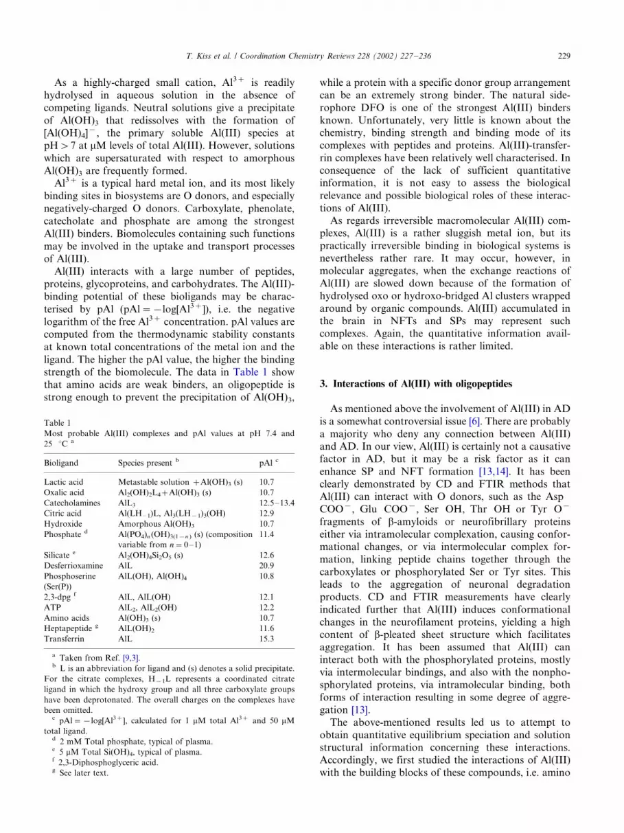
strength of the biomolecule. The data in Table 1 show
that amino acids are weak binders, an oligopeptide is
strong enough to prevent the precipitation of Al(OH)3,
while a protein with a specific donor group arrangement
can be an extremely strong binder. The natural side-
rophore DFO is one of the strongest Al(III) binders
known. Unfortunately, very little is known about thechemistry, binding strength and binding mode of its
complexes with peptides and proteins. Al(III)-transfer-
rin complexes have been relatively well characterised. In
consequence of the lack of sufficient quantitative
information, it is not easy to assess the biological
relevance and possible biological roles of these interac-
tions of Al(III).
As regards irreversible macromolecular Al(III) com-plexes, Al(III) is a rather sluggish metal ion, but its
practically irreversible binding in biological systems is
nevertheless rather rare. It may occur, however, in
molecular aggregates, when the exchange reactions of
Al(III) are slowed down because of the formation of
hydrolysed oxo or hydroxo-bridged Al clusters wrapped
around by organic compounds. Al(III) accumulated in
the brain in NFTs and SPs may represent suchcomplexes. Again, the quantitative information avail-
able on these interactions is rather limited.
3. Interactions of Al(III) with oligopeptides
As mentioned above the involvement of Al(III) in AD
is a somewhat controversial issue [6]. There are probably
a majority who deny any connection between Al(III)and AD. In our view, Al(III) is certainly not a causative
factor in AD, but it may be a risk factor as it can
enhance SP and NFT formation [13,14]. It has been
clearly demonstrated by CD and FTIR methods that
Al(III) can interact with O donors, such as the Asp�/
COO�, Glu�/COO�, Ser�/OH, Thr�/OH or Tyr�/O�
fragments of b-amyloids or neurofibrillary proteins
either via intramolecular complexation, causing confor-mational changes, or via intermolecular complex for-
mation, linking peptide chains together through the
carboxylates or phosphorylated Ser or Tyr sites. This
leads to the aggregation of neuronal degradation
products. CD and FTIR measurements have clearly
indicated further that Al(III) induces conformational
changes in the neurofilament proteins, yielding a high
content of b-pleated sheet structure which facilitatesaggregation. It has been assumed that Al(III) can
interact both with the phosphorylated proteins, mostly
via intermolecular bindings, and also with the nonpho-
sphorylated proteins, via intramolecular binding, both
forms of interaction resulting in some degree of aggre-
gation [13].
The above-mentioned results led us to attempt to
obtain quantitative equilibrium speciation and solutionstructural information concerning these interactions.
Accordingly, we first studied the interactions of Al(III)
with the building blocks of these compounds, i.e. amino
Table 1
Most probable Al(III) complexes and pAl values at pH 7.4 and
25 8C a
Bioligand Species present b pAl c
Lactic acid Metastable solution �Al(OH)3 (s) 10.7
Oxalic acid Al2(OH)2L4�Al(OH)3 (s) 10.7
Catecholamines AlL3 12.5�/13.4
Citric acid Al(LH � 1)L, Al3(LH�1)3(OH) 12.9
Hydroxide Amorphous Al(OH)3 10.7
Phosphate d Al(PO4)n (OH)3(1�n ) (s) (composition
variable from n�0�/1)
11.4
Silicate e Al2(OH)4Si2O5 (s) 12.6
Desferrioxamine AlL 20.9
Phosphoserine
(Ser(P))
AlL(OH), Al(OH)4 10.8
2,3-dpg f AlL, AlL(OH) 12.1
ATP AlL2, AlL2(OH) 12.2
Amino acids Al(OH)3 (s) 10.7
Heptapeptide g AlL(OH)2 11.6
Transferrin AlL 15.3
a Taken from Ref. [9,3].b L is an abbreviation for ligand and (s) denotes a solid precipitate.
For the citrate complexes, H�1L represents a coordinated citrate
ligand in which the hydroxy group and all three carboxylate groups
have been deprotonated. The overall charges on the complexes have
been omitted.c pAl��log[Al3�], calculated for 1 mM total Al3� and 50 mM
total ligand.d 2 mM Total phosphate, typical of plasma.e 5 mM Total Si(OH)4, typical of plasma.f 2,3-Diphosphoglyceric acid.g See later text.
T. Kiss et al. / Coordination Chemistry Reviews 228 (2002) 227�/236 229

acids, such as Asp, Glu, Ser, etc. [15], and also with
phosphorylated Ser and Tyr [16]. These amino acids are
rather weak Al(III) binders; depending on the metal ion
concentration, Al(OH)3 precipitates at pH 3�/4. Belowthis precipitation pH, monodentate COO� coordina-
tion is the most obvious binding mode, but the NH2
group can also take part in the coordination if it is
surrounded by O� donors, as in the case of Asp and
Ser(P).
We continued with small peptide model systems, first
the Al(III)�/AspAsp and Al(III)�/AspAspAsp systems.
Depending on the Al(III) to ligand ratio, precipitationoccurred at pH 5�/6 in both systems. Monodentate and
chelating coordination of the COO� functions can be
assumed in the acidic pH range, but, the early precipita-
tion indicates that the terminal COO� groups of Asp or
Glu are not sufficiently strong in such a small peptide to
keep Al(III) in solution and to prevent the precipitation
of Al(OH)3. To achieve this, a more specific arrange-
ment of suitable side-chain donors is necessary.We studied several synthetic fragments of mid-size
neurofilament (NFM) peptides too. These were rich in
Glu and Ser. The pentapeptides AcProGluValSer-
GlyNH2 and HNProGluValSerGlyNH2 had a protected
C-terminus and a free or protected N-terminus. We
found that the Al(III)-binding abilities of the peptides
were stronger when they had a free ProNH function at
the N-terminus. This is rather surprising as Al(III) isknown [15,17] to have a low affinity for monodentate
amines, and the N-terminal Pro-NH group is, therefore,
not expected to be a strong binding site. As the C-
terminal COO� was blocked in both peptides, we would
expect coordination via the side-chain Glu carboxylate
and Ser alcoholate groups. Instead, coordination seems
to be much more likely through the N-terminal donor
groups.
4. The Al(III)�/AcLysSerProValValGluGly heptapeptide
system
This heptapeptide is also a synthetic NFM fragment
and contains the repeating tetramer LysSerProVal,
which is the recognition site of NFM [18]. CD [19]
and FTIR [20] studies were carried out on such NFMpeptides to detect any effects of Al(III) or other metal
ions. It was found that the effect of Al(III) depended
strongly on the solvent and the sequence of the peptides.
In aqueous solution below pH 7, Al(III) did not give rise
to any definite changes in the spectra. This shows that,
even if it does bind to side-chain functionalities, Al(III)
does not have an effect on the backbone conformation.
However, in trifluorethanol, a solvent with uniquesolvating properties, Al(III) resulted in significant
spectral effects, clearly indicating conformation changes
in the peptide. In this work we used pH-potentiometric
and multinuclear NMR techniques in order to detect
any interactions between this peptide and Al(III) in
aqueous solution.
4.1. Experimental
4.1.1. Materials
The peptide was synthesised on a BioSearch SAM II
automated peptide synthesiser as described earlier [21],
using the Fmoc�/N-terminal protecting strategy and a
modified M-BHA resin. The peptide was characterised
by amino acid analysis, 1H-NMR and positive ion FAB-
MS [22]. The Al(III) stock solution was prepared fromrecrystallised AlCl3 �/ 6H2O, and its concentration was
determined gravimetrically through its oxinate. The
stock solution contained 0.1 M HCl to prevent hydro-
lysis of the metal ion. The ionic strength of all solutions
was adjusted to 0.20 M KCl and the temperature was
25.09/0.1 8C.
4.1.2. Potentiometric measurements
The stability constants of the proton and the Al(III)
complexes of the ligand were determined by pH-
potentiometric titrations of 5 ml samples in the pH
range 2�/10 or until precipitation. The ligand concentra-
tion was 0.002 M and the metal ion to ligand ratio was
0:2, 1:2 or 2:2. In the consequence of the very small
amount of ligand available, all three titrations were
performed in the same sample; the solution titrated with0.2 M KOH solution was reacidified to the starting pH,
with a known amount of 0.2 M HCl acid solution. The
pH was measured with a Molspin pH-meter with a
Methrom combined glass electrode, which was cali-
brated for hydrogen ion concentration according to
Irving et al. [23]. The concentration stability constants
bpqr �/[MpLqHr ]/[M]p [L]q [H]r were calculated with the
aid of the PSEQUAD computer program [24]. Thestability constants used for the hydroxo species of
Al(III) were taken from Ref. [25] and corrected to I�/
0.2 by using the Davies equation: �/5.49 for [AlH�1]2�,
�/13.54 for [Al3H�4]5�, �/108.6 for [Al13H�32]7� and
�/23.40 for [AlH�4]�.
4.1.3. NMR spectroscopy1H- and 13C-NMR spectra were recorded at 25 8C
with a Bruker Avance DRX400 spectrometer. Chemical
shifts were referenced to the signal of TMS as an
external standard. The samples were prepared in water
containing 10% of D2O to provide an NMR lock signal.
The spectra of a 0.004 M solution of the peptide were
measured at pH 4 and 7 in the absence and in the
presence of an equimolar amount of the metal ion. The
two-dimensional homocorrelated (1H-COSY) and het-erocorrelated (HSQC, HMBC) spectra were measured
on a Bruker DRX500 spectrometer, using the standard
Bruker microprogram.
T. Kiss et al. / Coordination Chemistry Reviews 228 (2002) 227�/236230

4.1.4. Molecular modelling calculations
The structure of the AcLysSerProValValGluGly hep-
tapeptide system was studied by molecular mechanics/
molecular dynamics (MM/MD) methods in water withthe application of implicit and explicit solvation models
in the presence or in the absence of any counterions. The
effects of the cations Na�, Zn2� and Al3� on the
structure were followed on the trajectory generated
during 4 ns. Due to the lack of the necessary parameters
the model of Al3� ion was generated on the basis of the
Mg2� parameters. Na� served as a counterion to the
negatively charged peptide molecule. Accordingly, theNa-peptide conformation may be considered as the
conformation of the peptide free of metal ion coordina-
tion. Zn2� was also studied, partly because of its similar
nature to Al3� (Zn2� is not a real transition metal ion
due to its filled d subshell), and partly because of its
potential involvement of peptide aggregation processes
[1].
4.1.5. Set-up of the MD simulation
The initial structure of AcLysSerProValValGluGly
heptapeptide for the calculation of the structure with the
lowest energy was built up as an extended structure by
applying the TINKER package [26]. The structure was
optimised with a convergence criterion of 0.001 kcal
mol�1, with the application of the AMBER-95 all-atom
force field [27] and the GB/SA implicit solvation model
[28]. By means of simulation annealing, 200 structureswere generated by TINKER [26] with the following
protocol: 10 ps thermal equilibration at 1500 K,
followed by exponential cooling of the system to 50 K
during 10 ps. The cut-off of the charge and the van der
Waals interactions was 0.9 nm. The final structure was
geometry optimised similarly to the initial one. This
structure was used as the initial one for the next step.
The steps were repeated 200 times. The structures of thelowest energy conformers were analysed. The lowest
energy conformer was used as the initial structure in the
isothermal molecular dynamics simulation.
The MD simulations were performed with the GRO-
MACS program package [29], with a modified force field
of GROMOS87, applying the united atom model. The
ionizable residues were assigned formal charges appro-
priate to pH�/7 as Lys�/NH3�, Glu�/COO� and the C-
terminal Gly�/COO�. The peptide was placed in a cubic
box with 3.25 nm sides and solvated with 1000 SPC/E
water molecules. The box was large enough to contain
the peptide molecule and 1.0 nm of solvent on all sides.
The system was energy minimised with a criterion of
1000 kJ mol�1. The water molecules at the lowest
positive and/or negative potential energy positions were
replaced by the counterions (a) Na�, (b) Zn2� and 1Cl� and (c) Al3��/2Cl� in order to neutralise the
system by means of GENION included in GROMACS [29].
The parameters for Al3� were not available, and
approximated values were, therefore, applied: all the
parameters for Mg2� were applied during the simula-
tion except for the charge and molar mass, which were
taken �/3 and 26.9815, respectively. After the systemhad again been energy minimised, a 40 ps NVT
restrained simulation was performed at 300 K.
The simulations were performed in the NPT ensemble
with periodic box conditions at 300 K and 1 bar, using
the weak coupling method [30], with relaxation times for
the temperature and pressure of 0.1 and 0.5 ps,
respectively. The dielectric constant used and the
compressibility of water were 1.0 and 4.5 10�5 bar�1,respectively. Peptide, solvent and counterions were
independently coupled to the heat bath. The length of
the time steps was 2 fs. The high order vibrational
motions were eliminated by using LINCS [31] with a
relative tolerance of 10�4. For the SPC/E water
molecules SETTLE [32] was applied, while the long-
ranged electrostatic interactions were treated by the
Particle Mesh Ewald (PME) method [33]. A distance of0.9 nm was used for the electrostatic and van der Waals
cut-offs. The trajectories of the 4000 ps molecular
dynamics simulations were analysed starting at 20 ps.
4.1.6. Trajectory analysis
The total and potential energies, the root mean square
deviations (RMSD) of the backbone (N�/Ca�/C) atoms
relative to the initial structure, and fluctuation of the
RMS of the backbone (N�/Ca9�/C) atoms were calcu-
lated. The distances of the cations (Na�, Zn2� and
Al3�) and the charged groups on the side chain werealso analysed.
The secondary structures of the peptides were ana-
lysed by sampling the whole trajectory every 10 ps with
the DSSP program [34]. The secondary structural ele-
ments were assigned via the definitions given in [35].
4.2. Results and discussion
4.2.1. Speciation
The N-protected heptapeptide AcLysSerProValVal-
GluGly ([H2L], contains three protons that dissociate in
the measurable pH range. That with the highest
log K([HL]�) value of 10.33 can be unambiguouslyascribed to the Lys�/NH3
� group. The acidities of the
terminal COOH and the Glu�/g-COOH groups are
comparable, and they lose two protons in parallel,
overlapping processes in the acidic pH range. From a
comparison of the macroscopic protonation constants
(see Table 2) with those of other Glu-containing
oligopeptides [36], the higher log K([H2L]) value of
4.55 can be ascribed to the Glu carboxylate, and theother value, log K([H3L]�)�/3.39 to the terminal car-
boxylate, although these last two protons liberate in
overlapping processes. The Ser�/OH is very weakly
T. Kiss et al. / Coordination Chemistry Reviews 228 (2002) 227�/236 231

acidic and does not dissociate in the measurable pH
range.
The Al(III)�/peptide system could be titrated up to
pH�/10 without observable precipitation, although
complexation slowed down in the pH range 7�/9. In
this pH range 10 min was not sufficient for pH
equilibrium to be attained, and these points were,
therefore, omitted from the speciation calculations. No
measurements were made at a metal ion excess. Titra-
tion data were evaluated by the assumption of various
mononuclear complexes in different protonation states.
The best fit between the experimental and the calculated
titration curves was obtained with the species and
stability constants listed in Table 2. Complexes with
stoichiometries other than 1:1 were rejected by the
computer program. Species distribution curves are
depicted in Fig. 1. The speciation diagram indicates
that the interaction between Al(III) and the peptide
molecule is fairly weak in the acidic pH range, as in
equimolar solution only 20�/30% of the ligand is bound
to Al(III). The complex [AlLH2]3� liberates four pro-
tons, with stepwise deprotonation constants of
pK([AlLH2]3�)�/3.81, pK([AlLH]2�)�/3.94,
pK([AlL]�)�/4.64 and pK([AlLH�1])�/5.14, which
can be ascribed to deprotonation of the side-chain
donor groups of the peptide and, in consequence ofthe hydrolytic tendency of Al(III), to dissociation of the
coordinated water molecules around the metal ion.
Complex formation becomes predominant only in the
neutral pH range when the species [AlLH�2]� is
formed. It is worth mentioning that practically no
binary hydroxo complexes of Al(III) are formed at a
fourfold or higher excesses of the ligand in the pH range
studied.
4.2.2. NMR features of the Al(III)-ligand system
In order to specify the metal binding sites in thecomplexes formed, detailed NMR measurements were
carried out. As a first step, the 1H- and 13C-NMR
signals of the peptide were fully assigned by using the
two-dimensional techniques listed in Section 4.1. The1H-NMR spectra of the peptide in the absence and in
the presence of equimolar Al(III) are depicted in Figs. 2
and 3. The only singlet resonance at 2.03 ppm in the 1H-
NMR spectrum (Fig. 2a) can be unambiguously attrib-uted to the methyl protons of the acetyl group from the
N-terminus. This resonance served as our starting point
in the signal assignment. The doublets of the methyl
protons of the two Val appear at the highest fields (�/
1.1 ppm) as partly overlapping resonances (not shown in
figure). The multiplets of the methylene groups of the
peptide are present in a wide chemical shift range,
between 1.3 and 4.0 ppm. The protons of the b-CH2
group in Lys (1.81 and 1.72 ppm), Ser (3.84 and 3.90
ppm) and Glu (2.13 and 1.98 ppm) are magnetically
non-equivalent, as often happens when the methylene
group is situated near a chiral carbon. Similar splitting
of the resonances can be observed for the two methylene
groups (1 and 3) of the Pro ring, each of them giving rise
to two multiplets, centred at 3.84 and 3.75 ppm, and
2.29 and 1.92 ppm, respectively. The signals of the
Fig. 1. Speciation curves for the complexes formed in the Al(III)�/
AcLysSerProValValGluGly system at a 1:4 metal ion to ligand ratio,
with cAl(III)�/1 mM.
Fig. 2. 1H-NMR spectra (d range 0.5�/4.5 ppm) of AcLysSerProVal-
ValGluGly at 4 mM at pH 4 (a) in the absence and (b) in the presence
of 4 mM Al(III).
Table 2
Stability constants of proton (log K) and Al(III) complexes (log b) of
the heptapeptide AcLysSerProValValGluGly at 25 8C and I�0.2 M
(KCl)
log K /log b
log K ([HL]�) 10.33(5)
log K ([H2L]) 4.55(3)
log K ([H3L]�) 3.39(3)
[AlLH2]3� 17.11(8)
[AlLH]2� 13.30(7)
[AlL]� 9.36(6)
[AlLH�1] 4.72(6)
[AlLH�2]� �0.42(6)
Fitting (Dml) a 0.0032
Number of points 128
a The average difference between the experimental and the calcu-
lated titration curves, expressed in ml of the titrant.
T. Kiss et al. / Coordination Chemistry Reviews 228 (2002) 227�/236232

methyne protons of the peptide chain appear in the
chemical shift range 4.0�/4.5 ppm, except for that of Ser,
which lies under the water signal. It has to be mentioned
here that in Fig. 2, only the relatively well separated
signals are marked; the peaks of the remaining groups
occur as overlapping resonances in the chemical shift
range 1.9�/2.2 and 3.7�/3.9 ppm. Their assignment and
chemical shift values could be unambiguously obtained
from the two-dimensional spectra but is difficult to
indicate their assignment in the 1H-NMR spectrum. At
lower fields, between 8.0 and 8.5 ppm the spectrum
reveals the resonances of the amide NH protons of the
peptide bonds (Fig. 3a). The triplet at 8.09 ppm is due to
Gly, while the remaining NH protons appear as doub-
lets, since each of them is in the vicinity of a methyne
group. The 1H signal of the NH2 group from the side-
chain of Lys could not be detected, because of its rapid
proton exchange with the solvent.Fig. 2b and Fig. 3b depict the 1H-NMR spectrum of
the peptide at pH�/4 in the presence of equimolar
Al(III). Selective broadening of some signals (+) occurs
as compared with the signals of the free ligand,
indicating interactions between the peptide and the
metal ion. The facts that neither separate resonances
can be observed for the bound and unbound peptide,
nor significant shifts in the position of the resonances
occur (with exception of the amide protons), indicate
that the rate of ligand exchange is roughly comparable
with the 1H-NMR timescale. In the chemical shift range
of aliphatic protons, the resonances of Glu (b-CH2, g-
CH2 and CH groups) and of the CH2 group of Gly are
affected by the presence of Al(III) (Fig. 2b). At low
fields (Fig. 3b), the signal of the amide�/NH group of
Gly exhibits an upfield shift and there is a considerable
broadening as compared with the spectrum of the free
peptide, while the signal of Val1 is shifted to higher
chemical shift values without any change in its aspect.
Slight downfield shifts of the other NH resonances can
also be observed.
Similar results were obtained from the 13C-NMR
measurements. Fig. 4a and Fig. 5a show the J-MOD13C-NMR spectrum of the free ligand: the resonances of
the CH and CH3 groups appear as negative peaks, while
the positive signals are those of the CH2 and CO groups.
In the aliphatic carbon region, the four peaks of the
methyl carbons of the two Val show up at the highest
fields, followed by the signal of the acetyl methyl group
at 21.8 ppm (not shown in Fig. 4). The CH and CH2
signals appear at higher chemical shift values, between
22 and 61 ppm. Their correct assignments are presented
in Fig. 4a. In the carbonyl region (Fig. 5a), the
resonances of the amide CO groups are present in a
quite narrow range of 1.5 ppm, except that of Ser, which
appears more separated at higher field: 170.2 ppm. The
signals of the carboxylic groups of the C-terminal Gly
and Glu can be located at lower fields: 175.2 and 178.1
ppm.
In the spectrum of the Al(III)-containing sample (Fig.
4b and Fig. 5b), the signals of the aliphatic C atoms of
Glu (b-CH2, g-CH2 and CH carbons) and Gly (CH2
carbon) could not be detected; they probably fall into
the baseline due to the their high linewidth. These
resonances show up in the spectrum of the free peptide
at 26.8, 30.8 and 53.2 ppm for Glu and 42.9 ppm for
Gly. Similar broadening of the CO peaks of these two
amino acids could be observed in the CO region of the
same spectra (Fig. 5b).
This broadening of the above-mentioned signals in
the 1H- and 13C-NMR spectra suggests the involvement
of the corresponding amino acids in the coordination of
Al(III), i.e. Al(III) is most probably bound at the C-
terminus of the peptide through the terminal carbox-
ylate group and the side-chain carboxylic function of
Glu. Accordingly, it can be assumed that coordination
Fig. 3. 1H-NMR spectra (amide d range) of AcLysSerProValVal-
GluGly at 4 mM at pH 4 (a) in the absence and (b) in the presence of 4
mM Al(III).
Fig. 4. 13C-NMR spectra of AcLysSerProValValGluGly at 4 mM at
pH 4 (a) in the absence and (b) in the presence of 4 mM Al(III). The
symbols of the signals which disappear in the presence of Al(III) are
placed in black boxes.
T. Kiss et al. / Coordination Chemistry Reviews 228 (2002) 227�/236 233

starts at the terminal COO�, ([AlLH2]3�), and Al(III)
then chelates through the terminal COO� of the peptide
and the side chain-COO� of Glu, with participation of
the central peptide carbonyl, through the formation of ajoint chelate system ([AlLH]2�), and then parallel
overlapping deprotonation of the non-coordinating
protonated amino donor and a coordinated water
molecule subsequently takes place ([AlL]�, [AlLH�1]
and [AlLH�2]�).
The resonances of the two Val also suffer some
smaller changes in the presence of Al(III) (see Figs. 3
and 4). This cannot be explained by the participation ofthese amino acids in binding of the metal ion, but more
likely by some conformational changes of the peptide,
induced by Al(III) (vide infra).
4.2.3. Molecular dynamics calculations
In order to confirm the proposed binding modes of
Al(III) and also to examine whether Al(III) coordina-
tion results in any conformational change in the peptide
backbone, molecular modelling calculations were car-ried out.
The optimal conformation of the free peptide was first
calculated. Among the results of the simulated annealing
calculations, five structures were found with an energy
of less than �/295.0 kcal mol�1 (AMBER-95 and GB/
SA). The secondary structures of this peptide contain
mainly an a-helix. In only one case was a turn structure
found for SerProVal residues. The structure with thelowest energy is depicted in Fig. 6. The negative COO�
groups of Glu and the C-terminal Gly are localised on
one side of the peptide, which can make binding of the
cations possible. The distance between the protonated
positively charged Lys and the negatively charged side-
chain Glu is 0.5�/0.7 nm; a bend/turn structure is
stabilised in this small flexible peptide. The distance
between the same Lys�/NH3� and the C-terminal Gly�/
COO� group is greater: it varies between 1.2 and 1.5 nm
in all cases (Fig. 7).
The effects of the cations Na�, Zn2� and Al3� on
the structure of the peptide were also followed. In the
evaluation of the data, we have to consider that the
model for Al3� is only an approximation. The change in
the secondary structure during the molecular dynamics
calculations at 20�/4000 ps (see Fig. 8) relates to the
destruction of the initial helical structure and the
formation and stabilisation of bend (in the presence of
Na�) and turn (in the presence of Zn2� and Al3�)
structures. The distances between the cations and the
negatively charged COO� functions varied significantly
and revealed specific alterations, depending on the
charge of the cations. With a great fluctuation, Na�
Fig. 6. Most stable structure of AcLysSerProValValGluGly obtained
in the simulated annealing calculations (AMBER95 and GB/SA).
Fig. 7. Distance vs. simulation time plot: solid line, distance between
Lys�/NH3� and Glu�/COO�; the dotted line, distance between Lys�/
NH3� and Gly�/COO�.
Fig. 5. 13C-NMR spectra (carbonyl d range) of AcLysSerProValVal-
GluGly at 4 mM at pH 4 (a) in the absence and (b) in the presence of 4
mM Al(III). The symbols of the signals which disappear in the
presence of Al(III) are placed in black boxes.
T. Kiss et al. / Coordination Chemistry Reviews 228 (2002) 227�/236234

was found to be closer to the C-terminal COO� of the
peptide than to the negative side chain of the Glu. The
RMSD in the positions of the backbone atoms relative
to the structure at 20 ps, was ca. 0.3 nm at the end of the
simulation. The greatest root mean square fluctuation
(S.D.) calculated for the individual backbone atomic/
group positions was 0.15 nm for the residues 4 and 5
(Val, Val) during the simulation. The variations in the
distances during the simulation were similar for Zn2�
and Al3� too, but during ca. 50% (Zn2�) and ca. 80%
(Al3�) of the simulation time the distance between the
C-terminus and the cations is almost constant: 0.4�/0.6
nm. In order to illustrate the effects of the cations on the
peptide structure, the distances of Al3� from thenegatively charged Glu�/COO� and C-terminal Gly�/
COO� as a function of time are depicted in Fig. 9.
The fluctuation in the backbone atom distances (RMS)
is significantly smaller in the simulation of the systems
containing Zn2� and Al3� (0.07 nm for residues 4 and 5
(Val, Val) and 3 (Pro)).
These molecular dynamics calculations suggest that
the greater positive charge of the cations results in amore stable turn structure and a ‘geometrically more
stable’ metal complex in the solution. They indicate
favoured arrangements of the C-terminal COO� and
the side-chain COO� of Glu for the chelation of Al(III).
The average number of water molecules around Al3�
was found to be ca. six in a shell with a radius of 0.20 to
0.36 nm. The stability of the water shell around the
Al3� hindered the approach of the counterion in ashorter distance. This suggests that the interaction
between the oppositely charged hydrated ions is mostly
ionic character.1H-NMR measurements, similar as discussed above,
were carried out at pH�/7 too. It is interesting,
however, that at pH�/7 the two spectra (in the presence
and absence of Al(III)) barely differ, and the direct
coordination of Al(III) is, therefore, not confirmed.However, since no precipitation is observed in the
solution, it is very likely that Al(III)-hydroxo species
in some metastable state (which occurs fairly commonly
in aqueous Al(III) solutions) form outer-sphere com-
plexes through hydrogen-bonding with the hydrated
oligopeptides.
5. Conclusions
To summarise the above studies, interactions between
Al(III) and the heptapeptide AcLysSerProValVal-
GluGly were unambiguously detected by pH-potentio-
metric and multinuclear NMR measurements in weakly
acidic solutions. Molecular dynamics calculations con-
firmed the possible chelating binding of Al(III) to the
negatively charged C-terminal Gly�/COO� and the side-chain Glu�/COO� groups. The coordination of Al(III)
induced significant conformational changes from a more
or less a-helical to a more turned conformation. These
studies confirm that neurofilament non-phosphorylated
peptides can bind Al(III), and this can trigger confor-
mational changes which may result in an enhancement
of aggregation processes of b-amyloids and NFTs. We
intend to continue such investigations with similaroligopeptides, and also their phosphorylated derivatives,
as interchain links between oligopeptides through
Al(III) ions may play an even more important role in
Fig. 9. Distance vs. simulation time plot: solid line, distance between
Al3� and Glu�/COO�; dotted line, distance between Al3� and Gly�/
COO�.
Fig. 8. Changes in the secondary structure during the simulation in the
AcLysSerProValValGluGly�/ (a) Na� (b) Zn2� and (c) Al3�
systems: dark-grey: a-helical structure; grey: bend structure; and
light-grey: turn structure.
T. Kiss et al. / Coordination Chemistry Reviews 228 (2002) 227�/236 235

the aggregation of neuropeptides than can intramole-
cular Al(III) binding. It may be assumed that binding of
Al(III) at the carboxylate sites of neuropeptides influ-
ences the accessibility of Ser, Thr and Tyr phosphoryla-tion sites via posttranslataional mechanisms, which may
make the effects of Al(III) on brain peptides and
proteins even more complex.
Acknowledgements
The work was carried out in the frame of a COST D8collaboration and was supported by the Hungarian
Research Fund (OTKA 23776/97) and the Hungarian
Ministry of Education (FKFP 0013/97).
References
[1] C. Exley (Ed.),Aluminium and Alzheimer’s Disease, Elsevier,
Basel, 2001.
[2] R.B. Martin, Aluminium in Biology and Medicine, Wiley,
Chichester, 1992, p. 5.
[3] T. Kiss, E. Farkas, Perspecitves on Bioinorganic Chemistry, 1996,
p. 199.
[4] D.R.C. McLachlan, A.J. Dalton, T.P. Kruck, M.Y. Bell, W.L.
Smith, W. Kalow, D.F. Andrews, Lancet 337 (1991) 1304.
[5] P. Davies, M.A. Weiner, D.R. Holleman, J.R. Goldstone, Lancet
338 (1991) 325.
[6] J. Savory, C. Exley, W.F. Forbes, Y. Huang, J.G. Joshi, T.
Kruck, D.R.C. McLachlan, I. Wakayama, J. Toxicol. Environ.
Health 48 (1996) 615.
[7] A.K. Powell, S.L. Heath, Coord. Chem. Rev. 149 (1996) 59.
[8] W.R. Harris, G. Berthon, J.P. Day, C. Exley, T.P. Flaten, W.F.
Forbes, T. Kiss, C. Orvig, P.F. Zatta, J. Toxicol. Environ. Health
48 (1996) 543.
[9] W.R. Harris, Coord. Chem. Rev. 149 (1996) 347.
[10] J.C. Rochet, P.T. Lansbury, Jr., Curr. Opinion Struct. Biol. 10
(2000) 60.
[11] Report of the American Alzheimer Association, New York, 1999.
[12] T.L. Macdonald, R.B. Martin, Trends Biochem. Sci. 13 (1988) 15.
[13] M. Hollosi, L. Urge, A. Perczel, J. Kajtar, I. Teplan, L. Otvos,
G.D. Fasman, J. Mol. Biol. 223 (1992) 673.
[14] C. Exley, N.C. Price, S.M. Kelly, J.D. Birchall, FEBS Lett. 324
(1993) 293.
[15] T. Kiss, I. Sovago, I. Toth, A. Lakatos, R. Bertani, A. Tapparo,
G. Bombi, R.B. Martin, J. Chem. Soc. Dalton Trans. (1997) 1967.
[16] E. Kiss, A. Lakatos, I. Banyai, T. Kiss, J. Inorg. Biochem. 69
(1998) 145.
[17] R.B. Martin, in: M. Nicolini, P.F. Zatta, B. Corain (Eds.),
Aluminum in Chemistry, Biology and Medicine, Cortina Inter-
national, Verona, 1991.
[18] V.M.-Y. Lee, L. .Otvos, Jr, M.J. Carden, M. Hollosi, B.
Dietzschold, R.A. Lazzarini, Proc. Natl. Acad. Sci. USA 85
(1988) 1998.
[19] M. Hollosi, L. .Urge, A. Perczel, J. Kajtar, I. Teplan, L. Otvos, Jr,
G.D. Fasman, J. Mol. Biol. 223 (1992) 673.
[20] M. Hollosi, S. Holly, Zs. Majer, I. Laczko, G.D. Fasman,
Biopolymers 36 (1995) 381.
[21] Z.M. Shen, A. Perczel, M. Hollosi, I. Nagypal, G.D. Fasman,
Biochemistry 33 (1994) 9627.
[22] L. .Otvos, I.A. Tangorem, K. Wroblewski, M. Hollosi, V.M.-Y.
Lee, J. Chromatogr. 512 (1990) 265.
[23] H.M. Irving, M.G. Miles, L.D. Pettit, Anal. Chim. Acta 38 (1967)
475.
[24] L. Zekany, I. Nagypal, G. Peintler, PSEQUAD for Chemical
Equilibria, Technical Software Distributions, Baltimore, 1991.
[25] L.O. .Ohman, S. Sjoberg, Acta Chem. Scand. A36 (1982) 47.
[26] J.W. Ponder, TINKER, Software Tools for Molecular Design,
Version 3.8, October 2000.
[27] W.D. Cornell, P. Cieplak, C.I. Bayly, I.R. Gould, K.M. Merz, Jr,
D.M. Ferguson, D.C. Spellmeyer, T. Fox, J.W. Caldwell, P.A.
Kollman, J. Am. Chem. Soc. 117 (1995) 5179.
[28] D. Qui, P.S. Shenkin, F.P. Hollinger, W.C. Still, J. Phys. Chem. A
101 (1997) 3005.
[29] (a) D. van der Spoel, A.R. van Buuren, E. Apol, P.J. Meulenhoff,
D.P. Tieleman, A.L.T.M. Sijbers, R. van Drunen, H.J.C.
Berendsen, GROMACS User Manual, University of Gronongen,
1996.;
(b) H.J.C. Berendsen, D. van der Spoel, R. van Drunen, Comp.
Phys. Commun. 91 (1995) 43.
[30] H.J.C. Berendsen, J.P.M. Postma, A. DiNola, J.R. Haak, J.
Chem. Phys. 81 (1984) 3684.
[31] B. Hess, H. Bekker, H.J.C. Berendsen, J.D.E.M. Fraaije, J.
Comp. Chem. 18 (1997) 1463.
[32] J.P. Ryckaert, G. Ciccotti, H.J.C. Berendsen, J. Comp. Phys. 23
(1977) 327.
[33] (a) T. Darden, D. York, L. Pedersen, J. Chem. Phys. 98 (1993)
10089;
(b) U. Essmann, L. Perera, M.L. Berkowitz, T. Darden, H. Lee,
L.G.A. Pedersen, J. Chem. Phys. 103 (1995) 8577.
[34] C.S. Kabsch, Biopolymers 22 (1983) 2577.
[35] T. Kortvelyesi, R.F. Murphy, S. Lovas, J. Biomol. Struct. Dyn.
17 (1999) 393.
[36] L.D. Pettit, H.K.J. Powell, The IUPAC Stability Constant
Database, Academic Software and IUPAC, Royal Society of
Chemistry, London, 1992�/1997.
T. Kiss et al. / Coordination Chemistry Reviews 228 (2002) 227�/236236


