Embed Size (px)
Citation preview
Transpl Int (1996) 9:175-183 �9 Springer-Verlag 1996
Nicolau Beckmann Joanne Joergensen Konrad Bruttel Markus Rudin Henk-Jan Schuurman
Magnetic resonance imaging for the evaluation of rejection of a kidney allograft in the rat
Received: 18 July 1995 Received after revision: 16 November 1995 Accepted: 20 December 1995
N. Beckmann �9 J. Joergensen - K. Bruttel M. Rudin �9 H-J. Schuurman ( ~ ) Preclinical Research, Sandoz Pharma Ltd., Building 386/101B, CH-4002 Basel, Switzerland Fax: + 41 61 324 2379
Abstract Orthotopic DA (RT1 a) into Lewis (RT11) rat kidney allo- grafts and control Lewis-into-Lewis grafts were assessed by magnetic resonance imaging (MRI) and per- fusion measurement after intrave- nous injection of a superparamag- netic contrast agent. MRI anatomi- cal scores (range 1-6) and perfusion rates were compared with graft his- tology (rank of rejection score 1-6). Not only acute rejection, but also chronic events were monitored after acute rejection was prevented by daily cyclosporine (Sandimmune) treatment during the first 2 weeks after transplantation. In acute al- lograft rejection (n = 11), MRI scores reached the maximum value of 6 and perfusion rates were se- verely reduced within 5 days after transplantation; histology showed severe acute rejection (histologic score 5-6). In the chronic phase
(100-130 days after transplanta- tion), allografts (n = 5) manifested rejection (in histology cellular rejec- tion and vessel changes), accompa- nied by MRI scores of around 2-3 and reduced perfusion rates. Both in the acute and chronic phases, the MRI anatomical score correlated significantly with the histological score (Spearman rank correlation coefficient r s 0.89, n = 30, P < 0.01), and perfusion rates correlated sig- nificantly with the MRI score or histological score (r, values between -0.60 and -0.87, n = 23, P < 0.01). It is concluded that MRI represents an interesting tool for assessing the an- atomical and hemodynamical status of a kidney allograft in the acute and chronic phases after transplantation.
Key words MRI, rejection, kidney. Kidney, MRI, rejection �9 Rejection, MRI, kidney
Introduction
In the preclinical evaluation of immunosuppressive drugs developed for clinical transplantation, organ allo- grafting in rodents is a relevant in vivo experimental ap- proach. One of the models applied is orthotopic kidney allografting in rats using the strain combination of DA (RT1 a) into Lewis (RTll). This strain combination is generally considered to be a very stringent one because of the major histocompatibility mismatch between do- nor and recipient. In the experimental design, a con- tralateral nephrectomy is performed at day 7 after trans- plantation and the graft is inspected. At that time point,
untreated animals show macroscopic signs of rejection, such as enlargement of the graft, a reddening of the graft related to hemorrhage, and perigraft inflamma- tion. If left untreated, animals with acute rejection die at around day 11 after transplantation due to the loss of graft function. Long-term survival of the graft is achieved in recipients treated with cyclosporine (Sand- immune) starting from the day of transplantation until day 14 after transplantation. Usually, experiments with long-term survival are terminated between 100 and 130 days after transplantation [2].
In histological assessment, grafts with acute rejection show mononuclear cell infiltration and tubular damage.

176
A graft with long-term survival resembles a normal kid- ney with some mononuclear cell infiltration but without damage of the renal parenchyma. Some grafts may show a variable extent of cellular rejection accompanied by vasculopathy, including thickening of vascular walls with proliferation of smooth muscle cells and (neo)in- tima formation, in severe cases with obstruction of the blood vessel. These features are compatible with chron- ic rejection and may occur in animals that are appar- ently healthy and do not show signs of decreased renal function. Such animals may show an increase in serum creatinine; however, in our experience, serum creatinine is not a suitable parameter of graft function due to a lack of sensitivity. Increased creatinine levels, up to 200 ~tmol/1, may occur after kidney transplantation in animals that show no signs of renal disease when in- spected.
When determining graft survival and, hence, the effi- cacy of the immunosuppressive treatment, animal health associated with graft function (i. e., survival) and histology of the graft at autopsy are the main parame- ters. In our search for other parameters that are not in- vasive and that do not require sacrificing the animal, we evaluated nuclear magnetic resonance imaging (MRI). MRI and MR spectroscopy have been applied to study heart and liver transplantation in the rat [3, 5, 7, 8, 10, 14, 18] and kidney grafting in dogs [4]. For kid- ney allografts in rats, 311~ nuclear magnetic resonance has been applied to obtain biochemical data related to parenchymal tissue injury (increase in inorganic phos- phate, decrease in ~-ATP); for accurate measurements, the graft has had to be implanted in the groin of the re- cipient animal [13]. This application of nuclear magnetic resonance differs from our approach in which anatomi- cal imaging of orthotopic grafts was the first aim. In ad- dition, we assessed kidney perfusion using a bolus track- ing method originally developed to measure local cere- bral blood flow [9, 17]. The intravascular paramagnetic or superparamagnetic contrast agent leads to local chan- ges in magnetic susceptibility, which manifests itself as a decrease in the signal intensity in gradient-recalled echo experiments. This signal attenuation in a specific region- of-interest is related to the local tracer concentration and, thus, to the tissue blood volume from which perfu- sion rates can be derived using the central volume prin- ciple. The determination of absolute perfusion rates, however, is not feasible due to fundamental problems in the determination of the mean transit time or the mean residence time of the tracer in the tissue. In fact, it has been shown that the first moment of the signal in- tensity versus time profile is not equal to the mean tran- sit or residence times [19]. This aside, it is possible to as- sess relative perfusion rates with a bolus tracking meth- od.
In the experimental design of this study, acute al- lograft rejection was induced in Lewis rat recipients of
a DA kidney and compared with syngeneic Lewis kid- ney grafts. Chronic rejection was studied in Lewis recip- ients of a DA kidney that had received a 14-day course of cyclosporine to prevent acute allograft rejection. Also, in this case, the kidney allograft was compared with a syngeneic Lewis kidney graft. To enable an ade- quate comparison of the graft with a normal kidney, contralateral nephrectomy was not included in the ex- perimental design.
Materials and methods
Animals and experimental design
The study was performed in accordance with the Swiss Animal Welfare Act, dated 9 March 1978, and the accompanying Animal Welfare Regulation dated 28 May 1981. Male DA (RT1 a) rats and Lewis (RT11) rats were obtained from Olac (Bicester, UK) at about 8-10 weeks of age. Two studies were performed. The first study ad- dressed acute rejection and included three groups of animals. Four Lewis rats received a kidney graft from a syngeneic Lewis rat, 11 animals received an allograft with no treatment, and 7 received an allograft and were treated with cyclosporine (Sandimmune) at a dose of 7.5 mg/kg per day p.o. for the duration of the experiment. Animals were analyzed via MRI on days 1, 2, 3, 5, 6, 8, 10, 12, and 14, and autopsy was carried out on days 3, 4, 7, 8, 10, or 14. In the second study, long-term survival was addressed in two groups. Three Lewis rats received a syngeneic kidney graft; five others re- ceived a kidney allograft and were treated with cyctosporine (Sandimmune) at a dose of 7.5 mg/kg per day p. o. for 14 days start- ing from the day of transplantation. MR images were recorded on days 3, 7, 10, and 14 and then at weekly intervals up to day 133 after transplantation. Autopsy was carried out on days 100, 119, 120, 126, or 133. In all animals relative graft perfusion was assessed im- mediately before sacrifice.
Surgery
Orthotopic kidney transplantation was performed under anesthe- sia with isofiurane, 1 ml 5 % chloral hydrate i.p., and 0.05 mg atro- pine sulphate s.c. After removal of the recipient's kidney, the do- nor kidney was placed in position with end-to-end anastomoses of the renal arteries using 10/0 Ethilon interrupted sutures and of the veins using 10/0 continuous suture. Ureters were anastomosed end-to-end with 10/0 Ethilon interrupted sutures. The ischemia time was about 25 rain, 15 min of which was cold; during the other 10 min, the graft was rewarmed. There was no ex vivo perfusion of the graft.
Magnetic resonance imaging
For MRI measurements, animals anesthetized with isoflurane 2 % in a nitrous oxide/oxygen (2:1) mixture administered via a face mask were placed in supine position on a plexiglass support. The respiratory signal, measured with an elastic belt containing a strain gauge fixed around the chest, was recorded with a Physiogard SM 785 monitoring device (Bruker, Karlsruhe, Germany). Conven- tional MR images were acquired using a Biospec 47/15 spectrome- ter (Bruker). A homebuilt resonator [1] with a 70-mm inner diam- eter was used as radiofrequency transmitter/receiver. Spin echo
177
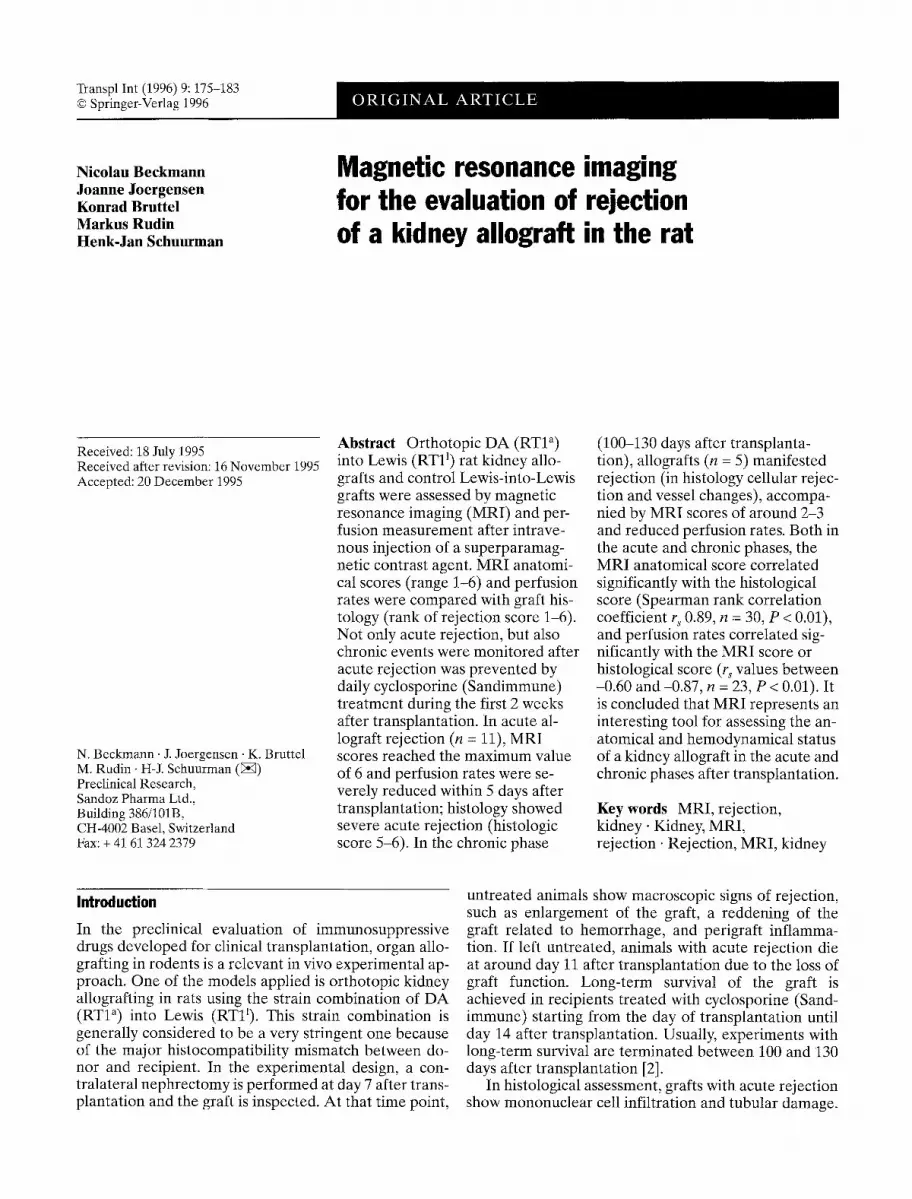
(SE) pulse sequences with repetition delays of 1000 and 2000 ms and echo delays of 21 and 60 ms [SE(1000/21) and SE(2000/60), re- spectively] were applied. In order to minimize breathing artifacts, data acquisition was triggered by respiration. The field-of-view was set at 60 • 60 mm 2, the slice thickness at 1 mm, and ten trans- verse slices were recorded. The matrix dimension was 256 x 128, corresponding to pixel dimensions of 0.23 • 0.47 mm 2. The total measurement time was approximately 4.3 rain for SE(1000/21) and 8.5 min for SE(2000/60), with two acquisitions being averaged.
MRI perfusion measurements
Perfusion measurements were carried out on a Biospec 47/40 spec- trometer (Bruker) equipped with a self-shielded gradient system. A birdcage resonator [16] with a 70-mm inner diameter was used as a radiofrequency transmitter and receiver. Images were re- corded in 1 s using a snapshot sequence [6] with an echo delay of 4.5 ms and a repetition time of 8.3 ms. The field-of-view was 60 x 60 mm 2, the slice thickness 1 mm, and one coronal slice was measured sequentially 64 times. After the 20th image, a superpara- magnetic contrast agent was injected via the tail vein for approxi- mately 1 s. Changes in signal intensity upon injection of the con- trast agent were monitored as a function of time. Superparamag- netic magnetite nanoparticles [15] coated with bovine serum albu- min were used as a contrast agent. The iron concentration was 5 mg/ml and the volume administered was 0.5 ml.
Histology
At autopsy, the grafted kidney was harvested, fixed in buffered for- malin, and embedded in paraffin. Sections 3.5 ~m thick were stained with hematoxylin and eosin. Histological changes were scored for the extent of rejection as follows: score 0, normal kidney architecture, no signs of rejection; score 1, slight infiltration of re- nal parenchyma without signs of rejection; score 2, infiltration with signs of cellular rejection with marginal destruction of renal parenchyma; score 3, cellular rejection with slight destruction of renal parenchyma; score 4, cellular rejection with moderate tissue destruction; score 5, cellular rejection with severe tissue destruc- tion; score 6, end-stage rejection with almost complete destruction of the graft. These scores are illustrated in Fig. 1 for acute rejection and in Fig. 2 for chronic rejection.
Data analysis
Anatomical MR images were analyzed by a person blinded to the treatment. Kidneys were scored as follows: 1, normal; 2, slightly en- larged with some loss of cortical/medullary contrast; 3, severely en- larged; 4, very large with predominantly medullary hemorrhages; 5, very large with focal hemorrhages in the cortex and involvement of adjacent tissue structures; and 6, very large with complete loss of internal structure and severe involvement of the adjacent tissue. Graft perfusion was assessed by measuring the signal profile dur- ing passage of an intravascular contrast agent. As the tracer passes the imaging plane, the signal is attenuated; the decrease in signal intensity is related to the amount of tracer in the tissue, thus being a measure for the local exchangeable tissue blood volume [11, 17]. The average signal intensity was determined in regions-of-interest in kidney cortex and medulla for both the transplanted and the contralateral native kidney. The average signal attenuation during the first pass of the contrast agent relative to the precontrast signal intensity was estimated for each region-of-interest. The attenua-
tion values of the transplanted kidney were then expressed as a percent of the values for the contralateral native kidney, i.e., flow(transpl)/flow(contralateral)attenuation(transpl)/attenuation (contralateral), and used as an index for relative perfusion.
Statistical analysis
The nonparametric Spearman rank correlation coefficient (rs), cor- rected for ties, was calculated.
Results
M R I scores are i l lustrated by the images in Fig. 3, which show transverse sections of t ransp lan ted kidneys in compar i son to the cont ra la tera l nat ive kidney. The cor- responding score is indicated in the respect ive image. C o m p a r e d to SE(1000/21) images, the SE(2000/60) im- ages general ly p rov ided a be t te r contras t and a l lowed dif ferent ia t ion be tween cor tex and medul la at the ex- pense of an increased susceptibil i ty to m o t i o n artifacts. A very intense (bright) signal was observed in the pelvis and was due to the presence of urine. Kidneys with acute and chronic re jec t ion showed no obvious differ- ences in gross m o r p h o l o g y using the two M R I pulse se- quences. Therefore , da ta are poo l ed for acute and chronic rejection.
His to logy of syngeneic (Lewis-der ived) k idney grafts at each t ime point assessed after t ransplan ta t ion showed a normal k idney archi tecture. Some infi l trat ion by m o n o n u c l e a r cells (score 1) was obse rved in grafts at day 8, 10, or 14 after t ransplantat ion. This was also found in one of the three long- te rm survivors. A l logene ic (DA- der ived) k idney grafts in animals that received no immu- nosuppress ion showed reject ion (score 3-4) at day 3 and 4 after t ransplanta t ion. O n day 7 af ter t ransplan ta t ion and later on, end-s tage re ject ion was obse rved (score 6). Al lograf ts f rom three of seven animals t rea ted with cy- c lospor ine dur ing days 3 and 14 after t ransp lan ta t ion showed some infi l trat ion wi thout signs of re ject ion (score 1); three others showed slight re jec t ion (score 2), and in one animal severe re jec t ion (score 5) was ob- served. In the series of five long- te rm survivors, two showed margina l re jec t ion (score 2, days 119 and 133 af- ter t ransplanta t ion) , one showed severe re ject ion (score 5, day 119), and two others showed end-s tage re jec t ion (score 6, bo th at day 100 after t ransplanta t ion) . Re jec t ion in long- te rm survivors was associated with vessel pathol- ogy, but in n o n e of the cases was a comple te obs t ruc t ion of b lood vessels evident in the histological section.
The corre la t ion be tween M R I scores and histological scores is shown in Fig. 4. M R I values co r r e sponded to the m e a s u r e m e n t on the same day that animals were au- topsied. A highly significant corre la t ion be tween M R I score and re ject ion score was calcula ted (r s = 0.89, P < 0.01). Changes in M R I score dur ing the t ime after

178
F i g . l a - f Histology of acute rejection shown at 100x (a,c,e) and 250x (b,d,f) magnification: a,b normal graft, histology score 0; e,fl slight cellular rejection, histology score 3, with infiltration of the tubular parenchyma and invasion of tubules by lymphocytes (at-
row); e,f end-stage cellular rejection, histology score 6, with almost complete tissue destruction, the structure of a glomerulus rela- tively maintained

179
Fig. 2a-c Histology of chronic rejection shown at 100 x magnifica- tion: a allograft after long-term survival without signs of rejection (score 0) showing an essentially normal kidney architecture; b se- vere cellular rejection (score 5); e end-stage rejection with almost complete destruction of tissue (score 6). Note the vessel change with thickening of the vessel wall and neointima formation; the lu- men is narrowed but not obstructed
transplantation are presented in Fig. 5. The appearance of syngeneic kidney grafts was about normal thoughout the experimental period; the maximum score noted was 3, in two out of ten cases. In contrast, kidney allografts in animals that did not receive cyclosporine reached the maximum score of 6 within 5 days after transplan- tation. The allografts in cylosporine-treated animals showed only a mild acute response with scores equal to or below 2 in all cases; this was not significantly different from the response in the syngeneic transplants. How- ever, abnormal MRI images indicative of rejection emerged at times longer than 1 month after transplanta- tion. At day 100, three of five animals had a score of 4 or higher. These three animals showed severe rejection at autopsy. The MRI score in two of five long-term surviv- ing allografts that showed marginal rejection at autopsy remained low (score 1) during the experimental period.
Examples of perfusion measurements in the cortex of a syngeneic and of an allogeneic kidney graft are shown in Fig. 6. After entry of the tracer in the tissue, the signal intensity is decreased to about 25 % of its original value within 2-5 s after the injection and then rapidly recovers to about 60 %. This partial recovery reflects recirculation and it takes several minutes before the tracer is cleared from the blood ( > 95 % of the original signal intensity). In almost all of the profiles, a second minimum occurred 7-10 s after the injection. The signal profile correspond- ing to the cortical (Fig. 6 a) and medullary regions-of-in- terest in syngeneic transplants was not different from that of the respective regions-of-interest in the contralat- eral native kidney of the animal. Kidney allografts during rejection showed a different picture (Fig. 6 b). The tracer uptake was severely compromised in these cases. The sig- nal minimum after 2-5 s, reflecting the first pass of the tracer, was not observed. Only at 20-25 s after tracer in- fusion were tracer plasma equilibrium levels reached in the region-of-interest. This profile is indicative of se- verely affected kidney perfusion. There were no signifi- cant differences in perfusion rates between grafts with an acute or chronic rejection. Therefore the data of per- fusion measurements during the acute and chronic pha- ses after transplantation have been combined.
The correlation of the perfusion rate and the scores obtained with MRI and graft histology are shown in Fig. 7. In all combinations, the correlation between per- fusion rate of cortex or medulla and score by MRI or histology was statistically significant (r S values between -0.60 and -0.87, P < 0.01). For MRI scores 1 and 2, the perfusion rate in the transplanted kidney was essentially normal, but the blood flow was severely compromised in cases of MRI scores of 4-6. Of all rats measured, only one had an MRI score of 3 at the time point of perfusion measurement and a perfusion rate of 53 %. Looking at the histological scores, slight (score 3) to moderate (score 4) rejection proved to be associated with a re- duced kidney blood flow measured by perfusion.

180
Fig.3 Transverse sections of the rat kidney, extracted from multi- slice (ten slices) MRI data sets. The images on the top correspond to the contralateral native kidney. The other images are transverse sections of transplanted kidneys and are presented in opposite ori- entation. MRI scores indicated in the figure are as follows: 1, nor- mal; 2, slightly enlarged kidney with some loss of contrast between cortex and medulla; 3, severely enlarged kidney; 4, very large kid- ney with predominantly medullary hemorrhages; 5, very large kid- ney with focal hemorrhages in the cortex and involvement of adja- cent tissue structures; 6, very large kidney with loss of cortical and medullary structure and severe involvement of the adjacent tissue. For each illustration, the image on the left side corresponds to an SE(1000/21) acquisition, and that on the right side corresponds to an SE(2000/60) acquisition. The data acquisition was triggered by respiration
Discussion
Respira tory-gated M R I yields images of good quality f rom the rat kidney, enabling one to differentiate be- tween cortex and medulla areas. The SE(1000/21) im-
ages essentially revealed proton density maps, allow- ing us to assess the overall kidney dimensions. The SE(2000/60) images provided a bet ter tissue contrast, and structures like cortex, medulla, and pelvis (urine) could be identified (Fig.3). Due to the longer echo t ime (60 ms), however, the latter sequence was more prone to artifacts (respiration, pulsatile blood flow, in- testinal motility). Therefore, both sequences were used in the present study. In syngeneic kidney grafts, M R I did not reveal any morphological changes. This was con- f i rmed by normal kidney histology at autopsy. There was only one exception, in which T2-weighted M R im- ages attained using the SE(2000/60) pulse sequence re- vealed a very large kidney pelvis, indicative of an ob- structed ureter. The kidney tissue itself seemed unaf- fected, i. e., neither hemorrhage nor loss of cortical/me- dullar contrast was observed.
Kidney allografts in rats receiving no t rea tment man- ifested severe rejection within 7 days, and this was con- f irmed by histology (Fig. 2). In these cases, M R imaging

181
4 o
3
2
A A , &
i i I i i i 0 1 2 3 4 5 6
Histology score
ttt
A,L
Fig.4 Correlation between MRI score and histologic score of re- jection. The nonparametric Spearman rank correlation coefficient (r,), corrected for ties, was 0.89 (n : 30, P < 0.01)
/'{- - .o- - .syngeneie 6- I ~ . m �9 --�9 no treatment
�9 / --�9 cyctesporine treatment
E:
lJ,,,~o k . . . , ~ " "o" o..o..o..o..o..o..o ]
o �9 , , / , ( i , 5 10 20 4'0 6L0 8'0 100
T ime af ter t ransp lanta t ion (days)
Fig.5 Changes in MRI scores of kidney grafts in time after trans- plantation presented for syngeneic transplantation and allogeneic transplantation with or without cyclosporine treatment. Data pre- sented represent mean values + SEM
revealed a striking feature of the involvement of the ad- jacent structures. The zone of hyperintense signals sur- rounding the kidney was due to increased T 2 values as- sociated with increased local tissue water (edema). This is indicative of an inflammatory process that is part of the rejection process.
According to our expectations, there was no, or only slight, cellular rejection in allografted recipients that re- ceived cyclosporine immunosuppression. The MRI score in these animals was low (< 2) during the initial period after transplantation (Fig. 5), and graft perfusion rate was as in the contralateral native kidney. Only in one case was a very large hemorrhagic kidney observed in MR images and severe rejection (score 5) in histol- ogy. This occurrence of rejection is related to the dose
a
s >.,
o3 c-
E
c
co
E ==
co
100'
80-
60.
40.
20
:-'I>,, - - u - - contra latera l k idney -- " - - t ransp lanted k idney
_ _ A . - �9
-5 0 5 1~0 1'5 2L0 2'5
T ime (s)
100-
80-
60-
40.
20.
- - ' - - cont ra la tera l k idney l �9 l ~ A,A,A ' - - " - - t ransp lanted k idney
i AkA �9 ~" �9 �9 A., . . . . . . . . . . . . .222;tk~,-ti,
%,
-; ; ; 1'o & 2; 2'5 Time (s)
Fig.6a,b Relative intensity of the MR signal averaged over a re- gion-of-interest in the kidney cortex for snapshot images acquired sequentially (acquisition time of one image was 1 s). At timepoint 0, 0.5 ml of the contrast agent ((Fe304)xBSA, 5 mg/ml) was in- jected into the tail vein for 1 s. In each figure, the relative intensi- ties of the transplanted kidney and the contralateral native kidney of the animal are presented: a syngeneic kidney graft without rejec- tion, 90 days after transplantation, showing a similar profile in time for the transplanted and native kidney; b kidney allograft with cel- lular rejection, 75 days after transplantation, showing a severely re- duced attenuation of the signal intensity in the graft
of cyclosporine administered; in our experience with this stringent Lewis/DA rat strain combination, a daily dose of 7.5 mg/kg per day given p.o. during the first 2 weeks after transplantation generally results in long- term survival, but some cases may show acute cellular rejection. This also underlies the emergence of chronic rejection at a later phase. Apparently, the dose of cy- closporine administered is just sufficient to prevent acute rejection in most cases, but not sufficient to induce tolerance in the animals in such a way that chronic rejec- tion is also prevented. Three of five animals with long- term survival showed severe to end-stage rejection at autopsy and MRI scores of 4 to 5. In these cases, there were no clear signs of the presence of hemorrhages on

182
o .o E
O gs
o_~
(a)
140 ~
120 ~
1 O0 ~
80-
60-
40-
20-
o-'
120 1
100-1
601 60
4i 1 2
i 2
zx
M R I s c o r e
�9 cortex
zx medulla
!
zx
n i n 2 ; 4 ~ 6 H i s t o l o g y s c o r e
�9 cortex
zx medulla
Fig.7a,b Correlation between graft perfusion rate in the cortex (ll; A) and the medulla and a MRI scores and b histological scores. The Spearman rank correlation coefficients (rs) were: per- fusion cortex vs MRI score -0.87 (n = 23, P < 0.01); perfusion me- dulla vs MRI score -0.80 (n = 23, P < 0.01); perfusion cortex vs his- tology score -0.82 (n = 23, P < 0.01); perfusion medulla vs histol- ogy score -0.60 (n = 23, P < 0.01)
MRI anatomical images, which contrasts to pictures in cases with acute rejection. However, all of the animals with chronic rejection had a significantly enlarged kid- ney pelvis, indicative of an imbalance between urine production and clearance. This difference from grafts showing acute rejection may be ascribed to the chronic- ity of the rejection process which, in addition to attack- ing the tubular parenchyma, involves glomeruli and ves- sel pathology. According to histology, the cellular type of rejection was invariably present, enabling us to apply the same scoring system to describe rejection in the acute phase and in long-term surviving grafts. Histologi- cal scoring, however, provides no information on al- tered perfusion due to vessel damage and graft size; both parameters can be assessed by MRI and the values recorded correlated with histological data.
The signal profiles from perfusion assessment shown in Fig. 6 indicate that the temporal resolution has to be of the order of 1 s or better to follow the first pass of the tracer bolus. The time dependence of the signal in- tensity in rejected kidneys is indicative of severely com-
promised graft perfusion, comparable to profiles ob- served in cerebral ischemia [12]. The relative perfusion rates, obtained from the comparison of grafted and con- tralateral native kidneys, significantly correlated with the scores by MR imaging and histology (Fig. 7). A sig- nificant lowering of perfusion already occurred with slight rejection (scores 3).
The present data on MRI parameters and histology were obtained in the experimental condition that the graft had no life-supporting function, i. e., the contralat- eral native kidney was kept in place. This design was chosen to enable a comparison between the animal's own native kidney, not under attack by the potential im- mune response, and the grafted kidney allograft. This implies that possible systemic, metabolic disturbances related to impaired renal function due to rejection and/ or adverse drug side effects (e.g., hypertension) pre- sumably did not occur in the animals and, hence, could not interfere with the graft status. We have extended the present studies to include an assessment of animals subjected to contralateral nephrectomy after kidney allografting with essentially similar results (data not shown).
In conclusion, we show that in vivo MRI represents an interesting tool for assessing the status of a kidney al- lograft, both in the acute and chronic phases after trans- plantation. The method provides relevant information on both anatomical and hemodynamic aspects, both of which correlated significantly with graft histology. This raises the possibility of assessing the status of the graft at multiple time points during an experimental period. This phenomenon has relevance for the preclinical eval- uation of drugs for their efficacy in treating ongoing re- jection reactions and in monitoring the graft more close- ly during treatment regimens. Thus, MRI serves as a valuable addition to the present armamentarium with which one can assess the rat kidney allograft status, even though the direct pathological-anatomical exami- nation is still the "gold standard". MR imaging is also of value in the assessment of graft function using con- trast agents that are predominantly cleared in the kid- ney, such as gadolinium-diethylenetriamine-pentaace- tate.

183
References
1. Alderman DW, Grant DM (1979) An efficient decoupler coil design which reduces heating on conductive samples in superconducting spectrometers. J Magn Reson Imaging 36:447-454
2. Borel JF (1989) Pharmacology of cy- closporine (Sandimmune). IV. Pharma- cological properties in vivo. Pharmacoi Rev 41:259-371
3. Bowers JL, Teramoto K, Khettry U, Clouse ME (1992) 31p NMR assessment of orthotopic rat liver transplant viabil- ity. The effect of warm ischemia. Trans- plantation 54:604-609
4. Bretan PN, Vigneron DB, Hricak I-I, Price DC, Yen TS, Luo JA, Tangho EA, James TL (1993) Assessment of in situ renal transplant viability by 3~P-MRS: experimental study in canines. Am Surg 59:182-187
5. Fraser CD, Chacko VR Jacobus WE, Mueller P, Soulen RL, Hutchins GM, Reitz BA Baumgartner WA (1990) Early phosphorus 31 nuclear magnetic resonance bioenergetic changes poten- tially predict rejection in heterotopic cardiac allografts. J Heart Transplant 9: 197-204
6. Haase A (1990) Snapshot FLASH MRI. Applications to T~, T 2, and chem- ical shift imaging. Magn Reson Med 13: 77-89
7. Konstam MA, Aronovitz MJ, Runge VM, Isner JM, Foster EA, Kaufman DM, Diehl JT, Dresdale AR, Katzen NA, Kaplan E (1991) Magnetic reso- nance imaging in ischemic injury after heart transplantation in rats. J Heart Lung Transplant 10:750-756
8. MUhler A, Freise CE, Kuwatsuru R, Rosenau W, Liu T, Mintorovitch J, Clement O, Vexler V, Mahboubi S, Lang P, Roberts JR Brasch RC (1993) Acute liver rejection: evaluation with cell-directed MR contrast agents in a rat transplantation model. Radiology 186:139-146
9. Rosen BR, Belliveau JW, Vevea JM, Brady TJ (1990) Perfusion imaging with NMR contrast agents. Magn Reson Med 14:249-265
10. Rozenman Y, Kantor HL (1990) Het- erotopic transplanted rat heart: a model for in vivo determination of phosphorus metabolites during ischemia and reper- fusion. Magn Reson Med 13:450-457
11. Rudin M, Sauter A (1991) Noninvasive determination of regional cerebral blood flow in rats using dynamic imag- ing with Gd(DTPA). Magn Reson Med 22:32-46
12. Rudin M, Beckmann N, Sauter A, Tie- fenauer L (1994) Assessment of brain perfusion in the rat using snapshot NMR and an intravascular contrast agent. In: Society of Magnetic Reso- nance, 2nd meeting, San Francisco, Au- gust 1994. Book of abstracts, p 1026
13. Shapiro JI, Haug CE, Shanley PF, Weil III R, Chan L (1988) 31p nuclear mag- netic resonance study of renal allograft rejection in the rat. Transplantation 45: 17-21
14. Suzuki S, Kanashiro M, Hayashi R, Kenmochi T, Fukuoka T, Amemiya H (1990) In vivo 31p nuclear magnetic res- onance findings on heterotopically allo- grafted hearts in rats treated with a novel immunosuppressant, FK 506. Heart Vessels 5:224-229
15. Tiefenauer LX, K~ihne G, Andres RY (1993) Antibody-magnetite nanoparti- cles: in vitro characterization of a po- tential tumor specific contrast agent for magnetic resonance imaging. Bioconjug Chem 4:347-352
16. Tropp J (1989) The theory of the bird- cage resonator. J Magn Reson Imaging 82:51-52
17. Villringer A, Rosen BR, Belliveau JW, Ackerman JL, Lauffer RB, Buxton RD, Chao YS, Wedeen VJ, Brady TJ (1988) Dynamic imaging with lanthanide che- lates in normal brain: contrast due to magnetic susceptibility effects. Magn Reson Med 6:164-174
18. Walpoth BH, Tschopp A, Lazeyras F, Galdikas J, Tschudi J, Altermatt H, Schaffner T, Aue WP, Althaus U (1993) Magnetic resonance spectroscopy for assessing myocardial rejection in the transplanted rat heart. J Heart Lung Transplant 12:271-282
19. Weisskoff RM, Chester D, Boxerman JL, Rosen BR (1993) Pitfalls in MR measurements of tissue blood flow with intravascular tracers: which mean tran- sit time? Magn Reson Med 29:553-558


