Embed Size (px)
Citation preview

New Strategy for Rapid Diagnosis and Characterizationof Fungal Infections: The Example of Corneal ScrapingsPablo Goldschmidt1*, Sandrine Degorge1, Patricia Che Sarria2, Djida Benallaoua1, Oudy Semoun1,
Vincent Borderie1, Laurent Laroche1, Christine Chaumeil1
1 Centre Hospitalier National d’Ophtalmologie des Quinze-Vingts, Paris, France, 2 Laboratoire Jean Dausset, Hopital Saint Louis, Paris, France
Abstract
Purpose: The prognosis of people infected with Fungi especially immunocompromised depends on rapid and accuratediagnosis to capitalize on time administration of specific treatments. However, cultures produce false negative results andnucleic-acid amplification techniques require complex post-amplification procedures to differentiate relevant fungal types.The objective of this work was to develop a new diagnostic strategy based on real-time polymerase-chain reaction high-resolution melting analysis (PCR-HRM) that a) detects yeasts and filamentous Fungi, b) differentiates yeasts from filamentousFungi, and c) discriminates among relevant species of yeasts.
Methods: PCR-HRM detection limits and specificity were assessed with a) isolated strains; b) human blood samplesexperimentally infected with Fungi; c) blood experimentally infected with other infectious agents; d) corneal scrapings frompatients with suspected fungal keratitis (culture positive and negative) and e) scrapings from patients with suspectedbacterial, viral or Acanthamoeba infections. The DNAs were extracted and mixed with primers diluted in the MeltDoctorHHRM Master Mix in 2 tubes, the first for yeasts, containing the forward primer CandUn (5’CATGCCTGTTTGAGCGTC) and thereverse primer FungUn (5’TCCTCCGCTT ATTGATATGCT) and the second for filamentous Fungi, containing the forwardprimer FilamUn (5’TGCCTGTCCGAGCGTCAT) and FungUn. Molecular probes were not necessary. The yields of DNAextraction and the PCR inhibitors were systematically monitored.
Results: PCR-HRM detected 0.1 Colony Forming Units (CFU)/ml of yeasts and filamentous Fungi, differentiated filamentousFungi from yeasts and discriminated among relevant species of yeasts. PCR-HRM performances were higher thanhaemoculture and sensitivity and specificity was 100% for culture positive samples, detecting and characterizing Fungi in 7out 10 culture negative suspected fungal keratitis.
Conclusions: PCR-HRM appears as a new, sensitive, specific and inexpensive test that detects Fungi and differentiatesfilamentous Fungi from yeasts. It allows direct fungal detection from clinical samples and experimentally infected blood inless than 2.30 h after DNA extraction.
Citation: Goldschmidt P, Degorge S, Che Sarria P, Benallaoua D, Semoun O, et al. (2012) New Strategy for Rapid Diagnosis and Characterization of FungalInfections: The Example of Corneal Scrapings. PLoS ONE 7(7): e37660. doi:10.1371/journal.pone.0037660
Editor: Richard C. Willson, University of Houston, United States of America
Received November 30, 2011; Accepted April 23, 2012; Published July 2, 2012
Copyright: � 2012 Goldschmidt et al. This is an open-access article distributed under the terms of the Creative Commons Attribution License, which permitsunrestricted use, distribution, and reproduction in any medium, provided the original author and source are credited.
Funding: This work was funded by the Laboratoire du Centre Hospitalier National d’Ophtalmologie des Quinze-Vingts, Ministry of Public Health, Paris, France.The funders had no role in study design, data collection and analysis, decision to publish, or preparation of the manuscript.
Competing Interests: The authors have declared that no competing interests exist.
* E-mail: [email protected]
Introduction
The frequency of fungal infections has been increasing for the
last 30 years due to viral or iatrogenic immunodeficiencies, the
efficiency in treating bacterial infections, the development of in-
dwelling devices, and the massive use of contact lenses.[1–8] The
incidence of fungal keratitis (keratomycosis) is also on the rise, and
filamentous Fungi are the most frequently reported pathogens.
[3,8] From yeasts, Candida albicans is the most frequently associated
with disease. However, C. glabrata, C. tropicalis, C. krusei, and C.
parapsilosis have gained greater significance. [1,2,4,8].
Fungal infection management requires timely diagnosis for
rapid onset of treatments, but approximately one half of the
samples remain culture negative and/or negative by fungal
antigen detection using immunosorbent assays (ELISA).[9–10]
Improved detection performances were reported by amplifying
fungal genomic regions (polymerase chain reactions, PCRs).
[11,12] However, the classic PCRs do not differentiate filamentous
Fungi from yeasts and require post amplification procedures
(restriction enzyme digestion and analysis; single-base extension;
hybridization probes or molecular sequencing).[11–15] The ‘‘gold
standard’’ for fungal characterization is DNA sequencing, but this
method is laborious, expensive and cannot be performed routinely
for daily diagnosis. [15].
The real-time Taqman PCR using fluorogenic labelled
Taqman-probes facilitates the detection and partial character-
ization of Fungi but requires a series of expensive labelled probes
(each probe detects a single fungal type or one species per
reaction). [12,16–17].
Because the first-line therapy is different for filamentous Fungi
and yeasts as well as for different yeasts, rapid and accurate
PLoS ONE | www.plosone.org 1 July 2012 | Volume 7 | Issue 7 | e37660

information is required to target the treatments according to
natural fungal susceptibilities.[18–20].
The availability of improved fluorescent DNA binding dyes with
highly predictable saturation properties allows precise assessment
of sequence length by High Resolution Melting real-time PCR
(PCR-HRM). [21,22] Recently, a diagnosis test based on PCR-
HRM technology was reported for vaginal samples, detecting and
identifying 8 Candida at species level. [23] Nevertheless, it was
unable to differentiate Candida from filamentous Fungi and did not
detect and characterise S. cervisiae and Trichosporon.
The goal of the present work is to develop a new test able to
detect in 1 run the equivalent of at least 1 fungal colony forming
unit (CFU) per reaction. In addition this molecular approach
should simultaneously differentiate yeasts from filamentous Fungi
and discriminate among relevant species of yeasts in clinical
samples and in blood experimentally infected with fungal
suspensions.
Materials and Methods
Investigations were conducted according to the principles
expressed in the Declaration of Helsinki (http://www. wma.net/
e/policy/) and were approved by the Institutional Review Board
of the Centre Hospitalier National des Quinze-Vingts (CHNO),
Ministry of Public Health, Paris-France. Written informed consent
was obtained from all participants for the use of each sample.
Forms with written consent were drafted according to the
requirements of the CHNO Review Board and the National
Health Authorities were double checked, validated and signed by
the physician in charge of the sampling and sent to the laboratory.
The preliminary studies were performed with characterized strains
isolated from patients presenting corneal ulcers in the National
Eye Hospital in Paris (CHNO des Quinze-Vingts) or from strains
isolated from blood stream infections (generous gift from Dr
Christophe Hennequin’s laboratory, CHU Saint-Antoine, Paris,
France).
One colony of each fungal species was scraped from the surface
after 48 h of culture on Sabouraud’s dextrose agar, suspended in
Phosphate Buffer Solution (PBS) and replated. To reduce the over
representation of fungal DNA from non viable organisms, one
colony was scraped from the second dish 48 hours later,
suspended in PBS and tenfold diluted. Each dilution was divided
in several aliquots; three were plated on Sabouraud’s dextrose agar
to assess the number of colonies (equivalent CFU/ml) and the
others kept as calibrators. For each series of experiments the PCR-
HRM detection limits were validated with serial dilutions of fungal
suspensions diluted in PBS and simultaneously titrated by plating.
Blood samples were collected in 10 ml citrate tubes from vein
puncture of healthy subjects and transported to the laboratory
within 1 h. After white cell count to assess the cell load of
inoculums (white cell counts .12.000/ml were excluded) random-
ized aliquots were spiked with different titrated fungal suspensions.
Negative controls consisted in non infected blood or leukocyte
suspensions from the same individuals.
For each fungal strain, haemoculture bottles were inoculated
with 10 ml of saline or blood spiked or not with Fungi and
incubated up to 12 days before discarded. Fungal isolates were
phenotypically characterized by conventional tests [Chromagar
Candida BR, ref 257480 (Becton Dickinson, France); API 20AUX
ref. 20210 (Biomerieux, France); API Candida ref 10500
(Biomerieux, France) and Lactophenol Blue, ref 363060-0125,
RAL Advanced Chromatic, France)]. The confirmation of fungal
species was carried out by sequencing the DNA from both ends
using flanking vector primers. [12,15] Bacteria were cultured and
characterized with routine diagnosis tests; Acanthamoeba and
Herpesviridae were detected by real-time PCR. [24,25].
Corneal scrapings from 38 patients, 13 with proven fungal
culture positive, 10 with suspected fungal keratitis culture negative
and 15 with non suspected fungal keratitis (bacterial, viral or
Acanthamoeba) were tested masked. Sampling from patients
presenting corneal ulcers and requiring microbiological diagnosis
was performed by deep corneal scraping by certified ophthalmol-
ogists with sterile stainless steel blades after rinsing of fluorescein
and topical anaesthetic from the eye surface. [24] Slides with
aliquots of scrapings were fixed and stained (Giemsa pH: 7.4) for
direct microscopic examination and the presence of Fungi was
confirmed by Grocott’s methenamine silver reaction. The second
aliquots were cultured within 30 minutes after collection up to
30 days before discarded as culture negative and remnants of
blades were frozen dry at –80uC for further molecular diagnosis.
PCR-HRM was carried out after thawing and addition of 200 ml
of sterile Phosphate Buffer Solution (PBS) to the tubes containing
the dry blades.
The DNA extraction was carried out in a vertical safety laminar
flow cabinet in a dedicated room. To monitor the extraction yields
and the absence of PCR inhibitors the internal control (IC)
consisting of 5 ml of a whole virus preparation of seal herpes virus
(gift from G. J. van Doornum, Dept. of Virology Erasmus MC,
Rotterdam, The Netherlands) was added to 200 ml of each
suspension (scraping, blood, leukocytes or saline) before extraction
(final concentration of 1000 to 2000 viral particles/ml). [24,25] In
order to obtain spheroplasts, each specimen (sample + IC) was
mixed with tris-EDTA buffer and 10 U recombinant lyticase
(Sigma-Aldrich, France)/100 ml of suspension and incubated at
37uC for 60 min. After incubation, the suspensions were vortexed
thoroughly and 100 ml were used for DNA extraction using the
MagNA Pure compact nucleic acid isolation kit IH as described by
the manufacturer in the MagNA Pure Compact automateH(Roche Diagnostics, Meylan, France) and eluted in 100 ml of
elution buffer. To monitor the DNA extraction yields and the
PCR inhibitors the seal herpes virus internal control (IC) was
amplified in an independent real-time PCR run. [24,25] The
primer sequences were respectively: 59GGGCGAATCACA-
GATTGA ATC and 59GCGGTTCCAAACGTACCAA and
VIC-TTTTTATGTGTCCGCCACCATCT GGATC-TAMRA
for the probe. Amplification and detection of the IC was carried
out in a separate tube containing 18.5 ml of the TaqManH FAST
Universal PCR Mastermix (2X no AmperaseH UNG) (Applied
Biosystems-France ABI Ref. 4352042), the forward and the
reverse primers (0.5 uM each) with or without the fluorophore-
labelled TaqManH probe (0.5 uM). This solution was mixed with
5 ml of the DNA eluted in DNA and RNA-free solution. The PCR
cycling program consisted of one cycle at 95uC for 20 sec and 45
cycles at 95uC for 3 sec and 30 sec at 60uC. [24].
Kits (stable at 220uC for at least 12 weeks) for fungal detection
consist of 2 tubes, the first for detection, semi quantification and
identification of yeasts; the second for detection and semi
quantification of filamentous Fungi. Each tube contains 10 ml of
MeltDoctorH HRM Master Mix (MDHRM) (ABI Applied
Biosystems-France Ref 4415440), and 1 mL of the forward and
1 ml of the reverse primer, each at 300 nM (final concentration).
For the detection, quantification and characterization of yeasts
and Filamentous Fungi the primers were selected in a region
bracketing significant polymorphisms of multicopy ribosomal
genes of the 18 S ribosomal RNA gene. The Primer 1: HRM
CandUn1:59CATGCCTGTTTGAGCGTC (conserved sequenc-
es of yeasts,) and the Primer 2: HRM FungUn:
59TCCTCCGCTTATTGATATGCT (conserved regions of all
Fungal Infections Diagnosis and Characterization
PLoS ONE | www.plosone.org 2 July 2012 | Volume 7 | Issue 7 | e37660

Fungi) allow obtaining profiles for the different yeasts according to
the sizes of amplicons (alignment of sequences according to EMBL
data library). The amplicon sizes (nucleotides bracketed by the
primers CandUn + FungUn) are 189 for Candida albicans, 192 for
C. dubliniensis (isolate M334a), 270 for C. Glabrata, 199 for
Issatchenkia orientalis (C. krusei), 269 for C. nivariensis (isolate VPCI
1293), 166 for C. metapsilosis (strain CBS-2916), 162 for C.
parapsilopsis, 179 for C. tropicalis and 225 for C. zeylanoides (strain
TJY13a 2).
For filamentous Fungi the selected sequences for HRM are
FilamUn: 59TGCCTGTTCCGAGCGTCAT (forward primer)
and HRM FungUn: 59TCCTCCGCTTAT TGATATGCT. The
amplicons sizes are 189 for A. versicolor and A. heteromorphus, 190 for
A. sidowi; A. carbonarius and Aspergillus sp., 191 for A. oryzae and A.
niger, and 192 for A. brasiliensis; A. flavus; A. toxicariu and A. bombycis.
For Fusarium napiforme and for F. solani (isolates FMR 799; FMR
4389; 4391; FMR 7338-42; FMR 7991; FMR 7993; FMR 7989;
7994; FMR isolates 7995 to 8000 and FMR 8013) the amplicons
are sizes are ranged between 194 and 196 nucleotides. For F.
polyphialidicum; F. redolens; F. proliferatum; F. proliferatum; F. beomiforme
and F. fujikuroi amplicon sizes are 206, and for F. proliferatum; F.
fujikuroi; F. dlaminii 201 nucleotides.
For PCR-HRM the DNA extracts (10 ml) were introduced in 2
tubes, the first containing CandUn + FungUn in the MDHRM the
second FilamUn + FungUn. During PCR-HRM, the amplicons
were automatically measured in a closed tube format using
integrated cycler/fluorimeter ABI 7500 upgraded equipment and
monitored using fluorescent DNA intercalating dyes present in the
MDHRM. The PCR program started with a denaturation of
10 min at 95uC, followed by 55 cycles of amplification (15 s at
95uC, 30 s at 60uC and 30 s at 72uC). The PCR-HRM curve was
obtained by denaturation at 95uC for 15 sec, cooling to 50uC for
1 min and a temperature increase until 60uC for 15 sec with a
2.2uC/s ramp rate. Samples with fluorescence of less than the
100% of the maximum were excluded from the analysis. Each run
contained negative controls with no template and DNA extracts
from the reactants. Linearity, sensitivity and detection limit
(Equivalent CFU/ml from the Ct versus dilution curves) and
reproducibility were assessed by diluting fungal suspensions in
distilled water before DNA extraction. The melting temperature
(Tm) at which 50% of the DNA is in the double stranded state was
assessed by taking the derivative of the melting curve. The melting
curves shapes depend on PCR product (amplicon) length. The
DNA patterns of the derivative plot (difference plot) were used for
amplicon analysis.
Results
Preliminary experiments were performed to assess the best
conditions for extraction of DNA from spores: a- heat for 10 min
at 94uC; b- proteinase K at 37uC for 60 min and heat at 94uC for
10 min; c- proteinase K at 37uC for 60 min, heat at 94uC for
10 min and extraction with the MagNA Pure compact nucleic
acid isolation kit IH as described by the manufacturer in the
Table 1. High-resolution melting analysis (PCR-HRM) detection limits (fungal spore suspensions titrated by plating) anddiscrimination among fungal species using the primers CandUn + FungUn and FilamUn + FungUn.
Set of primers
CandUn* + FungUn** FilamUn6 + FungUn**
HRM detectionlimit (CFU/ml) Differential profiles
HRM detectionlimit (CFU/ml) Differential profiles
I from III from IIIII from IIIDifferential profiles forIa Ib; Ic; Id; Ie;If and Ig HRMProfiles were similarfor II h; II i; II j and II k
I from III from IIIII from III
Ref Strain
I a Candida tropicalis #0.1 $1
b Candida parapsilosis #0.1 $1
c Candida albicans #0.1 $1
d Candida glabrata #0.1 $1
e Candida krusei #0.1 $1
f Saccharomyces cervisiae #0.1 $1
g Trichosporon #0.1 $1
II h Aspergillus nidulans $5 #0.1
i Aspergillus niger $5 #0.1
j Penicillium piccum $5 #0.1
k Aspergillus sp. $5 #0.1
III l Fusarium solani $5 #0.1
I: yeast; II and III: Filamentous Fungi;*CandUn sequence: 5’ CATGCCTGTTTGAGCGTC;uFilamUn sequence: 5’ TGCCTGTCCGAGCGTCAT;**FungUn sequence: 5’ TCCTCCGCTTATTGATATGCT.doi:10.1371/journal.pone.0037660.t001
Fungal Infections Diagnosis and Characterization
PLoS ONE | www.plosone.org 3 July 2012 | Volume 7 | Issue 7 | e37660

MagNA Pure CompactH automate (Roche Diagnostics, Meylan,
France); d- shaked in presence of beads and extraction with
MagNA Pure; e- shaked in presence of beads with or without
proteinase K at 37uC for 60 min, heat at 94uC for 10 min and
extraction with Magna Pure; f- shaked in presence of beads with or
without lyticase at 37uC for 60 min and heat at 94uC for 10 min;
g- shaked in presence of beads with lyticase at 37uC for 60 min,
heat at 94uC for 10 min and extracted with Magna Pure; or h-
lyticase at 37uC for 60 min, heat at 94uC for 10 min and
extraction with Magna Pure. The highest fungal DNA extraction
rates were obtained using the procedures g or h (results not
shown).
The detection limits have been obtained by dilution of fresh
titrated fungal suspensions. PCR-HRM with the primers CandUn
+ FungUn detected 0.1 CFU/ml of Candida albicans, C. krusei, C.
glabrata, C. tropicalis, Saccharomyces cervisiae and Trichosporon and
1 CFU/ml of filamentous Fungi suspended in PBS. As shown in
Table 1 PCR-HRM detection capacities were repeatedly higher
for yeasts (10 to 100 times) using the set CandUn + FungUn, and
more than 10 times higher for filamentous Fungi using the set
FilamUn + FungUn (Aspergillus nidulans, A. niger, A. versicolor, A.
terreus, Penicillium piccum and Fusarium solani). These results suggest
that the optimization of fungal detection requires the simultaneous
amplification of DNA extracts in 2 tubes, the first with the set
CandUn + FungUn and the second with FilamUn + FungUn.
Under these conditions, the PCR-HRM coefficient of variation for
the interassay reproducibility in the complete linear range of
detection [105 to 1021 colony forming units (CFU)/ml] for 5 runs
was less than 10%. The patterns of the first derivative (difference
plot) permitted differentiation of yeasts from filamentous Fungi and
the divergence between amplicon sizes of closely related species
allowed PCR-HRM to easily discriminate among yeasts (Figure 1).
According to the amplicon sizes bracketed by the set of primers
FilamUn + FungUn (generally $194 nucleotides for Fusarium sp.
versus #192 for most species of Aspergillus) the melting curve
shapes were repeatedly different for Fusarium solani (Figure 2).
Sensitivity and specificity of PCR-HRM (while comparing with
corneal scraping cultures) was of 100%. PCR-HRM allowed rapid
diagnosis of keratomycosis differentiating clinical relevant species
of yeasts, and produced negative results for all the samples
obtained from patients with non suspected fungal keratitis and for
all the negative controls [DNA extracted from 106 CFU/ml of
Bacteria, 106 PFU (plaque forming units)/ml of Herpes simplex virus
type 1 or 106 PFU/ml of Herpes simplex virus type 2, and 105
Acanthamoeba cysts/ml suspended in saline] (Table 2). Human cells
(106 human epithelial cells or fibroblasts) did not interfere with the
PCR-HRM performances, confirming the in-silico specificity
predictions. In patients with clinically suspected fungal keratitis
(samples 1; 3; 5; 6; 8; 10; 12; 13; 17; 18; 20; 23; 24) culture was
positive in 55% and images evoking Fungi were detected in 65% of
the clinical samples by direct microscopic examination (Table 2).
In 4 out 10 patients with clinically suspected fungal keratitis and
culture negative (samples 29–38), Fungi were detected by direct
microscopic examination of deep corneal scrapings (filaments in 2;
Figure 1. PCR-HRM profiles obtained with yeasts and filamentous Fungi using the primers CandUn + FungUn. In red: Candida tropicalis;green: C. parapsilopsis; violet: C. albicans; black: C. glabrata; yellow: C. krusei; blue: Filamentous Fungi (Aspergillus fumigatus and Fusarium solani.).doi:10.1371/journal.pone.0037660.g001
Fungal Infections Diagnosis and Characterization
PLoS ONE | www.plosone.org 4 July 2012 | Volume 7 | Issue 7 | e37660

budding yeasts in 1 and pseudohypha in 1) (samples 29; 31; 32 and
36). In addition, for these same 10 culture negative patients, PCR-
HRM detected and characterized Fungi in 7 out of 10, including
the 4 detected by direct microscopic examination. The relative
high number of cornea negative cultures could be partially the
result of residual eye drop preservatives carried with the samples
from the eye surface. PCR-HRM was negative for all the controls
carried out with the samples obtained from the air, surfaces,
reactants and the blood of healthy donors (total blood or buffy
coats) (Table 2).
Table 3 shows the recovery and detection time for haemocul-
ture bottles spiked with Fungi. For fungal inoculums containing
100 CFU/bottle, the time for positivity was ranged between 16
and 36 hours of incubation for yeasts and between 36–48 hours for
filamentous Fungi. Positivity was obtained after 24 to 48 hours of
incubation of blood spiked with yeasts and after 24 to 72 hours
with filamentous Fungi. For inoculums of 10 CFU/bottle the time
for positivity was ranged between 24–48 hours for yeasts and
between 48 and 72 hours for filamentous Fungi. For yeasts
suspended in blood cultures were positive after 24–48 hours and
after 24–72 h for filamentous Fungi. Only C. albicans, Trichosporon
and Penicillium piccum could be detected for inoculums containing
1 CFU per bottle (after 96 h of culture) in saline. Inoculums
containing 1 CFU per bottle or less of 7 different yeasts or 4
filamentous Fungi suspended in blood were negative. PCR-HRM
detected 100% of the samples containing the equivalent of
0.1 CFU/ml of yeasts and filamentous Fungi and generated
reproducible melt-curves. When challenged against profiles
obtained from referenced strains run in parallel the melting
profiles obtained with all the samples allowed differentiating yeasts
from filamentous Fungi and discriminating among 7 different
strains of yeasts. PCR-HRM performances were equivalent for
Fungi suspended in saline or blood at concentrations significantly
lower (10 times or more) than the detection limits of fungal
cultures (Table 3).
Discussion
The automatic melting analysis of fungal sequences amplified
with 2 sets of primers diluted in a mix containing a DNA
intercalating dye (SYTO9) allowed rapid detection of Fungi. The
differences in amplicon sizes between species were suited to fungal
differentiation of yeasts from filamentous Fungi and to speciation
among yeasts. By adapting the existing real-time PCR instrumen-
tation for data acquisition it was possible to carry out reproducible
diagnosis in less than 2.30 h after DNA extraction, with detection
limits of at least 0.1 CFU of filamentous Fungi and yeasts per ml of
sample with no need for molecular probes (radioactive, enzymatic
or fluorogenic) or post amplification procedures (sequencing,
amplicon restriction enzyme analysis, etc.). Corneal samples could
Figure 2. PCR-HRM profiles obtained with yeasts and filamentous Fungi using the primers FilamUn + FungUn. In red: Aspergillus sp.;black: Fusarium solani; violet, blue and green: Candida sp.doi:10.1371/journal.pone.0037660.g002
Fungal Infections Diagnosis and Characterization
PLoS ONE | www.plosone.org 5 July 2012 | Volume 7 | Issue 7 | e37660

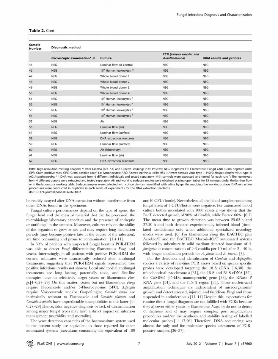
Table 2. Comparison of direct microscopic examination, culture and high-resolution melting analysis performances (PCR-HRM) oncorneal scrapings obtained from patients with keratitis.
SampleNumber Diagnostic method
microscopic examination* # CulturePCR (Herpes simplex andAcanthamoeba) HRM results and profiles
1 yeasts Candida glabrata NEG Candida glabrata
2 GNR Pseudomonas aeruginosa NEG NEG
3 budding yeasts Candida albicans NEG Candida albicans
4 GPC Staphylococcus aureus NEG NEG
5 yeasts Candida krusei NEG Candida krusei
6 Yeasts; pseudohypha Saccharomyces cervisiae NEG S. cervisiae
7 GPR Corynebacteria NEG NEG
8 filaments Aspergillus nidulans NEG Filamentous Fungi
9 LY; AEC NEG POS HSV1 NEG
10 filaments Penicillium piccum NEG Filamentous Fungi
11 LY; AEC NEG POS HSV2 NEG
12 NEG Candida tropicalis NEG Candida tropicalis
13 filaments Fusarium solani NEG Fusarium sp.
14 GPC Staphylococcus epidermidis NEG NEG
15 GNR Escherichia coli NEG NEG
16 GNR Acinetobacter baumannii NEG NEG
17 yeasts Trichosporon NEG POS; Trichosporon
18 yeasts Candida parapsilosis NEG Candida parapsilosis
19 GPC Streptococcus agalactiae NEG NEG
20 FF Aspergillus niger NEG Filamentous Fungi
21 GNR Pseudomonas aeruginosa NEG NEG
22 AEC NEG POS AC NEG
23 NEG Aspergillus niger NEG Filamentous Fungi
24 filaments Fusarium solani NEG Fusarium sp.
25 GPR Propionibacterium acnes NEG NEG
26 GPC Staphylococcus epidermidis NEG NEG
27 GNR Pseudomonas aeruginosa NEG NEG
28 GPC Streptococcus pneumoniae NEG NEG
29 filaments NEG NEG Filamentous Fungi
30 NEG NEG NEG NEG
31 filaments NEG NEG Fusarium sp.
32 budding yeasts NEG NEG Candida albicans
33 NEG NEG NEG NEG
34 NEG NEG NEG Filamentous Fungi
35 NEG NEG NEG NEG
36 filaments NEG NEG Filamentous Fungi
37 NEG NEG NEG Filamentous Fungi
38 NEG NEG NEG Candida albicans
Controls ##
39 NEG 106 Human epithelial cells NEG NEG
40 NEG Distilled water NEG NEG
41 NEG Saline NEG NEG
42 NEG DNA extraction reactants NEG NEG
43 NEG 106 Human fibroblasts NEG NEG
44 NEG Transport media NEG NEG
Fungal Infections Diagnosis and Characterization
PLoS ONE | www.plosone.org 6 July 2012 | Volume 7 | Issue 7 | e37660
be readily assayed after DNA extraction without interference from
other DNAs found in the specimens.
Fungal culture performances depend on the type of agent, the
fungal load and the mass of material that can be processed, the
microbiology laboratory capacities and the presence of antiseptic
or antifungal in the samples. Moreover, cultures rely on the ability
of the organism to grow ex vivo and may require long incubation
periods (may become positive late in the course of the infection),
are time consuming and prone to contamination. [1,4,11].
In 89% of patients with suspected fungal keratitis PCR-HRM
was able to detect Fungi differentiating filamentous Fungi and
yeasts. Interestingly, in all patients with positive PCR-HRM the
corneal infiltrates were dramatically reduced after antifungal
treatments, suggesting that PCR-HRM signals represented true
positive infections (results not shown). Local and topical antifungal
treatments are long lasting, potentially toxic, and first-line
therapies have to selectively target yeasts or filamentous Fun-
gi.[4–6,27–29] On this matter, yeasts but not filamentous Fungi
require Fluconazole and/or 5-Fluorocytosine (5FC). Aspergilli
require Voriconazole and/or Caspofungin; Candida krusei are
intrinsically resistant to Fluconazole and Candida glabrata and
Candida tropicalis have unpredictable susceptibilities to this latter.[4–
6,27–29] Hence, false negative diagnosis or lack of discrimination
among major fungal types may have a direct impact on infection
management (morbidity and mortality).
The yeast detection capacities of the haemoculture system used
in the present study are equivalent to those reported for other
automated systems (inoculums containing the equivalent of 100
and10 CFU/bottle). Nevertheless, all the blood samples containing
fungal loads of 1 CFU/bottle were negative. For automated blood
culture bottles inoculated with 1000 yeasts it was shown that the
BacT detected growth of 90% of Candida, while Bactec 66%. [6,7]
The mean time to growth detection was between 25.62 h and
27.30 h and both detected experimentally infected blood (simu-
lated candidemia) only when additional specialized mycology
media were used. [6] For filamentous Fungi the BACTEC plus
Aerobic/F and the BACTEC Mycosis-IC/F automated systems
followed by subculture in solid medium detected inoculums of A.
fumigatus at concentrations of .3 conidia per 10 ml after 21–40 h,
with longer incubation periods for A. flavus and A. terreus. [7].
For the detection and identification of Candida and Aspergillus
species a variety of real-time PCR assays based on species specific
probes were developed targeting the 18 S rDNA [16,30], the
mitochondrial cytochrome b [31], the 18 S and 28 S rDNA [32],
the CaMP65 (65-kDa mannoprotein) gene [33], the RNase P
RNA gene [34], and the ITS 2 region [35]. These nucleic-acid
amplification techniques are independent of microorganisms’
growth and detect stressed, injured, and fastidious Fungi and those
suspended in antimicrobials.[11–14] Despite this, expectations for
routine direct fungal diagnosis are not fulfilled with PCRs because
they a) cover either yeasts or filamentous Fungi, b) do not to detect
C. lusitaniae and c) may require complex post amplification
procedures and/or the synthesis and stability testing of labelled
molecular probes.[11–17,26] Therefore, DNA sequencing was
almost the only tool for molecular species assessment of PCR-
positive samples.[36–37].
Table 2. Cont.
SampleNumber Diagnostic method
microscopic examination* # CulturePCR (Herpes simplex andAcanthamoeba) HRM results and profiles
45 NEG Laminar-flow air control NEG NEG
46 NEG 106 Human leukocytes ** NEG NEG
47 NEG Whole blood donor 1 NEG NEG
48 NEG Whole blood donor 2 NEG NEG
49 NEG Whole blood donor 3 NEG NEG
50 NEG Whole blood donor 4 NEG NEG
51 NEG 106 Human leukocytes a NEG NEG
52 NEG 107 Human leukocytes a NEG NEG
53 NEG 108 Human leukocytes a NEG NEG
54 NEG 109 Human leukocytes a NEG NEG
55 NEG Air NEG NEG
56 NEG Laminar flow (air) NEG NEG
57 NEG Laminar flow (surface) NEG NEG
58 NEG DNA extraction reactants NEG NEG
59 NEG Laminar flow (surface) NEG NEG
60 NEG Air (laboratory) NEG NEG
61 NEG Laminar flow (air) NEG NEG
62 NEG DNA extraction reactants NEG NEG
HRM: high-resolution melting analysis; *: after Giemsa (pH: 7.4) and Grocott staining; POS: Positive; NEG: Negative; FF: Filamentous Fungi; GNR: Gram-negative rods;GPR: Gram-positive rods; GPC: Gram-positive cocci; LY: lymphocytes; AEC: Altered epithelial cells; HSV1: Herpes-simplex virus type 1; HSV2: Herpes-simplex virus type 2;AC: Acanthamoeba; **: DNA was extracted from 6 different individuals and tested separately; ##: controls were extracted and tested for each run; a: The leukocytesfrom 4 different donors were extracted and tested separately. Air and working surface samples were obtained placing open tubes for 15 minutes under the laminar flowor in the laboratory working table. Surface samples were collected with cotton devices humidified with saline by gentle swabbing the working surface. DNA extractionprocedures were conducted in duplicate in each series of experiments for the DNA extraction reactants.doi:10.1371/journal.pone.0037660.t002
Fungal Infections Diagnosis and Characterization
PLoS ONE | www.plosone.org 7 July 2012 | Volume 7 | Issue 7 | e37660

Table 3. Comparison of culture and high-resolution melting analysis (PCR-HRM) performances on experimentally infected blood.
Fungal load[a]CFU/ml Fluid Diagnosis method
Fungal Culture # HRM #
Result Hours for positivity Result Profile ##
Candida albicans 10 S POS POS 16 16 POS POS C. albicans
B POS POS 24 24 POS POS C. albicans
1 S POS POS 48 24 POS POS C. albicans
B POS POS 48 48 POS POS C. albicans
0.1 S POS NEG 96 – POS POS C. albicans
B NEG NEG – – POS POS C. albicans
Candida krusei 10 S POS POS 24 24 POS POS C. krusei
B POS POS 24 24 POS POS C. krusei
1 S POS POS 48 48 POS POS C. krusei
B POS POS 72 48 POS POS C. krusei
0.1 S POS NEG 96 – POS POS C. krusei
B NEG NEG – – POS NEG C.krusei
Candida glabrata 10 S POS POS 24 24 POS POS C. glabrata
B POS POS 24 24 POS POS C. glabrata
1 S POS POS 48 36 POS POS C. glabrata
B POS POS 48 72 POS POS C. glabrata
0.1 S NEG NEG – – POS POS C. glabrata
B NEG NEG – – POS POS C. glabrata
Candida tropicalis 10 S POS POS 36 36 POS POS C. tropicalis
B POS POS 48 48 POS POS C. tropicalis
1 S POS POS 48 48 POS POS C. tropicalis
B POS POS 48 72 POS POS C. tropicalis
0.1 S NEG POS – 96 POS POS C. tropicalis
B NEG NEG – – POS POS C. tropicalis
Candida parapsilopsis 10 S POS POS 24 24 POS POS C. parapsilopsis
B POS POS 24 24 POS POS C. parapsilopsis
1 S POS NEG 36 48 POS POS C. parapsilopsis
B NEG POS 48 36 POS POS C. parapsilopsis
0.1 S NEG NEG – – POS POS C. parapsilopsis
B NEG NEG – – POS POS C. parapsilopsis
Saccharomyces cervisiae 10 S POS POS 24 24 POS POS S. cervisiae
B POS POS 24 24 POS POS S. cervisiae
1 S POS POS 24 24 POS POS S. cervisiae
B POS POS 36 36 POS POS S. cervisiae
0.1 S NEG NEG – – POS POS S. cervisiae
B NEG NEG – – POS POS S. cervisiae
Trichosporon 10 S POS POS 24 36 POS POS Trichosporon
B POS POS 36 36 POS POS Trichosporon
1 S POS POS 48 36 POS POS Trichosporon
B NEG POS 48 48 POS POS Trichosporon
0.1 S POS NEG 96 – POS POS Trichosporon
B NEG NEG – – POS POS Trichosporon
Aspergillus niger 10 S POS POS 36 48 POS POS Filamentous Fungi
B POS POS 48 48 POS POS Filamentous Fungi
1 S POS POS 48 72 POS POS Filamentous Fungi
B POS NEG 72 96 POS POS Filamentous Fungi
Fungal Infections Diagnosis and Characterization
PLoS ONE | www.plosone.org 8 July 2012 | Volume 7 | Issue 7 | e37660

For diagnosis of candidemia in subjects with haematological
malignancies or various forms of immunodeficiency a real-time PCR
targeting the 18 S rRNA gene and requiring a series of labelled
molecular probes, yielded positive results in 58.3% of blood culture-
positive samples and detected in blood the genomes of Candida 3 days
earlier than culture. [37] In this series 27% of whole-blood were PCR
positive compared to 15% of haemoculture (92% of correlation of
positives). Other studies indicate that numerous pairs of primers and
labelled probes were required for each sample to identify 72% of
species of positive cultures. [38] For vaginal specimens it was reported
a test based on PCR-HRM technology, but it was unable to
differentiate yeasts from filamentous Fungi. [22] However, the strategy
developed here is different because PCR-HRM is able to detect and
differentiate yeasts and filamentous Fungi in one run with detection
limits of 0.1 CFU/ml or less. In addition to differentiating filamentous
Fungi from yeasts, PCR-HRM discriminated among relevant clinical
species of yeasts in less than 2.30 hours after DNA extraction. This
only required upgrading the available real-time PCR software of the
thermocyclers used in the routine microbiology laboratory.
Because PCR-HRM was able to produce consistent results
without the need for synthesizing and labelling molecular probes
and without post amplification procedures (restriction enzymes,
electrophoresis, gel analysis, hybridisation, sequencing reactants)
the cost for reactants for testing one DNA extract could be
reduced to less than 2 USD (2 primers and HRM mix). Compared
to classic PCR (amplification followed by electrophoreses and/or
hybridization and/or sequencing) this new PCR-HRM has the
additional advantage of minimizing risks for false positive results
due to cross contamination, because the targeted sequence
amplification, the signal detection, and the DNA melting analyses
are carried out in closed tubes. Moreover, PCR-HRM minimizes
risks for false negative results because the yields of extraction of the
DNA and the potential interference of PCR inhibitors are
systematically monitored in each run and for all the samples.
According to the results obtained in this study, if the future runs
generate reproducible melt-curves over time in different settings, a
reference database could be built to store PCR-HRM calculations
and shapes of the melting profiles for each family or species to be
challenged against profiles. Larger prospective multicentric trials
testing different types of samples (clinical and environmental) are
necessary to validate PCR-HRM usefulness as a diagnosis tool and
for environmental studies.
Acknowledgments
We thank the Centre de Ressources Biologiques du Centre Hospitalier
National d’Ophtalmologie des Quinze-Vingts, 75012 Paris, France, for
providing biological samples.
Author Contributions
Conceived and designed the experiments: PG SD. Performed the
experiments: PG SD PCS DB OS. Analyzed the data: PG SD DB OS
CC. Contributed reagents/materials/analysis tools: PG PC VB LL CC.
Wrote the paper: PG.
Table 3. Cont.
Fungal load[a]CFU/ml Fluid Diagnosis method
Fungal Culture # HRM #
Result Hours for positivity Result Profile ##
0.1 S NEG NEG – – POS POS Filamentous Fungi
B NEG NEG – – POS POS Filamentous Fungi
Aspergillus nidulans 10 S POS POS 48 48 POS POS Filamentous Fungi
B POS POS 48 72 POS POS Filamentous Fungi
1 S POS POS 48 48 POS POS Filamentous Fungi
B POS POS 72 72 POS POS Filamentous Fungi
0.1 S POS NEG – – POS POS Filamentous Fungi
B NEG NEG – – POS POS Filamentous Fungi
Penicillium piccum 10 S POS POS 24 36 POS POS Filamentous Fungi
B POS POS 36 36 POS POS Filamentous Fungi
1 S POS POS 72 72 POS POS Filamentous Fungi
B POS POS 72 96 POS POS Filamentous Fungi
0.1 S POS NEG – – POS POS Filamentous Fungi
B NEG NEG – – POS POS Filamentous Fungi
Fusarium solani 10 S POS POS 36 48 POS POS * Filamentous Fungi
B POS POS 48 48 POS POS * Filamentous Fungi
1 S POS POS 48 72 POS POS * Filamentous Fungi
B POS POS 72 48 POS POS * Filamentous Fungi
0.1 S NEG NEG – – POS POS * Filamentous Fungi
B NEG NEG – – POS POS * Filamentous Fungi
[a]Haemoculture bottles were inoculated with 10 ml; S: saline; B: blood; #: results from 2 independent experiments; POS: Positive; NEG: Negative after 240 hours; ##:reference strains and negative controls were extracted and tested for each run;*: melting-curve profiles consistently superimposed on those obtained with Fusariumsolani.doi:10.1371/journal.pone.0037660.t003
Fungal Infections Diagnosis and Characterization
PLoS ONE | www.plosone.org 9 July 2012 | Volume 7 | Issue 7 | e37660

References
1. Edmond MB, Wallace S, McClish D, Pfaller MA, Jones RN, et al. (1999)
Nosocomial bloodstream infections in United States hospitals: a three-yearanalysis. Clin Infect Dis 29: 239–44.
2. Ascioglu S, Rex JH, de Pauw B, Bennett JE, Bille J, et al. (2002) Invasive FungalInfections Cooperative Group of the European Organization for Research and
Treatment of Cancer, Mycoses Study Group of the National Institute of Allergy
and Infectious Diseases. Defining opportunistic invasive fungal infections inimmunocompromised patients with cancer and hematopoietic stem cell
transplants: an international consensus. Clin Infect Dis 34: 7–14.3. Szczotka-Flynn LB, Pearlman E, Ghannoum M (2010) Microbial contamination
of contact lenses, lens care solutions and their accessories: a literature review.
Eye Contact Lens 36: 116–29.4. Marr KA, Carter R, Crippa F, Wald A, Corey L (2002) Epidemiology and
outcome of mould infections in hematopoietic stem cell transplant recipients.Clin Infect Dis 34: 909–17.
5. Pagano L, Caira M, Candoni A, Offidani M, Fianchi L, et al. (2006) Theepidemiology of fungal infections in patients with hematologic malignancies: the
SEIFEM-2004 study. Haematologica 91: 1068–75.
6. Horvath L, George B, Murray C, Harrison L, Hospenthal D (2004) Directcomparison of the BACTEC 9240 and BacT/ALERT 3D automated blood
culture systems for Candida growth detection. J Clin Microbiol 42: 115–8.7. Rosa C, Araujo R, Rodrigues A, Pinto-de-Sousa M, Pina-Vaz C (2011)
Detection of Aspergillus species in BACTEC blood cultures. J Med Microbiol
60: 1467–71.8. Hall BJ, Jones L (2010) Contact lens cases: the missing link in contact lens safety?
Eye Contact Lens 36: 101–5.9. Allan EK, Jordanides NE, McLintock LA, Copland M, Devaney M, et al. (2005)
Poor performance of galactomannan and mannan sandwich enzyme-linkedimmunosorbent assays in the diagnosis of invasive fungal infection. Br J
Haematol. 128: 578–9.
10. Maertens J, Verhaegen J, Lagrou K, Van Eldere J, Boogaerts M (2001)Screening for circulating galactomannan as a noninvasive diagnostic tool for
invasive aspergillosis in prolonged neutropenic patients and stem celltransplantation recipients: a prospective validation. Blood 97: 1604–10.
11. Jordanides NE, Allan EK, McLintock LA, Copland M, Devaney M, et al. (2005)
A prospective study of real-time panfungal PCR for the early diagnosis ofinvasive fungal infection in haemato-oncology patients. Bone Marrow
Transplant 35: 389–95.12. Baskova L, Landlinger C, Preuner S, Lion T (2007) The Pan-AC assay: a single-
reaction real-time PCR test for quantitative detection of a broad range ofAspergillus and Candida species. J Med Microbiol 56: 1167–73.
13. Ririe K, Rasmussen R, Wittwer C (1997) Product differentiation by analysis of
DNA melting curves during the polymerase chain reaction. Anal Biochem 245:154–60.
14. Loeffler J, Hebart H, Magga S, Schmidt D, Klingspor L, et al. (2000)Identification of rare Candida species and other yeasts by polymerase chain
reaction and slot blot hybridization. Diagn Microbiol Infect Dis 38: 207–12.
15. Rakeman J, Bui U, Lafe K, Chen C, Honeycutt T, et al. (2005) Multilocus DNAsequence comparisons rapidly identify pathogenic molds. J Clin Microbiol 43:
3324–33.16. Klingspor L, Jalal S (2006) Molecular detection and identification of Candida
and Aspergillus spp. from clinical samples using real-time PCR. Clin MicrobiolInfect 12: 745–53.
17. Schabereiter-Gurtner C, Selitsch B, Rotter M, Hirschl A, Willinger B (2007)
Development of novel real-time PCR assays for detection and differentiation ofeleven medically important Aspergillus and Candida species in clinical
specimens. J Clin Microbiol 45: 906–14.18. Ostrosky-Zeichner L, Rex J, Pappas P, Hamill R, Larsen R (2003) Antifungal
susceptibility survey of 2,000 bloodstream Candida isolates in the United States.
Antimicrob Agents Chemother 47: 3149–54.19. Srinivasan M (2004) Fungal keratitis. Curr Opin Ophthalmol 15: 321–7.
20. White PL, Archer A, Barnes R (2005) Comparison of non-culture-based
methods for detection of systemic fungal infections, with an emphasis on invasiveCandida infections. J Clin Microbiol 43: 2181–7.
21. Monis P, Giglio S, Saint C (2005) Comparison of SYT09 and SYBR Green I forreal-time polymerase chain reaction and investigation of the effect of dye
concentration on amplification and DNA melting curve analysis. Anal Biochem
340: 24–34.22. Reed G, Kent JO, Wittwer C (2007) High-resolution DNA melting analysis for
simple and efficient molecular diagnostics. Pharmacogenomics 8: 597–608.23. Mandviwala T, Shinde R, Kalra A, Sobel JD, Akins R (2010) High-throughput
identification and quantification of Candida species using high resolution
derivative melt analysis of panfungal amplicons. J Mol Diagn 12: 91–101.24. Goldschmidt P, Rostane H, Saint-Jean C, Batellier L, Alouch C, et al. (2006)
Effects of topical anaesthetics and fluorescein on the real-time PCR used for thediagnosis of Herpesviruses and Acanthamoeba keratitis. Br J Ophthalmol 90:
1354–6.25. van Doornum G, Guldemeester J, Osterhaus A, Niesters H (2003) Diagnosing
herpesvirus infections by real-time amplification and rapid culture. J Clin
Microbiol 41: 576–80.26. Selvarangan R, Bui U, Limaye, Cookson B (2003) Rapid identification of
commonly encountered Candida species directly from blood culture bottles.J Clin Microbiol 41: 5660–4.
27. Vanden Bossche H, Dromer F, Improvisi I, Lozano-Chiu M, Rex H, et al.
(1998) Antifungal drug resistance in pathogenic fungi. Med Mycol 36(suppl):119–28.
28. Sullivan D, Haynes K, Bille J, Boerlin P, Rodero L, et al. (1997) Widespreadgeographic distribution of oral Candida dubliniensis strains in human
immunodeficiency virus-infected individuals. J Clin Microbiol 35: 960–4.29. Fidel Jr P, Vazquez J, Sobel J (1999) Candida glabrata: review of epidemiology,
pathogenesis, and clinical disease with comparison to C. albicans. Clin Microbiol
Rev 12: 80–96.30. Gomez-Lopez A, Martin-Gomez M, Martin-Davila P, Lopez-Onrubia P,
Gavalda J, et al. (2006) Detection of fungal DNA by real-time polymerase chainreaction: evaluation of 2 methodologies in experimental pulmonary aspergillosis.
Diagn. Microbiol. Infect. Dis 56: 387–393.
31. Spiess B, Buchheidt D, Baust C, Skladny H, Seifarth W, et al. (2003)Development of a LightCycler PCR assay for detection and quantification of
Aspergillus fumigatus DNA in clinical samples from neutropenic patients. J ClinMicrobiol 41: 1811–1818.
32. Hsu M, Chen K, Lo H, Chen Y, Liao M, et al. (2003) Species identification ofmedically important fungi by use of realtime LightCycler PCR. J. Med.
Microbiol 52: 1071–1076.
33. Arancia S, Carattoli A, La Valle R, Cassone A, De Bernardis F (2006) Use of65 kDa mannoprotein gene primers in real time PCR identification of Candida
albicans in biological samples. Mol. Cell Probes 20: 263–268.34. Innings A, Ullberg M, Johansson A, Rubin C, Noreus N, et al. (2007) Multiplex
real-time PCR targeting the RNase P RNA gene for detection and identification
of Candida species in blood. J. Clin. Microbiol 45: 874–880.35. Schabereiter-Gurtner C, Selitsch B, Rotter M, Hirschl A, Willinger B (2007)
Development of novel real-time PCR assays for detection and differentiation of11 medically important Aspergillus and Candida species in clinical specimens. J
Clin Microbiol 45: 906–914.36. Vollmer T, Stormer M, Kleesiek K, Dreier J (2008) Evaluation of novel broad-
range real-time PCR assay for rapid detection of human pathogenic fungi in
various clinical specimens. J Clin Microbiol 46: 1919–26.37. Wellinghausen N, Siegel D, Winter J, Gebert S (2009) Rapid diagnosis of
candidaemia by real-time PCR detection of Candida DNA in blood samples.J Med Microbiol 58: 1106–11.
38. Obara H, Aikawa N, Hasegawa N, Hori S, Ikeda Y, et al. (2011) The role of a
real-time PCR technology for rapid detection and identification of bacterial andfungal pathogens in whole-blood samples. J Infect Chemother 3: 327–33.
Fungal Infections Diagnosis and Characterization
PLoS ONE | www.plosone.org 10 July 2012 | Volume 7 | Issue 7 | e37660