Embed Size (px)
Citation preview

Retrieving accurate and distorted memories: Neuroimaging evidencefor effects of emotion
Elizabeth A. Kensinger* and Daniel L. Schacter
Department of Psychology, Harvard University, and the Athinoula A. Martinos Center for Biomedical Imaging, USA
Received 3 December 2004; revised 16 March 2005; accepted 28 March 2005
Available online 24 May 2005
While limbic activity is known to be associated with successful
encoding of emotional information, it is less clear whether it is related
to successful retrieval. The present fMRI study assessed the effects of
emotion on the neural processes engaged during retrieval of accurate
compared to distorted memories. Prior to the scan, participants (16
young adults) viewed names of neutral (e.g., frog) and emotional (e.g.,
snake) objects and formed a mental image of the object named. They
were shown photos of half of the objects. During the fMRI scan,
participants saw object names and indicated whether or not they had
seen the corresponding photo. Memory distortions (misattributions)
occurred when participants incorrectly indicated whether or not a
photo had been studied. Activity in some regions (e.g., L anterior
hippocampus) was related to accurate retrieval (correct attribution-
s>misattributions) for emotional and neutral items. However, activity
in other regions corresponded with accurate retrieval specifically for
emotional items (e.g., in R amygdala/periamygdaloid cortex and L
orbitofrontal cortex) or for neutral items (e.g., in lateral inferior
prefrontal cortex and R posterior hippocampus). Results indicate that
emotional salience modulates the processes engaged during accurate
retrieval and that activity in limbic regions corresponds with accurate
memory assignment for emotional items. To our knowledge, this study
is the first to demonstrate a link between limbic engagement at
retrieval and accurate memory attribution.
D 2005 Elsevier Inc. All rights reserved.
Keywords: Amygdala; Emotion; fMRI; Memory; Reality monitoring;
Retrieval
Memory retrieval often involves the reconstruction of a priorexperience rather than its verbatim replaying. Abundant evidencehas shown that this reconstruction process can be flawed, leading
to memory errors, illusions, and distortions (reviewed by Roedigerand McDermott, 2000; Schacter, 1999; Schacter et al., 1998). Arelatively common form of memory distortion arises whenindividuals must distinguish whether an item was previously
imagined or perceived (often referred to as reality monitoring;Johnson and Raye, 1981). Individuals sometimes incorrectlyattribute the source of an item’s familiarity; for example, a person
may believe an item was externally presented when it actually wasimagined.
Memory misattributions can occur less often for items that
contain emotional relevance than for those void of emotionalcontent (Kensinger and Corkin, 2004a; Kensinger and Schacter, inpress(b); Pesta et al., 2001), plausibly because emotional informa-
tion is more likely to be remembered with contextual details thannonemotional information (D’Argembeau and Van der Linden,2004; Doerksen and Shimamura, 2001; Kensinger and Corkin,2003). Neuroimaging studies have suggested that encoding
processes play a critical role in this memory enhancement foremotional memory. In particular, amygdalar engagement appears tobe critical for increasing the likelihood that verbal (e.g., Kensinger
and Corkin, 2004b) and nonverbal (e.g., Cahill et al., 1996; Canliet al., 2000) emotional information is remembered. Further, limbicactivity (in the amygdala and orbitofrontal cortex) during encoding
is related to subsequent correct memory attributions for emotionalitems (Kensinger and Schacter, in press(a)). These effects ofemotion occur even when individuals study information without
the intention to remember it (i.e., under incidental encodingconditions; Kensinger and Schacter, in press(a,b)), suggesting thatthe neural processes recruited to process emotional informationincrease the probability of successful and detailed encoding. This
increased encoding efficacy likely emerges in part throughinteractions between the amygdala and the hippocampal formationduring encoding and consolidation (reviewed by Phelps, 2004).
Consistent with this hypothesis, neuroimaging studies havedemonstrated correlations between amygdalar and hippocampalactivity (e.g., Dolcos et al., 2004; Kensinger and Corkin, 2004b;
Kensinger and Schacter, in press(a); Richardson et al., 2004) andincreases in functional connectivity of the two regions (Kilpatrickand Cahill, 2003) during encoding of emotional information.
In contrast to the rich neuroimaging literature examining theprocesses relating to encoding of emotional information, relativelyfew studies have examined retrieval processes. Two studies havesuggested that retrieval processes are modulated by emotional
content, with activity in visual cortex (Taylor et al., 1998) and
1053-8119/$ - see front matter D 2005 Elsevier Inc. All rights reserved.
doi:10.1016/j.neuroimage.2005.03.038
* Corresponding author. 33 Kirkland Street, Room 884, Cambridge, MA
02138, USA. Fax: +1 617 496 3122.
E-mail address: [email protected] (E.A. Kensinger).
Available online on ScienceDirect (www.sciencedirect.com).
www.elsevier.com/locate/ynimg
NeuroImage 27 (2005) 167 – 177

limbic regions (amygdala and anterior temporal lobe; Dolan et al.,2000) showing greater activity during retrieval of emotionalinformation than during retrieval of neutral information. Because
these studies utilized blocked designs, however, they cannotdistinguish state effects from item effects (e.g., Otten et al.,2002), nor can they pinpoint the neural activity that is related
specifically to successful retrieval.A few other event-related potential (Maratos and Rugg, 2001;
Smith et al., 2004a) and fMRI studies (Maratos et al., 2001; Smith
et al., 2004b) have examined how retrieval of neutral informationis affected by the emotional context in which it was presented.These studies again have suggested that limbic regions (e.g.,
amygdala and orbitofrontal cortex) play a role during retrieval ofinformation studied in emotional compared to neutral contexts. Thecritical comparison in these studies was between items correctlyrecognized from an emotional context and from a neutral context
(i.e., a comparison of activity for ‘‘hits’’) and thus these data do notspeak to whether limbic activity is related to accurate retrievalmore so for emotional than for neutral items.
To address this issue (i.e., whether activity in limbic regionsshows an interaction between memory accuracy and an item’semotional content), a more informative comparison is between
successful and unsuccessful retrieval of emotional and neutralitems. This comparison is especially appropriate for two reasons.First, this contrast mirrors that used during subsequent-memoryparadigms which, as outlined above, have been the primary focus
of neuroimaging studies of emotional memory. Second, thiscomparison also holds constant the emotional content of the itemsbeing retrieved, reducing the concern that differences in limbic
engagement during retrieval arise from emotional processing of theretrieval cue.
In neuroimaging investigations, such comparisons often are
made between items confidently or vividly remembered and thoseforgotten (e.g., Eldridge et al., 2000; Henson et al., 2000; Otten etal., 2001; Wheeler and Buckner, 2004). While these contrasts are
useful for neutral items, they introduce a potential difficulty whencomparing memory for emotional and neutral items: emotion canenhance subjective ratings of a memory’s vividness withoutincreasing the objective accuracy of the memory (e.g., Neisser
and Harsch, 1992; Talarico and Rubin, 2003; Winograd andNeisser, 1992). While emotion does not always have thisparadoxical effect on memory (e.g., Doerksen and Shimamura,
2001; Kensinger and Corkin, 2003), this possible dissociationbetween subjective confidence and objective accuracy of anemotional memory makes it important to assess the accuracy of
a memory directly, rather than relying only on subjective self-reports of a memory’s vividness or an individual’s confidence intheir memory. When only subjective report is obtained, amygdala
activity during vivid retrieval could be interpreted either asmodulating retrieval of rich, detailed memories or as enhancingthe subjective feeling that a memory is associated with contextualdetail without affecting the amount of detail retrieved (discussed by
Sharot et al., 2004).To assess memory accuracy for emotional and neutral items, the
present study employed a reality-monitoring paradigm previously
used with neutral items (Gonsalves and Paller, 2000; Okado andStark, 2003). Outside of the scanner, participants performed a taskthat required them to form mental images of objects. In the present
study, half of the stimuli contained negative emotional content andhalf were of neutral content. Participants were shown thecorresponding photo for half of those objects. The retrieval task,
performed during the fMRI scan, required participants to indicatewhether objects had been externally presented. Thus, activity couldbe compared for correct memory attributions and memory
misattributions. Two central questions were addressed: (1) whatneural processes support accurate retrieval of both emotional andneutral information? and (2) How does the emotional content of the
items influence the neural processes that are associated withaccurate retrieval?
With regard to the first question, we were particularly interested
in the relation between hippocampal activity and memoryaccuracy. Based on prior research implicating the hippocampalformation in retrieval of contextual details associated with a study
episode (e.g., Dobbins et al., 2003; Wheeler and Buckner, 2003),we hypothesized that hippocampal activity would correspond withcorrect memory attributions at retrieval. However, the few studiesthat examined retrieval of illusory memories found either no
hippocampal activity (Okado and Stark, 2003) or that hippocampalactivity did not discriminate true from false memories (Cabeza etal., 2001; Slotnick and Schacter, 2004). The tasks used by Cabeza
et al. (2001) and Slotnick and Schacter (2004) were quite differentfrom the present study: False recognition responses in those studiesindicated acceptance of an item that was semantically (Cabeza et
al., 2001) or visually (Slotnick and Schacter, 2004) associated withstudied items. Thus, it is not clear whether their findings generalizeacross recognition tasks or whether they are specific to tasks onwhich retrieval of gist information can support false recognition.
While the methods of Okado and Stark (2003) were similar tothose used here, their null finding (i.e., of no hippocampal activity)must be interpreted cautiously and does not provide compelling
evidence regarding whether the hippocampus contributes toaccurate memory attribution. In summary, the data have beenmixed with regard to the role of the hippocampus in accurate
retrieval, making it difficult to confidently predict the relationbetween hippocampal activity and accurate retrieval.
In regard to the second question, we were most interested in
whether activity in regions known to be important for theprocessing of emotional information (e.g., amygdala and orbito-frontal cortex) would show a relation to accurate retrieval foremotional items. As outlined above, these regions have been
implicated in subsequent-memory paradigms (reviewed byHamann, 2001; Phelps, 2004) and in retrieval of neutral itemspresented in an emotional context (Smith et al., 2004b). However,
it is not clear whether activity in these regions is relatedspecifically to accurate retrieval. This issue is of central importancein understanding the memory phases during which amygdalar
response modulates memory accuracy. While it is clear that theamygdala plays a role during early memory phases (encoding andconsolidation), its role during retrieval is debated (e.g., Nader,
2003; LeDoux, 2000).
Method
Participants
Participants comprised 17 native English speaking Harvardundergraduate or graduate students. The data from one participantwere excluded due to scanner malfunction. The remaining 16
young adults (8 women, 8 men) were ages 18–30. All were right-handed, native English speakers screened to exclude those withcontra-indicators for MRI scanning, or with a history of
E.A. Kensinger, D.L. Schacter / NeuroImage 27 (2005) 167–177168

depression. No participant was taking centrally-active medications.Informed consent was obtained from all participants in a mannerapproved by the Institutional Review Boards of Harvard University
and the Massachusetts General Hospital.
Materials and procedure
Materials comprised 450 concrete words and 450 photo objectsdepicting a single object on a white background (e.g., a baseball, a
tarantula; taken from Hemera Technologies Inc, 2002, Canada).Words and photo objects were selected as pairs, such that eachword named a photo object (e.g., ‘‘tarantula’’ and a picture of a
tarantula).Half of the words and objects were high in arousal (scores
>2.5 on a scale of !5 to +5, with negative values indicating thatan item was calming or soothing, and positive values indicating
that an item caused excitement or agitation). The other half of thewords and objects were neutral, having received arousal ratingslower than + 1. These items had been judged by a separate group
of 20 young adults (10 males) prior to the present study(Kensinger and Schacter, in press(a,b)). They also were ratedby the participants in this study, and the ratings agreed with those
from the separate group of participants. Neutral and emotionalobjects did not differ in the numbers that included animals,people, or objects, nor did the neutral and emotional words differin word length, word frequency, or word familiarity (Coltheart,
1981).One or two days before the scanning session, each participant
viewed a study list with 150 emotional words and 150 neutral
words presented for 2 s each (randomly intermixed). Participantswere instructed to make a button press to indicate whether eachword named an object that was bigger or smaller than a shoebox.
They were told that the study was examining mental imageryperformance and thus that they should use mental imagery toperform the size-judgment task. Half of the words were followed
by the corresponding photo object, presented for 2 s, and theremaining words were followed by a blank square shown for 2 s(design adapted from Gonsalves and Paller, 2000). Participantswere instructed to simply view the square or the photo object that
occurred after the word and were told that no response wasrequired to these items.
After a delay of 1–2 days, participants returned for the fMRI
scan. During the functional scans, participants performed a surpriserecognition task (Debriefing indicated that no participants realizedthat their memory would be tested for the items studied in the
laboratory). The recognition task was divided across three func-tional scans. In each scan, participants viewed 150 words. 50 of thewords corresponded to items that had been studied in the word-
only condition; 50 to items studied in the word–picture condition;and 50 to items that had not been studied. Emotional and neutralwords from the three conditions (items from word-only trials, itemsfrom word–picture trials, and novel items) were pseudorandomly
intermixed with one another and with fixation crosses (+) toprovide jitter (Dale, 1999). Stimuli were back-projected onto ascreen in the scanner bore, and participants viewed the words
through an angled mirror attached to the head coil. For each word,participants indicated whether or not the corresponding photoobject had been presented at study. Thus, a ‘‘no’’ response was
required both for novel words that had not been studied and forwords that had been presented without their corresponding photoobject at study.
Image acquisition and data analysis
Images were acquired on a 1.5-T Siemens Sonata MRI scanner.
Detailed anatomic data were acquired using a multiplanar rapidlyacquired gradient echo (MP-RAGE) sequence. Functional imageswere acquired using a T2*-weighted echo planar imaging (EPI)
sequence (TR = 3000 ms, TE = 40 ms, FOV = 200 mm; flip angle =90-). Twenty-one axial-oblique slices (5 mm thickness, 1 mm skipbetween slices), aligned along the anterior commissure/posterior
commissure line, were acquired in an interleaved fashion.All pre-processing and data analysis were conducted within
SPM99 (Wellcome Department of Cognitive Neurology). Standard
pre-processing was performed on the functional data, includingslice-timing correction, rigid body motion correction, normal-ization to the Montreal Neurological Institute template (re-sampling at 3 mm cubic voxels), and spatial smoothing (using an
8-mm full-width half maximum isotropic Gaussian kernel).For each participant, and on a voxel-by-voxel basis, an event-
related analysis was first conducted in which all instances of a
particular event type were modeled through convolution with acanonical hemodynamic response function. All participants had atleast 10 instances of every event type. Effects for each event type
were estimated using a subject-specific, fixed-effects model. Thesedata were then entered into a second-order, random-effectsanalysis. Analyses contrasted activation as a function of memoryperformance (comparing correct memory attributions and memory
misattributions) separately for each emotion type (emotional orneutral) and item history (from a word-only trial or a word–picturetrial). Analyses were conducted at the level of P < 0.001.
Conjunction analyses (using the masking function in SPM99) thenexamined what regions showed activation that varied as a functionof memory accuracy (a) regardless of emotion type or item history,
and (b) for a particular emotion type, regardless of item history.The threshold for each contrast entered into a conjunction analysiswas set at P < 0.05.
All activations are presented in neurological coordinates (i.e.activity on the right hemisphere is presented on the right side of thebrain images). Voxel coordinates are reported in Talairach coor-dinates (Talairach and Tournoux, 1998). We report coordinates of
the most significant voxel within the cluster of activation (peakvoxel) and the voxel located at the geographical center of the cluster(center voxel). Event-related time-courses were extracted from
active clusters by creating regions-of-interest (ROI) as 8mm spheresusing the ROI toolbox implemented in SPM99. Analysis ofvariance (ANOVA) was performed on these extracted time-
courses to examine whether the ROIs showed an interactionbetween memory accuracy and emotion type (i.e., activityrelating to memory accuracy for emotional but not neutral items,
or vice-versa).
Results
Behavioral data
ANOVA with response type (picture, no picture), item history(word-only, word–picture, new), and emotion type (emotional,neutral) as within-subject factors revealed a main effect of response
type (F(1,15) = 21.1, P < 0.001, partial eta-squared = 0.59) as wellas interactions between response type and item history (F(1,14) =393., P < 0.001, partial eta-squared = 0.85) and among response
E.A. Kensinger, D.L. Schacter / NeuroImage 27 (2005) 167–177 169

type, item history, and emotion type (F(1,14) = 13.0, P < 0.001,partial eta-squared = 0.65). This three-way interaction resultedbecause while the proportion of correct (‘‘no picture’’) responses did
not differ for new emotional and neutral items (0.85 and 0.84,respectively), participants were significantly more likely to makecorrect memory attributions for emotional items than for neutral
items from word–picture trials (0.67 versus 0.57) and from word-only trials (0.69 versus 0.65 for emotional and neutral, respec-tively). Thus, memory was more accurate (i.e., included more cor-rect attributions) for the emotional items than for the neutral items.
Neuroimaging data
Random-effects analyses contrasted activation as a function ofmemory performance (comparing correct memory attributions and
memory misattributions) separately for each emotion type (emo-tional or neutral) and item history type (from a word-only or word–picture trial; Tables 1–4). Because the goal of this study was to
examine how the emotional content of the stimuli (regardless oftheir item histories) affected the neural processes that wereassociated with accurate memory assignment, conjunction analyseswere performed to examine the regions in which activity was related
to accurate retrieval both for word-only and word–picture items.The results of these conjunctions are discussed below.
Accurate retrieval for both emotional and neutral items
A conjunction analysis was conducted to examine the regions
in which activity was related to memory accuracy for all items (i.e.,a conjunction of the four contrasts corresponding to correctattributions > misattributions for neutral word-only items, emo-tional word-only items, neutral word–picture items, and emotional
word–picture items1). The regions that showed this pattern ofactivation are in accord with those implicated in prior studies ofepisodic retrieval (Table 5). We had been particularly interested in
whether the hippocampus would show this pattern of response and,indeed, activity in a region centered in the left anterior hippo-campus2 was related to accurate retrieval for emotional and neutral
items (Fig. 1).
Accurate retrieval for emotional but not neutral items
We were additionally interested in activity that was associated
with accurate retrieval for the emotional items. To isolate theseregions, we conducted a conjunction analysis of emotional word-only correct attributions > misattributions and emotional word–
picture correct attributions > misattributions. Table 6 presents theregions that resulted in that conjunction, but not in theconjunction described above for both emotional and neutralitems. Our a priori regions of interest had been the amygdala and
orbitofrontal cortex, as activity in these regions has been shownto correspond with successful encoding of emotional items(Hamann, 2001; Phelps, 2004) and with retrieval-related pro-
cesses for emotional information (Dolan et al., 2000; Sharot etal., 2004; Smith et al., 2004b). Activity in these regions wasrelated to accurate retrieval specifically for the emotional items
(Fig. 2), with ANOVA indicating a significant interactionbetween response type (correct attribution, misattribution) andemotion (emotional, neutral).
Accurate retrieval for neutral but not emotional items
To examine the regions that were associated with accurate
retrieval for the neutral items, but not the emotional items, weperformed a conjunction of neutral word-only correct attributions >misattributions and neutral word–picture correct attributions >
misattributions. Table 7 presents the regions resulting from thatanalysis that were not included in the conjunction for bothemotional and neutral items. Of most interest, activity in the
inferior prefrontal cortex bilaterally and in the right posteriorhippocampus was related to memory accuracy specifically forthe neutral items and not for the emotional items [with ANOVAindicating a significant interaction between response type
(correct attribution, misattribution) and emotion (emotional,neutral); Fig. 3].
Discussion
The central aim of this study was to examine the processes thatwere related to accurate memory assignment for emotional andneutral items. The results suggest three principal conclusions. First,
activity in regions implicated in prior studies of episodic retrieval(including a region of activity centered in the anterior hippo-campus) corresponded with accurate retrieval regardless of theitem’s emotional content. Second, despite these commonalities,
emotional content modulates the neural processes recruited duringsuccessful retrieval. Third, activity in the amygdala/periamygda-
Table 1
Regions in which activity was related to accurate retrieval of emotional word–picture items ( P < 0.001)
Region Hemisphere Talairach coordinates
of peak voxel (x, y, z)
Talairach coordinates
of center voxel (x, y, z)
Approximate
Brodmann area
Middle frontal gyrus L !44, 48, !13 !41, 46, !11 BA 10/11/47
Precentral gyrus L !35, !26, 59 !35, !25, 58 BA 4
Postcentral gyrus L !53, !20, 18 !51, !22, 17 BA 2
Inferior parietal lobe L !36, !65, 42 !37, !63, 44 BA 7
!39, !41, 57 !40, !37, 53 BA 40
Amygdala/ periamygdaloid
cortex
R 18, !3, !17 17, !2, !18
Cerebellum
2 Although the activity did extend anteriorly to the border with the
amygdala, the cluster of activity was centered in the hippocampus.
1 The contrasts entered into the conjunction were analyzed at a threshold
of P < 0.05. Because the P value of each individual contrast was lower than
the standard threshold of P < 0.001, some regions that were not detected in
the initial analyses (reported in Tables 1–4) were revealed by the
conjunction analyses.
E.A. Kensinger, D.L. Schacter / NeuroImage 27 (2005) 167–177170

loid cortex and orbitofrontal cortex does not merely inflate the
confidence or vividness with which individuals believe that theyremember information, but it also corresponds with accuratememory attributions for emotional information. We elaborate on
each of these conclusions below.
Accurate retrieval for all items
A question posed by the present study regarded the neuralprocesses that were related to accurate retrieval for all items,regardless of their emotional content. We were particularly
interested in whether the hippocampus would show this patternof results. As discussed in Introduction, hippocampal activity doesnot discriminate true and false recognition responses when false
recognition signifies endorsement of an item semantically orvisually associated with studied items (Cabeza et al., 2001;Slotnick and Schacter, 2004). The present study allowed exami-
nation of whether this finding would generalize to a paradigm inwhich false recognition was not driven by retrieval of gist-based(i.e., global semantic or visual) information present in many itemsfrom the encoding episode.
In contrast to those prior studies, activity in a region centered inthe left anterior hippocampus was greater for correct attributions
than for misattributions regardless of the emotion type. These data
suggest that this region plays a role in retrieval of particularcontextual details required for accurate memory attribution (in thiscase, whether an item was externally presented). This finding
aligns well with prior data implicating the anterior hippocampus insuccessful retrieval of contextual details associated with anencoding episode (e.g., Dobbins et al., 2003; Eldridge et al.,2000; Wheeler and Buckner, 2003) and in the ability to recognize
items paired together at encoding (e.g., Giovanello et al., 2004).The present data further suggest that emotional content does notalter the relation of this region to accurate memory attribution: the
anterior hippocampus appears important for accurate retrieval ofemotional and neutral items.
More broadly, the network of regions that was related to
accurate retrieval for all items, regardless of their emotionalcontent, was consistent with that implicated in many prior studiesof episodic retrieval. For example, activity in the left anterior
prefrontal cortex has been found to be related to retrieval ofperceptual details (Ranganath et al., 2000), and activity in the leftparietal cortex also has been found to track the amount ofcontextual information retrieved (Cabeza et al., 2001; Henson et
al., 1999a,b; Wheeler and Buckner, 2004). In fact, Cabeza et al.(2001) found that parietal activity distinguished true from false
Table 3
Regions in which activity was associated with accurate retrieval of emotional word-only items ( P < 0.001)
Region Hemisphere Talairach coordinates
of peak voxel (x, y, z)
Talairach coordinates
of center voxel (x, y, z)
Approximate
Brodmann area
Orbitofrontal gyrus L !12, 34, !7 !7, 34, !11 BA 11
Middle frontal gyrus L !30, 37, 42 !27, 36, 41 BA 8
Precentral gyrus R 45, !9, 53 52, !5, 44 BA 4
Postcentral gyrus R 54, !18, 51 42, !12, 37 BA 1
Precuneus L !9, !50, 44 !10, !48, 46 BA 7
Superior temporal gyrus R 62, !43, 10 61, !42, 10 BA 22
Middle temporal gyrus R 47, !49, 0 50, !50, 0 BA 21
62, 0, !10 58, !1, !15 BA 21
45, 12, !33 44, 12, !33 BA 38
50, !1, !22 51, 0, !21 BA 20/21
L !56, !6, !22 !56, !6, !21 BA 20
Amygdala/periamygdaloid cortex R 20, 0, !18 24, 0, !12
Hippocampus L !27, !9, !22 !27, !12, !22
Hippocampus/parahippocampal
gyrus
R 21, !12, !22 22, !14, !24
Cerebellum
Table 2
Regions in which activity was related to accurate retrieval of neutral word–picture items ( P < 0.001)
Region Hemisphere Talairach coordinates
of peak voxel (x, y, z)
Talairach coordinates
of center voxel (x, y, z)
Approximate
Brodmann area
Middle frontal gyrus L !30, !9, 61 !30, !8, 60 BA 6
!27, 37, !7 !28, 42, !5
Medial frontal gyrus L !9, !6, 50 !14, !5, 50 BA 6
Precentral gyrus L !35, !20, 62 !34, !16, 56 BA 4
Postcentral gyrus L !48, !23, 51 !51, !21, 50 BA 1
Inferior parietal lobe L !33, !36, 43 !34, !35, 45 BA 7
Inferior temporal gyrus L !35, !64, !2 !37, !60, !4 BA 19/37
Cuneus R 18, !97, !1 15, !91, !1 BA 18
Posterior cingulate gyrus L !3, !31, 24 !1, !31, 24 BA 23/31
Anterior hippocampus L !27, !6, !15 !28, !12, !12
Putamen R 24, 9, 9 27, 10, 7
Thalamus
Cerebellum
E.A. Kensinger, D.L. Schacter / NeuroImage 27 (2005) 167–177 171
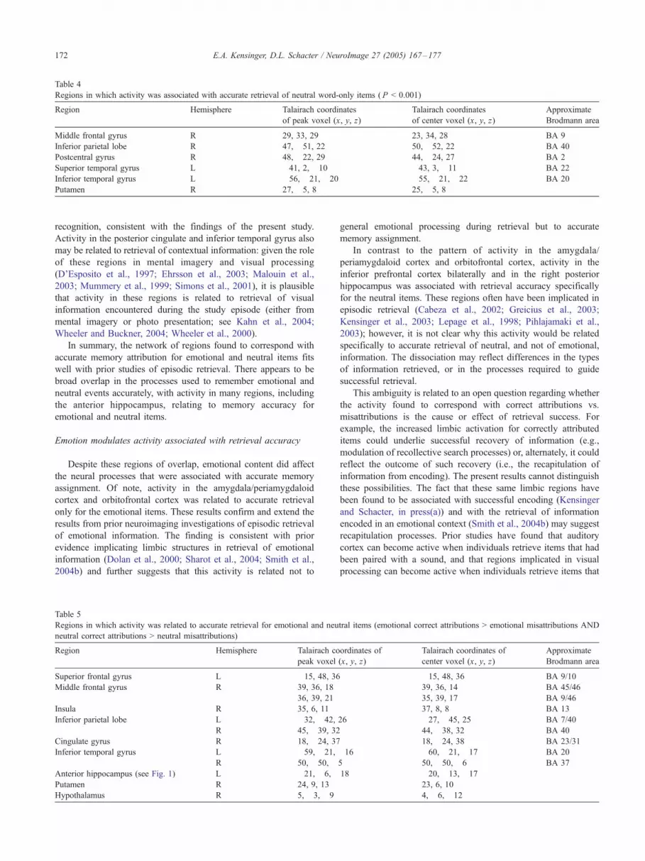
recognition, consistent with the findings of the present study.
Activity in the posterior cingulate and inferior temporal gyrus alsomay be related to retrieval of contextual information: given the roleof these regions in mental imagery and visual processing
(D’Esposito et al., 1997; Ehrsson et al., 2003; Malouin et al.,2003; Mummery et al., 1999; Simons et al., 2001), it is plausiblethat activity in these regions is related to retrieval of visualinformation encountered during the study episode (either from
mental imagery or photo presentation; see Kahn et al., 2004;Wheeler and Buckner, 2004; Wheeler et al., 2000).
In summary, the network of regions found to correspond with
accurate memory attribution for emotional and neutral items fitswell with prior studies of episodic retrieval. There appears to bebroad overlap in the processes used to remember emotional and
neutral events accurately, with activity in many regions, includingthe anterior hippocampus, relating to memory accuracy foremotional and neutral items.
Emotion modulates activity associated with retrieval accuracy
Despite these regions of overlap, emotional content did affect
the neural processes that were associated with accurate memoryassignment. Of note, activity in the amygdala/periamygdaloidcortex and orbitofrontal cortex was related to accurate retrieval
only for the emotional items. These results confirm and extend theresults from prior neuroimaging investigations of episodic retrievalof emotional information. The finding is consistent with prior
evidence implicating limbic structures in retrieval of emotionalinformation (Dolan et al., 2000; Sharot et al., 2004; Smith et al.,2004b) and further suggests that this activity is related not to
general emotional processing during retrieval but to accurate
memory assignment.In contrast to the pattern of activity in the amygdala/
periamygdaloid cortex and orbitofrontal cortex, activity in the
inferior prefrontal cortex bilaterally and in the right posteriorhippocampus was associated with retrieval accuracy specificallyfor the neutral items. These regions often have been implicated inepisodic retrieval (Cabeza et al., 2002; Greicius et al., 2003;
Kensinger et al., 2003; Lepage et al., 1998; Pihlajamaki et al.,2003); however, it is not clear why this activity would be relatedspecifically to accurate retrieval of neutral, and not of emotional,
information. The dissociation may reflect differences in the typesof information retrieved, or in the processes required to guidesuccessful retrieval.
This ambiguity is related to an open question regarding whetherthe activity found to correspond with correct attributions vs.misattributions is the cause or effect of retrieval success. For
example, the increased limbic activation for correctly attributeditems could underlie successful recovery of information (e.g.,modulation of recollective search processes) or, alternately, it couldreflect the outcome of such recovery (i.e., the recapitulation of
information from encoding). The present results cannot distinguishthese possibilities. The fact that these same limbic regions havebeen found to be associated with successful encoding (Kensinger
and Schacter, in press(a)) and with the retrieval of informationencoded in an emotional context (Smith et al., 2004b) may suggestrecapitulation processes. Prior studies have found that auditory
cortex can become active when individuals retrieve items that hadbeen paired with a sound, and that regions implicated in visualprocessing can become active when individuals retrieve items that
Table 5
Regions in which activity was related to accurate retrieval for emotional and neutral items (emotional correct attributions > emotional misattributions AND
neutral correct attributions > neutral misattributions)
Region Hemisphere Talairach coordinates of
peak voxel (x, y, z)
Talairach coordinates of
center voxel (x, y, z)
Approximate
Brodmann area
Superior frontal gyrus L !15, 48, 36 !15, 48, 36 BA 9/10
Middle frontal gyrus R 39, 36, 18 39, 36, 14 BA 45/46
36, 39, 21 35, 39, 17 BA 9/46
Insula R 35, 6, 11 37, 8, 8 BA 13
Inferior parietal lobe L !32, !42, 26 !27, !45, 25 BA 7/40
R 45, !39, 32 44, !38, 32 BA 40
Cingulate gyrus R 18, !24, 37 18, !24, 38 BA 23/31
Inferior temporal gyrus L !59, !21, !16 !60, !21, !17 BA 20
R 50, !50, !5 50, !50, !6 BA 37
Anterior hippocampus (see Fig. 1) L !21, !6, !18 !20, !13, !17
Putamen R 24, 9, 13 23, 6, 10
Hypothalamus R 5, !3, !9 4, !6, !12
Table 4
Regions in which activity was associated with accurate retrieval of neutral word-only items ( P < 0.001)
Region Hemisphere Talairach coordinates
of peak voxel (x, y, z)
Talairach coordinates
of center voxel (x, y, z)
Approximate
Brodmann area
Middle frontal gyrus R 29, 33, 29 23, 34, 28 BA 9
Inferior parietal lobe R 47, !51, 22 50, !52, 22 BA 40
Postcentral gyrus R 48, !22, 29 44, !24, 27 BA 2
Superior temporal gyrus L !41, 2, !10 !43, 3, !11 BA 22
Inferior temporal gyrus L !56, !21, !20 !55, !21, !22 BA 20
Putamen R 27, !5, 8 25, !5, 8
E.A. Kensinger, D.L. Schacter / NeuroImage 27 (2005) 167–177172

were paired with a visual image (Kahn et al., 2004; Nyberg et al.,2000; Wheeler and Buckner, 2004; Wheeler et al., 2000; Vaidya etal., 2002). Thus, retrieval-related activity in limbic regions may
reflect the repetition or bringing on-line of emotion-relevantinformation that was present during encoding. It also could reflectthe reinstantiation of the emotion evoked during encoding.However, it is also plausible that limbic activity enhances the
processes that guide episodic retrieval. Perhaps, the amygdalainfluences medial temporal-lobe regions during retrieval, as well asduring encoding and consolidation, increasing not only the
likelihood that a memory is encoded and stored, but also thelikelihood that the memory trace is reactivated at retrieval.
Similar logic can be used to contemplate the role of the
prefrontal and posterior hippocampal regions that were implicatedin accurate retrieval of the neutral, but not the emotional, items.These regions have been associated with successful encoding
(reviewed by Paller and Wagner, 2002), and thus activity in theseregions may represent recapitulation processes. However, it isequally plausible that these regions may be recruited in the serviceof information retrieval. Future studies will be required to
disentangle the extent to which emotional content modulatesprocesses that lead to, versus result from, recovery of informationat retrieval.
Amygdalar activity is related to retrieval success
The results of the present study are important in indicating that
activity in limbic regions (particularly the amygdala/periamygda-loid cortex and orbitofrontal cortex) can correspond with accurateretrieval. As outlined in Introduction, it has been unclear whetherlimbic engagement during memory retrieval would increase only
the subjective richness associated with a memory (Dolcos et al.,2005; Sharot et al., 2004), or also its objective accuracy. Behavioraldemonstrations of ‘‘flashbulb memories’’ have suggested that
perhaps limbic engagement enhances primarily the former, asindividuals often report high confidence in their memories ofemotional events even when they are not remembered accurately
(Talarico and Rubin, 2003; Winograd and Neisser, 1992; Neisserand Harsch, 1992). In contrast, demonstrations of enhanced sourcememory for emotional stimuli have suggested that limbic engage-
ment may increase memory accuracy (e.g., Davidson and Glisky,2002; Doerksen and Shimamura, 2001). The present study shedslight on this issue, indicating that limbic activity does not act solelyon confidence assessments while leaving memory accuracy
unchanged. Rather, amygdala/periamygdaloid activity duringretrieval does correspond with increased memory accuracy. Whilethe mechanisms underlying this relation require clarification, this
Table 6
Regions in which activity corresponded with accurate retrieval of emotional items (emotional correct attributions > emotional misattributions)
Region Hemisphere Talairach coordinates
of peak voxel (x, y, z)
Talairach coordinates
of center voxel (x, y, z)
Approximate
Brodmann area
Inferior frontal/ orbitofrontal gyrus L !35, 43, !17 !35, 43, !17 BA 10/11/47
Superior temporal gyrus L !35, 13, !21 !35, 13, !21 BA 38
Middle temporal gyrus R 48, !50, 0 50, !49, !4 BA 21
Temporo-parietal junction L !56, !54, 25 !45, !48, 18 BA 22/39/40
Fusiform gyrus L !41, !61, !14 !40, !62, !15 BA 37
R 41, !70, !12 42, !68, !13 BA 37
Precuneus L !41, !71, 40 !40, !69, 40 BA 19
Amygdala/periamygdaloid
cortex (see Fig. 2)
R 15, !2, !20 12, !1, !16
Cerebellum
Fig. 1. Activity in a region centered in the anterior hippocampus was greater during retrieval of correctly-attributed (CA) as compared to misattributed (MA)
emotional (emo) and neutral (neu) items. (*Indicates significant difference at P < 0.05).
E.A. Kensinger, D.L. Schacter / NeuroImage 27 (2005) 167–177 173

study highlights the fact that amygdala engagement at retrieval, aswell as during encoding (Kensinger and Schacter, in press(a)) can
reduce the probability of memory misattributions.
Summary
By adopting a reality-monitoring paradigm that required
participants to indicate which memories were attributable toexternal presentation, the present study could examine theprocesses engaged during correct memory attributions versus
misattributions of emotional and neutral items. The resultsindicated that many of the regions found to correspond with
retrieval of contextual details for neutral items (e.g., Dobbins et al.,2003; Giovanello et al., 2004; Wheeler and Buckner, 2003) alsowere associated with accurate memory attribution for emotional
items. Despite the overlaps in the neural networks, however, theemotional content of items affected the regions in which activitycorresponded with accurate retrieval. Engagement of limbic
regions was related to accurate retrieval of emotional itemsspecifically, while engagement of additional prefrontal and medialtemporal-lobe regions corresponded with accurate retrieval of
Table 7
Regions with activity was associated with accurate retrieval of neutral items (neutral correct attributions > neutral misattributions)
Region Hemisphere Talairach coordinates
of peak voxel (x, y, z)
Talairach coordinates
of center voxel (x, y, z)
Approximate
Brodmann
area (BA)
Inferior frontal gyrus R 53, 20, 7 55, 21, 7 BA 45/47
R 60, 15, 24 59, 15, 21 BA 9/44
L !51, 7, 19 !50, 7, 18 BA 9/44
Anterior cingulate gyrus L !11, 20 !4 !11, 19, !4 BA 24/32
Insula L !35, 11, !5 36, 13, 9 BA 13
Fusiform gyrus/
middle occipital gyrus R 38, !70, !6 35, !61, 1 BA 19
Lingual gyrus L !24, !84, !6 !24, !84, !7 BA 18
Hippocampus (see Fig. 3) R 18, !24, !4 18, !24, !7
Fig. 2. Activity in the amygdala (A) and orbitofrontal cortex (B) was related to accurate retrieval for the emotional items, but not for the neutral items. CA =
correct attribution; MA = misattribution.
E.A. Kensinger, D.L. Schacter / NeuroImage 27 (2005) 167–177174

neutral items. Although the role of limbic structures duringsuccessful encoding of emotional information has been well
established (e.g., Hamann, 2001; Phelps, 2004), to our knowledge,this study is the first to demonstrate a link between limbicengagement and accurate memory attribution at retrieval.
Acknowledgments
We thank Ronnie Bryan and Mariko Jameson for help withparticipant recruitment and testing. This research was supported
by grants MH60941 (to D.L.S.) and MH070199 (to E.A.K.) fromthe National Institutes of Health and by a MassachusettsBiomedical Research Corporation Tosteson Postdoctoral Fellow-ship (to E.A.K.). Figures depicting the medial temporal-lobe
regions reported in Tables 1–4 are available by contacting thefirst author.
References
Cabeza, R., Rao, S.M., Wagner, A.D., Mayer, A.R., Schacter, D.L., 2001.
Can medial temporal lobe regions distinguish true from false? An event-
related functional MRI study of veridical and illusory recognition
memory. Proc. Natl. Acad. Sci. U. S. A. 98, 4805–4810.
Cabeza, R., Dolcos, F., Graham, R., Nyberg, L., 2002. Similarities and
differences in the neural correlates of episodic memory retrieval and
working memory. Neuroimage 16, 317–330.
Cahill, L., et al., 1996. Amygdala activity at encoding correlated with long-
term, free recall of emotional information. Proc. Natl. Acad. Sci. 93,
8016–8021.
Canli, T., Zhao, Z., Brewer, J., Gabrieli, J.D., Cahill, L., 2000. Event-related
activation in the human amygdala associates with later memory for
individual emotional experience. J. Neurosci. 20, RC99.
Coltheart, M., 1981. The MRC psycholinguistic database. Q. J. Exp.
Psychol. 33A, 497–505.
D’Argembeau, A., Van der Linden, M., 2004. Influence of affective
meaning on memory for contextual information. Emotion 4, 173–188.
D’Esposito, M., Detre, J.A., Aguirre, G.K., Stallcup, M., Alsop, D.C.,
Tippet, L.J., Farah, M.J., 1997. A functional MRI study of mental image
generation. Neuropsychologia 35, 725–730.
Dale, A.M., 1999. Optimal experimental design for event-related fMRI.
Hum. Brain Mapp. 8, 109–114.
Davidson, P.S., Glisky, E.L., 2002. Is flashbulb memory a special instance
of source memory? Evidence from older adults. Memory 10, 99–111.
Dobbins, I.G., Rice, H.J., Wagner, A.D., Schacter, D.L., 2003. Memory
orientation and success: separable neurocognitive components under-
lying episodic recognition. Neuropsychologia 41, 318–333.
Doerksen, S., Shimamura, A.P., 2001. Source memory enhancement for
emotional words. Emotion 1, 5–11.
Dolan, R.J., Lane, R., Chua, P., Fletcher, P., 2000. Dissociable temporal
lobe activations during emotional episodic memory retrieval. Neuro-
image 11, 203–209.
Dolcos, F., LaBar, K.S., Cabeza, R., 2004. Interaction between the
amygdala and the medial temporal lobe memory system predicts better
memory for emotional events. Neuron 5, 855–863.
Dolcos, F., LaBar, K.S., Cabeza, R., 2005. Remembering one year later:
role of the amygdala and the medial temporal lobe memory system in
Fig. 3. Activity in the inferior prefrontal cortex bilaterally (A) and in the right posterior hippocampus (B) was associated with accurate memory retrieval for the
neutral, but not the emotional, items. CA = correct attribution; MA = misattribution.
E.A. Kensinger, D.L. Schacter / NeuroImage 27 (2005) 167–177 175

retrieving emotional memories. Proc. Natl. Acad. Sci. U. S. A. 102,
2626–2631.
Ehrsson, H.H., Geyer, S., Naito, E., 2003. Imagery of voluntary movement
of fingers, toes, and tongue activates corresponding body-part-specific
motor representations. J. Neurophysiol. 90, 3304–3316.
Eldridge, L.L., Knowlton, B.J., Furmanski, C.S., Bookheimer, S.Y., Engel,
S.A., 2000. Remembering episodes: a selective role for the hippo-
campus during retrieval. Nat. Neurosci. 3, 1149–1152.
Giovanello, K.S., Schnyer, D.M., Verfaellie, M., 2004. A critical role for
the anterior hippocampus in relational memory: evidence from an
fMRI study comparing associative and item recognition. Hippocampus
14, 5–8.
Gonsalves, B., Paller, K.A., 2000. Neural events that underlie remembering
something that never happened. Nat. Neurosci. 3, 1316–1321.
Greicius, M.D., Krasnow, B., Boyett-Anderson, J.M., Eliez, S., Schatzberg,
A.F., Reiss, A.L., Menon, V., 2003. Regional analysis of hippocampal
activation during memory encoding and retrieval: fMRI study. Hippo-
campus 13, 164–174.
Hamann, S., 2001. Cognitive and neural mechanisms of emotional memory.
Trends Cogn. Sci. 5, 394–400.
Henson, R.N., Rugg, M.D., Shallice, T., Josephs, O., Dolan, R.J.,
1999a. Recollection and familiarity in recognition memory: an event-
related functional magnetic resonance imaging study. J. Neurosci. 19,
3962–3972.
Henson, R.N., Shallice, T., Dolan, R.J., 1999b. Right prefrontal cortex and
episodic memory retrieval: a functional MRI test of the monitoring
hypothesis. Brain 122, 1367–1381.
Henson, R.N., Rugg, M.D., Shallice, T., Dolan, R.J., 2000. Confidence in
recognition memory for words: dissociating right prefrontal roles in
episodic retrieval. J. Cogn. Neurosci. 12, 913–923.
Johnson, M.K., Raye, C.L., 1981. Reality monitoring. Psych. Rev. 88,
67–85.
Kahn, I., Davachi, L., Wagner, A.D., 2004. Functional–neuroanatomic
correlates of recollection: implications for models of recognition
memory. J. Neurosci. 24, 4172–4180.
Kensinger, E.A., Corkin, S., 2003. Memory enhancement for emotional
words: are emotional words more vividly remembered than neutral
words? Mem. Cogn. 31, 1169–1180.
Kensinger, E.A., Corkin, S., 2004a. The effects of emotional content
and aging on false memories. Cogn. Affect. Behav. Neurosci. 4,
1–9.
Kensinger, E.A., Corkin, S., 2004b. Two routes to emotional memory:
distinct neural processes for valence and arousal. Proc. Natl. Acad. Sci.
U. S. A. 101, 3310–3315.
Kensinger, E.A., Schacter, D.L., in press-a. Emotional content and reality-
monitoring ability: FMRI evidence for the influence of encoding
processes. Neuropsychologia.
Kensinger, E.A., Schacter, D.L., in press-b. Reality monitoring and memory
distortion: effects of negative, arousing content. Mem. Cognit.
Kensinger, E.A., Clarke, R.J., Corkin, S., 2003. What neural correlates
underlie successful encoding and retrieval? A functional magnetic
resonance imaging study using a divided attention paradigm.
J. Neurosci. 23, 2407–2415.
Kilpatrick, L., Cahill, L., 2003. Amygdala modulation of parahippocampal
and frontal regions during emotionally influenced memory storage.
NeuroImage 20, 2091–2099.
LeDoux, J.E., 2000. Emotion circuits in the brain. Annu. Rev. Neurosci. 23,
155–184.
Lepage, M., Habib, R., Tulving, E., 1998. Hippocampal PET activations of
memory encoding and retrieval: the HIPER model. Hippocampus 8,
313–322.
Malouin, F., Richards, C.L., Jackson, P.L., Dumas, F., Doyon, J., 2003.
Brain activations during motor imagery of locomotor-related tasks: a
PET study. Hum. Brain Mapp. 19, 47–62.
Maratos, E.J., Rugg, M.D., 2001. Electrophysiological correlates of the
retrieval of emotional and non-emotional context. J. Cogn. Neurosci.
13, 877–891.
Maratos, E.J., Dolan, R.J., Morris, J.S., Henson, R.N., Rugg, M.D., 2001.
Neural activity associated with episodic memory for emotional context.
Neuropsychologia 39, 910–920.
Mummery, C.J., Patterson, K., Wise, R.J., Vandenbergh, R., Price, C.J.,
Hodges, J.R., 1999. Disrupted temporal lobe connections in semantic
dementia. Brain 122, 61–73.
Nader, K., 2003. Memory traces unbound. Trend Neurosci. 26, 65–72.
Neisser, U., Harsch, H.N., 1992. Phantom flashbulbs: false recollections of
hearing the news about the challenger. In: Winograd, E., Neisser, U.
(Eds.), Affect and Accuracy in Recall: Studies of ‘‘Flashbulb’’
Memories, vol. 4. Cambridge University Press, New York.
Nyberg, L., Habib, R., McIntosh, A.R., Tulving, E., 2000. Reactivation of
encoding-related brain activity during memory retrieval. Proc. Natl.
Acad. Sci. U. S. A. 97, 11120–11124.
Okado, Y., Stark, C., 2003. Neural processing associated with
true and false memory retrieval. Cogn. Affect. Behav. Neurosci. 3,
323–334.
Otten, L.J., Henson, R.N., Rugg, M.D., 2001. Depth of processing effects
on neural correlates of memory encoding: relationship between findings
from across- and within-task comparisons. Brain 124, 399–412.
Otten, L.J., Henson, R.N.A., Rugg, M.D., 2002. State-related and item-
related neural correlates of successful memory encoding. Nat. Neurosci.
5, 1339–1344.
Paller, K.A., Wagner, A.D., 2002. Observing the transformation of
experience into memory. Trends Cogn. Sci. 6, 93–102.
Pesta, B.J., Murphy, M.D., Sanders, R.E., 2001. Are emotionally charged
lures immune to false memory? J. Exper. Psychol., Learn., Mem., Cogn.
27, 328–338.
Phelps, E.A., 2004. Human emotion and memory: interactions of the
amygdala and hippocampal complex. Curr. Opin. Neurobiol. 14,
198–202.
Pihlajamaki, M., Tanila, H., Hanninen, T., Kononen, M., Mikkonnen, M.,
Jalkanen, V., Partanen, K., Aronen, H.J., Soininen, H., 2003. Encoding
of novel picture pairs activates the perirhinal cortex: an fMRI study.
Hippocampus 13, 67–80.
Ranganath, C., Johnson, M.K., D’Esposito, M., 2000. Left anterior
prefrontal activation increases with demands to recall specific percep-
tual information. J. Neurosci. 20 (1–5), RC108.
Richardson, M.P., Strange, B.A., Dolan, R.J., 2004. Encoding of emotional
memories depends on amygdala and hippocampus and their interac-
tions. Nat. Neurosci. 7, 278–285.
Roediger, H.L., McDermott, K.B., 2000. Tricks of memory. Curr. Dir.
Psychol. Sci. 9, 123–127.
Schacter, D.L., 1999. The seven sins of memory. Insights from psychology
and cognitive neuroscience. Am. Psychol. 54, 182–203.
Schacter, D.L., Norman, K.A., Koutstaal, W., 1998. The cognitive neuro-
science of constructive memory. Annu. Rev. Psychol. 49, 289–318.
Sharot, T., Delgado, M.R., Phelps, E.A., 2004. How emotion enhances the
feeling of remembering. Nat. Neurosci. 7, 1376–1380.
Simons, J.S., Graham, K.S., Owen, A.M., Patterson, K., Hodges, J.R.,
2001. Perceptual and semantic components of memory for objects and
faces: a PET study. J. Cogn. Neurosci. 13, 430–443.
Slotnick, S.D., Schacter, D.L., 2004. A sensory signature that distinguishes
true from false memories. Nat. Neurosci. 7, 664–672.
Smith, A.P., Dolan, R.J., Rugg, M.D., 2004a. Event-related potential
correlates of the retrieval of emotional and nonemotional context.
J. Cogn. Neurosci. 16, 760–775.
Smith, A.P.R., Henson, R.N.A., Dolan, R.J., Rugg, M.D., 2004b. fMRI
correlates of episodic retrieval of emotional contexts. Neuroimage 22,
868–878.
Talairach, J., Tournoux, P., 1998. Co-planar Stereotaxic Axis of the Human
Brain. Thieme, New York.
Talarico, J.M., Rubin, D.C., 2003. Confidence, not consistency, character-
izes flashbulb memories. Psychol. Sci. 14, 455–461.
Taylor, S.F., Liberzon, I., Fig, L.M., Decker, L.R., Minoshima, S., Koeppe,
R.A., 1998. The effect of emotional content on visual recognition
memory: a PET activation study. NeuroImage 8, 188–197.
E.A. Kensinger, D.L. Schacter / NeuroImage 27 (2005) 167–177176

Vaidya, C.J., Zhao, M., Desmond, J.E., Gabrieli, J.D., 2002. Evidence
for cortical encoding specificity in episodic memory: memory-
induced re-activation of picture processing areas. Neuropsychologia
40, 2136–2143.
Wheeler, M.E., Buckner, R.L., 2003. Functional dissociation among
components of remembering: control, perceived oldness, and content.
J. Neurosci. 23, 3869–3880.
Wheeler, M.E., Buckner, R.L., 2004. Functional–anatomic correlates of
remembering and knowing. Neuroimage 21, 1337–1349.
Wheeler, M.E., Petersen, S.E., Buckner, R.L., 2000. Memory’s echo: vivid
remembering reactivates sensory-specific cortex. Proc. Natl. Acad. Sci.
U. S. A. 97, 11125–11129.
Winograd, E., Neisser, U. (Eds.), Affect and Accuracy in Recall: Studies of
‘‘Flashbulb’’ Memories, vol. 4. Cambridge University Press, New York.
E.A. Kensinger, D.L. Schacter / NeuroImage 27 (2005) 167–177 177