Embed Size (px)
Citation preview
AaiAod
FAAlaTAa2T
Journal of the American College of Cardiology Vol. 55, No. 12, 2010© 2010 by the American College of Cardiology Foundation ISSN 0735-1097/10/$36.00P
Heart Rhythm Disorders
Reverse Remodeling of the Atria AfterTreatment of Chronic Stretch in HumansImplications for the Atrial Fibrillation Substrate
Bobby John, MD, PHD,*† Martin K. Stiles, MBCHB, PHD,* Pawel Kuklik, PHD,*Anthony G. Brooks, PHD,* Sunil T. Chandy, MD,† Jonathan M. Kalman, MBBS, PHD,‡Prashanthan Sanders, MBBS, PHD*
Adelaide and Melbourne, Australia; and Vellore, India
Objectives The aim of this report was to study the effect of chronic stretch reversal on the electrophysiological characteris-tics of the atria in humans.
Background Atrial stretch is an important determinant for atrial fibrillation. Whether relief of stretch reverses the substratepredisposed to atrial fibrillation is unknown.
Methods Twenty-one patients with mitral stenosis undergoing mitral commissurotomy (MC) were studied before and afterintervention. Catheters were placed at multiple sites in the right atrium (RA) and sequentially within the leftatrium (LA) to determine: effective refractory period (ERP) at 10 sites (600 and 450 ms) and P-wave duration(PWD). Bi-atrial electroanatomic maps determined conduction velocity (CV) and voltage. In 14 patients, RA stud-ies were repeated �6 months after MC.
Results Immediately after MC, there was significant increase in mitral valve area (2.1 � 0.2 cm2, p � 0.0001) with de-crease in LA (23 � 7 mm Hg to 10 � 4 mm Hg, p � 0.0001) and pulmonary arterial pressures (38 � 16 mm Hgto 27 � 12 mm Hg, p � 0.0001) and LA volume (75 � 20 ml to 52 � 18 ml, p � 0.0001). This was associ-ated with reduction in PWD (139 � 19 ms to 135 � 20 ms, p � 0.047), increase in CV (LA: 1.3 � 0.3 mm/msto 1.7 � 0.2 mm/ms, p � 0.006; and RA: 1.0 � 0.1 mm/ms to 1.3 � 0.3 mm/ms, p � 0.002) and voltage(LA: 1.7 � 0.6 mV to 2.5 � 1.0 mV, p � 0.005; and RA: 1.8 � 0.6 mV to 2.2 � 0.7 mV, p � 0.09), and nochange in ERP. Late after MC, mitral valve area remained at 2.1 � 0.3 cm2 (p � 0.7) but with further decreasein PWD (113 � 19 ms, p � 0.04) and RA ERP (at 600 ms, p � 0.0001), with increase in CV (1.0 � 0.1 mm/msto 1.3 � 0.2 mm/ms, p � 0.006) and voltage (1.8 � 0.7 mV to 2.8 � 0.6 mV, p � 0.002).
Conclusions The atrial electrophysiologic and electroanatomic abnormalities that result from chronic stretch due to MS re-verses after MC. These observations suggest that the substrate predisposing to atrial arrhythmias might bereversed. (J Am Coll Cardiol 2010;55:1217–26) © 2010 by the American College of Cardiology Foundation
ublished by Elsevier Inc. doi:10.1016/j.jacc.2009.10.046
ttcrka
HDUNSrra
common feature of many of the conditions predisposed totrial fibrillation (AF) is chronic atrial “stretch.” Atrial sizes known to be an important marker for the development ofF (1). Electrical remodeling caused by short-term stretchf the atria due to asynchronous ventricular pacing has beenemonstrated to be reversible with restoration of atrioven-
rom the *Cardiovascular Research Center, Department of Cardiology, Royaldelaide Hospital and the Disciplines of Medicine and Physiology, University ofdelaide, Adelaide, Australia; †Department of Cardiology, Christian Medical Col-
ege, Vellore, India; and the ‡Department of Cardiology, Royal Melbourne Hospitalnd the Department of Medicine, University of Melbourne, Melbourne, Australia.his work was presented in part by Dr. John, who received the Young Investigatorward, at the 3rd Asia-Pacific AF Symposium, October 2007, Taipei, Taiwan; and
t the Annual Scientific Sessions of the American Heart Association, November
007, Orlando, Florida; and published in abstract form (Circulation 2007;116:II438).his work was supported in part by a Grant-in-Aid (G 08A 3646) from the National aricular synchrony (2). However, several studies have iden-ified structural change as an important component ofhronic conditions predisposed to AF (3–10). Whethereversal of chronic atrial stretch in a clinical conditionnown to have such structural and electrophysiologicalbnormalities alters the AF substrate is not known.
eart Foundation of Australia and by the Australia-India Strategic Research Fund.r. John is supported by the Biosense-Webster Electrophysiology Scholarship,niversity of Adelaide. Dr. Stiles is supported by the National Heart Foundation ofew Zealand and the Dawes Scholarship, Royal Adelaide Hospital. Drs. Brooks andanders are supported by the National Heart Foundation of Australia. Dr. Sanderseports having served on the advisory board of and having received lecture fees andesearch funding from St. Jude Medical, Bard Electrophysiology, Biosense-Webster,nd Medtronic.
Manuscript received June 28, 2009; revised manuscript received October 22, 2009,ccepted October 26, 2009.
ncOmuc
waapfptEwp(�ffiM(“(wsoflScp
cWpsp1aaq
doosHs
E
pas1dtdabi47ja
V
iAwEcCaeotCwc�tolpaemq
1218 John et al. JACC Vol. 55, No. 12, 2010Atrial Reverse Remodeling and Chronic Stretch March 23, 2010:1217–26
Severe mitral stenosis (MS) isan established substrate for AFand results in chronically ele-vated atrial pressures and atrialenlargement (8). Mitral commis-surotomy (MC) is now the treat-ment of choice for patients withsevere MS and favorable valvemorphology. We aimed to char-acterize the immediate and lateeffects of chronic stretch reversalby performing high-density elec-trophysiological and electroana-tomical mapping of the atria be-fore and serially after MC.
Methods
Study population. The studycomprised 21 patients with se-vere MS due to rheumatic heartdisease, mitral valve area (MVA)of �1.5 cm2 associated with sig-
ificant symptoms (New York Heart Association functionallass �II), and mitral valve morphology suitable for MC.ther structural heart disease, hypertension, atrial arrhyth-ias (by history and monitoring for �48 h), amiodarone
se, or the presence of left atrial thrombus formed exclusionriteria.
All patients gave written informed consent to the study,hich was approved by each institutional clinical research
nd ethics committee. All antiarrhythmic drugs were ceasedt least 5 half lives before the study. Echocardiography waserformed to determine the atrial volumes with the equationor prolate ellipsoid (10). Patients underwent either electro-hysiological or electroanatomical mapping as detailed inhe following text.lectrophysiological mapping. Electrophysiological studyas performed in the fasted state with sedation. Electro-hysiological evaluation of the right (RA) and left atriaLA) was performed before and immediately after MC. At6-month follow-up electrophysiological study was per-
ormed in the RA. The following catheters were positionedor the study protocol: 1) 10-pole catheter (2-5-2 mmnter-electrode spacing, Daig Electrophysiology, St. Jude
edical, St. Paul, Minnesota) within the coronary sinusCS) with the proximal bipole at the CS ostium; 2) 20-polecrista” catheter with 1-3-1 mm interelectrode spacingBiosense-Webster, Diamond Bar, California) positionedith the aid of a long sheath along the crista terminalis and
tandardized such that the second bipole lay at the junctionf the superior vena cava with RA as determined byuoroscopy and intracardiac echocardiography (Acunav,iemens Medical, Mountain View, California); 3) 20-poleatheter (2-5-2 mm interelectrode spacing, Daig Electro-
Abbreviationsand Acronyms
AF � atrial fibrillation
CL � cycle length(s)
CS � coronary sinus
CV � conduction velocity
DP � double potential(s)
ECG � electrocardiogram
ERP � effective refractoryperiod
FS � fractionated signals
LA � left atrium
LAT � local activation time
MC � mitralcommissurotomy
MS � mitral stenosis
MVA � mitral valve area
PWD � P-wave duration
RA � right atrium
hysiology) placed along the lateral RA; and 4) 10-pole P
atheter (2-5-2 mm inter-electrode spacing, Biosense-ebster) was positioned within the LA via transseptal
uncture. This catheter was stabilized with the use of a longheath (Preface, Biosense-Webster, or SL0, Daig Electro-hysiology) and sequentially positioned as follows at the:) LA roof; 2) inferior LA; 3) mid-posterior LA; 4) LAppendage; and 5) high RA septum (8). Digital images wererchived to help standardize catheter locations for subse-uent studies.Surface electrocardiograms (ECGs) and bipolar endocar-
ial electrograms were continuously monitored and storedn a computer-based digital amplifier/recorder system forffline analysis (Bard Electrophysiology, Lowell, Massachu-etts). Intracardiac electrograms were filtered from 30 to 500z and measured with computer-assisted calipers at a sweep
peed of 200 mm/s.
FFECTIVE REFRACTORINESS. Atrial effective refractoryeriod (ERP) was evaluated at cycle lengths (CL) of 600nd 450 ms with an 8-beat drive followed by an extra-timulus, starting with an extra-stimulus coupling interval of50 ms increasing in 10-ms increments. The ERP wasefined as the longest coupling interval failing to propagateo the atrium. At each site the ERP was measured 3 timesuring each CL and averaged. If ERP varied by �10 ms, andditional 2 measurements were made, and the total num-er was averaged. The ERP was measured from the follow-ng sites: 1) distal-CS; 2) proximal-CS; 3) low lateral RA;) high lateral RA; 5) high septal RA; 6) LA appendage;) posterior LA; 8) right superior pulmonary vein–LA roof
unction; 9) left superior pulmonary vein–LA roof junction;nd 10) inferior LA.
ULNERABILITY FOR AF. Vulnerability to AF was defined asrregular atrial activity �30 s during the study protocol.trial fibrillation lasting �5 min was considered sustained;hen this occurred, no further data were acquired.lectroanatomic mapping. Electroanatomic maps were
reated of the LA and RA during sinus rhythm with theARTO mapping system (Biosense-Webster) both before
nd immediately after MC. At �6 months after MC,lectroanatomic mapping was performed in sinus rhythmnly in the RA. Endocardial contact during point acquisi-ion was facilitated by fluoroscopy, the catheter icon on theARTO system, and intracardiac echocardiography. Pointsere acquired in the auto-freeze mode, if the stability
riteria in space (�6 mm) and local activation time (LAT;5 ms) were met. Mapping was performed with a fill-
hreshold of 15 mm. Editing of points was performedffline. The LAT was manually annotated to the peak of theargest amplitude deflection on bipolar electrograms. In theresence of double potentials (DP), the LAT was annotatedt the largest potential. If the bipolar electrogram displayedquivalent maximum positive and negative deflections, theaximum negative deflection on the simultaneously ac-
uired unipolar electrogram was used to annotate the LAT.
oints not conforming to the 12-lead ECG P-wave mor-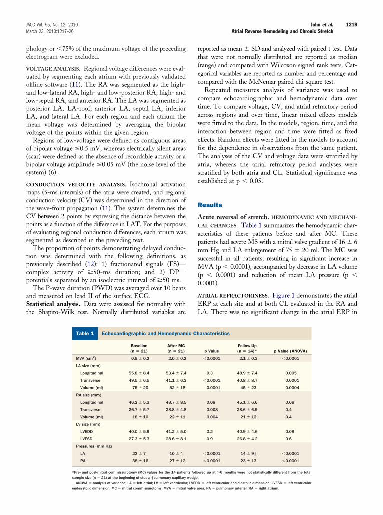
pe
V
uoalpLmv
o(bs
C
mctCpos
tpcp
aSt
rt(ec
ctawiefTase
R
AC
apmsM(0
A
EL
1219JACC Vol. 55, No. 12, 2010 John et al.March 23, 2010:1217–26 Atrial Reverse Remodeling and Chronic Stretch
hology or �75% of the maximum voltage of the precedinglectrogram were excluded.
OLTAGE ANALYSIS. Regional voltage differences were eval-ated by segmenting each atrium with previously validatedffline software (11). The RA was segmented as the high-nd low-lateral RA, high- and low-posterior RA, high- andow-septal RA, and anterior RA. The LA was segmented asosterior LA, LA-roof, anterior LA, septal LA, inferiorA, and lateral LA. For each region and each atrium theean voltage was determined by averaging the bipolar
oltage of the points within the given region.Regions of low-voltage were defined as contiguous areas
f bipolar voltage �0.5 mV, whereas electrically silent areasscar) were defined as the absence of recordable activity or aipolar voltage amplitude �0.05 mV (the noise level of theystem) (6).
ONDUCTION VELOCITY ANALYSIS. Isochronal activationaps (5-ms intervals) of the atria were created, and regional
onduction velocity (CV) was determined in the direction ofhe wave-front propagation (11). The system determines theV between 2 points by expressing the distance between theoints as a function of the difference in LAT. For the purposesf evaluating regional conduction differences, each atrium wasegmented as described in the preceding text.
The proportion of points demonstrating delayed conduc-ion was determined with the following definitions, asreviously described (12): 1) fractionated signals (FS)—omplex activity of �50-ms duration; and 2) DP—otentials separated by an isoelectric interval of �50 ms.The P-wave duration (PWD) was averaged over 10 beats
nd measured on lead II of the surface ECG.tatistical analysis. Data were assessed for normality with
he Shapiro-Wilk test. Normally distributed variables are
Echocardiographic and Hemodynamic CharacterTable 1 Echocardiographic and Hemodynam
Baseline(n � 21)
After MC(n � 21)
MVA (cm2) 0.9 � 0.2 2.0 � 0.2
LA size (mm)
Longitudinal 55.8 � 8.4 53.4 � 7.4
Transverse 49.5 � 6.5 41.1 � 6.3
Volume (ml) 75 � 20 52 � 18
RA size (mm)
Longitudinal 46.2 � 5.3 48.7 � 8.5
Transverse 26.7 � 5.7 28.8 � 4.8
Volume (ml) 18 � 10 22 � 11
LV size (mm)
LVEDD 40.0 � 5.9 41.2 � 5.0
LVESD 27.3 � 5.3 28.6 � 8.1
Pressures (mm Hg)
LA 23 � 7 10 � 4
PA 38 � 16 27 � 12
*Pre- and post-mitral commissurotomy (MC) values for the 14 patiensample size (n � 21) at the beginning of study; †pulmonary capillary
ANOVA � analysis of variance; LA � left atrial; LV � left ventricular; LVEDDend-systolic dimension; MC � mitral commissurotomy; MVA � mitral valve a
eported as mean � SD and analyzed with paired t test. Datahat were not normally distributed are reported as medianrange) and compared with Wilcoxon signed rank tests. Cat-gorical variables are reported as number and percentage andompared with the McNemar paired chi-square test.
Repeated measures analysis of variance was used toompare echocardiographic and hemodynamic data overime. To compare voltage, CV, and atrial refractory periodcross regions and over time, linear mixed effects modelsere fitted to the data. In the models, region, time, and the
nteraction between region and time were fitted as fixedffects. Random effects were fitted in the models to accountor the dependence in observations from the same patient.he analyses of the CV and voltage data were stratified by
tria, whereas the atrial refractory period analyses weretratified by both atria and CL. Statistical significance wasstablished at p � 0.05.
esults
cute reversal of stretch. HEMODYNAMIC AND MECHANI-
AL CHANGES. Table 1 summarizes the hemodynamic char-cteristics of these patients before and after MC. Theseatients had severe MS with a mitral valve gradient of 16 � 6m Hg and LA enlargement of 75 � 20 ml. The MC was
uccessful in all patients, resulting in significant increase inVA (p � 0.0001), accompanied by decrease in LA volume
p � 0.0001) and reduction of mean LA pressure (p �.0001).
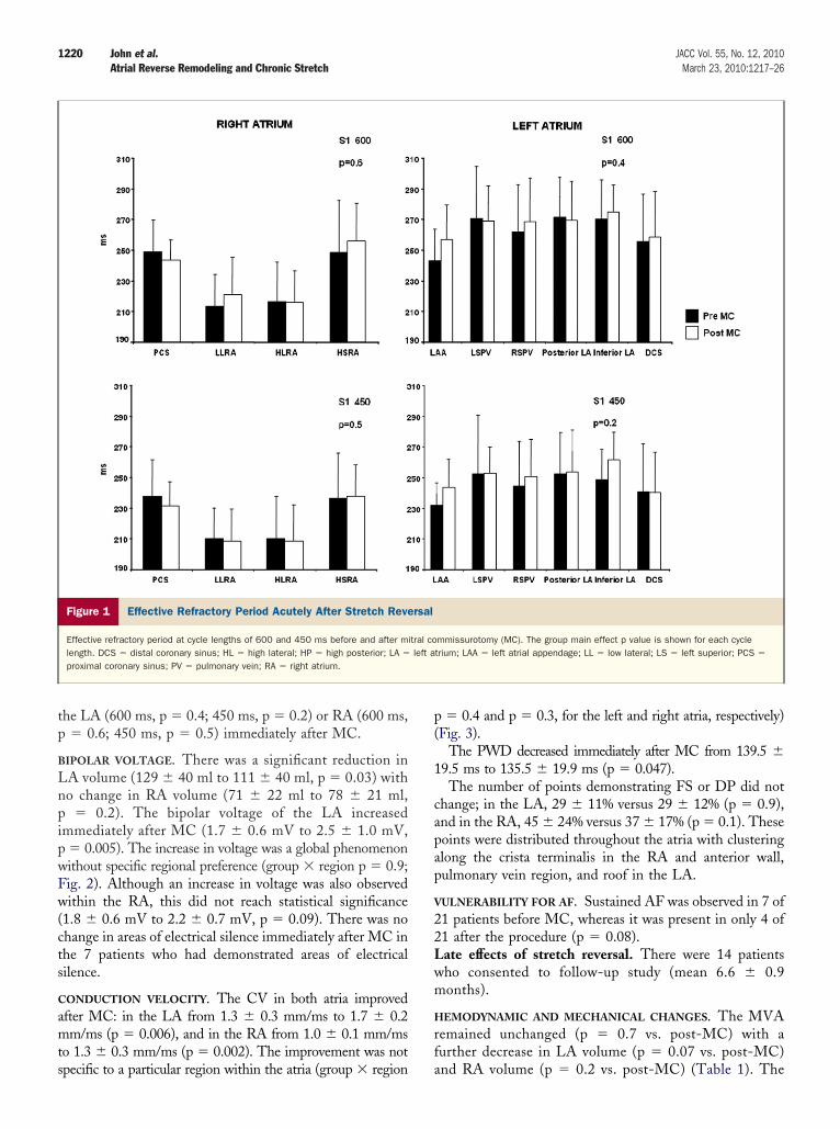
TRIAL REFRACTORINESS. Figure 1 demonstrates the atrialRP at each site and at both CL evaluated in the RA andA. There was no significant change in the atrial ERP in
aracteristics
p ValueFollow-Up(n � 14)* p Value (ANOVA)
�0.0001 2.1 � 0.3 �0.0001
0.3 48.9 � 7.4 0.005
�0.0001 40.8 � 8.7 0.0001
0.0001 45 � 23 0.0004
0.08 45.1 � 6.6 0.06
0.008 28.6 � 6.9 0.4
0.004 21 � 12 0.4
0.2 40.9 � 4.6 0.08
0.9 26.8 � 4.2 0.6
�0.0001 14 � 9† �0.0001
�0.0001 23 � 13 �0.0001
wed up at �6 months were not statistically different from the total
isticsic Ch
ts followedge.
� left ventricular end-diastolic dimension; LVESD � left ventricularrea; PA � pulmonary arterial; RA � right atrium.
tp
B
LnpipwFw(cts
C
amts
p(
1
capap
V
22Lwm
H
rf
1220 John et al. JACC Vol. 55, No. 12, 2010Atrial Reverse Remodeling and Chronic Stretch March 23, 2010:1217–26
he LA (600 ms, p � 0.4; 450 ms, p � 0.2) or RA (600 ms,� 0.6; 450 ms, p � 0.5) immediately after MC.
IPOLAR VOLTAGE. There was a significant reduction inA volume (129 � 40 ml to 111 � 40 ml, p � 0.03) witho change in RA volume (71 � 22 ml to 78 � 21 ml,
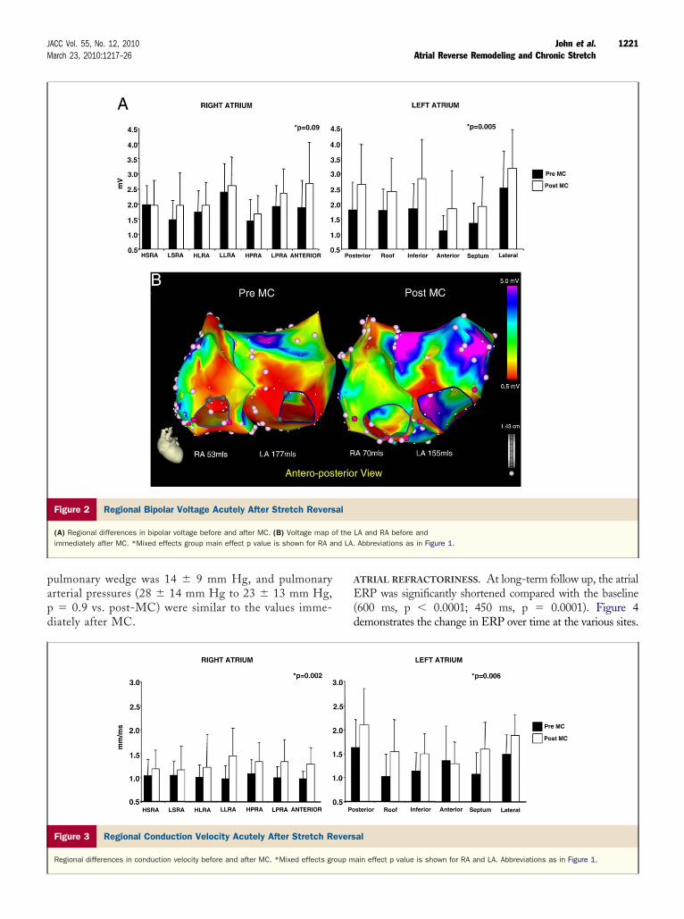
� 0.2). The bipolar voltage of the LA increasedmmediately after MC (1.7 � 0.6 mV to 2.5 � 1.0 mV,� 0.005). The increase in voltage was a global phenomenonithout specific regional preference (group � region p � 0.9;ig. 2). Although an increase in voltage was also observedithin the RA, this did not reach statistical significance
1.8 � 0.6 mV to 2.2 � 0.7 mV, p � 0.09). There was nohange in areas of electrical silence immediately after MC inhe 7 patients who had demonstrated areas of electricalilence.
ONDUCTION VELOCITY. The CV in both atria improvedfter MC: in the LA from 1.3 � 0.3 mm/ms to 1.7 � 0.2m/ms (p � 0.006), and in the RA from 1.0 � 0.1 mm/ms
o 1.3 � 0.3 mm/ms (p � 0.002). The improvement was not
Figure 1 Effective Refractory Period Acutely After Stretch Reve
Effective refractory period at cycle lengths of 600 and 450 ms before and after mlength. DCS � distal coronary sinus; HL � high lateral; HP � high posterior; LA �
proximal coronary sinus; PV � pulmonary vein; RA � right atrium.
pecific to a particular region within the atria (group � region a
� 0.4 and p � 0.3, for the left and right atria, respectively)Fig. 3).
The PWD decreased immediately after MC from 139.5 �9.5 ms to 135.5 � 19.9 ms (p � 0.047).
The number of points demonstrating FS or DP did nothange; in the LA, 29 � 11% versus 29 � 12% (p � 0.9),nd in the RA, 45 � 24% versus 37 � 17% (p � 0.1). Theseoints were distributed throughout the atria with clusteringlong the crista terminalis in the RA and anterior wall,ulmonary vein region, and roof in the LA.
ULNERABILITY FOR AF. Sustained AF was observed in 7 of1 patients before MC, whereas it was present in only 4 of1 after the procedure (p � 0.08).ate effects of stretch reversal. There were 14 patientsho consented to follow-up study (mean 6.6 � 0.9onths).
EMODYNAMIC AND MECHANICAL CHANGES. The MVAemained unchanged (p � 0.7 vs. post-MC) with aurther decrease in LA volume (p � 0.07 vs. post-MC)
mmissurotomy (MC). The group main effect p value is shown for each cycletrium; LAA � left atrial appendage; LL � low lateral; LS � left superior; PCS �
rsal
itral coleft a
nd RA volume (p � 0.2 vs. post-MC) (Table 1). The
papd
A
E(d
1221JACC Vol. 55, No. 12, 2010 John et al.March 23, 2010:1217–26 Atrial Reverse Remodeling and Chronic Stretch
ulmonary wedge was 14 � 9 mm Hg, and pulmonaryrterial pressures (28 � 14 mm Hg to 23 � 13 mm Hg,� 0.9 vs. post-MC) were similar to the values imme-
iately after MC.
Figure 2 Regional Bipolar Voltage Acutely After Stretch Revers
(A) Regional differences in bipolar voltage before and after MC. (B) Voltage map oimmediately after MC. *Mixed effects group main effect p value is shown for RA a
Figure 3 Regional Conduction Velocity Acutely After Stretch R
Regional differences in conduction velocity before and after MC. *Mixed effects gr
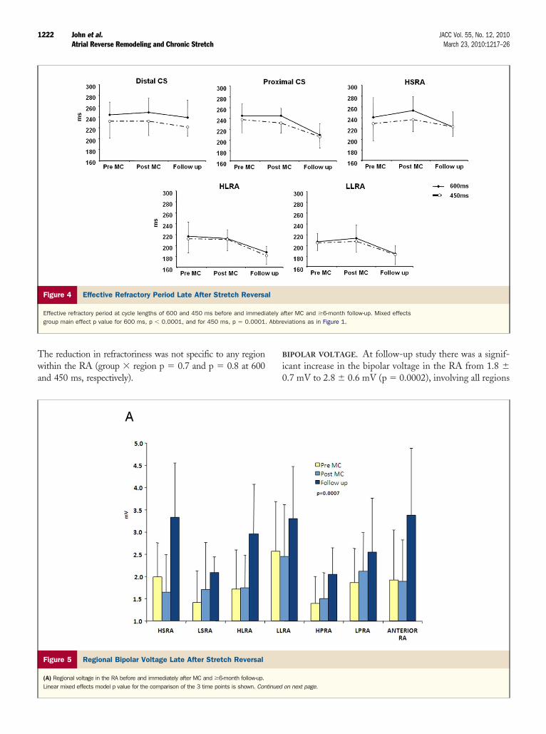
TRIAL REFRACTORINESS. At long-term follow up, the atrialRP was significantly shortened compared with the baseline
600 ms, p � 0.0001; 450 ms, p � 0.0001). Figure 4emonstrates the change in ERP over time at the various sites.
LA and RA before andAbbreviations as in Figure 1.
al
ain effect p value is shown for RA and LA. Abbreviations as in Figure 1.
al
f thend LA.
evers
oup m
Twa
B
i0
1222 John et al. JACC Vol. 55, No. 12, 2010Atrial Reverse Remodeling and Chronic Stretch March 23, 2010:1217–26
he reduction in refractoriness was not specific to any regionithin the RA (group � region p � 0.7 and p � 0.8 at 600
nd 450 ms, respectively).
Figure 5 Regional Bipolar Voltage Late After Stretch Reversal
(A) Regional voltage in the RA before and immediately after MC and �6-month follow-up.Linear mixed effects model p value for the comparison of the 3 time points is shown. Con
Figure 4 Effective Refractory Period Late After Stretch Reversa
Effective refractory period at cycle lengths of 600 and 450 ms before and immedigroup main effect p value for 600 ms, p � 0.0001, and for 450 ms, p � 0.0001.
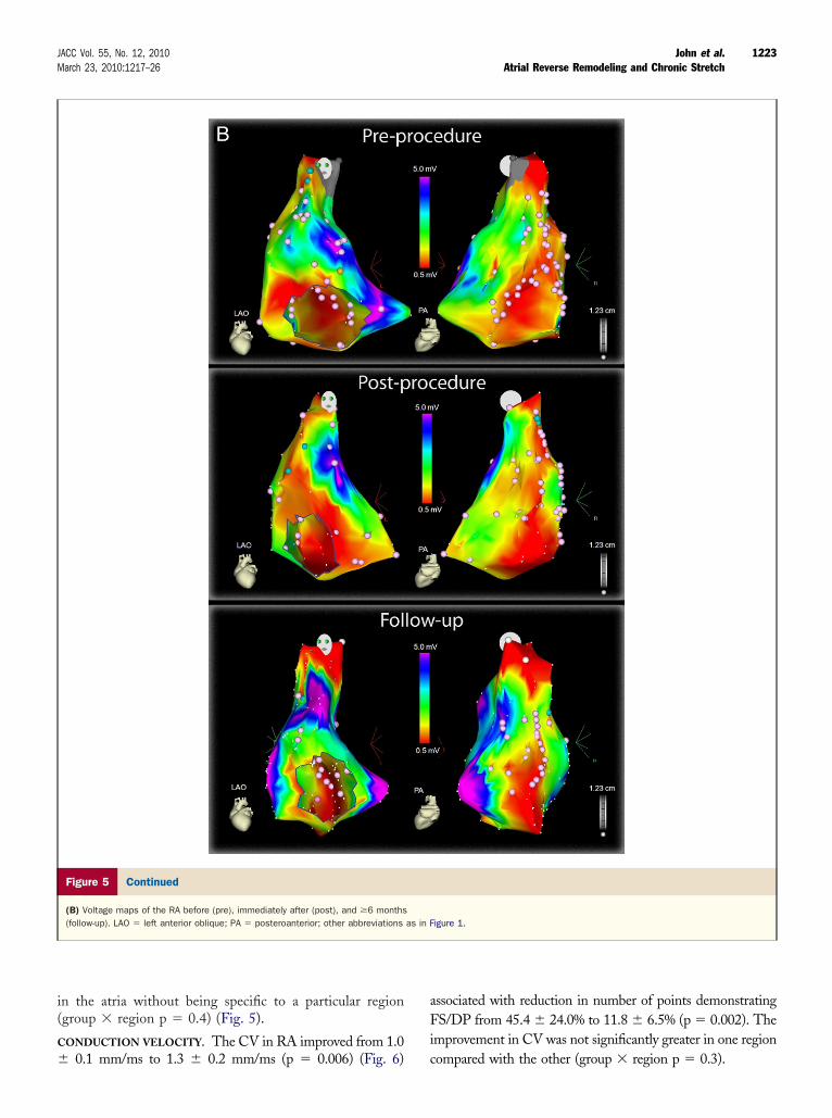
IPOLAR VOLTAGE. At follow-up study there was a signif-cant increase in the bipolar voltage in the RA from 1.8 �.7 mV to 2.8 � 0.6 mV (p � 0.0002), involving all regions
on next page.
fter MC and �6-month follow-up. Mixed effectsviations as in Figure 1.
tinued
l
ately aAbbre
i(
C
�
aFi
1223JACC Vol. 55, No. 12, 2010 John et al.March 23, 2010:1217–26 Atrial Reverse Remodeling and Chronic Stretch
n the atria without being specific to a particular regiongroup � region p � 0.4) (Fig. 5).
ONDUCTION VELOCITY. The CV in RA improved from 1.0
Figure 5 Continued
(B) Voltage maps of the RA before (pre), immediately after (post), and �6 months(follow-up). LAO � left anterior oblique; PA � posteroanterior; other abbreviations
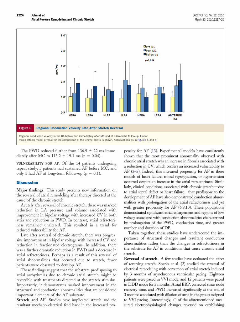
0.1 mm/ms to 1.3 � 0.2 mm/ms (p � 0.006) (Fig. 6) c
ssociated with reduction in number of points demonstratingS/DP from 45.4 � 24.0% to 11.8 � 6.5% (p � 0.002). The
mprovement in CV was not significantly greater in one region
igure 1.
as in Fompared with the other (group � region p � 0.3).
d
V
ro
D
Mtc
rianr
srwaap
arIsiSr
pscaAmoltdmwdvbn
patsRoebpir3t
1224 John et al. JACC Vol. 55, No. 12, 2010Atrial Reverse Remodeling and Chronic Stretch March 23, 2010:1217–26
The PWD reduced further from 136.9 � 22 ms imme-iately after MC to 113.2 � 19.1 ms (p � 0.04).
ULNERABILITY FOR AF. Of the 14 patients undergoingepeat study, 5 patients had sustained AF before MC, andnly 1 had AF at long-term follow-up (p � 0.1).
iscussion
ajor findings. This study presents new information onhe reversal of atrial remodeling after therapy directed at theause of the chronic stretch.
Acutely after reversal of chronic stretch, there was markededuction in LA pressure and volume associated withmprovement in bipolar voltage with increased CV in bothtria and reduction in PWD. In contrast, atrial refractori-ess remained unaltered. This resulted in a trend foreduced vulnerability for AF.
Late after reversal of chronic stretch, there was progres-ive improvement in bipolar voltage with increased CV andeduction in fractionated electrograms. In addition, thereas a further dramatic reduction in PWD and a decrease in
trial refractoriness. Perhaps as a result of this reversal oftrial abnormalities that occurred due to stretch, feweratients were observed to develop AF.These findings suggest that the substrate predisposing to
trial arrhythmias due to chronic atrial stretch might beeversible with treatments directed at the stretch stimulus.mportantly, it demonstrates marked improvement in thetructural and conduction abnormalities that are consideredmportant elements of the AF substrate.tretch and AF. Studies have implicated stretch and the
Figure 6 Regional Conduction Velocity Late After Stretch Reve
Regional conduction velocity in the RA before and immediately after MC and at �6mixed effects model p value for the comparison of the 3 time points is shown. Ab
esultant mechano-electrical feed back in the increased pro- s
ensity for AF (13). Experimental models have consistentlyhown that the most prominent abnormality observed withhronic atrial stretch was an increase in fibrosis associated withreduction in CV, which confers an increased vulnerability toF (3–5). Indeed, this increased propensity for AF in theseodels of heart failure, mitral regurgitation, or hypertension
ccurred despite an increase in the atrial refractoriness. Simi-arly, clinical conditions associated with chronic stretch—dueo atrial septal defect or heart failure—that predispose to theevelopment of AF have also demonstrated conduction abnor-alities with prolongation of the atrial refractoriness and yetith greater propensity for AF (6,9,10). These populationsemonstrated significant atrial enlargement and regions of lowoltage associated with conduction abnormalities characterizedy prolongation of the PWD, conduction time, and greaterumber and duration of DP.Taken together, these studies have underscored the im-
ortance of structural changes and resultant conductionbnormalities rather than the changes in refractoriness inhe substrate for AF in conditions that cause chronic atrialtretch.eversal of stretch. A few studies have evaluated the effect
f reversing stretch. Sparks et al. (2) studied the reversal oflectrical remodeling with correction of atrial stretch inducedy 3 months of asynchronous ventricular pacing. Eighteenatients were paced in VVI mode, and 12 patients were pacedn DDD mode for 3 months. Atrial ERP, corrected sinus nodeecovery time, and PWD increased significantly at the end ofmonths associated with dilation of atria in the group assigned
o VVI pacing. Interestingly, all of the aforementioned mea-
hs follow-up. Linearions as in Figures 1 and 4.
rsal
-montbreviat
ured electrophysiological changes reversed on establishing
atwcedrs
esag2amiaswi
stnssostMdcdarmcdw
srsdc
bfbacctio
cprtttCciTtdrtsSmererobea
ATfU
RC55
R
1225JACC Vol. 55, No. 12, 2010 John et al.March 23, 2010:1217–26 Atrial Reverse Remodeling and Chronic Stretch
trioventricular synchrony with dual-chamber pacing. Al-hough the aforementioned study demonstrated reversal after 3eeks of stretch, Morton et al. (10) observed persistence of
onduction abnormality along the crista terminalis at repeatlectrophysiological studies after closure of the atrial septalefect, suggesting that changes in conduction would noteverse by treating the underlying condition. However, thistudy was limited by the late evaluation of only 4 of 12 patients.
In patients with MS and AF, Fan et al. (14) described thelectrophysiological changes in the RA after MC. Repeattudy at 3 months after MC demonstrated an increase in thetrial ERP to become comparable to the sinus rhythmroup. Soylu et al. (15) also reported similar observations in5 patients in sinus rhythm, after MC. They measured ERPt 3 RA sites, but conduction was evaluated by surrogatearkers. Immediately after MC, they demonstrated an
ncrease in atrial ERP with improvement in the intra-atrialnd inter-atrial conduction time. Neither of these lattertudies presented any data on electroanatomic evaluation,hich provides information on CV and voltage, both
mportant elements of the AF substrate.The current study extends these observations by demon-
trating in a population in sinus rhythm, without the poten-ially confounding effects of reverse-remodeling due to termi-ation of AF, reversal of the changes due to chronic atrialtretch. Importantly, it demonstrates marked reversal of thetructural and conduction abnormalities with the consequencef reduced fractionation and AF. These data suggest that theubstrate predisposing to AF can be reversed by treatinghe underlying condition.
echanisms of atrial reverse remodeling. This study hasemonstrated marked improvement in the structuralhanges associated with chronic atrial stretch by therapyirected at the stretch stimulus. It is feasible, at least in thecute setting, that this observation might be consequent toeduction in atrial volume in the presence of constant atrialass. Thus, the increase in voltage and improvement in
onduction might in part be attributable to a mechanicaliminution of the atrial size or a reduction in the pressuresithin the chamber.There is emerging data to suggest that therapy directed at
tructural change might reduce the incidence of AF. Theenin-angiotensin system promotes cardiac fibrosis. Noturprisingly, both animal models and multicenter trials haveemonstrated reduced incidence of AF with angiotensin-onverting enzyme inhibitors (16,17).
At the molecular level, stretch activated ion channels haveeen identified as playing a crucial role in mechano-electricaleedback, which promotes AF (18,19). However, these haveeen demonstrated to be reversible; perhaps one of the mech-nisms for the observed reverse-remodeling (20). Furthermore,onnexins are known to be important determinants ofonduction and might be altered in models of AF (21). Inhe present study we observed an acute and then progressivencrease in the CV. This might relate to reverse-remodeling
f connexins. The absence of change in the number ofomplex signals immediately after stretch reversal but im-rovement at follow-up suggests that some elements of theeverse-remodeling occurs over a longer time course. Al-hough it is uncertain whether this coincides with theime-course of reverse-remodeling of connexins, it is likelyhat some of this improvement is due to such remodeling.
linical implications. Structural remodeling and the asso-iated conduction abnormalities are increasingly recognized asmportant precursors for the substrate predisposing to AF.hus, reversal of these changes could potentially have impor-
ant implications for the prevention of AF. The present studyemonstrates such marked reversal by treatment directed ateversing chronic atrial stretch. It lends weight to a strategy ofreatment of the predisposing condition to modify the AFubstrate to prevent arrhythmogenesis.tudy limitations. Although this study has demonstratedarked reversal of the AF substrate, it was only possible to
valuate the late findings in the RA. Whether similareverse remodeling would be observed in the LA was notvaluated (but suggested by the findings in immediateeversal). Finally, it is well-recognized that the developmentf clinical AF is complex and depends not only on substrateut also on other factors such as triggers and initiators. Theffect of reversal of stretch on these other factors was notddressed by this study.
cknowledgmenthe authors acknowledge the statistical assistance received
rom Mr. Thomas Sullivan, BMaCompSc (Hons), from theniversity of Adelaide.
eprint requests and correspondence: Dr. Prashanthan Sanders,ardiovascular Research Centre, Department of Cardiology, Level, McEwin Building, Royal Adelaide Hospital, Adelaide, SA000, Australia. E-mail: [email protected].
EFERENCES
1. Henry WL, Morganroth J, Pearlman AS, et al. Relation betweenechocardiographically determined left atrial size and atrial fibrillation.Circulation 1976;53:273–9.
2. Sparks PB, Mond HG, Vohra JK, Jayaprakash S, Kalman JM.Electrical remodeling of the atria following loss of atrioventricularsynchrony: a long-term study in humans. Circulation 1999;100:1894–900.
3. Li D, Fareh S, Leung TK, Nattel S. Promotion of atrial fibrillation byheart failure in dogs: atrial remodeling of a different sort. Circulation1999;100:87–95.
4. Kistler PM, Sanders P, Dodic M, et al. Atrial electrical and structuralabnormalities in an ovine model of chronic blood pressure elevationafter prenatal corticosteroid exposure: implications for development ofatrial fibrillation. Eur Heart J 2006;27:3045–56.
5. Verheule S, Wilson E, Everett T, Shanbhag S, Golden C, Olgin J.Alterations in atrial electrophysiology and tissue structure in a caninemodel of chronic atrial dilatation due to mitral regurgitation. Circu-lation 2003;107:2615–22.
6. Sanders P, Morton JB, Davidson NC, et al. Electrical remodeling ofthe atria in congestive heart failure: electrophysiological and electro-anatomic mapping in humans. Circulation 2003;108:1461–9.
7. Sanders P, Morton JB, Kistler PM, et al. Electrophysiological and
electroanatomic characterization of the atria in sinus node disease:evidence of diffuse atrial remodeling. Circulation 2004;109:1514–22.
1
1
1
1
1
1
1
1
1
1
2
2
K
1226 John et al. JACC Vol. 55, No. 12, 2010Atrial Reverse Remodeling and Chronic Stretch March 23, 2010:1217–26
8. John B, Stiles MK, Kuklik P, et al. Electrical remodelling of the leftand right atria due to rheumatic mitral stenosis. Eur Heart J 2008;29:2234–43.
9. Roberts-Thomson KC, John B, Worthley SG, et al. Left atrialremodeling in patients with atrial septal defects. Heart Rhythm2009;6:1000–6.
0. Morton JB, Sanders P, Vohra JK, et al. Effect of chronic right atrialstretch on atrial electrical remodeling in patients with an atrial septaldefect. Circulation 2003;107:1775–82.
1. Kuklik P, Szumowski L, Zebrowski JJ, Walczak F. The reconstructionand analysis of the interior surface of the heart chamber from a set ofpoints. Physiol Meas 2004;25:617–27.
2. Jais P, Shah DC, Haissaguerre M, et al. Mapping and ablation of leftatrial flutters. Circulation 2000;101:2928–34.
3. Kaseda S, Zipes DP. Contraction-excitation feedback in the atria: a causeof changes in refractoriness. J Am Coll Cardiol 1988;11:1327–36.
4. Fan K, Lee KL, Chow WH, Chau E, Lau CP. Internal cardioversionof chronic atrial fibrillation during percutaneous mitral commissurot-omy: insight into reversal of chronic stretch-induced atrial remodeling.Circulation 2002;105:2746–52.
5. Soylu M, Demir AD, Ozdemir O, et al. Evaluation of atrial refracto-riness immediately after percutaneous mitral balloon commissurotomyin patients with mitral stenosis and sinus rhythm. Am Heart J
2004;147:741–5. r6. Li D, Shinagawa K, Pang L, et al. Effects of angiotensin-convertingenzyme inhibition on the development of the atrial fibrillation sub-strate in dogs with ventricular tachypacing-induced congestive heartfailure. Circulation 2001;104:2608–14.
7. Pedersen OD, Bagger H, Kober L, Torp-Pedersen C. Trandolaprilreduces the incidence of atrial fibrillation after acute myocardialinfarction in patients with left ventricular dysfunction. Circulation1999;100:376–80.
8. Hagiwara N, Masuda H, Shoda M, Irisawa H. Stretch-activated anioncurrents of rabbit cardiac myocytes. J Physiol 1992;456:285–302.
9. Ruknudin A, Sachs F, Bustamante JO. Stretch-activated ion channelsin tissue-cultured chick heart. Am J Physiol Heart Circ Physiol1993;264:H960–72.
0. Gaborit N, Steenman M, Lamirault G, et al. Human atrial ion channeland transporter subunit gene-expression remodeling associated withvalvular heart disease and atrial fibrillation. Circulation 2005;112:471–81.
1. van der Velden HM, van Kempen MJ, Wijffels MC, et al. Alteredpattern of connexin40 distribution in persistent atrial fibrillation in thegoat. J Cardiovasc Electrophysiol 1998;9:596–607.
ey Words: atrial fibrillation y rheumatic mitral stenosis y reverse
emodeling y substrate.