Embed Size (px)
Citation preview
J Physiol 559.1 (2004) pp 205–214 205
Activation of Na+–H+ exchange and stretch-activatedchannels underlies the slow inotropic response to stretchin myocytes and muscle from the rat heart
Sarah Calaghan and Ed White
School of Biomedical Sciences, University of Leeds, Leeds LS2 9NQ, UK
We present the first direct comparison of the major candidates proposed to underlie theslow phase of the force increase seen following myocardial stretch: (i) the Na+–H+ exchanger(NHE) (ii) nitric oxide (NO) and the ryanodine receptor (RyR) and (iii) the stretch-activatedchannel (SAC) in both single myocytes and multicellular muscle preparations from therat heart. Ventricular myocytes were stretched by approximately 7% using carbon fibres.Papillary muscles were stretched from 88 to 98% of the length at which maximum tensionis generated (Lmax). Inhibition of NHE with HOE 642 (5 µM) significantly reduced (P < 0.05)the magnitude of the slow force response in both muscle and myocytes. Neither inhibitionof phosphatidylinositol-3-OH kinase (PtdIns-3-OH kinase) with LY294002 (10 µM) nor NOsynthase with L-NAME (1 mM) reduced the slow force response in muscle or myocytes(P > 0.05), and the slow response was still present in the single myocyte when the sarcoplasmicreticulum was rigorously inhibited with 1 µM ryanodine and 1 µM thapsigargin. We sawa significant reduction (P < 0.05) in the slow force response in the presence of the SACblocker streptomycin in both muscle (80 µM) and myocytes (40 µM). In fura 2-loaded myocytes,HOE 642 and streptomycin, but not L-NAME, ablated the stretch-induced increase in [Ca2+]i
transient amplitude. Our data suggest that in the rat, under our experimental conditions, thereare two mechanisms that underlie the slow inotropic response to stretch: activation of NHE;and of activation of SACs. Both these mechanisms are intrinsic to the myocyte.
(Resubmitted 28 May 2004; accepted after revision 29 June 2004; first published online 2 July 2004)Corresponding author S. Calaghan: School of Biomedical Sciences, University of Leeds, Leeds LS2 9NQ, UK.Email: [email protected]
When cardiac muscle is stretched, the force of contractionincreases allowing the intact heart to adjust cardiac outputto meet demand. The change in force upon stretch isbiphasic (for recent reviews see Calaghan et al. 2003;Cingolani et al. 2003b). Contractility increases within onebeat following stretch; this is often referred to as theFrank-Starling mechanism and is due primarily to anincrease in the sensitivity of the myofilaments to Ca2+
(Allen & Kurihara, 1982; Kentish & Wrzosek, 1998). Overthe following minutes there is a further slow increase inforce that is associated with an increase in the magnitudeof the intracellular Ca2+ ([Ca2+]i) transient (Allen &Kurihara, 1982).
This slow positive inotropic response to stretch is seen inthe intact heart (Tucci et al. 1984), in isolated ventricularand atrial muscle (Parmley & Chuck, 1973; Tavi et al.1998), and in single ventricular myocytes (Hongo et al.1996). Thus, the mechanism underlying the slow responseis intrinsic to the cardiac cell itself, although in intact
cardiac muscle it may be modified by non-myocytes suchas fibroblasts and endothelial cells. There is evidence thatcyclic AMP contributes to the slow response to stretch(e.g. Calaghan et al. 1999), although the target of proteinkinase A phosphorylation has yet to be identified. Morerecently, two candidate mechanisms for the slow responsehave received attention: the Na+–H+ exchanger (NHE;Alvarez et al. 1999; Perez et al. 2001; von Lewinski et al.2003) and nitric oxide (NO; Vila-Petroff et al. 2001).
Inhibition of NHE reduces the magnitude of the slowresponse in ventricular muscle from the rat, cat and rabbit(Alvarez et al. 1999; Perez et al. 2001; von Lewinski et al.2003) and in the failing human myocardium (von Lewinskiet al. 2004). Stretch-activation of NHE will raise [Na+]i
and there is evidence to support a subsequent stimulationof Ca2+ influx via reverse-mode Na+–Ca2+ exchange(NCX) (Perez et al. 2001; von Lewinski et al. 2003, 2004).We have previously shown that endothelin 1 plays a rolein the slow response in ferret cardiac muscle (Calaghan &
C© The Physiological Society 2004 DOI: 10.1113/jphysiol.2004.069021
206 S. Calaghan and E. White J Physiol 559.1
White, 2001), and it has been suggested that activation ofNHE is secondary to stimulation by endothelin 1 of proteinkinase C (Alvarez et al. 1999; Perez et al. 2001). However,in rabbit cardiac muscle and failing human myocardium,activation of NHE following stretch is independent of end-othelin 1 (von Lewinski et al. 2003, 2004).
Vila-Petroff et al. (2001) have presented evidence thatNO is important during the slow response. These workersobserved a slow increase in Ca2+ spark frequency and[Ca2+]i transient amplitude in single rat ventricularmyocytes stretched within an agarose gel, which wassensitive to inhibitors of NO synthase and PtdIns-3-OHkinase. A NO-dependent stimulation of RyR activityvia s-nitrosylation was proposed as the mechanism ofaction.
We consider a third contributor to the slow responseto stretch deserves attention: non-selective cationicstretch-activated channels (SACs) (see Calaghan et al.2003). Like the NHE and NCX, non-selective cationic SACsmay be responsible for bringing Na+ and/or Ca2+ intothe cardiac myocyte. Several studies have used gadolinium(Gd3+) to block SACs and from these there is evidence toboth support (Lab et al. 1994; Tavi et al. 1996) and refute(Lamberts et al. 2002b; von Lewinski et al. 2003) the roleof the SAC in the length-dependent modulation of force.
Comparison of previous studies is hampered bydifferences in species, preparation, parameters measuredand mechanisms tested for. The effect on the slow inotropicresponse to stretch of blocking NHE, NO signalling,the sarcoplasmic reticulum (SR) or SACs in myocyteshas not been measured to date. Perhaps because ofthis, a hypothesis has arisen that the major mechanismsunderlying the slow response are different in single andmulticellular preparations (Kentish, 1999; Vila-Petroffet al. 2001; Calaghan et al. 2003; Cingolani et al. 2003b).Furthermore, there are inconsistencies in the literatureregarding the role of the SR; Vila-Petroff et al. (2001) andvon Lewinski et al. (2004) suggested a major role for theSR in the slow response, whereas others (e.g. Bluhm &Lew, 1995; Kentish & Wrzosek, 1998) showed that the slowresponse is not attenuated by inhibition of SR function.
In order to resolve the above issues we have comparedthe involvement of NHE, NO signalling and SACs,under the same experimental conditions, in both singlemyocytes and multicellular preparations from the ratheart.
Methods
Male Wistar rats (250–400 g) were killed humanely bycervical dislocation following stunning and hearts wereremoved in accordance with the Home Office Guidanceon the Operation of the Animals (Scientific Procedures)Act of 1986.
Cell isolation
Ventricular myocytes were isolated enzymaticallyaccording to established methods (Calaghan et al.1998). Excised hearts were perfused sequentially withHepes-buffered physiological isolation solution at37◦C containing 0.75 mm Ca2+ (to clear the coronarycirculation), 0.1 mm EGTA (4 min), 0.8 mg ml−1 Type 1collagenase and 0.08 mg ml−1 type XIV protease (7 min).Ventricles were then excised from the heart, minced andgently shaken at 37◦C in collagenase/protease-containingisolation solution supplemented with 1% bovine serumalbumin (BSA). Myocytes were filtered from this solutionat 5 min intervals and re-suspended in isolation solutioncontaining 0.75 mm Ca2+.
Stretch of single myocytes
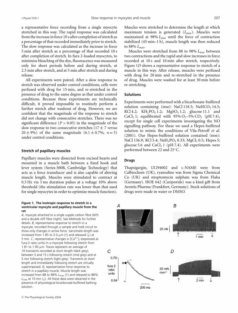
Carbon fibres were used to stretch single myocytes (seeLe Guennec et al. 1990; Hongo et al. 1996; Belus &White, 2003) in the experimental chamber of an invertedmicroscope. The base of the chamber was pre-coated witha 10% solution of BSA to reduce adherence of cells tothe chamber. One end of each myocyte was attached toa flexible single carbon fibre (diameter, 12 µm; length,1.3 mm; compliance, 22 mN−1). The other end of themyocyte was attached to a stiff double fibre (length,0.8 mm; compliance, 0.3 mN−1) (see Fig. 1). Fibreswere mounted in the ends of microelectrodes whichwere attached to micromanipulators. The degree ofdisplacement of the single fibre during auxotonic contra-ctions was used to calculate the force developed. The cellwas stretched by displacement of the stiff fibre. An image ofthe cell from a CCD camera enabled the position of fibresto be continuously monitored via a video edge-detectionsystem (Crescent Electronics, UT, USA). On-line fastFourier transformation of the cell image gave a measureof sarcomere length (Gannier et al. 1993). Change insarcomere length was used as our index of stretch.
In some cells, simultaneous measurements of [Ca2+]i
and force were made upon stretch. For these experiments,myocytes were incubated in a 2.4 µm solution of theacetoxymethyl ester form of fura 2 (fura 2-AM; MolecularProbes, OR, USA) for 10 min at room temperature,followed by resuspension in fura 2-free solution for30 min (see Calaghan et al. 1998). Fura 2 fluorescencewas measured using an Optoscan Monochromator (CairnResearch, Kent, UK). As each cell acted as its owncontrol, [Ca2+]i was expressed as the ratio of fluorescencemeasured at 510 nm following excitation at 340 nm and380 nm.
Cells were stimulated to contract at 1 Hz. We stretchedmyocytes to the greatest extent possible (within a periodof 5 s); this typically increased sarcomere length by around7%. Stretch was maintained for 5 min, after which time thecell was returned to its pre-stretch length. Figure 1B shows
C© The Physiological Society 2004
J Physiol 559.1 Slow response in myocytes and muscle 207
a representative force recording from a single myocytestretched in this way. The rapid response was calculatedfrom the increase in force 10 s after completion of stretch asa percentage of that recorded immediately prior to stretch.The slow response was calculated as the increase in force5 min after stretch as a percentage of that recorded 10 safter completion of stretch. In fura 2-loaded myocytes, tominimize bleaching of the dye, fluorescence was measuredonly for short periods before and during stretch, at2.5 min after stretch, and at 5 min after stretch and duringrelease.
All experiments were paired. After a slow response tostretch was observed under control conditions, cells wereperfused with drug for 15 min, and re-stretched in thepresence of drug to the same degree as that under controlconditions. Because these experiments are technicallydifficult, it proved impossible to routinely perform afurther stretch after washout of drug. However, we areconfident that the magnitude of the response to stretchdid not change with consecutive stretches. There was nosignificant difference (P > 0.05) in the magnitude of theslow response to two consecutive stretches (17 ± 7 versus20 ± 9%) of the same magnitude (6.1 ± 0.7%; n = 5)under control conditions.
Stretch of papillary muscles
Papillary muscles were dissected from excised hearts andmounted in a muscle bath between a fixed hook andlever system (Series 300B, Cambridge Technology) thatacts as a force transducer and is also capable of alteringmuscle length. Muscles were stimulated to contract at0.5 Hz via 5 ms duration pulses at a voltage 10% abovethreshold (the stimulation rate was lower than that usedfor single myocytes in order to optimize muscle function).
Figure 1. The inotropic response to stretch in aventricular myocyte and papillary muscle from theratA, myocyte attached to a single supple carbon fibre (left)and a double stiff fibre (right). See Methods for furtherdetails. B, representative response to stretch in amyocyte, recorded through a sample and hold circuit toshow only changes in active force. Sarcomere length wasincreased from 1.85 to 2.0 µm (↑) and released (↓) at5 min. C, representative changes in [Ca2+]i (expressed asfura-2 ratio units) in a myocyte following stretch from1.81 to 1.90 µm. Traces represent an average of10 transients recorded at short length (dark grey),between 5 and 15 s following stretch (mid grey) and at5 min following stretch (light grey). Transients at shortlength and immediately following stretch are virtuallysuperimposed. D, representative force response tostretch in a papillary muscle. Muscle length wasincreased from 88 to 98% Lmax (↑) and released to 88%Lmax at 10 min (↓). All these data were obtained in thepresence of physiological bicarbonate-buffered bathingsolution.
Muscles were stretched to determine the length at whichmaximum tension is generated (Lmax). Muscles weremaintained at 98% Lmax until the force of contractionstabilized (45 min–1 h), muscle length was then reducedto 88% Lmax.
Muscles were stretched from 88 to 98% Lmax betweentwo contractions and the rapid and slow increases in forcerecorded at 10 s and 10 min after stretch, respectively.Figure 1D shows a representative response to stretch of amuscle in this way. After release, muscles were perfusedwith drug for 20 min and re-stretched in the presenceof drug. Muscles were washed for at least 30 min beforere-stretching.
Solutions
Experiments were performed with a bicarbonate-bufferedsolution containing (mm): NaCl 118.5; NaHCO3 14.5;KCl 4.2; KH2PO4 1.2; MgSO4 1.2; glucose 11.1 andCaCl2 1; equilibrated with 95% O2–5% CO2 (pH 7.4),except for single cell experiments investigating the NOsignalling pathway. For these we used a Hepes-bufferedsolution to mimic the conditions of Vila-Petroff et al.(2001). Our Hepes-buffered solution contained (mm):NaCl 136.9; KCl 5.4; NaH2PO4 0.33; MgCl2 0.5; Hepes 5;glucose 5.6 and CaCl2 1 (pH 7.4). All experiments wereperformed between 22 and 25◦C.
Drugs
Thapsigargin, LY294002 and l-NAME were fromCalbiochem (UK), ryanodine was from Sigma ChemicalCo (UK) and streptomycin sulphate was from Fluka(Germany). HOE 642 (Cariporide) was a kind gift fromAventis Pharma (Frankfurt, Germany). Stock solutions ofdrugs were made in water or DMSO.
C© The Physiological Society 2004
208 S. Calaghan and E. White J Physiol 559.1
Table 1. The effect of antagonists on the force of contraction at short length and the rapid responseto stretch in rat papillary muscle
Force at short Rapid responselength (mN mm−2) to stretch (%)
Drug Conc (µM) n Control Drug Control Drug
HOE 642 5 6 11.2 ± 3.6 7.1 ± 5.7∗ 186 ± 58 156 ± 64L-NAME 1000 6 11.2 ± 2.3 10.4 ± 2.0 163 ± 39 161 ± 36LY294002 10 6 7.9 ± 1.8 7.0 ± 1.4 220 ± 32 256 ± 50Streptomycin 80 7 9.3 ± 2.1 8.1 ± 2.1 132 ± 24 130 ± 19
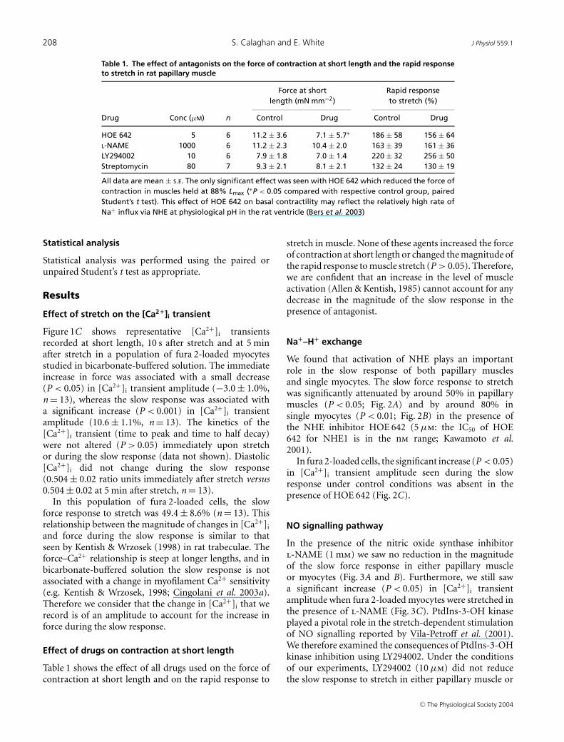
All data are mean ± S.E. The only significant effect was seen with HOE 642 which reduced the force ofcontraction in muscles held at 88% Lmax (∗P < 0.05 compared with respective control group, pairedStudent’s t test). This effect of HOE 642 on basal contractility may reflect the relatively high rate ofNa+ influx via NHE at physiological pH in the rat ventricle (Bers et al. 2003)
Statistical analysis
Statistical analysis was performed using the paired orunpaired Student’s t test as appropriate.
Results
Effect of stretch on the [Ca2+]i transient
Figure 1C shows representative [Ca2+]i transientsrecorded at short length, 10 s after stretch and at 5 minafter stretch in a population of fura 2-loaded myocytesstudied in bicarbonate-buffered solution. The immediateincrease in force was associated with a small decrease(P < 0.05) in [Ca2+]i transient amplitude (−3.0 ± 1.0%,n = 13), whereas the slow response was associated witha significant increase (P < 0.001) in [Ca2+]i transientamplitude (10.6 ± 1.1%, n = 13). The kinetics of the[Ca2+]i transient (time to peak and time to half decay)were not altered (P > 0.05) immediately upon stretchor during the slow response (data not shown). Diastolic[Ca2+]i did not change during the slow response(0.504 ± 0.02 ratio units immediately after stretch versus0.504 ± 0.02 at 5 min after stretch, n = 13).
In this population of fura 2-loaded cells, the slowforce response to stretch was 49.4 ± 8.6% (n = 13). Thisrelationship between the magnitude of changes in [Ca2+]i
and force during the slow response is similar to thatseen by Kentish & Wrzosek (1998) in rat trabeculae. Theforce–Ca2+ relationship is steep at longer lengths, and inbicarbonate-buffered solution the slow response is notassociated with a change in myofilament Ca2+ sensitivity(e.g. Kentish & Wrzosek, 1998; Cingolani et al. 2003a).Therefore we consider that the change in [Ca2+]i that werecord is of an amplitude to account for the increase inforce during the slow response.
Effect of drugs on contraction at short length
Table 1 shows the effect of all drugs used on the force ofcontraction at short length and on the rapid response to
stretch in muscle. None of these agents increased the forceof contraction at short length or changed the magnitude ofthe rapid response to muscle stretch (P > 0.05). Therefore,we are confident that an increase in the level of muscleactivation (Allen & Kentish, 1985) cannot account for anydecrease in the magnitude of the slow response in thepresence of antagonist.
Na+–H+ exchange
We found that activation of NHE plays an importantrole in the slow response of both papillary musclesand single myocytes. The slow force response to stretchwas significantly attenuated by around 50% in papillarymuscles (P < 0.05; Fig. 2A) and by around 80% insingle myocytes (P < 0.01; Fig. 2B) in the presence ofthe NHE inhibitor HOE 642 (5 µm: the IC50 of HOE642 for NHE1 is in the nm range; Kawamoto et al.2001).
In fura 2-loaded cells, the significant increase (P < 0.05)in [Ca2+]i transient amplitude seen during the slowresponse under control conditions was absent in thepresence of HOE 642 (Fig. 2C).
NO signalling pathway
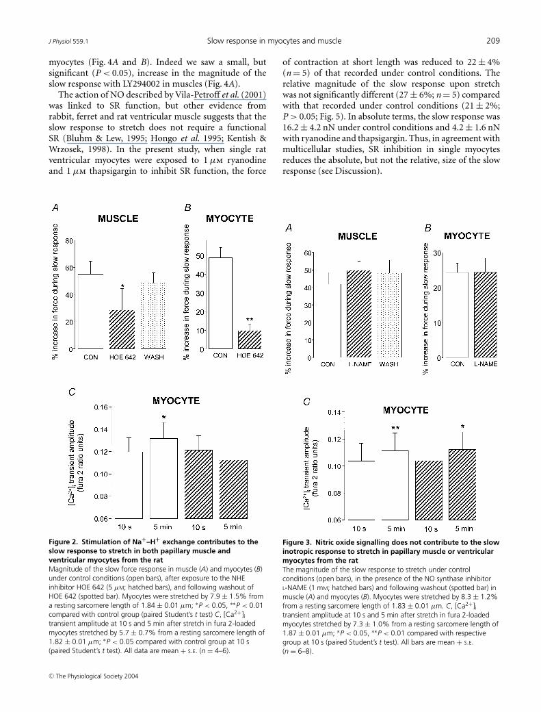
In the presence of the nitric oxide synthase inhibitorl-NAME (1 mm) we saw no reduction in the magnitudeof the slow force response in either papillary muscleor myocytes (Fig. 3A and B). Furthermore, we still sawa significant increase (P < 0.05) in [Ca2+]i transientamplitude when fura 2-loaded myocytes were stretched inthe presence of l-NAME (Fig. 3C). PtdIns-3-OH kinaseplayed a pivotal role in the stretch-dependent stimulationof NO signalling reported by Vila-Petroff et al. (2001).We therefore examined the consequences of PtdIns-3-OHkinase inhibition using LY294002. Under the conditionsof our experiments, LY294002 (10 µm) did not reducethe slow response to stretch in either papillary muscle or
C© The Physiological Society 2004
J Physiol 559.1 Slow response in myocytes and muscle 209
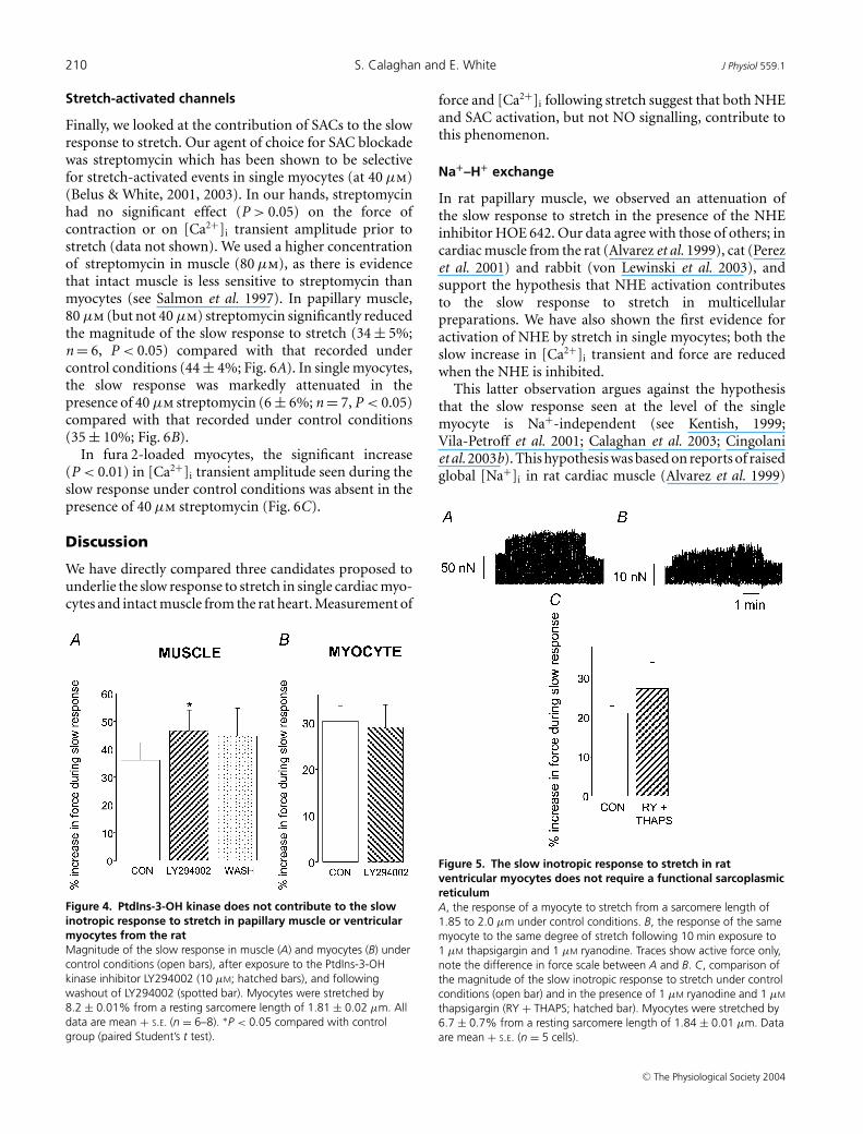
myocytes (Fig. 4A and B). Indeed we saw a small, butsignificant (P < 0.05), increase in the magnitude of theslow response with LY294002 in muscles (Fig. 4A).
The action of NO described by Vila-Petroff et al. (2001)was linked to SR function, but other evidence fromrabbit, ferret and rat ventricular muscle suggests that theslow response to stretch does not require a functionalSR (Bluhm & Lew, 1995; Hongo et al. 1995; Kentish &Wrzosek, 1998). In the present study, when single ratventricular myocytes were exposed to 1 µm ryanodineand 1 µm thapsigargin to inhibit SR function, the force
Figure 2. Stimulation of Na+–H+ exchange contributes to theslow response to stretch in both papillary muscle andventricular myocytes from the ratMagnitude of the slow force response in muscle (A) and myocytes (B)under control conditions (open bars), after exposure to the NHEinhibitor HOE 642 (5 µM; hatched bars), and following washout ofHOE 642 (spotted bar). Myocytes were stretched by 7.9 ± 1.5% froma resting sarcomere length of 1.84 ± 0.01 µm; ∗P < 0.05, ∗∗P < 0.01compared with control group (paired Student’s t test) C, [Ca2+]itransient amplitude at 10 s and 5 min after stretch in fura 2-loadedmyocytes stretched by 5.7 ± 0.7% from a resting sarcomere length of1.82 ± 0.01 µm; ∗P < 0.05 compared with control group at 10 s(paired Student’s t test). All data are mean + S.E. (n = 4–6).
of contraction at short length was reduced to 22 ± 4%(n = 5) of that recorded under control conditions. Therelative magnitude of the slow response upon stretchwas not significantly different (27 ± 6%; n = 5) comparedwith that recorded under control conditions (21 ± 2%;P > 0.05; Fig. 5). In absolute terms, the slow response was16.2 ± 4.2 nN under control conditions and 4.2 ± 1.6 nNwith ryanodine and thapsigargin. Thus, in agreement withmulticellular studies, SR inhibition in single myocytesreduces the absolute, but not the relative, size of the slowresponse (see Discussion).
Figure 3. Nitric oxide signalling does not contribute to the slowinotropic response to stretch in papillary muscle or ventricularmyocytes from the ratThe magnitude of the slow response to stretch under controlconditions (open bars), in the presence of the NO synthase inhibitorL-NAME (1 mM; hatched bars) and following washout (spotted bar) inmuscle (A) and myocytes (B). Myocytes were stretched by 8.3 ± 1.2%from a resting sarcomere length of 1.83 ± 0.01 µm. C, [Ca2+]itransient amplitude at 10 s and 5 min after stretch in fura 2-loadedmyocytes stretched by 7.3 ± 1.0% from a resting sarcomere length of1.87 ± 0.01 µm; ∗P < 0.05, ∗∗P < 0.01 compared with respectivegroup at 10 s (paired Student’s t test). All bars are mean + S.E.(n = 6–8).
C© The Physiological Society 2004
210 S. Calaghan and E. White J Physiol 559.1
Stretch-activated channels
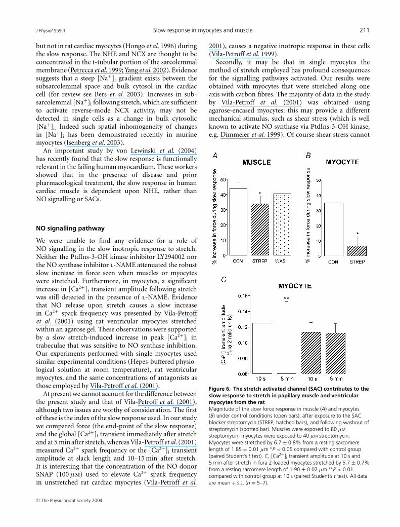
Finally, we looked at the contribution of SACs to the slowresponse to stretch. Our agent of choice for SAC blockadewas streptomycin which has been shown to be selectivefor stretch-activated events in single myocytes (at 40 µm)(Belus & White, 2001, 2003). In our hands, streptomycinhad no significant effect (P > 0.05) on the force ofcontraction or on [Ca2+]i transient amplitude prior tostretch (data not shown). We used a higher concentrationof streptomycin in muscle (80 µm), as there is evidencethat intact muscle is less sensitive to streptomycin thanmyocytes (see Salmon et al. 1997). In papillary muscle,80 µm (but not 40 µm) streptomycin significantly reducedthe magnitude of the slow response to stretch (34 ± 5%;n = 6, P < 0.05) compared with that recorded undercontrol conditions (44 ± 4%; Fig. 6A). In single myocytes,the slow response was markedly attenuated in thepresence of 40 µm streptomycin (6 ± 6%; n = 7, P < 0.05)compared with that recorded under control conditions(35 ± 10%; Fig. 6B).
In fura 2-loaded myocytes, the significant increase(P < 0.01) in [Ca2+]i transient amplitude seen during theslow response under control conditions was absent in thepresence of 40 µm streptomycin (Fig. 6C).
Discussion
We have directly compared three candidates proposed tounderlie the slow response to stretch in single cardiac myo-cytes and intact muscle from the rat heart. Measurement of
Figure 4. PtdIns-3-OH kinase does not contribute to the slowinotropic response to stretch in papillary muscle or ventricularmyocytes from the ratMagnitude of the slow response in muscle (A) and myocytes (B) undercontrol conditions (open bars), after exposure to the PtdIns-3-OHkinase inhibitor LY294002 (10 µM; hatched bars), and followingwashout of LY294002 (spotted bar). Myocytes were stretched by8.2 ± 0.01% from a resting sarcomere length of 1.81 ± 0.02 µm. Alldata are mean + S.E. (n = 6–8). ∗P < 0.05 compared with controlgroup (paired Student’s t test).
force and [Ca2+]i following stretch suggest that both NHEand SAC activation, but not NO signalling, contribute tothis phenomenon.
Na+–H+ exchange
In rat papillary muscle, we observed an attenuation ofthe slow response to stretch in the presence of the NHEinhibitor HOE 642. Our data agree with those of others; incardiac muscle from the rat (Alvarez et al. 1999), cat (Perezet al. 2001) and rabbit (von Lewinski et al. 2003), andsupport the hypothesis that NHE activation contributesto the slow response to stretch in multicellularpreparations. We have also shown the first evidence foractivation of NHE by stretch in single myocytes; both theslow increase in [Ca2+]i transient and force are reducedwhen the NHE is inhibited.
This latter observation argues against the hypothesisthat the slow response seen at the level of the singlemyocyte is Na+-independent (see Kentish, 1999;Vila-Petroff et al. 2001; Calaghan et al. 2003; Cingolaniet al. 2003b). This hypothesis was based on reports of raisedglobal [Na+]i in rat cardiac muscle (Alvarez et al. 1999)
Figure 5. The slow inotropic response to stretch in ratventricular myocytes does not require a functional sarcoplasmicreticulumA, the response of a myocyte to stretch from a sarcomere length of1.85 to 2.0 µm under control conditions. B, the response of the samemyocyte to the same degree of stretch following 10 min exposure to1 µM thapsigargin and 1 µM ryanodine. Traces show active force only,note the difference in force scale between A and B. C, comparison ofthe magnitude of the slow inotropic response to stretch under controlconditions (open bar) and in the presence of 1 µM ryanodine and 1 µM
thapsigargin (RY + THAPS; hatched bar). Myocytes were stretched by6.7 ± 0.7% from a resting sarcomere length of 1.84 ± 0.01 µm. Dataare mean + S.E. (n = 5 cells).
C© The Physiological Society 2004
J Physiol 559.1 Slow response in myocytes and muscle 211
but not in rat cardiac myocytes (Hongo et al. 1996) duringthe slow response. The NHE and NCX are thought to beconcentrated in the t-tubular portion of the sarcolemmalmembrane (Petrecca et al. 1999; Yang et al. 2002). Evidencesuggests that a steep [Na+]i gradient exists between thesubsarcolemmal space and bulk cytosol in the cardiaccell (for review see Bers et al. 2003). Increases in sub-sarcolemmal [Na+]i following stretch, which are sufficientto activate reverse-mode NCX activity, may not bedetected in single cells as a change in bulk cytosolic[Na+]i. Indeed such spatial inhomogeneity of changesin [Na+]i has been demonstrated recently in murinemyocytes (Isenberg et al. 2003).
An important study by von Lewinski et al. (2004)has recently found that the slow response is functionallyrelevant in the failing human myocardium. These workersshowed that in the presence of disease and priorpharmacological treatment, the slow response in humancardiac muscle is dependent upon NHE, rather thanNO signalling or SACs.
NO signalling pathway
We were unable to find any evidence for a role ofNO signalling in the slow inotropic response to stretch.Neither the PtdIns-3-OH kinase inhibitor LY294002 northe NO synthase inhibitor l-NAME attenuated the robustslow increase in force seen when muscles or myocyteswere stretched. Furthermore, in myocytes, a significantincrease in [Ca2+]i transient amplitude following stretchwas still detected in the presence of l-NAME. Evidencethat NO release upon stretch causes a slow increasein Ca2+ spark frequency was presented by Vila-Petroffet al. (2001) using rat ventricular myocytes stretchedwithin an agarose gel. These observations were supportedby a slow stretch-induced increase in peak [Ca2+]i intrabeculae that was sensitive to NO synthase inhibition.Our experiments performed with single myocytes usedsimilar experimental conditions (Hepes-buffered physio-logical solution at room temperature), rat ventricularmyocytes, and the same concentrations of antagonists asthose employed by Vila-Petroff et al. (2001).
At present we cannot account for the difference betweenthe present study and that of Vila-Petroff et al. (2001),although two issues are worthy of consideration. The firstof these is the index of the slow response used. In our studywe compared force (the end-point of the slow response)and the global [Ca2+]i transient immediately after stretchand at 5 min after stretch, whereas Vila-Petroff et al. (2001)measured Ca2+ spark frequency or the [Ca2+]i transientamplitude at slack length and 10–15 min after stretch.It is interesting that the concentration of the NO donorSNAP (100 µm) used to elevate Ca2+ spark frequencyin unstretched rat cardiac myocytes (Vila-Petroff et al.
2001), causes a negative inotropic response in these cells(Vila-Petroff et al. 1999).
Secondly, it may be that in single myocytes themethod of stretch employed has profound consequencesfor the signalling pathways activated. Our results wereobtained with myocytes that were stretched along oneaxis with carbon fibres. The majority of data in the studyby Vila-Petroff et al. (2001) was obtained usingagarose-encased myocytes: this may provide a differentmechanical stimulus, such as shear stress (which is wellknown to activate NO synthase via PtdIns-3-OH kinase;e.g. Dimmeler et al. 1999). Of course shear stress cannot
Figure 6. The stretch activated channel (SAC) contributes to theslow response to stretch in papillary muscle and ventricularmyocytes from the ratMagnitude of the slow force response in muscle (A) and myocytes(B) under control conditions (open bars), after exposure to the SACblocker streptomycin (STREP; hatched bars), and following washout ofstreptomycin (spotted bar). Muscles were exposed to 80 µM
streptomycin; myocytes were exposed to 40 µM streptomycin.Myocytes were stretched by 6.7 ± 0.8% from a resting sarcomerelength of 1.85 ± 0.01 µm ∗P < 0.05 compared with control group(paired Student’s t test). C, [Ca2+]i transient amplitude at 10 s and5 min after stretch in fura 2-loaded myocytes stretched by 5.7 ± 0.7%from a resting sarcomere length of 1.90 ± 0.02 µm ∗∗P < 0.01compared with control group at 10 s (paired Student’s t test). All dataare mean + S.E. (n = 5–7).
C© The Physiological Society 2004
212 S. Calaghan and E. White J Physiol 559.1
account for the observation that NO synthase inhibitionabolishes the slow increase in Ca2+ transient seen followingstretch in rat trabeculae (Vila-Petroff et al. 2001). However,our work does not stand alone in refuting the central roleof the SR (and by implication NO signalling) in the slowresponse (see below).
Our data suggest that PtdIns-3-OH kinase, NO ands-nitrosylation of the RyR do not play a pivotal role inthe slow inotropic response in the rat heart. In supportof this conclusion, the relative magnitude of the slowresponse is unaffected by inhibition of SR function inintact muscle from the rabbit and rat (Bluhm & Lew, 1995;Kentish & Wrzosek, 1998) and in the single rat cardiacmyocyte (the present study). If there were an increase inSR gain as a result of s-nitrosylation of the RyR, we wouldexpect the relative magnitude of the slow response to bereduced when the SR was non-functional. Furthermore,Eisner & Trafford (2000) have shown that enhanced RyRactivity results in only transient changes in [Ca2+]i andcontraction, and for the temporal characteristics of theslow response to be fulfilled, there would have to be aconcurrent increase in SR Ca2+ loading to compensate forthe increase in SR Ca2+ release. This could occur throughactivation of NHE (and SACs, see below) and stimulationof reverse mode NCX activity. However, in that case it issurprising that near total ablation of the slow response isseen when either the NHE (Perez et al. 2001) or the NOpathway (Vila-Petroff et al. 2001) is inhibited. This doesnot suggest that these two mechanisms are operating inparallel.
Whilst the data obtained under our experimentalconditions suggest that the SR is not required for theslow response to stretch, it is logical that a functional SRwill handle any extra Ca2+ that is brought into the cell(either via reverse mode NCX or via SACs, see below).This conclusion is supported by the stretch-inducedincrease in SR load inferred from rapid cooling contracturestudies of Bluhm & Lew (1995) and von Lewinski et al.(2003).
Stretch-activated channels
Using a mathematical model of the atrial myocyte, Taviet al. (1998) have shown that the slow increase in[Ca2+]i seen upon stretch of rat atrial muscle can bemimicked by introducing a non-selective cationic SACconductance. Experimentally, several studies have reliedon the SAC blocker Gd3+ to examine the role of SACsin the stretch-dependent modulation of cardiac contra-ctility. Lab et al. (1994) and Tavi et al. (1996) reporteda length-dependent depression of contractility by Gd3+
under conditions where the slow response mechanismswould be activated (i.e. after several minutes of continuousstretch). However, also using Gd3+, Lamberts et al. (2002b)and von Lewinski et al. (2003, 2004) concluded that there
was no role for SACs in the slow response in rat, rabbitor failing human cardiac muscle. Interpreting positiveand negative results with Gd3+ is difficult because freeGd3+ blocks many types of ion channels and exchangersand binds to anions such as HCO3−, and also becausethere is evidence from several studies that the Gd-anioncomplex can block stretch-activated events, perhaps withmore specificity than free Gd3+ (see Caldwell et al. 1998;Lamberts et al. 2002a).
In an alternative approach, we elected to usestreptomycin to block SACs. Streptomycin blocksstretch-activated currents at 40 µm (Belus & White, 2003),but does not affect the action potential duration (thesum of all electrogenic processes), [Ca2+]i transient orcontraction amplitude at short length in myocytes at50 µm (Belus & White, 2001, 2003). We also saw nosignificant effect of 40 µm streptomycin on force or [Ca2+]i
in unstretched cells. Neither did 80 µm streptomycinmodulate contractility in the absence of stretch in muscle;Salmon et al. (1997) have reported similar findings with200 µm streptomycin in the intact heart.
We saw a significant attenuation of the slow responsein both myocytes and intact papillary muscles in thepresence of streptomycin. Using arterially perfused ratpapillary muscle, Lamberts et al. have shown that theincrease in developed force seen in response to increasedperfusion pressure (the Gregg effect) is attenuated by40 µm streptomycin (2002a), although a reduction in theamplitude of the slow response by streptomycin (40 and100 µm) was not statistically significant (2002b).
The effect of streptomycin that we observe is consistentwith activation of non-selective cationic SACs whichconduct Na+, K+ and Ca2+. In the present study we sawno change in diastolic [Ca2+]i during the slow responsein the rat, in agreement with Kentish & Wrzosek (1998),Hongo et al. (1996) and Alvarez et al. (1999). Thisreinforces the idea that increased Ca2+ loading takes placeduring systole. The effects of KB-R7943 on the slowresponse from both feline and rabbit cardiac muscle (Perezet al. 2001; von Lewinski et al. 2003) are consistent withincreased Ca2+ influx via reverse-mode NCX, secondary toNa+ loading (potentially through both SACs and theNHE). The sequential change in [Na+]i then [Ca2+]i
may account for the temporal characteristics of increased[Ca2+]i and force during the slow response (see Bluhmet al. 1998). Han et al. (2002) have shown, using computermodelling, that activation of non-selective cationic SACsand stimulation of NHE have qualitatively similar effectson Ca2+ handling in cardiac cells. Ca2+ which entersthrough SACs may also contribute to the slow increasein Ca2+ transient seen following stretch.
Conclusions
We have compared directly the major candidatemechanisms for the slow response in both multicellular
C© The Physiological Society 2004
J Physiol 559.1 Slow response in myocytes and muscle 213
and single myocyte preparations. Under our experimentalconditions we find that the slow response to stretch isa sarcolemmal-based response that relies on activationof two pathways, NHE and SACs. The mechanisms thatunderlie the slow response are qualitatively similar in thesingle myocyte and intact muscle.
References
Allen DG & Kentish JC (1985). The cellular basis of the length-tension relation in cardiac muscle. J Mol Cell Cardiol 17,821–840.
Allen DG & Kurihara S (1982). The effects of muscle length onintracellular calcium transients in mammalian cardiacmuscle. J Physiol 327, 79–94.
Alvarez BV, Perez NG, Ennis IL, Camilion de Hurtado MC &Cingolani HE (1999). Mechanisms underlying the increasein force and Ca2+ transient that follow stretch of cardiacmuscle: a possible explanation of the Anrep effect. Circ Res85, 716–722.
Belus A & White E (2001). Effects of antibiotics on thecontractility and Ca2+ transients of rat cardiac myocytes.Eur J Pharmacol 412, 121–126.
Belus A & White E (2003). Streptomycin and intracellularcalcium modulate the response of single guinea-pigventricular myocytes to axial stretch. J Physiol 546,501–509.
Bers DM, Barry WH & Despa S (2003). Intracellular Na+regulation in cardiac myocytes. Cardiovasc Res 57, 897–912.
Bluhm WF & Lew WYW (1995). Sarcoplasmic reticulum incardiac length-dependent activation in rabbits. Am J Physiol269, H965–H972.
Bluhm WF, Lew WY, Garfinkel A & McCulloch AD (1998).Mechanisms of length history-dependent tension in an ionicmodel of the cardiac myocyte. Am J Physiol 274,H1032–H1040.
Calaghan SC, Belus A & White E (2003). Do stretch-inducedchanges in intracellular calcium modify the electrical activityof cardiac muscle? Prog Biophys Mol Biol 82, 81–95.
Calaghan SC, Colyer J & White E (1999). Cyclic AMP but notphosphorylation of phospholamban contributes to the slowinotropic response to stretch in ferret papillary muscle.Pflugers Arch 437, 780–782.
Calaghan SC & White E (2001). Contribution of angiotensin II,endothelin 1 and the endothelium to the slow inotropicresponse to stretch in ferret papillary muscle. Pflugers Arch441, 514–520.
Calaghan SC, White E & Colyer J (1998). Co-ordinated changesin cAMP, phosphorylated phospholamban, Ca2+ andcontraction following beta-adrenergic stimulation of ratheart. Pflugers Arch 436, 948–956.
Caldwell RA, Clemo HF & Baumgarten CM (1998). Usinggadolinium to identify stretch-activated channels: technicalconsiderations. Am J Physiol 275, C619–C621.
Cingolani HE, Chiappe GE, Ennis IL, Morgan PG, Alvarez BV,Casey JR, Dulce RA, Perez NG & Camilion de Hurtado MC(2003a). Influence of Na+-independent Cl− HCO3
−exchange on the slow force response to myocardial stretch.Circ Res 93, 1082–1088.
Cingolani HE, Perez NG, Pieske B, von Lewinski D & Camilionde Hurtado MC (2003b). Stretch-elicited Na+/H+ exchangeractivation: the autocrine/paracrine loop and its mechanicalcounterpart. Cardiovasc Res 57, 953–960.
Dimmeler S, Fleming I, Fisslthaler B, Hermann C, Busse R &Zeiher AM (1999). Activation of nitric oxide synthase inendothelial cells by Akt-dependent phosphorylation. Nature399, 601–605.
Eisner DA & Trafford AW (2000). No role for the ryanodinereceptor in regulating cardiac contraction? News Physiol Sci15, 275–279.
Gannier F, Bernengo JC, Jacquemond V & Garnier D (1993).Measurements of sarcomere dynamics simultaneously withauxotonic force in isolated cardiac cells. IEEE Trans BiomedEng 40, 1226–1232.
Han C, Tavi P & Weckstrom M (2002). Modulation of actionpotential by [Ca2+]i in modelled rat atrial and guinea pigventricular myocytes. Am J Physiol 282,H1047–H1054.
Hongo K, White E, Le Guennec JY & Orchard CH (1996).Changes in [Ca2+]i, [Na+]i and Ca2+ current in isolated ratventricular myocytes following an increase in cell length.J Physiol 491, 609–619.
Hongo K, White E & Orchard CH (1995). Effect of stretch oncontraction and the Ca2+ transient in ferret ventricularmuscles during hypoxia and acidosis. Am J Physiol 269,C690–C697.
Isenberg G, Kazanski V, Kondratev D, Gallitelli MF, Kiseleva I& Kamkin A (2003). Differential effects of stretch andcompression on membrane currents and [Na+]c inventricular myocytes. Prog Biophys Mol Biol 82, 43–56.
Kawamoto T, Kimura H, Kusumoto K, Fukumoto S, ShiraishiM, Watanabe T & Sawada H (2001). Potent and selectiveinhibition of the human Na+/H+ exchanger isoform NHE1by a novel aminoguanidine derivative T-162559.Eur J Pharmacol 240, 1–8.
Kentish JC (1999). A role for the sarcolemmal Na+/H+exchanger in the slow force response to myocardial stretch.Circ Res 85, 658–660.
Kentish JC & Wrzosek A (1998). Changes in force and cytosolicCa2+ concentration after length changes in isolated ratventricular trabeculae. J Physiol 506, 431–444.
Lab MJ, Zhou BY, Spencer CI, Horner SM & Seed WA (1994).Effects of gadolinium on length-dependent force inguinea-pig papillary muscle. Exp Physiol 79, 249–255.
Lamberts RR, Rijen MH, Sipkema P, Fransen P, Sys SU &Westerhof N (2002a). Increased coronary perfusionaugments cardiac contractility in the rat throughstretch-activated ion channels. Am J Physiol 282,H1334–H1340.
Lamberts RR, Van Rijen MH, Sipkema P, Fransen P, Sys SU &Westerhof N (2002b). Coronary perfusion and musclelengthening increase cardiac contraction: differentstretch-triggered mechanisms. Am J Physiol 283,H1515–H1522.
Le Guennec JY, Peineau N, Argibay JA, Mongo KG & Garnier D(1990). A new method of attachment of isolated mammalianventricular myocytes for tension recording: lengthdependence of passive and active tension. J Mol Cell Cardiol22, 1083–1093.
C© The Physiological Society 2004
214 S. Calaghan and E. White J Physiol 559.1
Parmley WW & Chuck L (1973). Length-dependent changes inmyocardial contractile state. Am J Physiol 224, 1195–1199.
Perez NG, de Hurtado MC & Cingolani HE (2001). Reversemode of the Na+-Ca2+ exchange after myocardial stretch:underlying mechanism of the slow force response. Circ Res88, 376–382.
Petrecca K, Atanasiu R, Grinstein S, Orlowski J & Shrier A(1999). Subcellular localization of the Na+/H+ exchangerNHE1 in rat myocardium. Am J Physiol 276, H709–H717.
Salmon AH, Mays JL, Dalton GR, Jones JV & Levi AJ (1997).Effect of streptomycin on wall-stress-induced arrhythmias inthe working rat heart. Cardiovasc Res 34, 493–503.
Tavi P, Han C & Weckstrom M (1998). Mechanisms ofstretch-induced changes in [Ca2+]i in rat atrial myocytes.Role of increased troponin C affinity and stretch-activatedion channels. Circ Res 83, 1165–1177.
Tavi P, Laine M & Weckstrom M (1996). Effect of gadoliniumon stretch-induced changes in contraction andintracellularly recorded action- and afterpotentials of ratisolated atrium. Br J Pharmacol 118, 407–413.
Tucci PJF, Bregagnollo EA, Spadaro J, Cicogna AC & RibeiroMCL (1984). Length-dependence of activiation in theisovolumic blood-perfused dog heart. Circ Res 55, 59–66.
Vila-Petroff MG, Kim SH, Pepe S, Dessy C, Marban E,Balligand JL & Sollott SJ (2001). Endogenous nitric oxidemechanisms mediate the stretch dependence of Ca2+ releasein cardiomyocytes. Nat Cell Biol 3, 867–873.
Vila-Petroff MG, Younes A, Egan J, Lakatta EG & Sollott SJ(1999). Activation of distinct cAMP-dependent andcGMP-dependent pathways by nitric oxide in cardiacmyocytes. Circ Res 84, 1020–1031.
von Lewinski D, Stumme B, Fialka F, Luers C & Pieske B(2004). Functional relevance of the stretch-dependent slowforce response in failing human myocardium. Circ Res 94,1392–1398.
von Lewinski D, Stumme B, Maier LS, Luers C, Bers DM &Pieske B (2003). Stretch-dependent slow force response inisolated rabbit myocardium is Na+ dependent. CardiovascRes 57, 1052–1061.
Yang Z, Pascarel C, Steele DS, Komukai K, Brette F & OrchardCH (2002). Na+-Ca2+ exchange activity is localized in theT-tubules of rat ventricular myocytes. Circ Res 91, 315–322.
Acknowledgements
This work was sponsored by the British Heart Foundation.
C© The Physiological Society 2004