Embed Size (px)
Citation preview
Structure-based discovery of an inhibitor of Arfactivation by Sec7 domains through targetingof protein–protein complexesJulien Viaud*†, Mahel Zeghouf‡, Helene Barelli§, Jean-Christophe Zeeh‡, Andre Padilla*†, Bernard Guibert‡,Pierre Chardin§, Catherine A. Royer*†, Jacqueline Cherfils‡¶, and Alain Chavanieu*†
*Institut National de la Sante et de la Recherche Medicale, U554 and †Universite Montpellier 1 et 2, Centre National de la Recherche Scientifique, UniteMixte de Recherche 5048, Centre de Biochimie Structurale, 34090 Montpellier, France; ‡Laboratoire d’Enzymologie et Biochimie Structurales, CentreNational de la Recherche Scientifique, Avenue de la Terrasse, 91198 Gif sur Yvette Cedex, France; and §Institut de Pharmacologie Moleculaire etCellulaire, Centre National de la Recherche Scientifique–Unite Mixte de Recherche 6097, 660 Route des Lucioles, 06560 Valbonne, France
Edited by Axel T. Brunger, Stanford University, Stanford, CA, and approved May 7, 2007 (received for review February 1, 2007)
Small molecules that produce nonfunctional protein–protein com-plexes are an alternative to competitive inhibitors for the inhibitionof protein functions. Here we target the activation of the smallGTP-binding protein Arf1, a major regulator of membrane traffic, bythe Sec7 catalytic domain of its guanine nucleotide exchange factorARNO. The crystal structure of the Arf1-GDP/ARNO complex, whichinitiates the exchange reaction, was used to discover an inhibitor,LM11, using in silico screening of a flexible pocket near the Arf1/ARNO interface. Using fluorescence kinetics and anisotropy, NMRspectroscopy and mutagenesis, we show that LM11 acts following anoncompetitive mechanism in which the inhibitor targets both Arf1-GDP and the Arf1-GDP/ARNO complex and produces a nonfunctionalArf-GDP/ARNO complex whose affinity is similar to that of the nativecomplex. In addition, LM11 recognizes features of both Arf and ARNOnear the Arf/Sec7 interface, a characteristic reminiscent of the para-digm interfacial inhibitor Brefeldin A. We then show that LM11 is acell-active inhibitor that impairs Arf-dependent trafficking structuresat the Golgi. Furthermore, LM11 inhibits ARNO-dependent migrationof Madin–Darby canine kidney (MDCK) cells, demonstrating thatARNO is a target of LM11 in cells. Remarkably, LM11 inhibits theactivation of Arf1 but not Arf6 in vitro, pointing to a possible synergybetween Arf1 and Arf6 activation by ARNO in cell migration. Ourdesign method shows that flexible regions in protein–protein com-plexes provide drugable sites with the potential to develop noveltools for investigating and inhibiting signaling pathways.
protein–protein interactions � inhibition � GTPase � guanine nucleotideexchage factor � Arf1 factor
The discovery of small molecules that affect protein interactionsis of crucial importance for the development of innovative
therapeutics (1–3) and for the investigation of molecular pathwaysin cells (4, 5). However, targeting protein interactions by compet-itive inhibitors is difficult, because small molecules must competewith large macromolecular partners (3). We recently proposed aconcept to inhibit protein functions, which we refer to as ‘‘interfacialinhibition’’ (6, 7). Interfacial inhibitors bind to protein–proteincomplexes in or near their interface in the course of structuraltransitions, thereby converting the complexes into abortive confor-mations rather than preventing the interaction of their components.Nature provides most examples of interfacial inhibitors, of whichsome are used in human therapeutics, such as colchicine, vinblas-tine, or camptothecin (7). The critical issue, however, is how todiscover or design inhibitors that promote inactive conformationsof protein–protein complexes. In this study, we focused on theactivation of a small GTP-binding protein (SMG) by its guaninenucleotide exchange factors (GEFs). This reaction is of particularinterest, because GEFs define the spatiotemporal specificity ofSMG activation by collecting activation signals and stimulating theintrinsically slow GDP/GTP exchange. GEFs are therefore emerg-ing as potential targets in diseases where SMGs are up-regulated,
such as cancer or infections (reviewed in refs. 8 and 9). This recentlyfostered the exploration of novel strategies for discovering com-petitive inhibitors acting either on the GEF (10–12) or on the SMG(13, 14). An alternative strategy, independent of the nature of theinhibition mechanism, was also devised by using an exchange assayreconstituted in yeast (15).
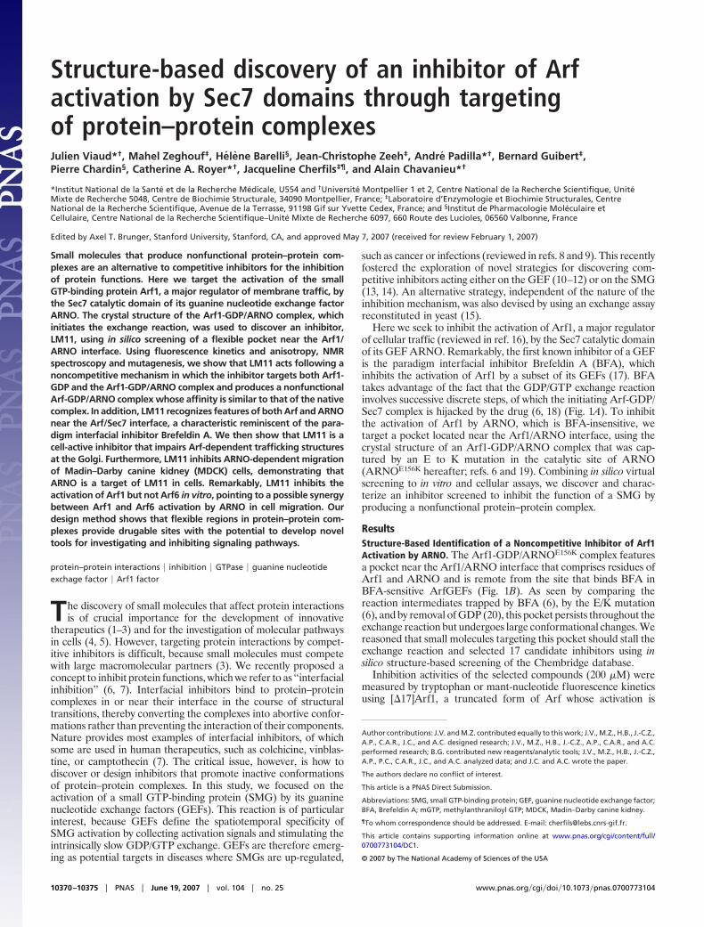
Here we seek to inhibit the activation of Arf1, a major regulatorof cellular traffic (reviewed in ref. 16), by the Sec7 catalytic domainof its GEF ARNO. Remarkably, the first known inhibitor of a GEFis the paradigm interfacial inhibitor Brefeldin A (BFA), whichinhibits the activation of Arf1 by a subset of its GEFs (17). BFAtakes advantage of the fact that the GDP/GTP exchange reactioninvolves successive discrete steps, of which the initiating Arf-GDP/Sec7 complex is hijacked by the drug (6, 18) (Fig. 1A). To inhibitthe activation of Arf1 by ARNO, which is BFA-insensitive, wetarget a pocket located near the Arf1/ARNO interface, using thecrystal structure of an Arf1-GDP/ARNO complex that was cap-tured by an E to K mutation in the catalytic site of ARNO(ARNOE156K hereafter; refs. 6 and 19). Combining in silico virtualscreening to in vitro and cellular assays, we discover and charac-terize an inhibitor screened to inhibit the function of a SMG byproducing a nonfunctional protein–protein complex.
ResultsStructure-Based Identification of a Noncompetitive Inhibitor of Arf1Activation by ARNO. The Arf1-GDP/ARNOE156K complex featuresa pocket near the Arf1/ARNO interface that comprises residues ofArf1 and ARNO and is remote from the site that binds BFA inBFA-sensitive ArfGEFs (Fig. 1B). As seen by comparing thereaction intermediates trapped by BFA (6), by the E/K mutation(6), and by removal of GDP (20), this pocket persists throughout theexchange reaction but undergoes large conformational changes. Wereasoned that small molecules targeting this pocket should stall theexchange reaction and selected 17 candidate inhibitors using insilico structure-based screening of the Chembridge database.
Inhibition activities of the selected compounds (200 �M) weremeasured by tryptophan or mant-nucleotide fluorescence kineticsusing [�17]Arf1, a truncated form of Arf whose activation is
Author contributions: J.V. and M.Z. contributed equally to this work; J.V., M.Z., H.B., J.-C.Z.,A.P., C.A.R., J.C., and A.C. designed research; J.V., M.Z., H.B., J.-C.Z., A.P., C.A.R., and A.C.performed research; B.G. contributed new reagents/analytic tools; J.V., M.Z., H.B., J.-C.Z.,A.P., P.C., C.A.R., J.C., and A.C. analyzed data; and J.C. and A.C. wrote the paper.
The authors declare no conflict of interest.
This article is a PNAS Direct Submission.
Abbreviations: SMG, small GTP-binding protein; GEF, guanine nucleotide exchange factor;BFA, Brefeldin A; mGTP, methylanthraniloyl GTP; MDCK, Madin–Darby canine kidney.
¶To whom correspondence should be addressed. E-mail: [email protected].
This article contains supporting information online at www.pnas.org/cgi/content/full/0700773104/DC1.
© 2007 by The National Academy of Sciences of the USA
10370–10375 � PNAS � June 19, 2007 � vol. 104 � no. 25 www.pnas.org�cgi�doi�10.1073�pnas.0700773104
independent of membranes (19), and the Sec7 domain of humanARNO carrying four mutations that sensitize it to BFA (ARNO4M
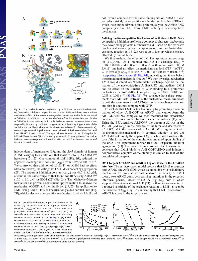
hereafter) (21, 22). One compound, LM11 (Fig. 1B), reduced theapparent exchange rate constant (kapp) from 0.028 to 0.0078 s�1.We controlled that addition of 0.01% Triton X-100 had no effect(data not shown), indicating that LM11 does not act by aggregation(23). The apparent inhibition constant (kiapp) was 49.7 � 8.0 �M,a value in the same range as that found for BFA using ARNO4M
(15.0 � 1.1 �M) or BIG1 (22) (Fig. 2A). The Michaelis–Mentenformalism has proven a convenient approximation to analyze themechanism of GEFs and their inhibition (19, 22). Its application toLM11 using Eadie–Hofstee linearization yielded parallel lines (Fig.2B), which rules out a competitive mechanism, in which LM11 and
Arf1 would compete for the same binding site on ARNO. It alsoexcludes a strictly uncompetitive mechanism such as that of BFA inwhich the compound would bind preferentially to the Arf1/ARNOcomplex (see Fig. 1A). Thus, LM11 acts by a noncompetitivemechanism.
Defining the Noncompetitive Mechanism of Inhibition of LM11. Non-competitive inhibition profiles are complex to characterize, becausethey cover many possible mechanisms (3). Based on the extensivebiochemical knowledge on the spontaneous and Sec7-stimulatedexchange reaction (6, 18–22), we set up to identify which steps areaffected by the inhibitor.
We first analyzed the effect of LM11 on spontaneous exchangeon [�17]Arf1. LM11 inhibited mGDP/GTP exchange (kapp �0.004 � 0.0002 and 0.0004 � 0.0004 s�1 without and with 150 �MLM11) but had no effect on methylanthraniloyl GTP (mGTP)/GTP exchange (kapp � 0.0006 � 0.0001 and 0.0005 � 0.0002 s�1)[supporting information (SI) Fig. 7A], indicating that it acts beforethe formation of nucleotide-free Arf. We then investigated whetherLM11 would inhibit ARNO-stimulated exchange beyond the for-mation of the nucleotide-free Arf1/ARNO intermediate. LM11had no effect on the kinetics of GTP binding to a preformednucleotide-free Arf1-ARNO complex (kapp � 0.088 � 0.011 and0.085 � 0.009 s�1) (SI Fig. 7B). We conclude from these experi-ments that LM11 acts upstream of the nucleotide-free intermediatein both the spontaneous and ARNO-stimulated exchange reaction,and that it does not compete with GTP.
To exclude that LM11 acts allosterically by promoting a confor-mation of either Arf1-GDP or ARNO that cannot form theArf1-GDP/ARNO complex, we then measured the dissociationconstants of this complex by fluorescence anisotropy (Fig. 2C).Using the BFA-sensitive ARNO4M, the apparent Kd was in the150–200 �M range in the absence of inhibitor and decreased to8.6 � 0.7 �M in the presence of BFA (100 �M), in agreement withits uncompetitive mechanism. In contrast, addition of 100 �MLM11 did not modify the apparent Kd, which would have been thecase if the formation of the complex was impaired upon binding ofthe drug. This experiment further rules out unspecific inhibitoraggregation (23). Exclusion of an allosteric effect allows us toconclude that LM11 binds to Arf1-GDP/ARNO and induces anonproductive complex whose stability is similar to that of thenoninhibited complex.
LM11 Targets Arf1-GDP and ARNO in Regions Close to the Arf/ARNOInterface. The in silico screen model predicts that LM11 recognizesboth ARNO and Arf1-GDP, which is compatible with its inhibitorymechanism. To probe it, we first analyzed the activity of LM11toward two ARNO constructs carrying mutations in the screenedinterfacial pocket, R152E or N201A (Fig. 1B), both of whichsupport efficient activation of Arf1 (24). Both mutations resulted ina reduced sensitivity of the exchange reaction to LM11 as seen bythe increase of kiapp (Fig. 3A), indicating that LM11 is sensitive toARNO features in the target pocket.
Fig. 1. The mechanism of Arf activation by its GEFs and its inhibition by LM11.(A) Comparison of the uncompetitive mechanism of BFA and the noncompetitivemechanism of LM11. Representative crystal structures are available for unboundArf-GDP and Arf-GTP, for the nucleotide-free Arf/Sec7 intermediate, and for theArf-GDP/Sec7 intermediate, which subdivides in two successive conformationstrapped by BFA and by the E to K charge reversal of the catalytic glutamate of theSec7 domain. (B) The pocket used for the structure-based in silico screen (circle),comprisingtheswitch1 (yellow)andstrand �2(red)of the interswitchofArf1andloop 198–202 (cyan) of ARNO. The approximate location of the binding site forBFA in BFA-sensitive ArfGEFs is shown by an asterisk. A closeup view of the pocketis shown in surface representation with LM11 docked. The chemical structure ofLM11 is shown in Inset.
Fig. 2. Analysis of the noncompetitive mechanism ofLM11. (A) Determination of the apparent inhibitionconstants (kiapp) of BFA and LM11 measured with[�17]Arf1 and either ARNOWT (BFA resistant) orARNO4M (BFA sensitive) as indicated and increasingconcentrations of the drug as in SI Fig. 7C. (B) Eadie–Hoffstee linearization of the Michaelis–Menten satu-ration curves obtained in the presence of the indicatedconcentrations of LM11 and by varying [�17]Arf1 con-centration between 0 and 5 �M. (C) LM11 does notinhibit the formation of the Arf1-GDP/ARNO complex.Anisotropy binding profiles were obtained from the titration of Alexa488-labeled [�17]Arf1-GDP with ARNOWT in the absence or in the presence of 100 �M LM11as indicated. Titration in the presence of 100 �M BFA was performed with the BFA-sensitive ARNO4M mutant. Anisotropy values measured with ARNOWT orARNO4M in the absence of drug were identical (data not shown).
Viaud et al. PNAS � June 19, 2007 � vol. 104 � no. 25 � 10371
BIO
CHEM
ISTR
Y
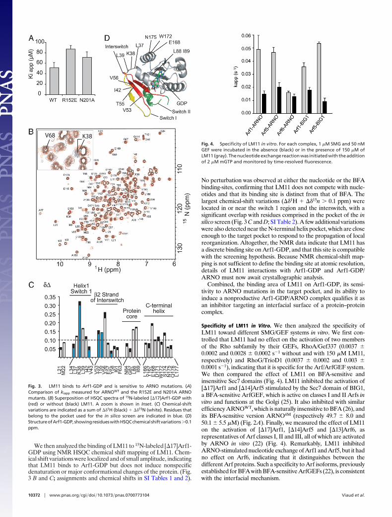
We then analyzed the binding of LM11 to 15N-labeled [�17]Arf1-GDP using NMR HSQC chemical shift mapping of LM11. Chem-ical shift variations were localized and of small amplitude, indicatingthat LM11 binds to Arf1-GDP but does not induce nonspecificdenaturation or major conformational changes of the protein. (Fig.3 B and C; assignments and chemical shifts in SI Tables 1 and 2).
No perturbation was observed at either the nucleotide or the BFAbinding-sites, confirming that LM11 does not compete with nucle-otides and that its binding site is distinct from that of BFA. Thelargest chemical-shift variations (��1H � ��15n � 0.1 ppm) werelocated in or near the switch 1 region and the interswitch, with asignificant overlap with residues comprised in the pocket of the insilico screen (Fig. 3 C and D; SI Table 2). A few additional variationswere also detected near the N-terminal helix pocket, which are closeenough to the target pocket to respond to the propagation of localreorganization. Altogether, the NMR data indicate that LM11 hasa discrete binding site on Arf1-GDP, and that this site is compatiblewith the screening hypothesis. Because NMR chemical-shift map-ping is not sufficient to define the binding site at atomic resolution,details of LM11 interactions with Arf1-GDP and Arf1-GDP/ARNO must now await crystallographic analysis.
Combined, the binding area of LM11 on Arf1-GDP, its sensi-tivity to ARNO mutations in the target pocket, and its ability toinduce a nonproductive Arf1-GDP/ARNO complex qualifies it asan inhibitor targeting an interfacial surface of a protein–proteincomplex.
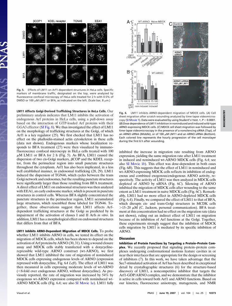
Specificity of LM11 in Vitro. We then analyzed the specificity ofLM11 toward different SMG/GEF systems in vitro. We first con-trolled that LM11 had no effect on the activation of two membersof the Rho subfamily by their GEFs, RhoA/Gef337 (0.0037 �0.0002 and 0.0028 � 0.0002 s�1 without and with 150 �M LM11,respectively) and RhoG/TrioD1 (0.0037 � 0.0002 and 0.003 �0.0001 s�1), indicating that it is specific for the Arf/ArfGEF system.We then compared the effect of LM11 on BFA-sensitive andinsensitive Sec7 domains (Fig. 4). LM11 inhibited the activation of[�17]Arf1 and [�14]Arf5 stimulated by the Sec7 domain of BIG1,a BFA-sensitive ArfGEF, which is active on classes I and II Arfs invitro and functions at the Golgi (25). It also inhibited with similarefficiency ARNOWT, which is naturally insensitive to BFA (26), andits BFA-sensitive version ARNO4M (respectively 49.7 � 8.0 and50.1 � 5.5 �M) (Fig. 2A). Finally, we measured the effect of LM11on the activation of [�17]Arf1, [�14]Arf5 and [�13]Arf6, asrepresentatives of Arf classes I, II and III, all of which are activatedby ARNO in vitro (22) (Fig. 4). Remarkably, LM11 inhibitedARNO-stimulated nucleotide exchange of Arf1 and Arf5, but it hadno effect on Arf6, indicating that it distinguishes between thedifferent Arf proteins. Such a specificity to Arf isoforms, previouslyestablished for BFA with BFA-sensitive ArfGEFs (22), is consistentwith the interfacial mechanism.
Fig. 3. LM11 binds to Arf1-GDP and is sensitive to ARNO mutations. (A)Comparison of kiapp measured for ARNOWT and the R152E and N201A ARNOmutants. (B) Superposition of HSQC spectra of 15N-labeled [�17]Arf1-GDP with(red) or without (black) LM11. A zoom is shown in Inset. (C) Chemical-shiftvariations are indicated as a sum of ��1H (black) � ��15N (white). Residues thatbelong to the pocket used for the in silico screen are indicated in blue. (D)StructureofArf1-GDP,showingresidueswithHSQCchemical shiftvariations�0.1ppm.
Fig. 4. Specificity of LM11 in vitro. For each complex, 1 �M SMG and 50 nMGEF were incubated in the absence (black) or in the presence of 150 �M ofLM11 (gray). The nucleotide exchange reaction was initiated with the additionof 2 �M mGTP and monitored by time-resolved fluorescence.
10372 � www.pnas.org�cgi�doi�10.1073�pnas.0700773104 Viaud et al.
LM11 Affects Golgi-Derived Trafficking Structures in HeLa Cells. Ourpreliminary analysis indicates that LM11 inhibits the activation ofendogenous Arf proteins in HeLa cells, using a pull-down assaybased on the interaction of GTP-loaded Arf proteins with theirGGA3 effector (SI Fig. 8). We thus investigated the effect of LM11on the morphology of trafficking structures at the Golgi, of whichArf1 is a key regulator (25). We first checked that LM11 has noeffect on the phalloidin-stained actin cytoskeleton in these cells(data not shown). Endogenous markers whose localization re-sponds to BFA treatment (27) were then visualized by immuno-fluorescence confocal microscopy in HeLa cells treated with 100�M LM11 or BFA for 2 h (Fig. 5). As BFA, LM11 caused thedispersion of two cis-Golgi markers, �COP and the KDEL recep-tor, from the perinuclear region into small punctate structuresthroughout the cytoplasm. Arf1 has also been implicated, in a lesswell established manner, in endosomal trafficking (28, 29). LM11induced the dispersion of TGN46, which cycles between the transGolgi network and endosomes, but the resulting punctate structureswere significantly larger than those resulting from BFA treatment.A direct effect of LM11 on endosomal structures was then analyzedwith EEA1, an early endosome marker, which is present in punctatestructures in control cells. Whereas BFA slightly concentrated thepunctate structures in the perinuclear region, LM11 accumulatedlarge structures, which resembled those labeled for TGN46. To-gether, these observations suggest that LM11 affects Arf-dependent trafficking structures at the Golgi as predicted by itsimpairment of the activation of classes I and II Arfs in vitro. Inaddition, LM11 has a morphological effect on endosomal structuresthat differs from that of BFA.
LM11 Inhibits ARNO-Dependent Migration of MDCK Cells. To probewhether LM11 inhibits ARNO in cells, we tested its effect on themigration of MDCK cells, which has been shown to depend on theactivation of Arf proteins by ARNO (30, 31). Using a wound closureassay and MDCK cells stably transfected with a doxycycline-repressible wild-type ARNO construct (wt-ARNO), we firstshowed that LM11 inhibited the rate of migration of noninducedMDCK cells expressing endogenous levels of ARNO (expressionrepressed with doxycycline, Fig. 6A Left). The effect of LM11 wasthen measured in cells expressing moderate levels of wt-ARNO(�8-fold over endogenous ARNO, without doxycycline). As pre-viously reported, the rate of migration was increased by 54% byexogenous wt-ARNO expression compared with noninduced wt-ARNO MDCK cells (Fig. 6A; see also SI Movie 1a). LM11 fully
inhibited the increase in migration rate resulting from ARNOexpression, yielding the same migration rate after LM11 treatmentin induced and noninduced wt-ARNO MDCK cells (Fig. 6A; seealso SI Movie 1b). This effect was dose-dependent in both cases(Fig. 6B). This suggests that the effect of LM11 in noninduced andwt-ARNO-expressing MDCK cells reflects its inhibition of endog-enous and combined exogenous/endogenous ARNO activity, re-spectively. The activity of LM11 was then analyzed in MDCK cellsdepleted of ARNO by siRNA (Fig. 6C). Silencing of ARNOinhibited the migration of MDCK cells after wounding to the sameextent as LM11 treatment in naive MDCK cells (Fig. 6C). Remark-ably, LM11 had no more effect in ARNO-depleted MDCK cells(Fig. 6A). Finally, we compared the effect of LM11 to that of BFA,which disrupts cis- and trans-Golgi structures in MCDK cells�15–20 �M (C. Jackson, personal communication). BFA treat-ment at this concentration had no effect on the migration rate (datanot shown), ruling out an indirect effect of LM11 on migrationbecause of its inhibition of Arf functions at the Golgi. Together,these experiments strongly suggest that the inhibition of MDCKcells migration by LM11 is mediated by its specific inhibition ofARNO.
DiscussionInhibition of Protein Functions by Targeting a Protein–Protein Com-plex. We recently proposed that signaling protein–protein com-plexes undergoing conformational motions feature cavities in ornear their interfaces that are appropriate for the design or screeningof inhibitors (7). In this work, we have taken advantage that theSec7-stimulated activation of Arf has been described in exceptionaldetail by crystallographic structures (6) for the structure-baseddiscovery of LM11, a noncompetitive inhibitor that targets theArf1-GDP/ARNO complex, and we demonstrate that the inhibitoris active in cells toward both Arf1 and ARNO functions. Based onour kinetics, fluorescence anisotropy, mutagenesis, and NMR
Fig. 5. Effects of LM11 on Arf1-dependent structures in HeLa cells. Specificmarkers of membrane traffic, designated on the top, were analyzed byfluorescence confocal microscopy of HeLa cells treated for 2 h with 0.5% ofDMSO or 100 �M LM11 or BFA, as indicated on the left. (Scale bar, 8 �m.)
Fig. 6. LM11 inhibits ARNO-dependent migration of MDCK cells. (A) Cellsheet migration after scratch wounding analyzed by time-lapse videomicros-copy (SI Movie 1). Data were evaluated by using Student’s t test. *, P � 0.0001.(B) Dose dependence of LM11 inhibition in noninduced and induced wild-typeARNO-expressing MDCK cells. (C) MDCK cell sheet migration was followed bytime-lapse videomicroscopy in the presence of a nonsilencing siRNA (Top), ofan ARNO siRNA (Middle), or of 100 �M LM11 and an ARNO siRNA (Bottom).Each colored line represents the hourly progression of the cell monolayerduring the first 6 h after wounding.
Viaud et al. PNAS � June 19, 2007 � vol. 104 � no. 25 � 10373
BIO
CHEM
ISTR
Y
analysis, we propose that LM11 binds to both Arf-GDP and theArf-GDP/ARNO complex, yielding a ternary complex of affinitysimilar to that of the normal reaction intermediate but impaired forthe conformational conversion that decreases the affinity for GDPand yields the nucleotide-free complex (Fig. 1A). Our data indicatethat LM11 recognizes both components of the complex, suggestingthat the expected increase in affinity of the Arf-GDP/ARNOcomplex because of complex/inhibitor interactions may be com-pensated for by a less-than-optimal protein/protein interactionbetween Arf-GDP and ARNO because of the inhibitor. Wesurmise that LM11 takes advantage of the flexibility of the switch1 region of Arf1-GDP, which can be observed in unbound Arf1-GDP using NMR dynamics (V. Buosi, C. van Heijenoort, and E.Guittet, personal communication), which results in favorable char-acteristics for binding a small molecule. In this respect, LM11probably uses, at the level of a protein–protein complex, adaptivephysicochemical characteristics similar to those previously de-scribed for unbound proteins within the surface that they use toform protein–protein interactions (32, 33). Remarkably, the effectof LM11 on both trafficking structures and MDCK migration wasstrong and rapid despite its modest kiapp. This highlights the kineticscomponent of the inhibitory response, as described for the inter-facial inhibitor BFA, in which the inhibitor, by yielding a nonfunc-tional protein–protein intermediate, stalls the reaction on a suffi-ciently long timescale for the biological effects to manifest. Thediscovery of this noncompetitive inhibitor emphasizes that flexible/dynamic regions appearing in protein–protein complexes provide‘‘drugable’’ sites.
A Tool to Investigate BFA-Insensitive Arf Pathways in Cells. To date,the substrate specificity of ARNO in vivo has proven difficult toresolve, notably because ARNO and Arf6 are not sensitive to BFA(22, 26). On the one hand, ARNO is active on both Arf1 and Arf6in vitro with a strong preference for Arf1 (22, 26, 34). On the otherhand, ARNO significantly activates Arf6 in cells and is recruited tosites of Arf6 activity at the plasma membrane (30), and it is involvedin the migration of MDCK cells (30, 31), a process believed toinvolve Arf6 preferentially to Arf1. The unique ability of LM11 toselectively inhibit Arf1, but not Arf6, activation by ARNO in vitro,while strongly inhibiting ARNO-dependent migration in vivo, ad-dresses the question of an unexpected involvement of Arf1 activa-tion by ARNO in addition to Arf6 in this process, as suggested inref. 35. LM11 should now provide a novel tool for future investi-gations of endogenous BFA-insensitive Arf pathways and of thepromiscuity of the ARNO family for Arf1 and Arf6 in cells. Inconclusion, our study provides the proof of principle that smallmolecules that target protein–protein complexes, including low-affinity intermediates, can yield cell-active inhibitors. Such mole-cules should be instrumental in deciphering cellular pathways andelucidating their specificity, with the potential for development intonew therapeutic compounds.
Materials and MethodsProteins and Reagents. All Arf and Sec7 constructs and mutantswere expressed and purified as described in ref. 22. 15N-labeled[�17]Arf1 was expressed as in ref. 36. GST-fused RhoG, RhoA,TRIO (DH1 domain), and GEF337 (DH domain) are a kind giftof A. Debant, A. Blangy, and P. Fort (Centre de Recherche deBiochimie Macromoleculaire, Centre National de la RechercheScientifique, Montpellier, France). BFA was purchased from Sigma(St. Louis, MO); N-mGTP from Euromedex (Souffelweyersheim,France); alkaline phosphatase from New England Biolabs (Ipswich,MA); molecules selected by virtual screening from Chembridge(San Diego, CA); mouse monoclonal antibodies (Abs) against�COPI and EEA1 from Sigma and Transduction Laboratories(Lexington, KY); and TRITC-labeled phalloidin and Alexa 488-conjugated secondary Abs from Sigma and Molecular Probes(Leiden, The Netherlands). Mouse Ab against KDEL receptor and
sheep Ab against TGN46 were a kind gift from C. Jackson(Laboratoire d’Enzymologie et Biochimie Structurales, CentreNational de la Recherche Scientifique).
Virtual Screening. Structure-based screening for inhibitors wastargeted at an interfacial pocket identified with the ‘‘binding site’’module in Insight II (Accelrys, Cambridge, U.K.). The pocket wasfirst screened for the binding of small fragments with the LUDImodule of Insight II. The five fragments with higher scores wereused to filter the Chembridge Express-Pick virtual library using theMDL.ISIS/Base software (Elsevier, Amsterdam, The Nether-lands), yielding 3,227 commercially available compounds of �500Da containing at least one fragment. These compounds were thendocked into the pocket (defined as all atoms within 10 Å of Asn-201in ARNO) by using FlexX 1.13.1 (Tripos Associates, Villebon,France). Standard parameters were used for iterative growing andsubsequent scoring of FlexX poses as described in ref. 37. Dockingpositions closer than 5 Å to Asn-201 and with a score smaller than�25 were reranked with X-Score (http://sw16.im.med.umich.edu/software/xtool), which has a more accurate estimation of bindingfree energies, from which 17 compounds were selected by visualinspection of the top 113 scores.
Kinetics Measurements. All kinetics experiments were performedwith Arf proteins truncated of their N-terminal helix and loadedwith GDP before the experiments. Activation of [�17]Arf1 wasmonitored by either tryptophan f luorescence (emission/excitation wavelengths of 292/340 nm), fluorescence of the N-methylanthraniloyl fluorophore (mGDP or mGTP) (360/440 nm),or FRET (292/440 nm). All measurements were performed at 37°Cin 50 mM Tris (pH 8)/50 mM NaCl/2 mM MgCl2/2 mM 2-mer-captoethanol. Inhibitors were incubated for 5 min before initiatingthe reaction with GTP or mGTP. Fluorescence data were fitted byusing the program Origin 6.1 (Microcal, Northampton, MA).Spontaneous mGDP/GTP exchange of [�17]Arf1 was measuredwith [�17]Arf1 (1 �M) loaded with mGDP (10 �M) in the presenceof EDTA with or without LM11 (150 �M) and followed by theaddition of GTP (200 �M). Spontaneous mGTP/GTP exchangewas measured as for the mGDP/GTP exchange. The nucleotide-free complex was formed by elimination of GDP by alkalinephosphatase (20 units) and 2 mM EDTA as described in ref. 38;binding of GTP to the complex (0.2 �M) was then initiated with 1�M mGTP in the presence or absence of 150 �M LM11. All valuesare means � SD of at least three independent experiments.
Fluorescence Anisotropy. [�17]Arf1-GDP was labeled with Alexa488 at pH 8.3 at 4°C for 2 h, conditions that favor unique labelingof the N terminus (labeling ratio 72%). Fluorescence anisotropyprofiles were obtained in the serial dilution format on a Beacon2000 (Panvera, Madison, WI) polarization instrument. Experi-ments were performed at 4°C by using Alexa 488-labeled[�17]Arf1-GDP at a fixed concentration (10 nM) in 50 mM Tris,pH 8/200 mM NaCl/2 mM MgCl2/100 �M GDP/10% glycerol/2mM 2-mercaptoethanol. Anisotropy values were similar at 4°C and37°C (data not shown). Data points were taken at equilibriumstarting from 120 �l of a solution of ARNO [initial concentration200 �M (BFA) or 400 �M (LM11, no ligand)] containing 10 nMAlexa 488-labeled [�17]Arf1-GDP with or without 100 �M inhib-itor. For each subsequent measurement, 30 �l was removed fromthe initial solution and replaced by 30 �l containing 10 nM Alexa488-labeled [�17]Arf1-GDP with or without 100 �M of inhibitor.Anisotropy data were fit by using the Bioeqs program (39).
NMR Spectroscopy. 2D and 3D NMR spectra (2D-HSQC, 3D-HSQC NOESY, and 3D-HSQC TOCSY) were recorded on aBruker (Billerica, MA) Avance 500 NMR spectrometer by using a5-mm TXI z-grad cryo-probe at 300 K. The sample contained 1 mM15N[�17]Arf1 in 50 mM Tris�HCl/150 mM NaCl/50 �M GDP/0.5
10374 � www.pnas.org�cgi�doi�10.1073�pnas.0700773104 Viaud et al.
mM 2-mercaptoethanol, pH 7.0 with 5% 2H2O for the lock. Amixing time of 100 msec was used for the NOESY and a 20-msecspin-lock was used for the TOCSY. Acquisition size for 3D spectrawas 400(1H) � 70(15N) � 1024(1H) with 8 scans. All data wereprocessed with the Bruker UXNMR package and analyzed withCINDY (40). We compared our data to the published chemical-shift assignments for 15N[�17]Arf1-GDP (36), of which 9 missingassignments were added, and 21 were corrected (SI Table 1). Forchemical-shift mapping of LM11, 2D-HSQC spectra of15N[�17]Arf1-GDP (210 �M in 50 mM Tris�HCl/30 mM NaCl/50�M GDP/0.5 mM 2-mercaptoethanol/5% 2H2O at pH 8.0) wererecorded without or with 400 �M LM11 at 300 K with a BrukerAvance 600 NMR spectrometer equipped with a 5-mm TXIz-gradient cryoprobe. In all experiments, the 1H carrier was cen-tered on the water resonance, and a WATERGATE sequence wasincorporated to suppress the solvent resonance. Spectral widths of13 ppm for 1H and 34 ppm for 15N were used.
Immunofluorescence Microscopy. HeLa cells were grown and fixedas described in ref. 41. Incubation with the first Ab was performedovernight at 4°C in PBS containing 0.25% BSA, 0.01% Tween 20,and 0.01% saponin. Abs against TGN46 and EEA1 were diluted1:200, and �COPI was used at 1:100 and KDEL receptor at 1:500dilution. Staining with secondary Ab was carried out for 20 min atroom temperature by using Alexa 488-conjugated goat anti-mouse(1:800) or donkey anti-sheep (1:500) IgG and TRITC-labeledphalloidin (1:100). Images were collected with a Leica (Rueil-Malmaison, France) TCS SP2 upright laser-scanning confocalmicroscope with an oil 63� (N.A. 1.32) objective. Different fluo-rochromes were detected sequentially in frame-interlace mode withthe acousto-optical tunable filter system using laser lines 488 nm(Alexa 488) and 514 nm (TRITC). Serial sections were acquiredsatisfying the Nyquist criteria for sampling and processed using theImajeJ 1.35 software (http://rsb.info.nih.gov/ij).
Wound-Healing/Cell Sheet Migration Assay. wt-ARNO fused to aN-terminal c-myc tag was expressed in MDCK cells under controlof the tetracycline-repressible transactivator. MDCK cells stablytransfected with wt-ARNO vector were plated on plastic dishescoated with collagen I at 3 �g/ml to form monolayers. Confluentmonolayers were wounded by scraping with a tip, rinsed with mediato remove dislodged cells, and placed back into MEM with 5% FBSwith or without 20 ng/ml doxycycline, to repress or allow, respec-tively, further transgene expression, and with or without LM11. Cellsheet migration into the cleared wound area (350-�m width �22-mm length) was recorded by using a Zeiss (Le Peck, France)
Axiovert 200M inverted microscope equipped with a thermostatedincubation chamber maintained at 37°C under 5% CO2. Digitalimages were acquired every 5 min for 16 h by using a CoolSnap HQCCD camera. The wounded area was measured on each timeframe, and the increment of the area recovered by cells over timewas determined by using Metamorph software. The migration rate(�m2/h) was calculated between two sequential frames separated bya time interval of 1 h during 8 h or until total wound closure. Theaverage migration rates are means � SD of at least three separateexperiments. Similar migration and inhibition rates were obtainedwith nontransfected MDCK and noninduced wt-ARNO-MDCKcell monolayers (data not shown).
SiRNA oligonucleotides were designed by using the Canis famil-iaris ARNO sequence and purchased from Eurogentec (Angers,France). A control nonsilencing siRNA duplex was used. MDCKcells were transfected with 150 pmol ARNO or control siRNAoligonucleotides by using Lipofectamine 2000. At 2 days aftertransfection, MDCK cells monolayers were wounded, and the levelof ARNO or cytohesin1 (negative control) was measured at the endof the wound-healing experiment by immunoblot after immuno-precipitation of the cell lysates by using anti-ARNO (A18) andanticytohesin 1 (139) Abs (kind gift from S. Bourgoin, CentreHospitalier, Universite de Quebec, Ste-Foy, PQ, Canada) withTrueblot kit (SI Fig. 9). Transfection efficiency was determined byepifluorescence microscopy by using Rhodamine-labeled ARNOsiRNA. Cell sheet migration was recorded and analyzed as de-scribed above for inducible MDCK cells.
This work was supported by the Association pour la Recherche Contre leCancer, the ACI-Biologie cellulaire, Moleculaire et Structurale of theFrench Ministere de la Recherche (J.C.) and the Agence Nationale de laRecherche (J.C. and A.C.). J.V. was supported by a grant from theFondation pour la Recherche Medicale. We thank the staff at the Imagingand Cell Biology Facility [Centre National de la Recherche Scientifique(CNRS), Gif-sur-Yvette, France] for assistance with the confocal micro-scopes; A. Cave and Y.S. Yang (Centre de Biochimie Structurale, CNRS,Montpellier, France) for help with NMR data acquisition and treatment; B.Olofsson and E. Dransart [Laboratoire d’Enzymologie et Biochimie Struc-turales (LEBS), CNRS] for help with confocal microscopy; F. Luton[Institut de Pharmcologie Moleculaire et Cellulaire (IPMC), CNRS,Sophia-Antipolis, France] for the gift of ARNO-expressing MDCK cells;Gregoire Malandain (INRIA, Sophia Antipolis, France) for producing thevideos; C. van Heijenoort, V. Buosi and E. Guittet (Institut de Chimie desSubstances Naturelles, CNRS) and C. Jackson (LEBS, CNRS) for sharingunpublished data; B. Antonny (IPMC, CNRS, Sophia-Antipolis, France),and A. Di Nardo (Ecole Normale Superieure, CNRS, Paris, France) forcritical reading of the manuscript.
1. Toogood PL (2002) J Med Chem 45:1543–1558.2. Berg T (2003) Angew Chem Int Ed Engl 42:2462–2481.3. Arkin MR, Wells JA (2004) Nat Rev Drug Discov 3:301–317.4. McCormick F (2000) Curr Opin Biotechnol 11:593–597.5. Pelish HE, Peterson JR, Salvarezza SB, Rodriguez-Boulan E, Chen JL, Stamnes M,
Macia E, Feng Y, Shair MD, Kirchhausen T (2006) Nat Chem Biol 2:39–46.6. Renault L, Guibert B, Cherfils J (2003) Nature 426:525–530.7. Pommier Y, Cherfils J (2005) Trends Pharmacol Sci 26:138–145.8. Rossman KL, Der CJ, Sondek J (2005) Nat Rev Mol Cell Biol 6:167–180.9. Zeghouf M, Guibert B, Zeeh JC, Cherfils J (2005) Biochem Soc Trans 33:1265–1268.
10. Schmidt S, Diriong S, Mery J, Fabbrizio E, Debant A (2002) FEBS Lett 523:35–42.11. Mayer G, Blind M, Nagel W, Bohm T, Knorr T, Jackson CL, Kolanus W, Famulok M
(2001) Proc Natl Acad Sci USA 98:4961–4965.12. Hafner M, Schmitz A, Grune I, Srivatsan SG, Paul B, Kolanus W, Quast T, Kremmer E,
Bauer I, Famulok M (2006) Nature 444:941–944.13. Gao Y, Dickerson JB, Guo F, Zheng J, Zheng Y (2004) Proc Natl Acad Sci USA 101:7618–7623.14. Desire L, Bourdin J, Loiseau N, Peillon H, Picard V, De Oliveira C, Bachelot F, Leblond
B, Taverne T, Beausoleil E, et al. (2005) J Biol Chem 280:37516–37525.15. Blangy A, Bouquier N, Gauthier-Rouviere C, Schmidt S, Debant A, Leonetti JP, Fort P
(2006) Biol Cell 98:1387�1398.16. D’Souza-Schorey C, Chavrier P (2006) Nat Rev Mol Cell Biol 7:347–358.17. Klausner RD, Donaldson JG, Lippincott-Schwartz J (1992) J Cell Biol 116:1071–1080.18. Peyroche A, Antonny B, Robineau S, Acker J, Cherfils J, Jackson CL (1999) Mol Cell 3:275–285.19. Beraud-Dufour S, Robineau S, Chardin P, Paris S, Chabre M, Cherfils J, Antonny B
(1998) EMBO J 17:3651–3659.20. Goldberg J (1998) Cell 95:237–248.21. Robineau S, Chabre M, Antonny B (2000) Proc Natl Acad Sci USA 97:9913–9918.
22. Zeeh JC, Zeghouf M, Grauffel C, Guibert B, Martin E, Dejaegere A, Cherfils J (2006)J Biol Chem 281:11805–11814.
23. McGovern SL, Caselli E, Grigorieff N, Shoichet BK (2002) J Med Chem 45:1712–1722.24. Cherfils J, Menetrey J, Mathieu M, Le Bras G, Robineau S, Beraud-Dufour S, Antonny
B, Chardin P (1998) Nature 392:101–105.25. Donaldson JG, Honda A, Weigert R (2005) Biochim Biophys Acta 1744:364–373.26. Chardin P, Paris S, Antonny B, Robineau S, Beraud-Dufour S, Jackson CL, Chabre M
(1996) Nature 384:481–484.27. Volpicelli-Daley LA, Li Y, Zhang CJ, Kahn RA (2005) Mol Biol Cell 16:4495–4508.28. Bonifacino JS, Jackson CL (2003) Cell 112:141–142.29. Shen X, Xu KF, Fan Q, Pacheco-Rodriguez G, Moss J, Vaughan M (2006) Proc Natl Acad
Sci USA 103:2635–2640.30. Santy LC, Casanova JE (2001) J Cell Biol 154:599–610.31. Santy LC, Ravichandran KS, Casanova JE (2005) Curr Biol 15:1749–1754.32. DeLano WL, Ultsch MH, de Vos AM, Wells JA (2000) Science 287:1279–1283.33. Arkin MR, Randal M, DeLano WL, Hyde J, Luong TN, Oslob JD, Raphael DR, Taylor L,
Wang J, McDowell RS, Wells JA, Braisted AC (2003) Proc Natl Acad Sci USA 100:1603–1608.34. Macia E, Chabre M, Franco M (2001) J Biol Chem 276:24925–24930.35. Cohen LA, Honda A, Varnai P, Brown FD, Balla T, Donaldson JG (2007) Mol Biol Cell
18:2244–2253.36. Seidel RD, 3rd, Amor JC, Kahn RA, Prestegard JH (2004) J Biol Chem 279:48307–48318.37. Guichou JF, Viaud J, Mettling C, Subra G, Lin YL, Chavanieu A (2006) J Med Chem 49:900–910.38. Beraud-Dufour S, Paris S, Chabre M, Antonny B (1999) J Biol Chem 274:37629–37636.39. Grillo AO, Brown MP, Royer CA (1999) J Mol Biol 287:539–554.40. Ponchon L, Dumas C, Fesquet D, Padilla A (2004) J Biomol NMR 28:299–300.41. Dransart E, Morin A, Cherfils J, Olofsson B (2005) J Biol Chem 280:4674–4683.
Viaud et al. PNAS � June 19, 2007 � vol. 104 � no. 25 � 10375
BIO
CHEM
ISTR
Y


