Embed Size (px)
Citation preview
The gastric HK-ATPase: structure, function, and inhibition
Jai Moo Shin,Department of Physiology, David Geffen School of Medicine, University of California at LosAngeles and VA Greater Los Angeles Healthcare System, Los Angeles, CA 90073, USA
Keith Munson,Department of Physiology, David Geffen School of Medicine, University of California at LosAngeles and VA Greater Los Angeles Healthcare System, Los Angeles, CA 90073, USA
Olga Vagin, andDepartment of Physiology, David Geffen School of Medicine, University of California at LosAngeles and VA Greater Los Angeles Healthcare System, Los Angeles, CA 90073, USA
George SachsDepartment of Physiology, David Geffen School of Medicine, University of California at LosAngeles and VA Greater Los Angeles Healthcare System, Los Angeles, CA 90073, USA.Membrane Biology, Room 324, Building 113, 11301 Wilshire Boulevard, Los Angeles, CA 90073,USAGeorge Sachs: [email protected]
AbstractThe gastric H,K-ATPase, a member of the P2-type ATPase family, is the integral membraneprotein responsible for gastric acid secretion. It is an α,β-heterodimeric enzyme that exchangescytoplasmic hydronium with extracellular potassium. The catalytic α subunit has tentransmembrane segments with a cluster of intramembranal carboxylic amino acids located in themiddle of the transmembrane segments TM4, TM5,TM6, and TM8. Comparison to the knownstructure of the SERCA pump, mutagenesis, and molecular modeling has identified these asconstituents of the ion binding domain. The β subunit has one transmembrane segment with Nterminus in cytoplasmic region. The extracellular domain of the β subunit contains six or seven N-linked glycosylation sites. N-glycosylation is important for the enzyme assembly, maturation, andsorting. The enzyme pumps acid by a series of conformational changes from an E1 (ion site in) toan E2 (ion site out) configuration following binding of MgATP and phosphorylation. Severalexperimental observations support the hypothesis that expulsion of the proton at 160 mM (pH 0.8)results from movement of lysine 791 into the ion binding site in the E2P configuration. Potassiumaccess from the lumen depends on activation of a K and Cl conductance via a KCNQ1/KCNE2complex and Clic6. K movement through the luminal channel in E2P is proposed to displace thelysine along with dephosphorylation to return the enzyme to the E1 configuration. This enzyme isinhibited by the unique proton pump inhibitor class of drug, allowing therapy of acid-relateddiseases.
KeywordsThe gastric H,K-ATPase; ATPase structure–function; Proton pump inhibitors; Acid pumpantagonists; Gastric acid secretion
Correspondence to: George Sachs, [email protected].
NIH Public AccessAuthor ManuscriptPflugers Arch. Author manuscript; available in PMC 2011 April 19.
Published in final edited form as:Pflugers Arch. 2009 January ; 457(3): 609–622. doi:10.1007/s00424-008-0495-4.
NIH
-PA Author Manuscript
NIH
-PA Author Manuscript
NIH
-PA Author Manuscript
IntroductionThe basis for success of modern methods of therapy of acid secretion has been theidentification of unique expression of target proteins by cells like the gastric parietal cell.For example, the major functional location of both the H2 receptor and the H,K-ATPase isthe parietal cell. Histamine binds to the H2 receptor and stimulates acid secretion throughthe cAMP-stimulated morphological changes of the parietal cell from the resting status tothe stimulated state and activation of the KCl pathway. The gastric H,K-ATPase is locatedin the canaliculus of the stimulated state and secretes gastric acid by an electroneutral ATP-dependent hydrogen–potassium exchange [41]. The introduction of H2 receptor antagonistsin 1977 followed by proton pump inhibitors targeted to the gastric H,K-ATPase in 1989reflected a major medical therapeutic breakthrough in the treatment of peptic ulcers andgastroesophageal reflux disease, resulting in more rapid healing of the lesions and symptomrelief [6,11]. The H2 receptor antagonist dramatically increased healing of peptic ulcerdisease, but not gastroesophageal reflux disease, which requires greater inhibition of gastricacid secretion that was then achieved by the proton pump inhibitors (PPIs), which inhibit thegastric H,K-ATPase.
The gastric H,K-ATPase is an α,β-heterodimeric enzyme. In this review, the structure andfunction of the H,K-ATPase and inhibitors of this enzyme will be discussed.
Cellular location of the gastric H,K-ATPaseThe gastric H,K-ATPase is found in the parietal cell of the stomach and a small amount inthe renal medulla. This enzyme appears to be in cytoplasmic tubular membranes in theresting state and then in the microvilli of the expanded secretory canaliculus in thestimulated state of the parietal cell [13,45]. This morphological change is assumed to resultfrom fusion of cytoplasmic vesicles with the rudimentary microvilli to form the elongatedmicrovilli of the expanded secretory canaliculus [13,45]. Once the enzyme moves to thecanaliculus, the enzyme secrets acid by the exchange of cytoplasmic hydronium withextracellular K. Genomic microarray analysis of genes specifically expressed in a pure(99%) parietal cell isolate identified the likely K efflux channel associated with the gastricH,K-ATPase. The function of this channel is to supply K to the luminal surface of the pumpto allow H for K exchange. KCNQ1-KCNE2 were the most highly expressed andsignificantly enriched members of the large variety of K channels expressed in the gastricepithelium. The function of this K channel in acid secretion was then shown by inhibition ofsecretion in isolated gastric glands with specific KCNQ inhibitors and by co-localization ofthe channel with the H,K-ATPase in the secretory canaliculus of the parietal cell. KCNQ1-KCNE2 appears to be the K efflux channel that is essential for gastric acid secretion [25].Enrichment of Clic6 appears to identify the necessary accompanying Cl channel [29,44].
The isolated hog gastric ATPase is obtained in relatively ion tight vesicles and was shown tobe a member of the P2-type ATPases where the first step of the reaction is phosphorylationof the catalytic subunit by MgATP with export of protons, and this is followed by luminalK-dependent dephosphorylation and K reabsorption. This results in electroneutral exchangeof cytoplasmic protons for exoplasmic potassium. The hog gastric vesicles as isolated didnot include a KCl pathway and required the addition of ionophores such as nigericin formaximal ATPase activity [41]. Isolation of the ATPase from stimulated rabbit providedevidence for the K and Cl conductance pathways [72].
The structure and function of the gastric H,K-ATPaseThe primary structure of the gastric H,K-ATPase α subunits containing the catalytic site wasfirst elucidated in the rat [54] and then in the hog [26], rabbit [4], dog [57], and human [27].
Shin et al. Page 2
Pflugers Arch. Author manuscript; available in PMC 2011 April 19.
NIH
-PA Author Manuscript
NIH
-PA Author Manuscript
NIH
-PA Author Manuscript
This catalytic subunit consists of 1,033 or 1,034 amino acids in length in all species. Theyare 98% homologous with each other. Examination of the catalytic subunit revealed that itwas highly homologous to the Na, K-ATPase (~63%) and less so to the SERCA Ca-ATPase(25%) [59].
Functional studies demonstrated that ATP catalyzed an electroneutral exchange of H for K,with a variable stoichiometry of 2H/2K/ATP at pH 6.1 that fell to 1H/1K/ATP as luminalpH fell to <3.0 [31,37].
Later, it was discovered that a second β subunit was required for proper maturation andtargeting of the enzyme to the apical membrane. This β subunit consists of 291 amino acidsand contains six or seven N-linked glycosylation sites [40,53,60]. The gastric H,K-ATPaseis fully assembled during biosynthesis in the endoplasmic reticulum and delivered to theapical membrane as a hetero-dimeric oligomer. N-glycosylation of the β subunit wasidentified to be responsible for the trafficking. The steady-state distribution of the H,K-ATPase β subunit in polarized cells depends on the balance between (a) direct sorting fromthe trans-Golgi network, (b) secondary associative sorting with a partner protein, and (c)selective trafficking [65,67,68].
In the α subunit, there is a cluster of intramembranal carboxylic amino acids located in themiddle of the transmembrane segments TM4, TM5,TM6, and TM8 that contain the ionbinding domain in this enzyme and the Na, K- and Ca-ATPases. In the gastric H,K-ATPase,there is also a lysine 791 located in the fifth transmembrane segment that replaces a serinepresent in the Na,K-ATPase isoforms. This lysine of the H,K-ATPase seems to characterizethe H,K-enzyme specificity for outward transport of the hydronium ion. A model of thegastric H,K-ATPase operating in the highly acidic secretory canaliculus of the parietal cell isshown in Fig. 1.
The crystal structures of the SERCA ATPase at high resolution by Toyoshima and hiscollaborators permitted understanding of the catalytic cycle of P2-type enzymes in bothgeneral and specific terms [61–63]. Using the crystal structure of the SERCA Ca-ATPase indifferent conformations as a template, computer-assisted homology modeling and site-directed mutation analysis resulted in not only an expanded picture of the ion transportmechanism of the enzyme but also explained how the covalently binding PPIs and the K+-competitive acid pump antagonists (APAs) inhibited the pump. As a high-resolution crystalstructure of the H,K-ATPase has not been described, the exact location of the β subunit isnot known. It is thought that there is close interaction on the luminal surface with the loopbetween TM7 and TM8 on the outside of the membrane domain [28] and that perhaps themembrane domain of the β subunit is interdigitated between TM9 and TM10 [8,36]. In acrystal of the Na,K-ATPase [30], the β subunit membrane domain is tilted and contacts TM7and TM10, consistent with the yeast two hybrid and biochemical data obtained for the H,K-ATPase [28,50]. One study, using fluorescence resonance energy transfer, suggested that theα and β subunits move toward each other during the E2 to E1 conformational transition [10].
The Na,K- and Ca-ATPases extrude Na+ and Ca2+ while utilizing generally the samecarboxylic acids as found in the ion binding site of the H,K-ATPase. Given a pKa for thesecarboxyl side chains between 3.0 and 5.0, how does the H, K-ATPase manage to extrudeprotons at pH 1.0? As illustrated in Fig. 2, this depends on displacement of one of the boundhydroniums by movement of the (ammonium) group of lysine 791 into thehydronium binding site [31,33]. This movement is generated by the reorientation of the M4and M6 helices during the E1 to E2P transition and favors stabilization of the hydrogenbonding of the of lysine 791 as compared to H3O+. The entry of K+ into the ion
Shin et al. Page 3
Pflugers Arch. Author manuscript; available in PMC 2011 April 19.
NIH
-PA Author Manuscript
NIH
-PA Author Manuscript
NIH
-PA Author Manuscript
binding pocket as a focal positive charge would then destabilize the interaction of K791 withglutamyls 795 and 820 and initiate the conversion to the E1 conformation [31].
The postulated displacement of hydronium from site H2 by Lys791 is strongly supported bythe effects of mutating the homologous lysine (Lys800) in the toad bladder H,K-ATPase [7].This electroneutral proton pump can also transport Na+ [16]. However, a K+-stimulatedoutward current was observed in Na+-loaded oocytes injected with mutants K800A andK800E, showing that loss of the lysine positive charge resulted in Na+ outward electrogenictransport. Conversely, the corresponding S782R mutant of the normally electrogenic toadNa,K-ATPase was electroneutral [7]. Thus, it appears that the positively charged side chainin the Na,K-ATPase mutant replaces one Na+ in the forward step, making the exchange 2Na+ for 2 K+, and electroneutral and mutation of the positively charged side chain at Lys800of toad bladder H,K-ATPase allows binding of the third Na+ to give 3 Na+ for 2 K+
exchange generating current. These data strongly support the role of the side chain group of Lys791 in ion transport by the H,K-ATPase and its postulated role in theelectroneutral mechanism of gastric acid secretion.
The overall general conformational changes of the enzyme are illustrated in Fig. 3.Emphasis is placed on changes occurring in the cytoplasmic domain as the enzyme movesfrom an E1 to an E2 configuration following binding of MgATP and phosphorylation. Thecrystal structure of the Ca-ATPase has shown that the cytoplasmic domain is divided intothree sectors named the N (nucleotide binding) domain, the A (actuator) domain, and the P(phosphorylation) domain. Movements induced by ion and ATP binding in these domainsare transmitted to the membrane domain to catalyze the ion displacements shown in Fig. 2.
Recently, a paper appeared describing the crystal structure of the SERCA Ca-ATPase in theabsence of inhibitors with BeF3 to provide the E2P conformation [35]. Homology modelingof the ion site structure of the Ca ATPase compared to the H,K-ATPase is shown in Fig. 4[31,33]. On the left is the ion site region of the SERCA and on the right the H,K ATPase,viewed from the same orientation in the membrane. The replacement of serine 767 withlysine 791 necessitates an expansion of the site and a different orientation of asparagine 792as compared to asparagine 768 of the Ca pump. The presence of this lysine is essential forproton pumping by the H,K-ATPase, with the residue of the lysine displacing thehydronium ion at the appropriate pKa <1.0, for outward proton transport.
The functional form of the gastric H,K-ATPase is a [αβ]2 heterodimer oligomer as identifiedbiochemically [1,48]. However, the recent crystal structure of the Na,K-ATPase suggests amonomeric heterodimer [30]. The large changes in conformation in the cytoplasmic domainprobably accounts for the finding that the enzyme functions as an out of phase oligomericheterodimer [48]. Thus, when one heterodimer is in the E1 form, the other is obligated to bein the E2 from. This has been most clearly demonstrated by measuring the stoichiometry ofATP binding, acid-stable phosphorylation and binding of APAs or PPIs. One mole of ATPbinds to 2 mol of ATPase forming 1 mol of phosphoenzyme at physiological ATPconcentration [48].
The E1 form of the enzyme allows access to the ion binding domain from the cytoplasmicsurface and following binding of ATP with two Mg ions, one to stabilize the αβ orientationof the first two phosphates of the nucleotide and the second in proximity of the acceptoraspartyl residue to allow transfer of the γ phosphate to the catalytic subunit of the proteinand initiate the change of conformation from the E1 form to the E1P conformer with the ionsites binding the hydronium ions. This is followed by conversion to the E2P form where theproton is released outward and K binds from the luminal surface following the path shown
Shin et al. Page 4
Pflugers Arch. Author manuscript; available in PMC 2011 April 19.
NIH
-PA Author Manuscript
NIH
-PA Author Manuscript
NIH
-PA Author Manuscript
in Fig. 2. ATP has dual roles in the transport cycle of the gastric H,K-ATPase. ATPphosphorylates the enzyme and promotes the K·E2→E1 + K transition [38].
The stoichiometry of protons extruded per ATP has been controversial. Two papers claim astoichiometry of 2H+ at neutral pH [37,56], another a stoichiometry of 1 [39]. At pH >3.0,two protons can be released in exchange for 2K without violating the amount of energyavailable from ATP hydrolysis, but at pH <3.0, only a single proton can be transported perATP hydrolyzed.. To explain the variation in stoichiometry, site H3 can release a proton atneutral pH, but at pH <3.0, this carboxylate in the ion binding domain, site H3 (E343),remains protonated, whereas site H2 (E820) is deprotonated due to displacement by Lys791, accounting for the change in stoichiometry from 2/ATP to 1/ATP as the luminal pHdecreases. Such a stoichiometric variation is not possible with transport of cations such asNa+ by the Na,K-ATPase or Ca2+ by the Ca-ATPases.
The resulting potassium occlusion site from homology modeling [32] showed distortedoctahedral geometry with K+ bound predominantly on the M4 helix with ligands contributedby backbone carbonyl oxygens of V338, A339, and V341 and by side chain oxygens ofE820 and E795. Long duration molecular dynamics (20 ns) after inclusion of explicit waterand lipid confirmed the stability of this ion bound conformation (manuscript in preparation).E343 does not participate directly in ion binding in the model but assumed an orientationfacing the ion site via interaction with water (H3 in Fig. 7a). This arrangement is verysimilar to the occluded form presented by Swarts et al. [58] with the exception that E343 ispredicted to participate in ion binding directly (Fig. 5).
Potassium pathwayThe hydrated model generated for the E2P conformation showed a channel for the passageof K+ from the luminal vestibule to the ion occlusion site near the middle of the membrane.The M5/M6 loop presents the first protein encounter for passage of the ion into the channel.The only pathway to the site accessed by the ion was between the carbonyl oxygens of L811and G812 and the sulfur of C813. This led to apparent binding to these two carbonyls (Fig.7b) and two molecules of water (not shown). This appears from the model to be the initialentry site into the channel.
At close to neutral pH with deprotonation of both E820 and E343, two K+ can bind toprovide equal stoichiometry to the two extruded protons. With the absence of deprotonationof E343, the stoichiometry will drop to 1K+/1ATP with the initial site of K+ occupancy atE820 and E795 and then movement to E343 with reprotonation of E820. Such a modelwould be consistent with the K+ independence of turnover of the E820 mutant [58].
A notable feature of the luminal face of the enzyme modeled in the E2 form is the presenceof a luminal vestibule bounded by TM4, TM5, and TM6 and the connecting exoplasmicloops between TM3-TM4 and TM5-TM6 containing cysteine 813 as part of the loopbetween TM5 and TM6. This vestibule would be a natural exit and entry point fortransported cations and also provide access to the different classes of inhibitors of the gastricH, K-ATPase and is illustrated in the model of Fig. 2.
The gastric H,K-ATPase as an acid pumpIn 1976, it was shown that gastric acid secretion was due to the action of an electroneutralATP-dependent hydrogen–potassium exchanger [41]. The H,K-ATPase is the final step ofacid secretion, which suggested that an inhibitor of the pump would be more effective insuppressing gastric acid secretion than a receptor antagonist [11]. Accordingly, if inhibition
Shin et al. Page 5
Pflugers Arch. Author manuscript; available in PMC 2011 April 19.
NIH
-PA Author Manuscript
NIH
-PA Author Manuscript
NIH
-PA Author Manuscript
of the pump were effective, hormonal stimulation of the parietal cell could not increasegastric acid secretion.
Proton pump inhibitorsA compound, timoprazole, had been developed in 1975 that inhibited acid secretionirrespective of the nature of the stimulus in vivo whether it was ligands acting viaextracellular receptors such as histamine or acetylcholine or the intracellular secondmessenger, cAMP. This compound is pyridylmethylsulfinyl benzimidazole. It was foundthat in the absence of acid transport by the ATPase, the compound was relatively ineffective,as was a successor, picoprazole synthesized in 1977. However, when the pump wastransporting acid, the compounds did inhibit acid secretion after a lag phase [71].Subsequently, omeprazole was synthesized and became the first drug of this class to beintroduced into clinical use in 1989. Omeprazole (Losecr) was followed by lansoprazole(Prevacidr), pantoprazole (Protonixr) or rabeprazole (Aciphexr), and more recently by the S-enantiomer of omeprazole (Nexiumr). All of these drugs inhibit the gastric H,K-ATPase bycovalent binding, and therefore, the duration of their effect is longer than expected fromtheir levels in the blood [51]. This class of drug has been named PPIs, and these drugs arenow the mainstay of treatment of all acid-related diseases. Although they all share a similarmechanism of action, there are subtle chemical differences between them that affect theprecise mechanism by which they inhibit the pump, which may result in clinical differencesin their effectiveness. The marketed proton pump inhibitors are shown in Fig. 6. Animidazopyridine PPI, tenatoprazole, is in development and differs from the benzimidazoleclass by having a long plasma half-life [14].
Chemistry of the proton pump inhibitorsProton pump inhibitors are weak bases with a pKa1 between 3.8 and 4.5. This weak base pKaenables proton pump inhibitors to accumulate in the acidic space of the secretory canaliculusof the stimulated parietal cell. When gastric acid is secreted, the extracellular space of thecanaliculus achieves a pH ~1.0. This acid space-dependent concentration of the PPIs is thefirst important property that determines their therapeutic index, giving a concentration at theluminal surface of the pump that is about, 1000-fold of that in the blood.
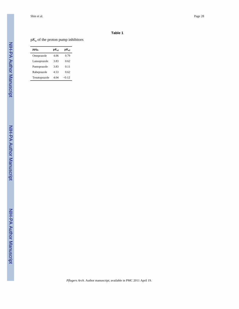
The second vital step is pH-dependent conversion from the accumulated prodrug to theactivated species that is a highly reactive thiophilic reagent. This means that a secondprotonation of these compounds is required for their activation to the compounds that formdisulfides with luminally accessible cysteines of the H,K-ATPase. The order of acid stabilityis tenatoprazole > pantoprazole > omeprazole > lansoprazole > rabeprazole [49]. Earlierwork, which had only considered the chemistry of omeprazole, suggested that the key sitethat was protonated was the N of the pyridine moiety, but this would not explain thedifferent rates of activation of the different PPIs with similar pyridine pKas. When the rate ofconversion of the different compounds was measured as a function of pH using UVabsorbance beginning at a relatively neutral pH, rather than with a pH electrode starting atpH 2.0, in the presence of a large excess of mercaptoethanol, it was found that the pHdependence of activation reflected protonation of the benzimidazole or pyridoimidazolemoiety. The rate of activation of some of the PPIs as a function of medium pH is shown inFig. 7. Table 1 shows the two pKas of all PPIs of Fig. 6 [47].
The actual inhibitory form of these prodrugs is somewhat controversial. In acidic solution,the form that is isolated is on the bottom left of Fig. 8 before reaction with one or morecysteines accessible from the luminal surface of the enzyme, a tetracyclic sulfenamide. Thisis a planar molecule; thus, any enantiomer of a PPI loses stereo-specifity upon activation.This was assumed for many years to be the actual active form of the drug, but more recent
Shin et al. Page 6
Pflugers Arch. Author manuscript; available in PMC 2011 April 19.
NIH
-PA Author Manuscript
NIH
-PA Author Manuscript
NIH
-PA Author Manuscript
data suggest that it is the precursor of the sulfenamide, the hydrated sulfenic acid on theright above the enzyme reaction step that is the reactive species forming directly from themono-protonated benzimidazole bound on the surface of the pump.
This hypothesis was formulated to explain the reaction with cysteine 813 or cysteine 321without reaction with cysteine 822; the likely first step is binding of the prodrug protonatedon the pyridine part of the compound to the vestibule with cysteine 813 at its apex. Withacid transport by the ATPase, the second proton is added, and then the compound can beactivated to the sulfenic acid. If this occurs rapidly as for omeprazole or lansoprazole,reaction with cysteine 813 or also 321 takes place, and no drug can access cysteine 822.However, if the activation is delayed, the drug can access cysteine 822 before activation tothe sulfenic acid and then when activated, both cysteine 813 and 822 are derivatized, as hasbeen shown when comparing the cysteines reacting with omeprazole, lansoprazole to thosereacting with pantoprazole or tenatoprazole [49,52].
Biology of proton pump inhibitorsProton pump inhibitors are prodrugs as discussed above. After accumulation in thestimulated secretory canaliculus of the parietal cell followed binding to the ATPase, thesecond protonation occurs, and they are then activated to form the thiophilic drug that reactswith luminally accessed cysteines on the pump. It clearly shows acid-catalyzed activation.Hence, it is recommended that they be given ~30 min before a meal to ensure that the pumpis active at peak levels of the drug. It is also necessary to protect them from gastric acidbefore absorption. Consequently, they are all formulated with an acid-resistant coating thatdoes not dissolve until pH ~5.0 to allow access to the duodenum for absorption withoutdestruction by gastric acid. PPIs have the half-life in blood that varies between 60 and 90min, but because they covalently bind to the pump, their half-life of inhibition of gastric acidsecretion is substantially longer than their half-life in blood [43].
Binding sites of proton pump inhibitors—The sites of reaction of the different protonpump inhibitors on the enzyme differs according to the particular proton pump inhibitor.However, all PPIs react with cysteine 813 in the loop between TM5 and TM6, fixing theenzyme in the E2 configuration. This cysteine, as well as cysteines 321 and 822, is on thedirect path from and to the ion binding site as illustrated in Figs. 2 and 3.
Omeprazole reacted with cysteine 813 and 892, the latter being peripheral to the iontransport domain [5]. Lansoprazole reacted with cysteine 813 and cysteine 321, these beingin the luminal vestibule [42], whereas pantoprazole and tenatoprazole react with cysteines813 and 822 [46,49,52]. The reaction with cysteine 822 confers a rather special property tothe covalently inhibited enzyme, namely irreversibility to reducing agents in vitro and invivo. The binding sites for pantoprazole are shown in Fig. 9 at cysteine 813, as for all thePPIs and cysteine 822 which is a particular property of pantoprazole and tenatoprazole.
Restoration of acid secretion after PPI inhibition—The PPIs are prodrugs, inactivein their native form. The PPIs are rapidly metabolized by the liver. There are no significantadverse effects during treatment with these medications. This may be ascribed to threefactors: their ability to accumulate selectively in the highly acidic space of the stimulatedsecretory canaliculus of the parietal cell due to the pKa of the pyridine group of the PPIs,their formation of the reactive cationic (hence relatively membrane impermeable) sulfenicacid on the pump itself, and the requirement for a pH <2.5 for a significant rate of activation.
Because of these properties, these drugs have a very large therapeutic index, but also, asnoted previously, the requirement for acid activation results in a delay in their suppression ofthe pump’s activity. Even though meal stimulates acid secretion and acid secretion activates
Shin et al. Page 7
Pflugers Arch. Author manuscript; available in PMC 2011 April 19.
NIH
-PA Author Manuscript
NIH
-PA Author Manuscript
NIH
-PA Author Manuscript
PPIs, PPIs cannot inhibit all pumps. About 70% of pump enzyme is inhibited, as PPIs haveshort half-life and not all pump enzymes are activated. It takes about 3 days to reach steady-state inhibition of acid secretion, as a balance is struck between covalent inhibition of activepumps, subsequent stimulation of inactive pumps after the drug has been eliminated fromthe blood, and de novo synthesis of new pumps. The pump protein has a half-life of about 54h in the rat [15] (and probably in man); thus, about 20% new pumps are synthesized over a24-h period, and it may be that there is greater pump synthesis at night than during the day.In addition, bedtime administration will not add to inhibition of nocturnal acid breakthrough,as the drug will have disappeared by the time nighttime acid secretion is evident. On theassumption that about 70% of pumps are activated by breakfast and that the PPI is given 30–60 min before, it can be calculated that steady-state inhibition on once a day dosage is about66% of maximal acid output. Increasing the dose has virtually no effect once optimal dosagehas been reached, but increasing dose frequency has some effect, so a morning dose andevening dose before meals results in about 80% inhibition of maximal acid output.
The effect of binding of the drug to cysteine 822 and the direct reversibility of PPI inhibitionwas examined. A prelude to this analysis was evident in measurement of the halftime ofpump protein biosynthesis in rats treated for 7 days with omeprazole (54 h in untreated andtreated rats) and the halftime of restoration of ATPase activity (15 h), suggesting a morerapid recovery of ATPase activity and acid secretion than if only de novo biosynthesis wasresponsible for restoration of ATPase activity [15]. In other experiments, the halftime ofrestoration of acid secretion in omeprazole-treated rats was 20 h [19,70]. An analysis of therate of restoration of acid secretion in man after omeprazole inhibition suggested that thehalftime was 24 h, whereas after pantoprazole, it was 46 h [12]. Apparently, only the latterdrug gave a rate of recovery compatible with restoration of acid secretion as due entirely topump turnover [9,23].
To find the difference of inhibitory activity among PPIs, two types of experiments werecarried out in rats to further investigate the problem. The animals were stimulatedmaximally by histamine and a large dose of radiolabel led omeprazole or pantoprazoleadministered IV, and either the stomach was removed and the ATPase isolated after 2 h orstomachs were removed at different times after drug administration (1 to 24 h), and againthe ATPase isolated. Using quantitative Western blot analysis, it was possible to determinethe stoichiometry of labeling, and by measuring acid secretion, it was ascertained that about90% inhibition had occurred after 1 h.
Then, the isolated enzyme was treated with the endogenous reducing agent, glutathione, thathas a concentration of about 3 mM in the parietal cell. As shown in Fig. 10, incubation ofthe inhibited ATPase with glutathione resulted in a different rate of loss of binding ofomeprazole and pantoprazole.
These observations suggest that removal of binding of the drug to cysteine 813 accounts forthe fast phase of recovery of acid secretion and the slow recovery occurs because of a delayin removal of the drug from cysteine 822. Both residues cysteine 813 and 822 are equallylabeled by pantoprazole in vivo. The small amount of cysteine 822 bound by omeprazole invivo is not seen in vitro [51,52], presumably because acidification is isolated gastric vesiclesis less than in vivo. In vivo, it is likely that a minor fraction of the omeprazole remainsprotonated at both the pyridine and benzimidazole nitrogen and is, as for pantoprazole,slowly activated, allowing some access to cysteine 822.
A similar differential was seen when the time course of labeling was followed by killing theanimals at different times and the stoichiometry of labeling determined as shown in Fig. 11.The differential stability of binding of pantoprazole and omeprazole to the ATPase where
Shin et al. Page 8
Pflugers Arch. Author manuscript; available in PMC 2011 April 19.
NIH
-PA Author Manuscript
NIH
-PA Author Manuscript
NIH
-PA Author Manuscript
loss of binding was biphasic with each phase having first-order kinetics was shown in Fig.11. The fast phase accounts for 84% of omeprazole bound, the slow phase for 16% of thedrug, whereas the fast and slow phases for pantoprazole bound are approximately equal,51% and 49%, respectively. These data are again explained by reversibility of almost all ofthe omeprazole labeling (to cysteine 813 and 892) and only partial reversibility ofpantoprazole binding, namely the 51% bound to cysteine 813.
The stoichiometry of labeling for full inhibition, namely 2.6 nmol/mg ATPase protein,reflects the oligomeric nature of the active functional enzyme, as 1 mg of protein containsabout 5 nmol of the gastric enzyme. Hence, binding to only half of the enzyme is sufficientfor full inhibition.
The above data are consistent with the slower recovery of acid secretion after pantoprazoleinhibition as compared to that after omeprazole inhibition deduced from human data [9,23].Whether this stability of labeling after pantoprazole labeling produces a clinical benefit,particularly in diminishing nocturnal acid breakthrough and night time GERD, remains to beestablished.
Acid pump antagonistsAn alternative to PPIs is the reversible K+-competitive APAs.
In the early 1980s, it was recognized that several tertiary amines were able to K+
competitively inhibit the H,K-ATPase, and an imidazopyridine compound, SCH28080, wasdeveloped for control of acid secretion [22]. In contrast to PPIs, this compound does notrequire acid secretion to inhibit the enzyme activity [69]. The APAs, including theimidazo[1,2α]pyridine, SCH28080, bind selectively to the E2P or E2 form of the enzyme[48,69], and binding to only half cycle was enough to block the enzyme activity [48].Inhibition by SCH28080 depended on binding of the protonated form of the inhibitor.
In body, unlike the PPIs, inhibition by APAs was expected to be fast and effective, as APAsdo not require acid activation which is necessary for PPI activation. However, their durationof action will be determined entirely by their level in the blood, and there will not be theprolongation of inhibition as found in the case of the PPIs where the half-life of the covalentbond is much longer than the half-life of the PPI in the blood. Data in man show theexpected rapid and virtually complete inhibition by an APA (PCAB), AZD0865, but ashorter duration of action as compared to S-omeprazole [21]. APAs require bid dosing toshow superiority to PPIs but may become the drugs of choice for rapid symptom relief.
As these compounds are reversible, their site of binding cannot be determined simply byidentification of the fragment of the ATPase that is labeled, but must rely on the effects ofsite-directed mutations on the affinity and K-competitive nature of the compound. A keyobservation was that inhibition by omeprazole and SCH2808 was mutually exclusive,indicating an overlap in the binding region of these two compounds, and this allowedorientation of the SCH28080 in the same general domain as omeprazole [18]. Site-directedmutations enabled a more exact docking structure for the K+-competitive antagonists thatcould then be confirmed by varying the structure of the inhibitor and correlating the Ki ofsuch compounds with the docking site model.
Mutations towards the exoplasmic surface of TM4, TM5 [3], TM6, the loop between TM5and TM6, and one site at the end of TM8 altered either the Ki or changed the nature ofinhibition from strictly competitive to mixed or even non-competitive without affecting ionaffinity. Mutation of lysine 791 to serine greatly reduced enzyme activity as well asincreased the Ki for SCH28080 [31,64,66]. The latter observations indicate that these
Shin et al. Page 9
Pflugers Arch. Author manuscript; available in PMC 2011 April 19.
NIH
-PA Author Manuscript
NIH
-PA Author Manuscript
NIH
-PA Author Manuscript
mutations allow simultaneous occupancy of the inhibitor binding site and the ion bindingdomain, which does not happen in the wild-type enzyme. These observations are interpretedas showing that the particular mutations generate a space whereby K+ can bypass theinhibitor, allowing access to the ion domain situated above the inhibitor binding region.
Using the various mutational data, it was possible to generate a model showing the dockingof the imidazonapthyridine to the membrane domain of the H,K-ATPase [32]. This fusedring structure was generated to mimic the calculated structure of the imidazopyridinesshown in Fig. 12. The Ki of BYK 36399 was found to be 47 nM similar to the 56 nMdetermined for SCH28080. However, when N-methyl group was used to replace thehydrogen of BYK 36399, the Ki rose to ~1 mM. This finding is explained in the model ofFig. 13 where there is interference with binding due to the presence of the methyl group onthe nitrogen of the tricyclic structure.
Many APAs with either imidazopyridines or benzimidazole core structures weresynthesized. These compounds provided very high affinity to the H,K-ATPase withexcellent inhibition [2,24,55]. However, despite superior inhibition, developments of manyAPAs including SCH28080 were dropped because of toxicity. A few APA compoundshaving other parent structures other than imidazopyridines or benzimidazoles are still beingdeveloped. One of these is revaprazan. Revaprazan is a pyrimidine derivative, N-(4-fluorophenyl)-4,5-dimethyl-6-(1-methyl-1,2,3,4-tetrahydroisoquinolin-2-yl)pyrimidin-2-amine hydrochloride. Revaprazan demonstrated dose-dependent inhibition and reachedmaximal effect at day 1 during the multiple-dose study, which is different from PPIs [17,73].Revaprazan is now clinically used in the Far East. Another structure of APA is CS-526.CS-526 has the pyrrolopyridazine structure and is also under development [20].
ConclusionThe gastric H,K-ATPase of the parietal cell pumps acid, which is the final step of gastricacid secretion in the stomach. Stimulation of the parietal cell moves the H,K-ATPase intothe secretory canaliculi, and the enzyme then can pump acid by becoming associated withK+ and Cl− conductance. One of the stimulants is histamine. Histamine stimulates theenzyme through binding to histamine H2 receptor. The initial success of H2RAs confirmedthat controlling acid secretion enables healing of acid-related diseases. To achieve moreeffective inhibition, the gastric H,K-ATPase became the rational target, and PPIs weredeveloped. These PPIs provided a major medical success story for the treatment of pepticulcer disease and GERD.
The gastric H,K-ATPase exchanges hydronium ion with potassium using ATP energy. Totransport the hydronium ion, the enzyme undergoes large conformational changes in thetransition between E1 EP and E2. The functional form of the gastric H,K-ATPase is anoligomeric dimer, [αβ]2. One heterodimer forms E1, while the other heterodimer forms E2.One mole of the inhibitor per 2 mol of the enzyme blocks the enzyme reaction. This enzymeis the target of PPIs, the drugs of choice for treatment of acid-related diseases. Covalentinhibitor binding sites, site mutagenesis for analysis of APA binding sites, and homologymodeling have provided a framework for understanding of the mechanism of this P2-typeATPase.
References1. Abe K, Kaya S, Taniguchi K, Hayashi Y, Imagawa T, Kikumoto M, Oiwa K, Sakaguchi K.
Evidence for a relationship between activity and the tetraprotomeric assembly of solubilized piggastric H/K-ATPase. J Biochem (Tokyo). 2005; 138:293–301. [PubMed: 16169880]
Shin et al. Page 10
Pflugers Arch. Author manuscript; available in PMC 2011 April 19.
NIH
-PA Author Manuscript
NIH
-PA Author Manuscript
NIH
-PA Author Manuscript
2. Andersson K, Carlsson E. Potassium-competitive acid blockade: a new therapeutic strategy in acid-related diseases. Pharmacol Ther. 2005; 108:294–307. [PubMed: 16000224]
3. Asano S, Yoshida A, Yashiro H, Kobayashi Y, Morisato A, Ogawa H, Takeguchi N, Morii M. Thecavity structure for docking the K(+)-competitive inhibitors in the gastric proton pump. J BiolChem. 2004; 279:13968–13975. [PubMed: 14699149]
4. Bamberg K, Mercier F, Reuben MA, Kobayashi Y, Munson KB, Sachs G. cDNA cloning andmembrane topology of the rabbit gastric H+/K(+)-ATPase alpha-subunit. Biochim Biophys Acta.1992; 1131:69–77. [PubMed: 1316171]
5. Besancon M, Shin JM, Mercier F, Munson K, Miller M, Hersey S, Sachs G. Membrane topologyand omeprazole labeling of the gastric H+,K(+)-adenosinetriphosphatase. Biochemistry. 1993;32:2345–2355. [PubMed: 8382947]
6. Black JW, Duncan WAM, Durant CJ, Ganellin CR, Parsons ME. Definition and antagonism ofhistamine H2 receptors. Nature. 1972; 236:385–390. [PubMed: 4401751]
7. Burnay M, Crambert G, Kharoubi-Hess S, Geering K, Horisberger JD. Electrogenicity of Na,K- andH,K-ATPase activity and presence of a positively charged amino acid in the fifth transmembranesegment. J Biol Chem. 2003; 278:19237–19244. [PubMed: 12637496]
8. Codina J, Li J, Dubose TD Jr. A carboxy-terminus motif of HKalpha2 is necessary for assembly andfunction. Kidney Int. 2004; 66:2283–2292. [PubMed: 15569317]
9. Dammann HG, Burkhardt F. Pantoprazole versus omeprazole: influence on meal-stimulated gastricacid secretion. Eur J Gastroenterol Hepatol. 1999; 11:1277–1282. [PubMed: 10563540]
10. Dempski RE, Hartung K, Friedrich T, Bamberg E. Fluorometric measurements of intermoleculardistances between the alpha- and beta-subunits of the Na+/K+-ATPase. J Biol Chem. 2006;281:36338–36346. [PubMed: 16980302]
11. Fellenius E, Berglindh T, Sachs G, Olbe L, Elander B, Sjostrand SE, Wallmark B. Substitutedbenzimidazoles inhibit gastric acid secretion by blocking (H+ + K+)ATPase. Nature. 1981;290:159–161. [PubMed: 6259537]
12. Ferron GM, McKeand W, Mayer PR. Pharmacodynamic modeling of pantoprazole’s irreversibleeffect on gastric acid secretion in humans and rats. J Clin Pharmacol. 2001; 41:149–156.[PubMed: 11210394]
13. Forte JG, Forte TM, Black JA, Okamoto C, Wolosin JM. Correlation of parietal cell structure andfunction. J Clin Gastroenterol. 1983; 5(Suppl 1):17–27. [PubMed: 6228573]
14. Galmiche JP, Bruley Des Varannes S, Ducrotte P, Sacher-Huvelin S, Vavasseur F, Taccoen A,Fiorentini P, Homerin M. Tenatoprazole, a novel proton pump inhibitor with a prolonged plasmahalf-life: effects on intragastric pH and comparison with esomeprazole in healthy volunteers.Aliment Pharmacol Ther. 2004; 19:655–662. [PubMed: 15023167]
15. Gedda K, Scott D, Besancon M, Lorentzon P, Sachs G. Turnover of the gastric H+,K(+)-adenosinetriphosphatase alpha subunit and its effect on inhibition of rat gastric acid secretion.Gastroenterology. 1995; 109:1134–1141. [PubMed: 7557078]
16. Grishin AV, Caplan MJ. ATP1AL1, a member of the non-gastric H,K-ATPase family, functions asa sodium pump. J Biol Chem. 1998; 273:27772–27778. [PubMed: 9774385]
17. Han KS, Kim YG, Yoo JK, Lee JW, Lee MG. Pharmacokinetics of a new reversible proton pumpinhibitor, YH1885, after intravenous and oral administrations to rats and dogs: hepatic first-passeffect in rats. Biopharm Drug Dispos. 1998; 19:493–500. [PubMed: 9840211]
18. Hersey SJ, Steiner L, Mendlein J, Rabon E, Sachs G. SCH28080 prevents omeprazole inhibition ofthe gastric H+/K+-ATPase. Biochim Biophys Acta. 1988; 956:49–57. [PubMed: 2841979]
19. Im WB, Blakeman DP, Davis JP. Irreversible inactivation of rat gastric (H+–K+)-ATPase in vivoby omeprazole. Biochem Biophys Res Commun. 1985; 126:78–82. [PubMed: 2982382]
20. Ito K, Kinoshita K, Tomizawa A, Inaba F, Morikawa-Inomata Y, Makino M, Tabata K, ShibakawaN. Pharmacological profile of novel acid pump antagonist 7-(4-fluorobenzyloxy)-2, 3-dimethyl-1-{[(1S,2S)-2-methyl cyclopropyl]methyl}-1H-pyrrolo [2,3-d] pyridazine (CS-526). J PharmacolExp Ther. 2007; 323:308–317. [PubMed: 17630360]
21. Kahrilas PJ, Dent J, Lauritsen K, Malfertheiner P, Denison H, Franzen S, Hasselgren G. Arandomized, comparative study of three doses of AZD0865 and esomeprazole for healing of refluxesophagitis. Clin Gastroenterol Hepatol. 2007; 5:1385–1391. [PubMed: 17950677]
Shin et al. Page 11
Pflugers Arch. Author manuscript; available in PMC 2011 April 19.
NIH
-PA Author Manuscript
NIH
-PA Author Manuscript
NIH
-PA Author Manuscript
22. Kaminski JJ, Bristol JA, Puchalski C, Lovey RG, Elliott AJ, Guzik H, Solomon DM, Conn DJ,Domalski MS, Wong SC, Gold EH, Long JF, Chiu PJ, Steinberg M, McPhail AT. Antiulceragents. 1. Gastric antisecretory and cytoprotective properties of substituted imidazo[1,2-a]pyridines. J Med Chem. 1985; 28:876–892. [PubMed: 4009611]
23. Katashima M, Yamamoto K, Tokuma Y, Hata T, Sawada Y, Iga T. Comparative pharmacokinetic/pharmacodynamic analysis of proton pump inhibitors omeprazole, lansoprazole and pantoprazole,in humans. Eur J Drug Metab Pharmacokinet. 1998; 23:19–26. [PubMed: 9625268]
24. Kirchhoff P, Andersson K, Socrates T, Sidani S, Kosiek O, Geibel JP. Characteristics of the K+-competitive H+,K+-ATPase inhibitor AZD0865 in isolated rat gastric glands. Am J PhysiolGastrointest Liver Physiol. 2006; 291:G838–G843. [PubMed: 16798725]
25. Lambrecht NW, Yakubov I, Scott D, Sachs G. Identification of the K efflux channel coupled to thegastric H–K-ATPase during acid secretion. Physiol Genomics. 2005; 21:81–91. [PubMed:15613615]
26. Maeda M, Ishizaki J, Futai M. cDNA cloning and sequence determination of pig gastric (H+ + K+)-ATPase. Biochem Biophys Res Commun. 1988; 157:203–209. [PubMed: 2848518]
27. Maeda M, Oshiman K, Tamura S, Futai M. Human gastric (H+ + K+)-ATPase gene. Similarity to(Na+ + K+)-ATPase genes in exon/intron organization but difference in control region. J BiolChem. 1990; 265:9027–9032. [PubMed: 2160952]
28. Melle-Milovanovic D, Milovanovic M, Nagpal S, Sachs G, Shin JM. Regions of associationbetween the alpha and the beta subunit of the gastric H,K-ATPase. J Biol Chem. 1998;273:11075–11081. [PubMed: 9556592]
29. Mizukawa Y, Nishizawa T, Nagao T, Kitamura K, Urushidani T. Cellular distribution of parchorin,a chloride intracellular channel-related protein, in various tissues. Am J Physiol Cell Physiol.2002; 282:C786–C795. [PubMed: 11880267]
30. Morth JP, Pedersen BP, Toustrup-Jensen MS, Sorensen TL, Petersen J, Andersen JP, Vilsen B,Nissen P. Crystal structure of the sodium-potassium pump. Nature. 2007; 450:1043–1049.[PubMed: 18075585]
31. Munson KB, Gutierrez C, Balaji VN, Ramnarayan K, Sachs G. Identification of anextracytoplasmic region of H+,K(+)-ATPase labeled by a K(+)-competitive photoaffinity inhibitor.J Biol Chem. 1991; 266:18976–18988. [PubMed: 1655768]
32. Munson K, Vagin O, Sachs G, Karlish S. Molecular modeling of SCH28080 binding to the gastricH,K-ATPase and MgATP interactions with SERCA- and Na,K-ATPases. Ann N Y Acad Sci.2003; 986:106–110. [PubMed: 12763782]
33. Munson K, Garcia R, Sachs G. Inhibitor and ion binding sites on the gastric H,K-ATPase.Biochemistry. 2005; 44:5267–5284. [PubMed: 15807521]
34. Munson K, Law RJ, Sachs G. Analysis of the gastric H,K ATPase for ion pathways and inhibitorbinding sites. Biochemistry. 2007; 46:5398–5417. [PubMed: 17425287]
35. Olesen C, Picard M, Winther AM, Gyrup C, Morth JP, Oxvig C, Moller JV, Nissen P. Thestructural basis of calcium transport by the calcium pump. Nature. 2007; 450:1036–1042.[PubMed: 18075584]
36. Purhonen P, Thomsen K, Maunsbach AB, Hebert H. Association of renal Na,K-ATPase alpha-subunit with the beta-and gamma-subunits based on cryoelectron microscopy. J Membr Biol.2006; 214:139–146. [PubMed: 17557166]
37. Rabon EC, McFall TL, Sachs G. The gastric [H,K]ATPase: H+/ATP stoichiometry. J Biol Chem.1982; 257:6296–6299. [PubMed: 6281267]
38. Reenstra WW, Forte JG. H+/ATP stoichiometry for the gastric (K+ + H+)-ATPase. J Membr Biol.1981; 61:55–60. [PubMed: 6267286]
39. Reenstra WW, Crothers J Jr, Forte JG. The conformation of H,K-ATPase determines thenucleoside triphosphate (NTP) selectivity for active proton transport. Biochemistry. 2007;46:10145–10152. [PubMed: 17696364]
40. Reuben MA, Lasater LS, Sachs G. Characterization of a beta subunit of the gastric H+/K(+)-transporting ATPase. Proc Natl Acad Sci U S A. 1990; 87:6767–6771. [PubMed: 2168558]
Shin et al. Page 12
Pflugers Arch. Author manuscript; available in PMC 2011 April 19.
NIH
-PA Author Manuscript
NIH
-PA Author Manuscript
NIH
-PA Author Manuscript
41. Sachs G, Chang HH, Rabon E, Schackman R, Lewin M, Saccomani G. A nonelectrogenic H+
pump in plasma membranes of hog stomach. J Biol Chem. 1976; 251:7690–7698. [PubMed:12175]
42. Sachs G, Shin JM, Besancon M, Prinz C. The continuing development of gastric acid pumpinhibitors. Aliment Pharmacol Ther. 1993; 7:4–12. discussion 29–31. [PubMed: 8387826]
43. Sachs G, Shin JM, Howden CW. Review article: the clinical pharmacology of proton pumpinhibitors. Aliment Pharmacol Ther. 2006; 23(Suppl 2):2–8. [PubMed: 16700898]
44. Sachs G, Shin JM, Vagin O, Lambrecht N, Yakubov I, Munson K. The gastric H,K ATPase as adrug target: past, present, and future. J Clin Gastroenterol. 2007; 41:S226–S242. [PubMed:17575528]
45. Sawaguchi A, Aoyama F, Ide S, Suganuma T. The cryofixation of isolated rat gastric mucosaprovides new insights into the functional transformation of gastric parietal cells: an in vitroexperimental model study. Arch Histol Cytol. 2005; 68:151–160. [PubMed: 16276021]
46. Shin JM, Sachs G. Identification of a region of the H,K-ATPase alpha subunit associated with thebeta subunit. J Biol Chem. 1994; 269:8642–8646. [PubMed: 8132592]
47. Shin JM, Sachs G. Restoration of acid secretion following treatment with proton pump inhibitors.Gastroenterology. 2002; 123:1588–1597. [PubMed: 12404233]
48. Shin JM, Sachs G. Differences in binding properties of two proton pump inhibitors on the gastric H+,K+-ATPase in vivo. Biochem Pharmacol. 2004; 68:2117–2127. [PubMed: 15498502]
49. Shin JM, Besancon M, Simon A, Sachs G. The site of action of pantoprazole in the gastric H+/K(+)-ATPase. Biochim Biophys Acta. 1993; 1148:223–233. [PubMed: 8389196]
50. Shin JM, Cho YM, Sachs G. Chemistry of covalent inhibition of the gastric (H+, K+)-ATPase byproton pump inhibitors. J Am Chem Soc. 2004; 126:7800–7811. [PubMed: 15212527]
51. Shin JM, Grundler G, Senn-Bilfinger J, Simon WA, Sachs G. Functional consequences of theoligomeric form of the membrane-bound gastric H,K-ATPase. Biochemistry. 2005; 44:16321–16332. [PubMed: 16331993]
52. Shin JM, Homerin M, Domagala F, Ficheux H, Sachs G. Characterization of the inhibitory activityof tenatoprazole on the gastric H+,K+-ATPase in vitro and in vivo. Biochem Pharmacol. 2006;71:837–849. [PubMed: 16405921]
53. Shull GE. cDNA cloning of the beta-subunit of the rat gastric H,K-ATPase. J Biol Chem. 1990;265:12123–12126. [PubMed: 2165052]
54. Shull GE, Lingrel JB. Molecular cloning of the rat stomach (H+ + K+)-ATPase. J Biol Chem.1986; 261:16788–16791. [PubMed: 3023364]
55. Simon WA, Herrmann M, Klein T, Shin JM, Huber R, Senn-Bilfinger J, Postius S. Soraprazan:setting new standards in inhibition of gastric acid secretion. J Pharmacol Exp Ther. 2007;321:866–874. [PubMed: 17369284]
56. Skrabanja AT, Asty P, Soumarmon A, Joep J, de Pont HH, Lewin MJ. H+ transport byreconstituted gastric (H+ + K+)-ATPase. Biochim Biophys Acta. 1986; 860:131–136. [PubMed:3015212]
57. Song I, Mortell MP, Gantz I, Brown DR, Yamada T. Molecular cloning and structural analysis ofcanine gastric H+,K (+)-ATPase. Biochem Biophys Res Commun. 1993; 196:1240–1247.[PubMed: 8250881]
58. Swarts HG, Koenderink JB, Willems PH, Krieger E, De Pont JJ. Asn792 participates in thehydrogen bond network around the K+-binding pocket of gastric H,K-ATPase. J Biol Chem. 2005;280:11488–11494. [PubMed: 15644331]
59. Sweadner KJ, Donnet C. Structural similarities of Na,K-ATPase and SERCA, the Ca(2+)-ATPaseof the sarcoplasmic reticulum. Biochem J. 2001; 356:685–704. [PubMed: 11389677]
60. Toh BH, Gleeson PA, Simpson RJ, Moritz RL, Callaghan JM, Goldkorn I, Jones CM, MartinelliTM, Mu FT, Humphris DC, et al. The 60- to 90-kDa parietal cell autoantigen associated withautoimmune gastritis is a beta subunit of the gastric H+/K(+)-ATPase (proton pump). Proc NatlAcad Sci U S A. 1990; 87:6418–6422. [PubMed: 1974721]
61. Toyoshima C, Nakasako M, Nomura H, Ogawa H. Crystal structure of the calcium pump ofsarcoplasmic reticulum at 2.6 A resolution. Nature. 2000; 405:647–655. [PubMed: 10864315]
Shin et al. Page 13
Pflugers Arch. Author manuscript; available in PMC 2011 April 19.
NIH
-PA Author Manuscript
NIH
-PA Author Manuscript
NIH
-PA Author Manuscript
62. Toyoshima C, Asahi M, Sugita Y, Khanna R, Tsuda T, MacLennan DH. Modeling of theinhibitory interaction of phospholamban with the Ca2+ ATPase. Proc Natl Acad Sci U S A. 2003;100:467–472. [PubMed: 12525698]
63. Toyoshima C, Nomura H, Sugita Y. Crystal structures of Ca2+-ATPase in various physiologicalstates. Ann N Y Acad Sci. 2003; 986:1–8. [PubMed: 12763767]
64. Vagin O, Munson K, Lambrecht N, Karlish SJ, Sachs G. Mutational analysis of the K+-competitive inhibitor site of gastric H,K-ATPase. Biochemistry. 2001; 40:7480–7490. [PubMed:11412101]
65. Vagin O, Denevich S, Munson K, Sachs G. SCH28080, a K+-competitive inhibitor of the gastricH,K-ATPase, binds near the M5-6 luminal loop, preventing K+ access to the ion binding domain.Biochemistry. 2002; 41:12755–12762. [PubMed: 12379118]
66. Vagin O, Denevich S, Sachs G. Plasma membrane delivery of the gastric H,K-ATPase: the role ofbeta-subunit glycosylation. Am J Physiol Cell Physiol. 2003; 285:C968–C976. [PubMed:12773316]
67. Vagin O, Turdikulova S, Sachs G. The H,K-ATPase beta subunit as a model to study the role of N-glycosylation in membrane trafficking and apical sorting. J Biol Chem. 2004; 279:39026–39034.[PubMed: 15247221]
68. Vagin O, Turdikulova S, Yakubov I, Sachs G. Use of the H, K-ATPase beta subunit to identifymultiple sorting pathways for plasma membrane delivery in polarized cells. J Biol Chem. 2005;280:14741–14754. [PubMed: 15695513]
69. Wallmark B, Sachs G, Mardh S, Fellenius E. Inhibition of gastric (H+ + K+)-ATPase by thesubstituted benzimidazole, picoprazole. Biochim Biophys Acta. 1983; 728:31–38. [PubMed:6299338]
70. Wallmark B, Larsson H, Humble L. The relationship between gastric acid secretion and gastric H+,K+-ATPase activity. J Biol Chem. 1985; 260:13681–13684. [PubMed: 2997178]
71. Wallmark B, Briving C, Fryklund J, Munson K, Jackson R, Mendlein J, Rabon E, Sachs G.Inhibition of gastric H+,K+-ATPase and acid secretion by SCH 28080, a substituted pyridyl(1,2a)imidazole. J Biol Chem. 1987; 262:2077–2084. [PubMed: 3029064]
72. Wolosin JM, Forte JG. K+ and Cl− conductances in the apical membrane from secreting oxynticcells are concurrently inhibited by divalent cations. J Membr Biol. 1985; 83:261–272. [PubMed:2582127]
73. Yu KS, Bae KS, Shon JH, Cho JY, Yi SY, Chung JY, Lim HS, Jang IJ, Shin SG, Song KS, MoonBS. Pharmacokinetic and pharmacodynamic evaluation of a novel proton pump inhibitor,YH1885, in healthy volunteers. J Clin Pharmacol. 2004; 44:73–82. [PubMed: 14681344]
Shin et al. Page 14
Pflugers Arch. Author manuscript; available in PMC 2011 April 19.
NIH
-PA Author Manuscript
NIH
-PA Author Manuscript
NIH
-PA Author Manuscript
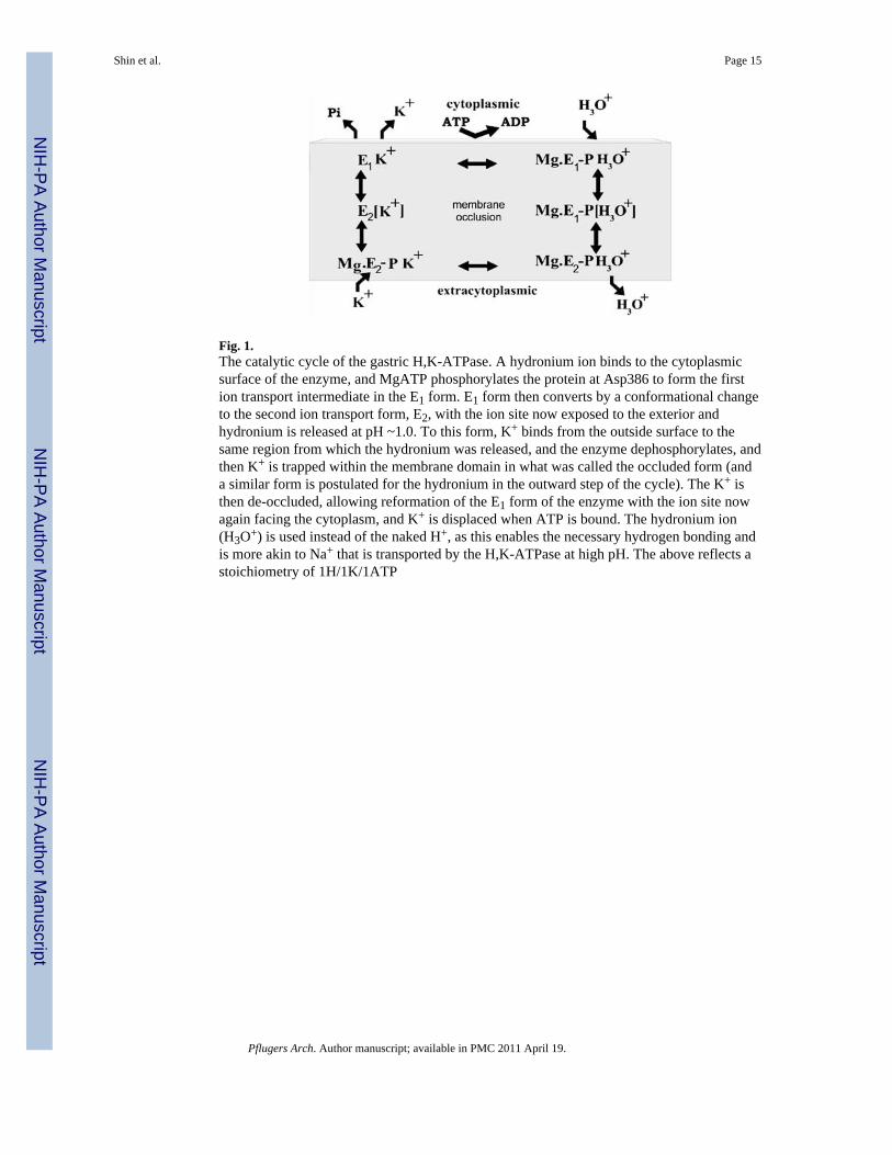
Fig. 1.The catalytic cycle of the gastric H,K-ATPase. A hydronium ion binds to the cytoplasmicsurface of the enzyme, and MgATP phosphorylates the protein at Asp386 to form the firstion transport intermediate in the E1 form. E1 form then converts by a conformational changeto the second ion transport form, E2, with the ion site now exposed to the exterior andhydronium is released at pH ~1.0. To this form, K+ binds from the outside surface to thesame region from which the hydronium was released, and the enzyme dephosphorylates, andthen K+ is trapped within the membrane domain in what was called the occluded form (anda similar form is postulated for the hydronium in the outward step of the cycle). The K+ isthen de-occluded, allowing reformation of the E1 form of the enzyme with the ion site nowagain facing the cytoplasm, and K+ is displaced when ATP is bound. The hydronium ion(H3O+) is used instead of the naked H+, as this enables the necessary hydrogen bonding andis more akin to Na+ that is transported by the H,K-ATPase at high pH. The above reflects astoichiometry of 1H/1K/1ATP
Shin et al. Page 15
Pflugers Arch. Author manuscript; available in PMC 2011 April 19.
NIH
-PA Author Manuscript
NIH
-PA Author Manuscript
NIH
-PA Author Manuscript
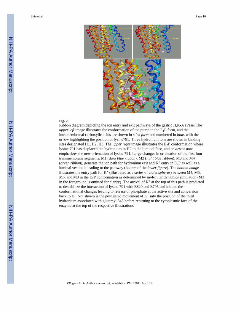
Fig. 2.Ribbon diagram depicting the ion entry and exit pathways of the gastric H,K-ATPase: Theupper left image illustrates the conformation of the pump in the E1P form, and theintramembranal carboxylic acids are shown in stick form and numbered in blue, with thearrow highlighting the position of lysine791. Three hydronium ions are shown in bindingsites designated H1, H2, H3. The upper right image illustrates the E2P conformation wherelysine 791 has displaced the hydronium in H2 to the luminal face, and an arrow nowemphasizes the new orientation of lysine 791. Large changes in orientation of the first fourtransmembrane segments, M1 (dark blue ribbon), M2 (light blue ribbon), M3 and M4(green ribbon), generate the ion path for hydronium exit and K+ entry in E2P as well as aluminal vestibule leading to the pathway (bottom of the lower figure). The bottom imageillustrates the entry path for K+ (illustrated as a series of violet spheres) between M4, M5,M6, and M8 in the E2P conformation as determined by molecular dynamics simulation (M3in the foreground is omitted for clarity). The arrival of K+ at the top of this path is predictedto destabilize the interaction of lysine 791 with E820 and E795 and initiate theconformational changes leading to release of phosphate at the active site and conversionback to E1. Not shown is the postulated movement of K+ into the position of the thirdhydronium associated with glutamyl 343 before returning to the cytoplasmic face of theenzyme at the top of the respective illustrations
Shin et al. Page 16
Pflugers Arch. Author manuscript; available in PMC 2011 April 19.
NIH
-PA Author Manuscript
NIH
-PA Author Manuscript
NIH
-PA Author Manuscript
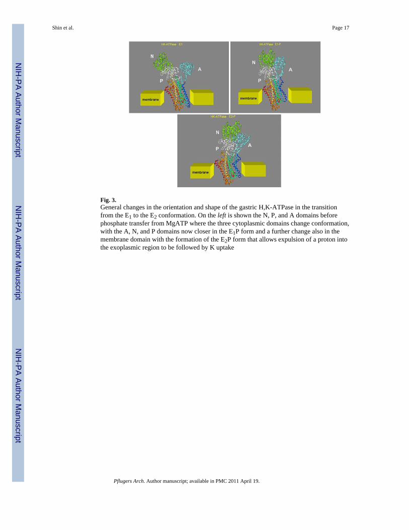
Fig. 3.General changes in the orientation and shape of the gastric H,K-ATPase in the transitionfrom the E1 to the E2 conformation. On the left is shown the N, P, and A domains beforephosphate transfer from MgATP where the three cytoplasmic domains change conformation,with the A, N, and P domains now closer in the E1P form and a further change also in themembrane domain with the formation of the E2P form that allows expulsion of a proton intothe exoplasmic region to be followed by K uptake
Shin et al. Page 17
Pflugers Arch. Author manuscript; available in PMC 2011 April 19.
NIH
-PA Author Manuscript
NIH
-PA Author Manuscript
NIH
-PA Author Manuscript
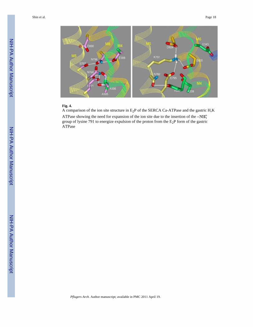
Fig. 4.A comparison of the ion site structure in E2P of the SERCA Ca-ATPase and the gastric H,KATPase showing the need for expansion of the ion site due to the insertion of the group of lysine 791 to energize expulsion of the proton from the E2P form of the gastricATPase
Shin et al. Page 18
Pflugers Arch. Author manuscript; available in PMC 2011 April 19.
NIH
-PA Author Manuscript
NIH
-PA Author Manuscript
NIH
-PA Author Manuscript
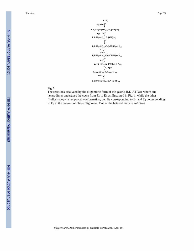
Fig. 5.The reactions catalyzed by the oligomeric form of the gastric H,K-ATPase where oneheterodimer undergoes the cycle from E1 to E2 as illustrated in Fig. 1, while the other(italics) adopts a reciprocal conformation, i.e., E2 corresponding to E1 and E1 correspondingto E2 in the two out of phase oligomers. One of the heterodimers is italicized
Shin et al. Page 19
Pflugers Arch. Author manuscript; available in PMC 2011 April 19.
NIH
-PA Author Manuscript
NIH
-PA Author Manuscript
NIH
-PA Author Manuscript
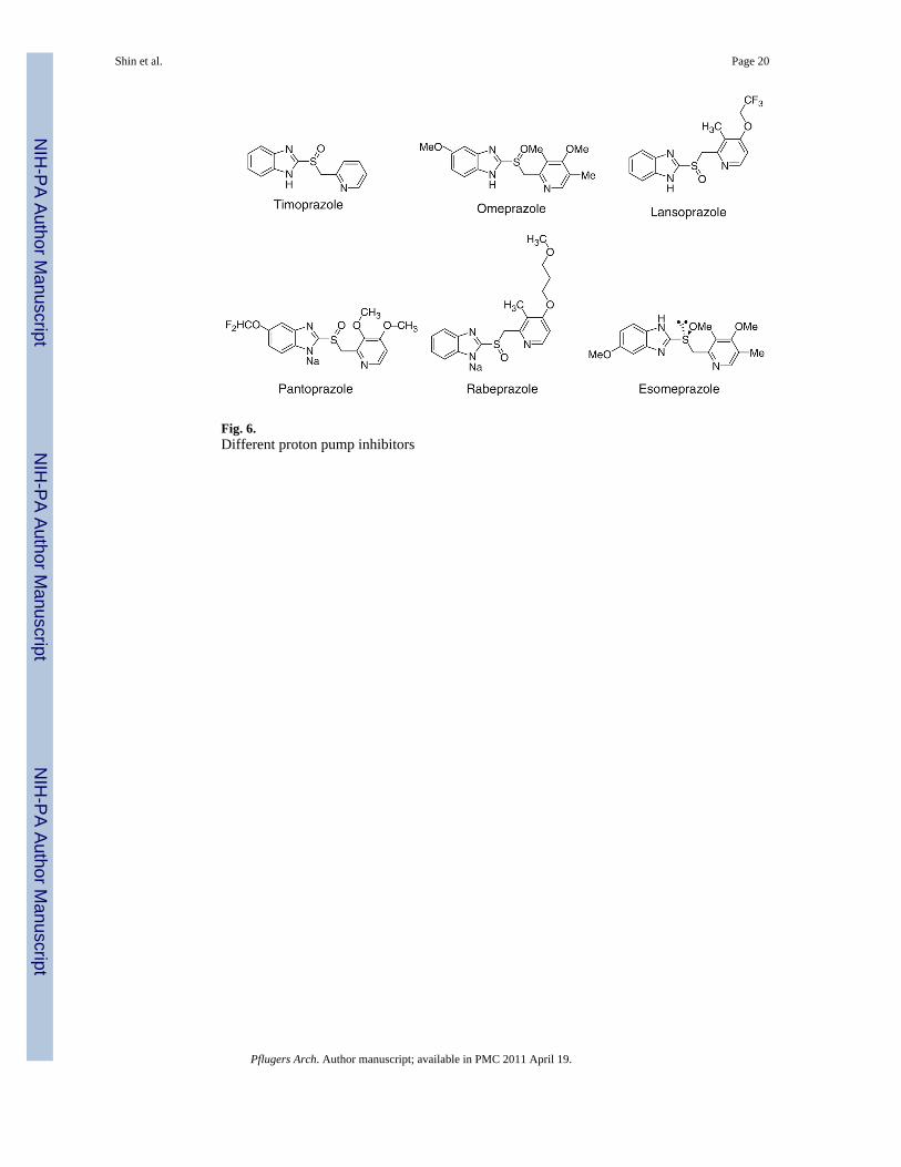
Fig. 6.Different proton pump inhibitors
Shin et al. Page 20
Pflugers Arch. Author manuscript; available in PMC 2011 April 19.
NIH
-PA Author Manuscript
NIH
-PA Author Manuscript
NIH
-PA Author Manuscript
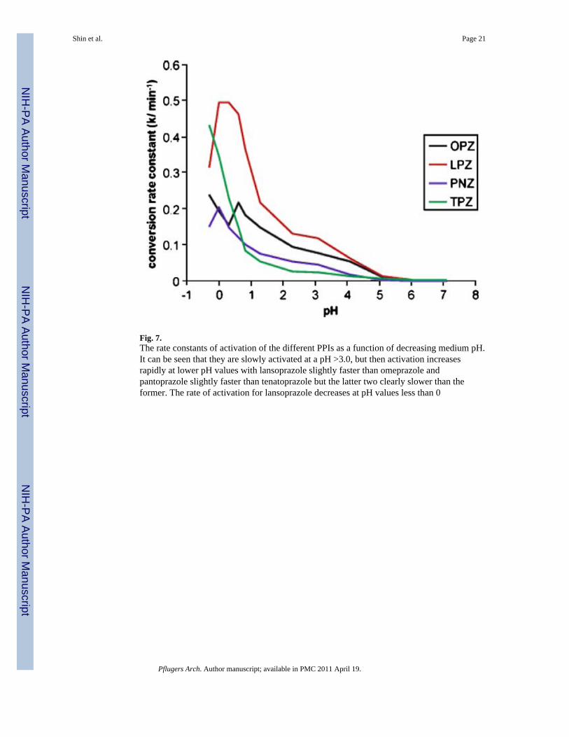
Fig. 7.The rate constants of activation of the different PPIs as a function of decreasing medium pH.It can be seen that they are slowly activated at a pH >3.0, but then activation increasesrapidly at lower pH values with lansoprazole slightly faster than omeprazole andpantoprazole slightly faster than tenatoprazole but the latter two clearly slower than theformer. The rate of activation for lansoprazole decreases at pH values less than 0
Shin et al. Page 21
Pflugers Arch. Author manuscript; available in PMC 2011 April 19.
NIH
-PA Author Manuscript
NIH
-PA Author Manuscript
NIH
-PA Author Manuscript
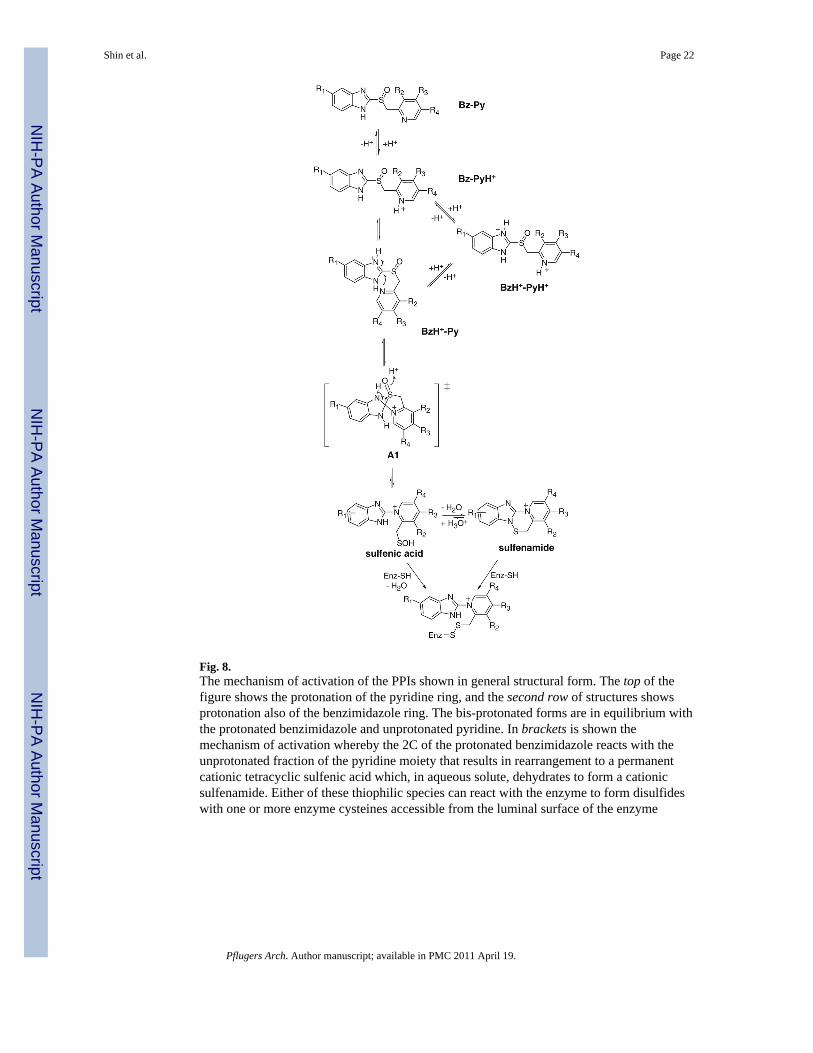
Fig. 8.The mechanism of activation of the PPIs shown in general structural form. The top of thefigure shows the protonation of the pyridine ring, and the second row of structures showsprotonation also of the benzimidazole ring. The bis-protonated forms are in equilibrium withthe protonated benzimidazole and unprotonated pyridine. In brackets is shown themechanism of activation whereby the 2C of the protonated benzimidazole reacts with theunprotonated fraction of the pyridine moiety that results in rearrangement to a permanentcationic tetracyclic sulfenic acid which, in aqueous solute, dehydrates to form a cationicsulfenamide. Either of these thiophilic species can react with the enzyme to form disulfideswith one or more enzyme cysteines accessible from the luminal surface of the enzyme
Shin et al. Page 22
Pflugers Arch. Author manuscript; available in PMC 2011 April 19.
NIH
-PA Author Manuscript
NIH
-PA Author Manuscript
NIH
-PA Author Manuscript
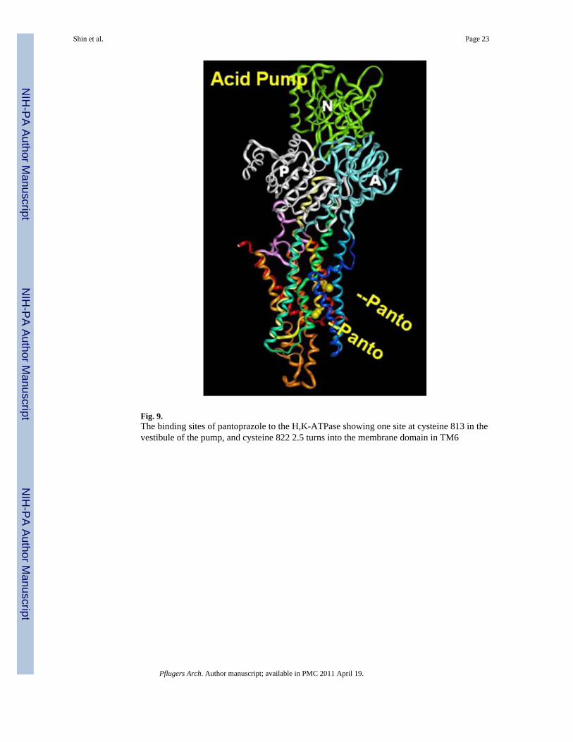
Fig. 9.The binding sites of pantoprazole to the H,K-ATPase showing one site at cysteine 813 in thevestibule of the pump, and cysteine 822 2.5 turns into the membrane domain in TM6
Shin et al. Page 23
Pflugers Arch. Author manuscript; available in PMC 2011 April 19.
NIH
-PA Author Manuscript
NIH
-PA Author Manuscript
NIH
-PA Author Manuscript
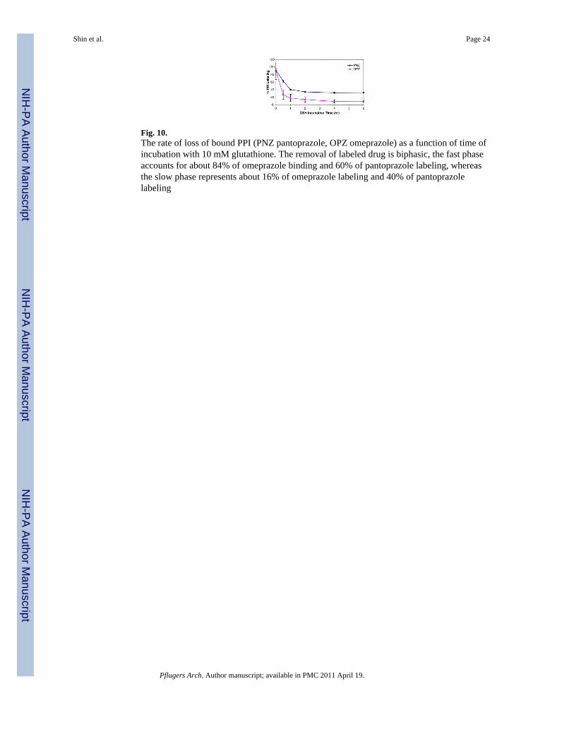
Fig. 10.The rate of loss of bound PPI (PNZ pantoprazole, OPZ omeprazole) as a function of time ofincubation with 10 mM glutathione. The removal of labeled drug is biphasic, the fast phaseaccounts for about 84% of omeprazole binding and 60% of pantoprazole labeling, whereasthe slow phase represents about 16% of omeprazole labeling and 40% of pantoprazolelabeling
Shin et al. Page 24
Pflugers Arch. Author manuscript; available in PMC 2011 April 19.
NIH
-PA Author Manuscript
NIH
-PA Author Manuscript
NIH
-PA Author Manuscript
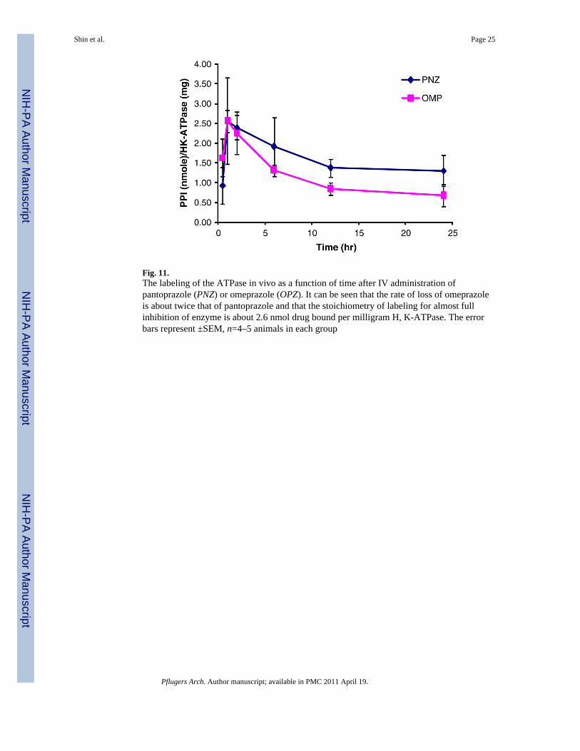
Fig. 11.The labeling of the ATPase in vivo as a function of time after IV administration ofpantoprazole (PNZ) or omeprazole (OPZ). It can be seen that the rate of loss of omeprazoleis about twice that of pantoprazole and that the stoichiometry of labeling for almost fullinhibition of enzyme is about 2.6 nmol drug bound per milligram H, K-ATPase. The errorbars represent ±SEM, n=4–5 animals in each group
Shin et al. Page 25
Pflugers Arch. Author manuscript; available in PMC 2011 April 19.
NIH
-PA Author Manuscript
NIH
-PA Author Manuscript
NIH
-PA Author Manuscript
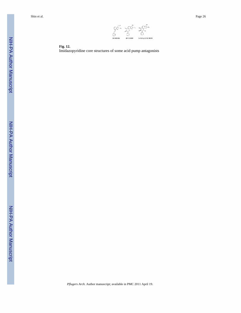
Fig. 12.Imidazopyridine core structures of some acid pump antagonists
Shin et al. Page 26
Pflugers Arch. Author manuscript; available in PMC 2011 April 19.
NIH
-PA Author Manuscript
NIH
-PA Author Manuscript
NIH
-PA Author Manuscript
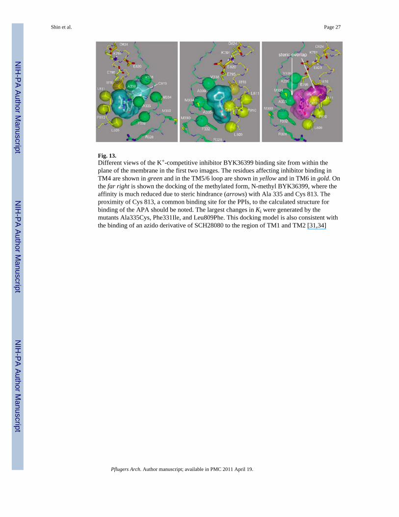
Fig. 13.Different views of the K+-competitive inhibitor BYK36399 binding site from within theplane of the membrane in the first two images. The residues affecting inhibitor binding inTM4 are shown in green and in the TM5/6 loop are shown in yellow and in TM6 in gold. Onthe far right is shown the docking of the methylated form, N-methyl BYK36399, where theaffinity is much reduced due to steric hindrance (arrows) with Ala 335 and Cys 813. Theproximity of Cys 813, a common binding site for the PPIs, to the calculated structure forbinding of the APA should be noted. The largest changes in Ki were generated by themutants Ala335Cys, Phe331Ile, and Leu809Phe. This docking model is also consistent withthe binding of an azido derivative of SCH28080 to the region of TM1 and TM2 [31,34]
Shin et al. Page 27
Pflugers Arch. Author manuscript; available in PMC 2011 April 19.
NIH
-PA Author Manuscript
NIH
-PA Author Manuscript
NIH
-PA Author Manuscript
NIH
-PA Author Manuscript
NIH
-PA Author Manuscript
NIH
-PA Author Manuscript
Shin et al. Page 28
Table 1
pKa of the proton pump inhibitors
PPIs pKa1 pKa2
Omeprazole 4.06 0.79
Lansoprazole 3.83 0.62
Pantoprazole 3.83 0.11
Rabeprazole 4.53 0.62
Tenatoprazole 4.04 −0.12
Pflugers Arch. Author manuscript; available in PMC 2011 April 19.