Embed Size (px)
Citation preview

167
©2009 European Journal of Histochemistry
Urocortin (UCN) is a 40 aminoacid peptide which belongs tocorticotropin-releasing factor (CRF) family. This family of pep-tides stimulates the secretion of proopiomelanocortin(POMC)-derived peptides, adrenocorticotropic hormone(ACTH), β-endorphin and melanocyte-stimulating hormone(MSH) in the pituitary gland. In the present study, usingWestern blotting and immunohistochemistry, the distributionof UCN in the primary lymphoid organs of the duck was inves-tigated at different ages. In the cloacal burse and thymus,Western blot demonstrated the presence of a peptide havinga molecular weight compatible with that of the mammalianUCN. In the cloacal burse, immunoreactivity was located in themedullary epithelial cells and in the follicular associated andcortico-medullary epithelium. In the thymus, immunoreactivitywas located in single epithelial cells. Double labellingimmunofluorescence studies showed that UCN immunoreac-tivity completely colocalised with cytokeratin immunoreactivityin both the thymus and cloacal burse. Statistically significantdifferences in the percentage of UCN immunoreactivity wereobserved between different age periods in the cloacal burse.The results suggest that, in birds, urocortin has an importantrole in regulating the function of the immune system.
Key words: cloacal burse, thymus, cytokeratin, medullaryreticular epithelial cells, CRF
Correspondence: Adriana De Luca,University of Naples Federico II, Napoli, ItalyTel.: +39.081.2536114.Fax +39.081.2536097.E-mail: [email protected]
Paper accepted on July 22, 2009
European Journal of Histochemistry2009; vol. 53 issue 3 (July-September): 167-176
Urocortin-like immunoreactivity in the primary lymphoid organsof the duck (Anas platyrhynchos)A. De Luca,1 C. Squillacioti,1 M.E. Pero,1 S. Paino,2 E. Langella,2 N. Mirabella1
1Department of Structures, Functions and biological Technologies, University of Naples Federico II,Napoli; 2Department of Sciences of Animal Production, University of Basilicata, Italy
Urocortin (UCN) is a 40-amino acid peptidebelonging to the mammalian corticotropin-releasing hormone (CRH) family which was
first discovered in the rat midbrain (Vaughan etal., 1995). On the basis of its selective ability tobind CRH-receptor type 2 (CRH-R2), differenttypes of UCN, i.e. UCN1, UCN2 and UCN3, havebeen identified (Lewis et al., 2001; Reyes et al.,2001). In mammals, UCN has been found in thecentral nervous (Vaughan et al., 1995), digestive(Muramatsu et al., 2000) and immune systems(Bamberger et al., 1998; Kageyama et al., 1999;Baigent et al., 2000), and in genital organs(Petraglia et al., 1996). UCN has also been foundto play a role in regulating some CRH-receptor-mediated effects (Turnbull et al., 1999). WhileUCN2 and UCN3 selectively bind to CRH-R2,UCN1 binds to both CRH-R1 and CRH-R2 andshows a greater affinity to CRH-R2 than CRHalone (Chalmers et al., 1996). Despite the abilityto interact with the same receptors, different func-tions are attributed to UCN and CRH. CRH is theprimary neuroregulator of the vertebrate stressresponse in so far as it has been shown to be themajor hypothalamic releasing factor for pituitaryadrenocorticotropic hormone, whereas UCNseems not to be involved in the activation of thehypothalamus- hypophysis-adrenal axis (Turnbullet al., 1999). Conversely, UCN influences the func-tion of the cardiovascular and nervous systems byincreasing anxiety, decreasing appetite and influ-encing behavioral activity (Latchman, 2001). Innon-mammalian vertebrates, few data have beenreported on the presence and the role of UCN.Themolecule, however, may have been conserved dur-ing vertebrate evolution, given that it has also beendetected in amphibians and birds (Kozicz et al.,2002; Cavani et al., 2003; Boorse et al., 2005;Calle et al., 2005). In amphibians, UCN and CRHreceptors have been found in the brain as well as
ORIGINAL PAPER

in many other organs and tissues, including thepituitary gland, heart, kidney and alimentarycanal (Kozicz et al., 2002; Boorse et al., 2005,2006); thus suggesting a potential role for diverseactions in tissue maintenance and function. InXenopus laevis, UCN injected in the third ventri-cle has been found to suppress food intake(Boorse et al. 2005). Moreover, it has been foundto act as a cytoprotective factor in tadpole tailduring metamorphosis (Boorse et al. 2006). Inbirds, UCN-ir has been found in neurons of thepigeon paramedian subgriseal mesencephalonwhich appear to be part of the brain circuitryinvolved in sympathetic nervous system-mediatedbehavioral responses to stress (Cavani et al.2003; Cunha et al., 2007). Intracerebro-ventricular administered UCN, moreover, has beenreported to decrease food intake in the chicken(Zhang et al., 2001). Up until now, however, nodata are available regarding the presence and roleof UCN in tissues and organs of birds outside thecentral nervous system (CNS). Since UCN and itsreceptors have been reported to be extensivelyexpressed in immune tissues and addressed to playimportant roles in the regulation of the immuneresponse (Baigent, 2001), the present study hasinvestigated the presence and distribution of UCNin the primary lymphoid organs of the duck bymeans of Western blotting and immunohistochem-istry. In addition, in order to verify if UCN alsoplays a role in the maturation of bird primary lym-phoid organs, UCN expression was evaluated atdifferent age periods.
Materials and Methods
Animals and tissue collectionCampbell khaki ducks (Anas platyrhynchos), of
both sexes, were used.They were anaesthetized byintramuscular injection of ketamine (25 mg/kg)and then killed by exsanguination. Thymus andcloacal burse from 10-, 30-, 50-, 70-, 90-, 110-,140- and 170-day-old ducks were collected. Fromeach thymus three lobes (one from cranial, onefrom median and one from caudal portion) wereutilized. Five animals for each group were used.Three adult Sprague-Dawley rats (13 weeks ofage; body weight: 380±20 g; purchased fromHarlan Italy) were used.They were housed in tem-perature- and lightcontrolled rooms and were
given water ad libitum.Three frogs (R. esculenta),captured in the vicinity of Naples were used. Ratswere anaesthetised by intraperitoneal injection ofurethane (1.2 g/kg), frogs by immersion in aMS222 solution (Sigma Chemical Co., St. Louis,MO). Both rats and frogs were killed by decapita-tion and brains were collected. All procedureswere approved by Italian laws regarding animaluse in research (art. 7 D.Lgs. 116/92).
Western blottingFresh pieces of duck thymus and cloacal burse,
frog brain and rat brain were collected, frozen ondry ice and stored at -80°C until extraction.Aliquots of thymus, cloacal burse, frog brain andrat brain extracts were lysed in ice cold-homoge-nization buffer, containing 2% Triton X-100, 150mM NaCl, 50 mM Tris HCl, pH 7.00, 5 mMEDTA, 1 mM PMSF, 10 µg/mL leupeptin and 0.1U/mL aprotinin. After 30 min on ice, lysates wereclarified by centrifugation at 10000 xg for 10 minat +4°C. The resulting supernatants were charac-terised for protein concentration by Bio-Rad DCprotein assay. Equal amounts of proteins wereimmunoprecipitated overnight at 4°C with anti-Urocortin antibody (cod. sc-1825, Santa CruzBiotechnologies, Santa Cruz, CA, USA) (1 µg/100µg proteins) previously bound to protein A/G-Agarose (Santa Cruz Biotechnologies, SantaCruz, CA, USA). The primary antibody was anaffinity-purified goat polyclonal antibody raisedagainst a peptide corresponding to amino acids102-121 mapping at the carboxy terminus of raturocortin precursor which is 122-amino acid pro-tein with a carboxy terminus including a putative40-amino acid peptide (Donaldson et al., 1996;Vaughan et al., 1995). Beads were sedimented bybrief centrifugation and washed extensively withice-cold homogenization buffer. Proteins, synthet-ic peptide UCN (cod U6631 Sigma, St Louis, MO,USA) and syntetic peptide CRF (cod C3042Sigma), solubilised in boiling SDS sample buffer(2% SDS, 5% L-mercaptoethanol, 66 mM Tris,pH 7.5, 10 mM EDTA), were separated on a 15%SDS-polyacrylamide gel (Bio-Rad, Hercules, CA,USA). After electrophoresis, gel was transferredto nitrocellulose using a semi-dry apparatus (Bio-Rad) according to the manufacturer’s instruc-tions. The membrane was incubated for 1 h at42°C in 5% bovine serum albumin (BSA; Sigma)in TBST (150 mMNaCl, 20 mM Tris/HCl pH 7.4,
168
A. De Luca, et al.
0.3% Tween 20), washed with TBST and incubat-ed for 1 h at room temperature with goat anti-serum specific for UCN (cod. sc-1825, Santa CruzBiotechnologies, Santa Cruz, CA, USA) diluited1:1000 in TBST containing 1% BSA. The mem-brane was washed three times with TBST and thanincubated with rabbit anti-goat peroxidase-conju-gated antibody (cod. PI-9500 Vector Labor-atories, Burlingame, Calif., USA) diluited 1:1000in TBST containing 1% BSA for 1 hour at roomtemperature. Proteins were visualized by anenhanced chemiluminescence kit (Amersham,Bucks, UK) Marker proteins (coloured proteinmolecular-weight markers; Rainbow, Amersham)were used to estimate the molecular weight ofeach band.
Immunohistochemistry
Tissue preparationFresh segments of thymus and cloacal burse
were collected from 10-, 30-, 50-, 70-, 90-, 110-,140- and 170-day-old ducks.
Frozen sectionsThe specimens were fixed in 4% paraformalde-
hyde in a 0.1 M phosphate-buffered saline (PBS)solution at pH 7.5 for 2-3 h and placed succes-sively in PBS containing 0.1% sodium azide and10% sucrose and stored overnight at 4°C.The fol-lowing day, the samples were transferred to a mix-ture of PBS-sucrose-azide and OCT compound(Tissue Tek, Elkhart, IN, USA) in a ratio of 1:1for 24 h before being embedded in 100% OCT.Coronal sections of 10-20 µm thickness were cut.These sections were treated using a doublelabelling fluorescence technique. They were prein-cubated in 2% Blocking Reagent (Cat No.1921673 Roche Applied Science, Germany) inPBS containing 0.1% Triton X-100 (PBS-T)overnight at 4°C. They were then rinsed threetimes for 10 min in PBS-T antisera overnight at4°C.The primary antibodies used were as follows:goat polyclonal anti-UCN (final dilution 1:50;cod. sc-1825, Santa Cruz Biotechnologies), mousemonoclonal anti-pan cytokeratin (final dilution1:1000 cod. C1801 Sigma). After washing withPBS, sections were incubated for 1 h at roomtemperature in a 1:1 mixture of secondary anti-bodies that had each an initial dilution of 1:50.The secondary antibodies used were as follows:
FITC-conjugated rabbit anti-goat (cod. 305-095-003, Jackson Immunoresearch, West Grove, PA,USA) and rhodamine-conjugated donkey anti-mouse IgG (cod. 715-025-151 JacksonImmunoresearch). Slides were cover-slipped usingimmuno-mount aqueous mounting medium anddried at 4°C overnight in the dark. To ensure thatno cross-reactivity of the secondary detection sys-tem occurred, the primary antisera were alterna-tively omitted in control tests. No cross-reactivitywas observed. The specimens were observed byusing a Leica TCS SP5 confocal microscopeequipped with epifluorescence and the appropriatefilter sets for viewing FITC and TRITC.
Paraffin-embedded sectionsThe specimens were immediately fixed by immer-
sion in Bouin’s fixative (6-24 h) and processed forparaffin embedding in a vacuum, and cut at a thick-ness of 5-7µm.The avidinbiotin-peroxidase complex(ABC) method was performed with the VectastainABC kit (cod. PK 4000, Vector Laboratories).Sections were deparaffinized in xylene and hydrat-ed in a graded solution of ethanol. After thequenching of endogenous peroxidase activity inwater containing 0.3% hydrogen peroxide for 30min, non specific binding was blocked by treatmentwith 1.5% normal rabbit serum (S-5000, VectorLaboratories) in 0.01 M PBS (phosphatebufferedsaline; pH 7.2) for 30 min. A polyclonal goat anti-UCN serum (cod. sc-1825, Santa CruzBiotechnologies) was applied to the sections at adilution of 1:1000 and each specimen was incubat-ed in a moist chamber overnight at 4°C. After thesections had been washed three times in PBS,biotinylated rabbit anti-goat IgG (cod. BA 5000,Vector Laboratories) was applied at a dilution of1:200. The sections were again incubated for 30min at room temperature. Freshly prepared ABCreagent was applied and incubated for 30 min afterthree washes in PBS.The localization of UCN wasvisualized by incubating the sections for 5 min infreshly prepared diaminobenzidine-nickel solution(cod. SK 4100,Vector Laboratories).The specifici-ty of the immunoreactions was tested by replacingthe primary antibody with buffer. No immunoreac-tion was detected in control tests.The specificity ofthe primary antibody anti-UCN was tested byadsorption the primary antibody with excess (up to50 µg/mL in the final dilution) homologous antigenpeptide (cod. SC-1825 P Santa Cruz Biotechno-
169
Original Paper

logies). The slides of the examinated organs wereindependently evaluated by two observers by usinga Leica DMRA2 microscope and scored accordingto the density of immunopositivities and intensity ofimmunoreactions.
Quantitative studyIn the cloacal burse, immunoreactive and nega-
tive follicles were counted from ducks in differentage periods and the percentage of the immunore-active follicles was calculated. In the cloacalburse and thymus of different age groups, the den-sity of UCN-ir was determinated by evaluating thearea occupied by ir-cells.The area was assessed byquantitative image analysis with the publicdomain NIH Image J software (http://rsb.info.nih.gov/nih-image/). Measure-ments were madeon ten serial sections (one every ten) for eachspecimen. In the cloacal burse, ten randomlyselected ir-follicles per section were measured. Inthe thymus, ten randomly ir-lobules per sectionwere measured. The area occupied by UCN-irstructures were manually delimited and the per-centage of immunoreactivity was calculated uponthe total area occupied by burse follicles orthymic lobules. Data were expressed as mean ±S.D., where n=numbers of animals. The signifi-cance of the differences across age groups wasevaluated by analysis of variance and by Student-Newman-Keuls’s Test for multiple comparisons. Avalue of p≤0.05 was considered significant. Theevaluation was done esclusively on paraffin-embedded sections processed with ABC method.
Results
Western blot analysisThe results of the Western blot analysis are
shown in Figure 1. Tissue extracts from the thy-mus (data not shown), cloacal burse, frog and ratbrain and the synthetic peptide UCN reacted withthe anti-UCN antibody, while the syntetic peptideCRF did not react. The antiserum recognised twoprotein bands from homogenates, one weighingabout 16.0 kDa and another about 6.5 kDa.
Immunohistochemistry and co-localization studiesUCN-like-ir was observed in both thymus and
cloacal burse in each of the examined age groups.UCN-like-ir was localized in the cortico-
medullary and medullary reticular epithelial(REC) cells (Figure 2b; 4 a,b,c). In addition,UCN-like-ir was localized in the follicle associat-ed epithelium (FAE) (Figure 2 a,c,c1), in theFAE supporting and in the interfollicular basalepithelial cells (Figure 2c).
The REC were stellate in morphology andformed a network showing an apparent continua-tion with the cortico-medullary epithelium(Figure 2b; 4 a,b,c) and the FAE (Figure 2c).UCN/CYT colocalization studies (Figure 2d-i)showed that UCN-like-ir completely colocalizedwith CYT-ir in the REC, in the cortico-medullaryepithelium, in the FAE, in the FAE supportingcells and in the interfullicular basal epithelialcells. In the thymus, UCN-like-ir was observed inmedullary epithelial cells (Figure 3a).These cellswere stellate in morphology (Figure 3b) and wereprimarily located in the zone separating the cor-tex from the medulla (Figure 3a) and around thethymic corpuscles (Figure 3c). UCN/CYT colo-calization studies showed that UCN-like-ir wasexpressed in CYT-ir cells (Figure 3d-f).
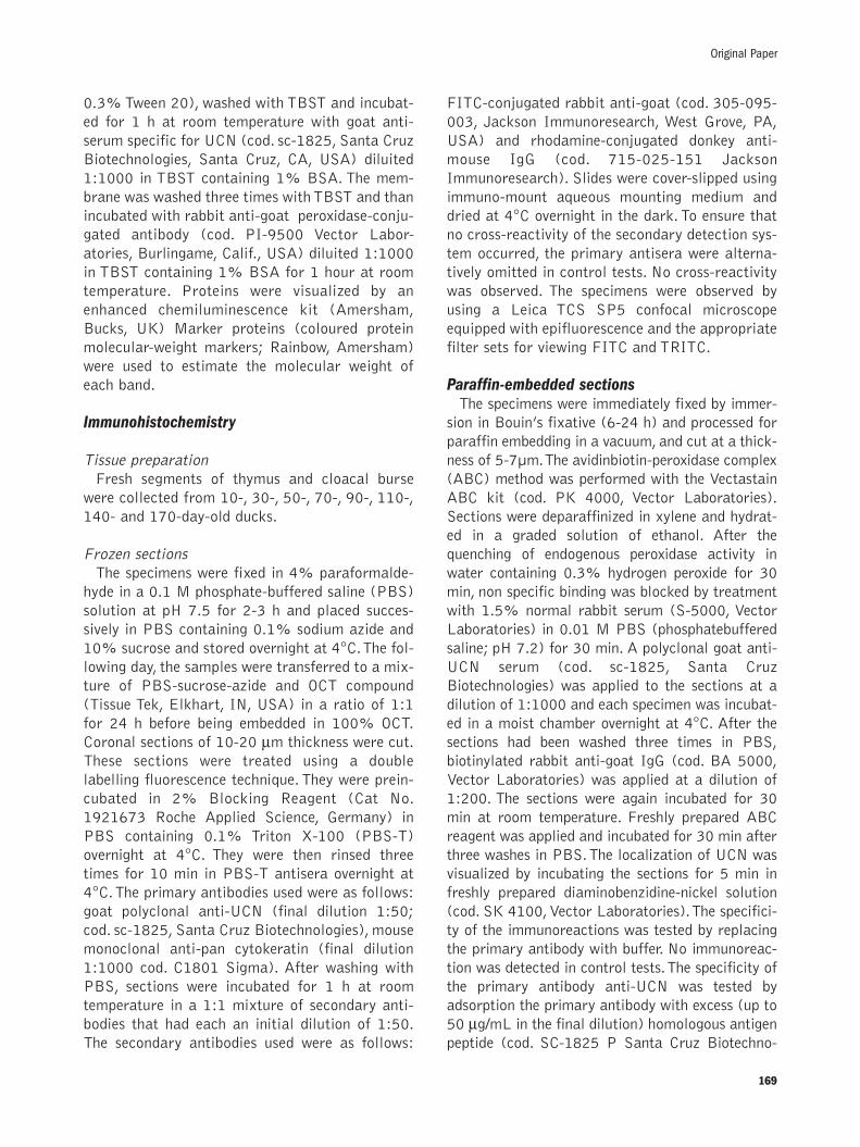
Quantitative resultsIn the cloacal burse the percentage of UCN-like
immunoreactive follicles was more than 90% inall the examined age groups (Figure 4 a,b,c,).Thedifferences in the percentage of ir-follicles among
170
A. De Luca, et al.
Figure 1. Detection of UCN-like immunoreactivity by Western blotanalysis. The anti-UCN antibody reacted with the tissue extracts(Lane A-C) and synthetic UCN (Lane D) but do not react with thesinthetic CRF (Lane E). Lane A, frog brain, Lane B rat brain, LaneC duck cloacal burse (140 days), lane D, synthetic UCN, Lane Esynthetic CRF. Protein markers are expressed in kDa. ãã,IgGheavy, IgG, light; ~~,, UCN Precursor; ����,, UCN.

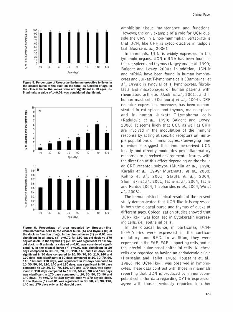
the groups were not statistically significant(Figure 5). In the cloacal burse, a significant andprogressive increase in the percentage of theimmunoreactive areas was observed between theage groups up until 140 days (Figure 6 A). Afterthis stage, a decrease was observed. In the thy-mus, a significant difference in the UCN-ir areawas observed between the first (T10) and the sec-ond (T30) stages. After 30 days no differenceswere observed (Figure 6 B).
Discussion
Western blot analysis showed that the anti-mammalian UCN antiserum used in the presentstudy recognized, in the duck cloacal burse andthymus, two protein bands having, respectively, amolecular weight of about 16.0 and 6.5 kDa.Thefirst protein is compatible with the UCN precur-sor which is a 122-amino acid protein.The secondis compatible with the putative 40-amino acidmature peptide of mammalian UCN (Donaldsonet al. 1996; Vaughan et al. 1995). The antiserum
171
Original Paper
Figure 2. Immunohistochemical localization of UCN-like in the duck cloacal burse of 140 day old animals. UCN-like-ir was found in the fol-licles in all the examined age groups (a). UCN-like-ir was observed in the cortico-medullary and medullary reticular epithelial cells (b).UCN-like-ir was also localized in the follicle associated epithelium (FAE) (c1), in the FAE supporting and in the interfollicular basal epithe-lial cells (c). UCN-like/CYT colocalization studies (d-i) showed that UCN-like-ir completely colocalized with CYT-ir in the REC, in the cor-tico-medullary epithelium, in the FAE, in the FAE supporting cells and in the interfullicular basal epithelial cells. M, Medulla; C, Cortex; ��,FAE; ����,, Cortico-medullary epithelium; ÇÇ, Medullary reticular epithelium; ��, FAE sc, FAE supporting cells; tt,, interfullicular basal epithe-lial cells. Bar= 50 µµm. Paraffin embedded sections.

also recognized a UCN-like protein in both frogand rat brains, suggesting that UCN is conservedamong vertebrates (Boorse et al., 2005). Theantiserum did not react with the syntetic peptideCRF, thus indicating that it is specific for UCN.
Up until now, the present paper is the firstreport regarding the presence of UCN in avian tis-sues outside the CNS. In mammals, UCN expres-sion has been observed in adipose tissue (Seres etal., 2004), heart (Nishikimi et al., 2000), enteric
plexus (Harada et al., 1999), testis and kidney(Kageyama et al., 1999), placenta (Petraglia etal., 1996) and adrenals (Fukuda et al., 2005),and has been addressed to regulate a variety offunctions (for reviews of the UCN functions inmammals see Oki and Sasano, 2004 and Feketeand Zorrilla, 2007). Among non-mammalian ver-tebrates, UCN has been found to be widelyexpressed in amphibians (Boorse et al., 2005),suggesting a potential for diverse actions in
172
A. De Luca, et al.
Figure 3. Immunohistochemical localization of UCN-like in the duck thymus of 140 day old animals. UCN-like-ir was observed in medullaryepithelial cells (a). These cells were stellate in morphology (b) and were primarily located in the zone separating the cortex from themedulla (a) and around the thymic corpuscles (c). UCN-like/CYT colocalization studies showed that UCN-like-ir was expressed in CYT-ircells (d-f). M, Medulla; C, Cortex; ��, Medullary thymic epithelial cells; xx, thymic corpuscle. Bar= 50 µµm. Paraffin embedded sections.
Figure 4. Immunohistochemical localization of UCN-like in the duck cloacal burse of 10 –70- 140 day old animals. UCN-like-ir was found inthe follicles in all the examined age groups (a, b, c). UCN-like-ir was observed in the cortico-medullary and medullary reticular epithelialcells (a,b,c). UCN-like-ir was also localized in the follicle associated epithelium (FAE) (c). M Medulla; C Cortex; ��,FAE; Bar= 50 µµm.Paraffin embedded sections.
amphibian tissue maintenance and functions.However, the only example of a role for UCN out-side the CNS in a non-mammalian vertebrate isthat UCN, like CRF, is cytoprotective in tadpoletail (Boorse et al., 2006).
In mammals, UCN is widely expressed in thelymphoid organs. UCN mRNA has been found inthe rat spleen and thymus (Kageyama et al. 1999;Baigent and Lowry, 2000). In addition, UCN-irand mRNA have been found in human lympho-cytes and Jurkatt T-lymphoma cells (Bamberger etal., 1998); in synovial cells, lymphocytes, fibrob-lasts and macrophages of human patients withrheumatoid arthritis (Uzuki et al., 2001); and inhuman mast cells (Kempuraj et al., 2004). CRFreceptor expression, moreover, has been demon-strated in rat spleen and thymus, mouse spleenand in human Jurkatt T-Lymphoma cells(Radulovic et al., 1999; Baigent and Lowry,2000). It seems likely that UCN as well as CRHare involved in the modulation of the immuneresponse by acting at specific receptors on multi-ple populations of immunocytes. Converging linesof evidence suggest that immune-derived UCNlocally and directly modulates pro-inflammatoryresponses to perceived environmental insults, withthe direction of this effect depending on the tissueor CRF receptor subtype (Muglia et al., 1994;Karalis et al., 1999; Muramatsu et al., 2000;Kohno et al., 2001; Saruta et al., 2004;Slominski et al., 2001; Tache et al., 2004; Tacheand Perdue 2004;Theoharides et al., 2004;Wu etal., 2006).
The immunohistochemical results of the presentstudy demonstrated that UCN-like-ir is expressedin both the cloacal burse and thymus of ducks atdifferent ages. Colocalization studies showed thatUCN-like-ir was localized in Cytokeratin express-ing cells, i.e., epithelial cells.
In the cloacal burse, in particular, UCN-like/CYT-irs were expressed in the cortico-medullary and REC. In addition, they wereexpressed in the FAE, FAE supporting cells, and inthe interfollicular basal epithelial cells. All thesecells are regarded as having an endodermic origin(Houssaint and Hallet, 1986; Houssaint et. al.,1986). No UCN-like-ir was observed in lympho-cytes.These data contrast with those in mammalsreporting that UCN is produced by immunocom-petent cells. Our data regarding CYT-ir expressionagree with those previously reported in other
173
Original Paper
Figure 5. Percentage of Urocortin-like-immunoreactive follicles inthe cloacal burse of the duck on the total as function of age. Inthe cloacal burse the values were not significant in all ages. n=5 animals; a value of p<0.01 was considered significant.
Figure 6. Percentage of area occupied by Urocortin-like-immunoreactive cells in the cloacal burse (A) and thymus (B) ofthe duck as function of age. In the cloacal burse (*) p< 0.01 wassignificant in all ages. (#) p=0.72 for 110 day-old duck vs 170day-old duck. In the thymus (*) p<0.01 was significant vs 10 day-old duck. n=5 animals; a value of p<0.01 was considered signifi-cant(*). In the cloacal burse (*) p<0.01 was significant in 10days compared to 30, 50, 70, 90, 110, 140 and 170 days, wassignificant in 30 days compared to 10, 50, 70, 90, 110, 140 and170 days, was significant in 50 days compared to 10, 30, 70, 90,110, 140 and 170 days, was significant in 70 days compared to10, 30, 50, 90, 110, 140 and 170 days, was significant in 90 dayscompared to 10, 30, 50, 70, 110, 140 and 170 days, was signif-icant in 110 days compared to 10, 30, 50,70, 90 and 140 dayswas significant in 170 days compared to 10, 30, 50, 70, 90 and140 days. (#) p=0,72 for 110 day-old duck vs 170 day-old duck.In the thymus (*) p<0.01 was significant in 30, 50, 70, 90, 110,140 and 170 days only vs 10 day-old duck.
10 30 50 70 90 110 140 170
10 30 50 70 90 110 140 170
10 30 50 70 90 110 140 170
Age (days)
Age (days)
Age (days)
1009080706050403020100
10
9
8
7
6
5
4
3
2
1
0
1
0.9
0.8
0.7
0.6
0.5
0.4
0.3
0.2
0.1
0
% o
f im
mun
orea
ctiv
e bu
rsal
folli
cles
% o
f uro
corti
n im
mun
orea
ctiv
e ce
lls%
of u
roco
rtin
imm
unor
eact
ive
cells
% o
f uro
corti
n im
mun
orea
ctiv
e ce
lls

avian species (Sanchez-Refusta et al., 1996; Olahand Glick, 1992).
The function of the bursal epithelium is believedto be connected to the maturation of B cells(Ciriaco et al., 2003; Baba & Okuno, 1976; Heller& Friedman, 1979; Toivanen & Toivanen, 1973).In particular, REC cells are addressed to partici-pate with macrophages, dendritic cells and ele-ments of the extracellular matrix in the formationof a bursal microenviroment for lymphoid cellmaturation (Glick, 1995). During the last decade,it was suggested that bursal medullary REC mightbe a source of bursin (Audhya et al., 1986;Viamontes et al., 1989), a peptide which selec-tively induces the differentiation of committed B-lymphocyte precursor cells (Audhya et al., 1986).The presence of UCN-like-ir in bursal epithelialcells suggests a role for this peptide in the bursallymphoid cell maturation. UCN produced byepithelial cells may act in a paracrine manner inregulating the activity of lymphoid cells. Thishypothesis is supported by the fact that both CRHreceptors are expressed by lymphoid cells(Baigent and Lowry, 2000). UCN, moreover, hasbeen demonstrated to stimulate proliferation oflymphocytes (McGillis et al., 1989).The presenceof UCN in FAE, considered an antigen-presentingarea (Lupetti and Dolfi, 1982), is relevant interms of its role in immune responses.
UCN-like-ir, in addition, was observed in thebasal cells of the interfollicular epithelium, thussuggesting a role for UCN in regulating theturnover of the interfollicular epithelial cells.
The results of the quantitative analysis showedthat the percentage of UCN-like ir areas signifi-cantly and progressively increased within the folli-cles up until 140 days of age.This partially agreeswith results observed in the chicken whereimmunoreactivity to POMC-derived peptides wasobserved in follicles from 2 months on (Franchiniand Ottaviani, 1999). Our findings indicate a phys-iological role of UCN during both the growing andinvolutive periods. Involution of the cloacal bursein ducks has been reported to start around 90days of age (Gille and Salomon, 1999). The pres-ence of an increasing UCN-like immunoreactivityafter this time suggest that UCN, in particular,acts as a factor in regulating the mechanismswhich control involutive processes. Findings inmammalian and non-mammalian vertebrate tis-sues have shown that CRH and related peptides
have important cytoprotective and proliferativeeffects (Brar et al., 2000, 1999; Fox et al., 1993;Radulovic et al., 2003; Boorse et al., 2006). Thedecrease of UCN-like expression after 140 days isconsistent with a rapid progression of the involu-tive process after this time.
No differences were found regarding the per-centage of ir-follicles between the age groups, thussuggesting that the number of follicles showingUCN-like-ir is not influenced by age.
In the thymus, UCN-like-ir was expressed inCYT-ir medullary cells in all the examined agegroups, suggesting a physiological role for UCNduring thymic maturation. This is in agreementwith findings describing the distribution of CRH-irin the thymus of the chicken and other mam-malian (rat) and non-mammalian (fish and frog)vertebrates (Ottaviani et al., 1998). In the chick-en, however, immunoreactivity to CRH has beenreported to be expressed also in interdigitatingvimentin expressing cells, in addition to CYT-irepithelial cells. Both these cell types have alsobeen reported to express POMC-derived peptides,cytokines and cortisol-like molecules (Franchiniand Ottaviani, 1999; Franchini et al., 1995;Ottaviani et al., 1995, 1997, 1998). As a result,the thymus has been addressed to constitute a welldefined structure that always present neuroen-docrine cells which are capable of producing amultitude of conserved molecules related to thestress response. Thymic epithelial cells, moreover,are addressed to be important elements in creat-ing an appropiate environment for thymic lympho-cyte differentiation and maturation (Kendall1980, 1991; Ciriaco et al., 2003). Similarly to thehypothesis regarding the bursa, thymic epithelialUCN-like producing cells may act in a paracrinemanner to regulate the differentiation and matu-ration of thymic lymphoid cells. It has been shownthat apoptosis occurrs in the same thymic areas inwhich neuroendocrine thymic epithelial cells arefound (Ottaviani et al., 1997), thus indicating thatthese neuroendocrine cells play a role in the mech-anism involved in the selection of thymic lympho-cytes. In the thymus, quantitative studies showed asignificant increase of the UCN like-ir between the10 and 30 day stages. From 30 days on, the per-centage of UCN like-ir did not change.These dataindicate that the role played by UCN in the thymicmaturation is less relevant in the first 10 days. Inconclusion, the results of the present study show
174
A. De Luca, et al.

that UCN like-ir is expressed in the primary lym-phoid organs of the duck at different age, and islocalized in CYT-ir epithelial cells. UCN may playan important role in the differentiation and matu-ration of lymphoid cells and in regulating themechanisms which control the growth and involu-tion of both the thymus and cloacal burse.
References
Audhya T, Kroon D, Heavner G, Viamontes G, Goldstein G. Tripeptidestructure of bursin, a selective B-cell-differentiating hormone of thebursa of fabricius. Science 1986;231:997-9.
Baba T, Okuno Y. Effect of bursa Fabricius extracts on antibody pro-duction in bursectomized or bursal cell autografted chickens.Immunology 1976;31:533-9.
Baigent SM. Peripheral corticotropin-releasing hormone and urocortinin the control of the immune response. Peptides 2001;22:809-20.Review.
Baigent SM, Lowry PJ. mRNA expression profiles for corticotrophin-releasing factor (CRF), urocortin, CRF receptors and CRF-bindingprotein in peripheral rat tissues. J Mol Endocrinol 2000;25:43-52.
Bamberger CM, Wald M, Bamberger AM, Ergun S, Beil FU, SchulteHM. Human lymphocytes produce urocortin, but not corticotropin-releasing hormone, J Clin Endocrinol Metab 1998;83:708-711.
Boorse GC, Denver RJ Widespread tissue distribution and diversefunctions of corticotropin-releasing factor and related peptides.General and Comparative Endocrinology 2006;146:9-18.
Boorse GC, Crespi EJ, Dautzenberg FM, Denver RJ. Urocortins of theSouth African clawed frog, Xenopus laevis: conservation of struc-ture and function in tetrapod evolution, Endocrinology 2005;146:4851-60.
Boorse GC, Kholdani CA, Seasholtz AF, Denver RJ. Corticotropin-releasing factor is cytoprotective in Xenopus tadpole tail:Integration of ligand, receptor and binding protein in tail muscle cellsurvival. Endocrinology 2006;147:1498-507.
Brar BK, Jonassen AK, Stephanou A, Santilli G, Railson J, Knight RA,et al. Urocortin protects against ischemic and reperfusion injury viaa MAPK-dependent pathway, J Biol Chem 2000;275:8508-14.
Brar BK, Stephanou A, Okosi A, Lawrence KM, Knight RA, MarberMS, et al. CRH-like peptides protect cardiac myocytes from lethalischaemic injury, Mol Cell Endocrinol 1999;158:55-63.
Calle M, Corstens GJ,Wang L, Kozicz T, Denver RJ, Barendregt HP, etal. Evidence that urocortin I acts as a neurohormone to stimulatealpha-MSH release in the toad Xenopus laevis. Brain Res 2005;1040:14-28.
Cavani JA, Reiner A, Cuthbertson SL, Bittencourt JC, Toledo CA.Evidence that urocortin is absent from neurons of the Edinger-Westphal nucleus in pigeons. Braz J Med Biol Res 2003;36:1695-700.
Chalmers DT, Lovenberg TW, Grigoriadis DE, Behan DP, De Souza EB.Corticotrophin-releasing factor receptors: from molecular biologyto drug design.Trends Pharmacol Sci 1996;17:166-172.
Ciriaco E, Píñera PP, Díaz-Esnal B, Laurà R. Age-related changes inthe avian primary lymphoid organs (thymus and bursa of Fabricius).Microsc Res Tech 2003;62:482-7.
Cunha RP, Reiner A,Toledo CA. Involvement of urocortinergic neuronsbelow the midbrain central gray in the physiological response torestraint stress in pigeons. Brain Res 2007;1147:175-83.
Donaldson CJ, Sutton SW, Perrin MH, Corrigan AZ, Lewis KA, RivierJE, et al. Cloning and characterization of human urocortin.Endocrinology 1996;137:2167-70.
Fekete EM, Zorrilla E P. Physiology, pharmacology, and therapeuticrelevance of urocortins in mammals: Ancient CRF paralogs.Frontiers in Neuroendocrinology 2007;28:1-27.
Fox MW, Anderson RE, Meyer FB. Neuroprotection by corticotropin-releasing factor during hypoxia in rat brain. Stroke 1993;24:1072-5.
Franchini A, Ottaviani E, Franceschi C. Presence of immunoreactivepro-opiomelanocortin-derived peptides and cytokines in the thymusof an anuran amphibian Rana esculenta.Tissue Cell 1995;27: 263-7.
Franchini A, and Ottaviani E. Immunoreactive POMC-Derived Peptidesand Cytokines in the Chicken Thymus and Bursa of FabriciusMicroenvironments: Age-Related Changes Journal ofNeuroendocrinology 1999;11;685-92.
Fukuda T, Takahashi K, Suzuki T, Saruta M, Watanabe M, et al.Urocortin 1, urocortin 3/stresscopin, and corticotropin-releasingfactor receptors in human adrenal and its disorders. J ClinEndocrinol Metab 2005;90:4671-8.
Gille U, Salomon FV. Growth of the cloacal bursa (bursa of Fabricius)and spleen in ducks. Anat Histol Embryol 1999;28:229-33.
Glick B. Embryogenesis of the bursa of Fabricius: stem cell, microen-vironment, and receptor-paracrine pathways. Poult Sci 1995;74:419-26.
Harada S, Imaki T, Naruse M, Chikada N, Nakajima K, Demura H.Urocortin mRNA is expressed in the enteric plexus of the rat,Neurosci Lett 1999;267, 125-8.
Heller DE, Friedman AR. The effect of crude bursa of Fabriciusextracts on the humoral immune response and its recovery in bur-sectomized chickens. Dev Comp Immunol 1979;3:667-81.
Houssaint E, Diez E, Hallet MM.The bursal microenvironment: pheno-typic characterization of the epithelial component of the bursa ofFabricius with the use of monoclonal antibodies. Immunology1986;58:43-9.
Houssaint E, Hallet MM. The follicle-associated epithelium in thebursa of Fabricius cell origin studied by means of quail-chickchimeras and monoclonal antibodies. J Leukoc Biol 1986;40:469-77.
Kageyama K, Bradbury MJ, Zhao L, Blount AL, Vale WW. Urocortinmessenger ribonucleic acid: tissue distribution in the rat and regula-tion in thymus by lipopolysaccharide and glucocorticoids.Endocrinology 1999;140:5651-8.
Karalis KP Kontopoulos E, Muglia LJ, Majzoub JA. Corticotropin-releasing hormone deficiency unmasks the proinflammatory effectof epinephrine. PNAS 1999;96:7093-97.
Kempuraj D, Papadopoulou N, Stanford EJ, Christodoulou S,Madhappan B, Sant GR, et al. Increased numbers of activated mastcells in endometriosis lesions positive for corticotropin-releasinghormone and urocortin. Am J Reprod Immunol 2004;52:267-75.
Kendall MD. Functional anatomy of the thymic microenvironment. J.Anat 1991;177:1-29.
Kendall MD. Avian thymus glands: a review. Dev Comp Immunol1980;4:191-209.
Kohno M, Kawahito Y,Tsubouchi Y, Hashiramoto A, Yamada R, InoueKI, et al. Urocortin expression in synovium of patients with rheuma-toid arthritis and osteoarthritis: relation to inflammatory activity, J.Clin. Endocrinol. Metab 2001;86:4344-52.
Kozicz T, Arimura A, Maderdrut JL, Lazar G. Distribution of uro-cortin-like immunoreactivity in the central nervous system of thefrog Rana esculenta, J. Comp. Neurol. 2002 ;453 , 185–198.
Latchman DS. Urocortin protects against ischemic injury via aMAPK-dependent pathway.Trends Cardiovasc 2001;11:167-9.
Lewis K, Li C, Perrin MH, Blount A, Kunitake K, Donaldson C, et al.Identification of urocortin III an additional member of the corti-cotropin-releasing factor (CRF) family with high affinity for theCRF2 receptor. Proc Natl Acad Sci USA 2001;98:7570-5.
Lupetti M, Dolfi A. A contribution to the study of the lymphoid-follicleassociated epithelial cells. Z mikrosk-anat Forsch 1982;96: 214-20.
McGillis JP, Park A, Rubin-Fletter P,Turck,C, Dallman MF, Payan, DG.Stimulation of rat B-lymphocyte proliferation by corticotropinreleasing factor. J Neurosci Res 1989;23:346-52.
Muglia LJ, Jenkins NA, Gilbert DJ, Copeland NG, Majzoub JA.Expression of the mouse corticotropin-releasing hormone gene invivo and targeted inactivation in embryonic stem cells. J Clin Invest1994;93:2066-72.
Muramatsu Y, Fukushima K, Iino K,Totsune K,Takahashi K, Suzuki T.,et al. Urocortin and corticotropin-releasing factor receptor expres-sion in the human colonic mucosa. Peptides 2000;21:1799-809.
Nishikimi T, Miyata A, Horio T, Yoshihara F, Nagaya N,Takishita S, etal. Urocortin, a member of the corticotropin-releasing factor fami-ly, in normal and diseased heart. Am J Physiol Heart Circ hysiol
175
Original Paper

2000;279:H3031-H3039.Oki Y, Sasano H. Localization and physiological roles of urocortin,
Peptides 2004;25:1745-9.Olah I, Glick B. Follicle-associated epithelium and medullary epithelial
tissue of the bursa of fabricius are two different compartments.Anat Rec 1992;233:577-87.
Ottaviani E, Capriglione T, Franceschi C. Invertebrate and vertebrateimmune cells express pro-opiomelanocortin POMC mRNA. BrainBehav Immun 1995;9:1-8.
Ottaviani E, Franchini A, Franceschi C. Evolution of neuroendocrinethymus: studies on POMC-derived peptides, cytokines and apoptosisin lower and higher vertebrates. Journal of Neuroimmunology.1997;72:67-74.
Ottaviani E, Franchini A, Franceschi C. Presence of immunoreactivecorticotropin-releasing hormone and cortisol molecules in inverte-brate haemocytes and lower and higher vertebrate thymus.Histochemical Journal 1998;30:61-7.
Petraglia F, Florio P, Gallo R, Simoncini T, Saviozzi M, Di Blasio AM,et al. Human placenta and fetal membranes express human uro-cortin mRNA and peptide. J Clin Endocrinol Metab 1996;81:3807-10.
Radulovic M, Hippel C, Spiess J. Corticotropin-releasing factor (CRF)rapidly suppresses apoptosis by acting upstream of the activation ofcaspases. J. Neurochem 2003;84:1074-85.
Radulovic J, Ruhmann A, Liepold T, Spiess J. Modulation of learningand anxiety by corticotropin-releasing factor (CRF) and stress: dif-ferential roles of CRF receptors 1 and 2. J Neurosci 1999;19:5016-25.
Reyes TM, Lewis K, Perrin MH, Kunitake KS, Vaughan J, Arias CA, etal. Urocortin II: A member of the corticotropin-releasing factor(CRF) neuropeptide family that is selectively bound by type 2 CRFreceptors. Proc Natl Acad Sci USA 2001;98:2843-8.
Sanchez-Refusta F, Ciriaco E, Germanà A, Germanà G, Vega JA. Age-related changes in the medullary reticular epithelial cells of thepigeon bursa of Fabricius. Anat Rec 1996;246:473-80.
Saruta M, Takahashi K, Suzuki T, Torii A, Kawakami M, Sasano H.Urocortin 1 in colonic mucosa in patients with ulcerative colitis. JClin Endocrinol Metab 2004;89:5352-61.
Seres J, Bornstein SR, Seres P, Willenberg HS, Schulte KM,Scherbaum WA, et al. Corticotropin-releasing hormone system in
human adipose tissue. J Clin Endocrinol Metab 2004;89:965-70.
Slominski A, Wortsman J, Pisarchik A, Zbytek B, Linton EA,Mazurkiewicz JE, et al. Cutaneous expression of corticotropin-releasing hormone (CRH), urocortin, and CRH receptors. FASEB J2001;15:1678-93.
Tache Y, Martinez V,Wang L, Million M. CRF1 receptor signaling path-ways are involved in stress-related alterations of colonic functionand viscerosensitivity: implications for irritable bowel syndrome. BrJ Pharmacol 2004;141:1321-30.
Tache Y, Perdue MH. Role of peripheral CRF signalling pathways instress-related alterations of gut motility and mucosal function,Neurogastroenterol. Motil 2004;16:137-42.
Theoharides TC, Donelan JM, Papadopoulou N, Cao J, Kempuraj D,Conti P. Mast cells as targets of corticotropin-releasing factor andrelated peptides.Trends Pharmacol Sci 2004;25:563-8.
Toivanen P, Toivanen A. Bursal and postbursal stem cells in chicken.Functional characteristics. Eur J Immunol 1973;3:585-95.
Turnbull AV,Vaughan J, Rivier JE,Vale WW, Rivier C. Urocortin is nota significant regulator of intermittent electrofootshock -inducedadrenocorticotropin secretion in the intact male rat. Endocrinology1999;140:71-8.
Uzuki M, Sasano H, Muramatsu,Y,Totsune K,Takahashi K, Oki Y, Ietal. Urocortin in the synovial tissue of patients with rheumatoidarthritis. Clin Sci (Lond) 2001;100577-89.
Vaughan J, Donaldson C, Bittencourt J, Perrin MH, Lewis K, Sutton S,et al. Urocortin, a mammalian neuropeptide related to fish urotensinI and to corticotropin-releasing factor. Nature 1995;378:287-92.
Viamontes GI, Audhya TK, Babu U, Goldstein G. Immunohistochemicallocalization of bursin in epithelial cells of the avian bursa ofFabricius. J Histochem Cytochem 1989;37:793-9.
Wu Y, Zhou H, Xu Y, Li S. Enhanced expression of urocortin in lung tis-sues of rats with allergic asthma. Biochem Biophys Res Commun2006;341:532-40.
Zhang R, Nakanishi T, Ohgushi A, Ando R,Yoshimatsu T, Denbow DM,et al. Suppression of food intake induced by corticotropin-releasingfactor family in neonatal chicks. Eur J Pharmacol 2001;427:37-41.
176
A. De Luca, et al.


