Embed Size (px)
Citation preview

REVIEW
Using proteomics to study sexual reproduction in angiosperms
Jan A. Miernyk • Anna Pretova • Adela Olmedilla •
Katarına Klubicova • Bohus Obert •
Martin Hajduch
Received: 14 April 2010 / Accepted: 21 August 2010 / Published online: 10 September 2010
� Springer-Verlag 2010
Abstract While a relative latecomer to the postgenomics
era of functional biology, the application of mass spec-
trometry-based proteomic analysis has increased exponen-
tially over the past 10 years. Some of this increase is the
result of transition of chemists, physicists, and mathemati-
cians to the study of biology, and some is due to improved
methods, increased instrument sensitivity, and better tech-
niques of bioinformatics-based data analysis. Proteomic
Biological processes are typically studied in isolation, and
seldom are efforts made to coordinate results obtained using
structural, biochemical, and molecular-genetic strategies.
Mass spectrometry-based proteomic analysis can serve as a
platform to bridge these disparate results and to additionally
incorporate both temporal and anatomical considerations.
Recently, proteomic analyses have transcended their initial
purely descriptive applications and are being employed
extensively in studies of posttranslational protein modifi-
cations, protein interactions, and control of metabolic net-
works. Herein, we provide a brief introduction to sample
preparation, comparison of gel-based versus gel-free
methods, and explanation of data analysis emphasizing
plant reproductive applications. We critically review the
results from the relatively small number of extant proteo-
mics-based analyses of angiosperm reproduction, from
flowers to seedlings, and speculate on the utility of this
strategy for future developments and directions.
Keywords Electrophoresis �Mass spectrometry � Pollen �Proteins � Proteomics � Seeds
Abbreviations
CM Central metabolism
CS Cell structure
1-DE One-dimensional electrophoresis
2-DE Two-dimensional electrophoresis
DIGE Difference in-gel electrophoresis
GelC- A 1-DE variant where after electrophoresis
the gel lane is cut into multiple slices
HS Hormones and signaling
LC- Liquid chromatography
MALDI- Matrix-assisted laser-desorption/ionization
MS Mass spectrometry
MS/MS Tandem MS
MudPIT Multi-dimensional protein identification
technology
MT Membrane transport
NA Nucleic acid metabolism
PF Protein folding
Communicated by Scott Russell.
J. A. Miernyk � A. Pretova � K. Klubicova � B. Obert �M. Hajduch (&)
Institute of Plant Genetics and Biotechnology,
Slovak Academy of Sciences, Akademicka 2,
P. O. Box 39A, 950 07 Nitra, Slovak Republic
e-mail: [email protected]
J. A. Miernyk
USDA, Agricultural Research Service,
Plant Genetics Research Unit, Department of Biochemistry,
Interdisciplinary Plant Group, University of Missouri,
Columbia, MO 65211, USA
Present Address:A. Pretova
Department of Botany and Genetics,
Faculty of Natural Sciences, The University of Constantine
the Philosopher, Trieda A. Hlinku, Nitra, Slovak Republic
A. Olmedilla
Department of Plant Biochemistry, Cell and Molecular Biology,
Estacion Experimental de Zaidın (C.S.I.C.), EEZ, Granada,
Spain
123
Sex Plant Reprod (2011) 24:9–22
DOI 10.1007/s00497-010-0149-5
PMF Peptide mass fingerprint
PS Protein synthesis
PT Protein targeting
PTM Posttranslational modifications
PUF Proteins of unknown function
SDS–PAGE Sodium dodecyl-sulfate polyacrylamide gel
electrophoresis
SR Stress response
SSP Seed storage protein
TOF Time of flight
TOF/TOF In tandem MS, the first mass analyzer
determines the TOF for the precursor
(parent) ions then selected ions are diverted
to a collision cell fragmented, and the
second TOF mass analyzer determines
masses of the fragmented ions
Introduction
The angiosperm reproductive cycle begins with develop-
ment of the diploid flower, which governs breeding sys-
tems and the development of the reduced haploid sexual
stages. Pollen grains germinate on the stigma, and pollen
tubes grow down the style and into the ovary, penetrating
the ovule and triggering fertilization (Borges et al. 2008).
One sperm fuses with the egg to create a diploid zygote,
while the other sperm cell fuses with the two polar nuclei to
produce the endosperm (Dumas and Rogowsky 2008). The
main function of the typically triploid endosperm is to
provide nutrients to the developing, and later germinating,
embryo (Sabelli and Larkins 2009). The typically diploid
embryo develops inside the embryo sac, with integuments
of the ovule forming a protective seed coat, and the mature
ovary forming a protective fruit around the seed. Eventu-
ally, the seed is shed, and the embryo temporarily suspends
development and enters dormancy, synchronizing its later
development with favorable conditions. Seeds subse-
quently germinate with the embryo growing into a mature
diploid sporophyte that produces flowers to complete one
cycle of the alternation of generations. In addition to
genomic (Le et al. 2007) and transcriptomic (Borges et al.
2008) research strategies, in recent years there has been an
increasing application of proteomic approaches to study
plant reproduction (Hochholdinger et al. 2006).
The term ‘‘proteome’’, a chimera of ‘‘protein’’ and
‘‘genome’’, was first used in 1997 by Wilkins et al. The
proteome has been defined as the entire complement of
proteins, including posttranslational modifications (PTM),
found in a cell, tissue, or organ. The term proteomics is
typically used to describe the coupling of high-resolution
mass spectrometry (MS) with search-and-match algorithms
to identify a protein based upon accurate peptide mass
(peptide mass fingerprinting, PMF) or peptide mass plus
fragmentation (MS/MS) information (Ahn et al. 2007).
Analyses where target proteins are digested prior to MS
analysis are referred to as ‘‘bottom-up proteomics’’ (Gundry
et al. 2009). Proteins can be identified directly from SDS gels
by excising the stained band and performing in-gel digestion,
typically with trypsin (Shevchenko et al. 1996). If the sample
contains relatively few proteins, they can be separated by
SDS–PAGE. However, analysis of samples containing a
relatively large number of proteins requires SDS–PAGE
coupled with liquid chromatography plus tandem MS
(LC–MS/MS). If the samples are very complex (containing
hundreds of proteins), then a 2-dimensional electrophoretic
(2-DE) separation, especially when using immobilized pH
gradient strips, in conjunction with LC–MS/MS, is the cur-
rent state-of-the-art for gel-based protein identification
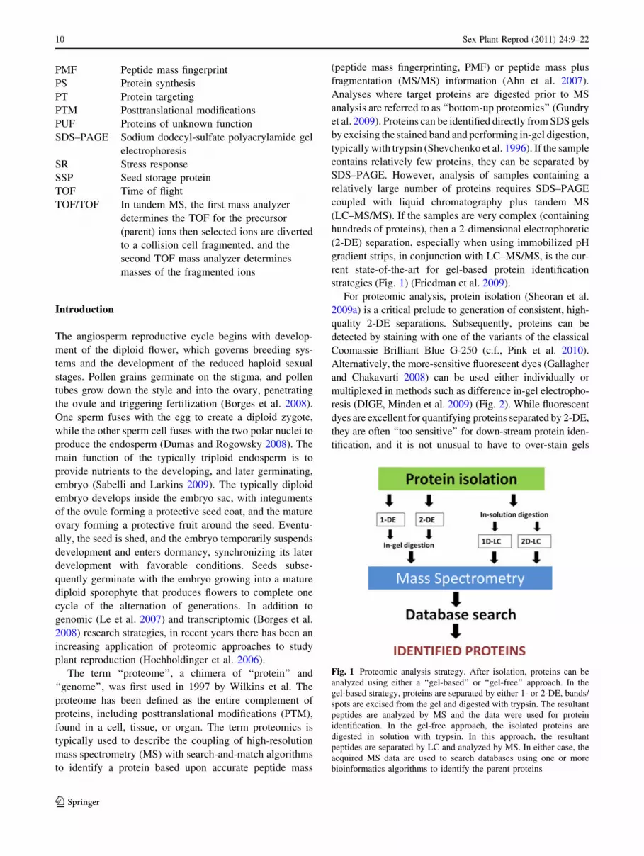
strategies (Fig. 1) (Friedman et al. 2009).
For proteomic analysis, protein isolation (Sheoran et al.
2009a) is a critical prelude to generation of consistent, high-
quality 2-DE separations. Subsequently, proteins can be
detected by staining with one of the variants of the classical
Coomassie Brilliant Blue G-250 (c.f., Pink et al. 2010).
Alternatively, the more-sensitive fluorescent dyes (Gallagher
and Chakavarti 2008) can be used either individually or
multiplexed in methods such as difference in-gel electropho-
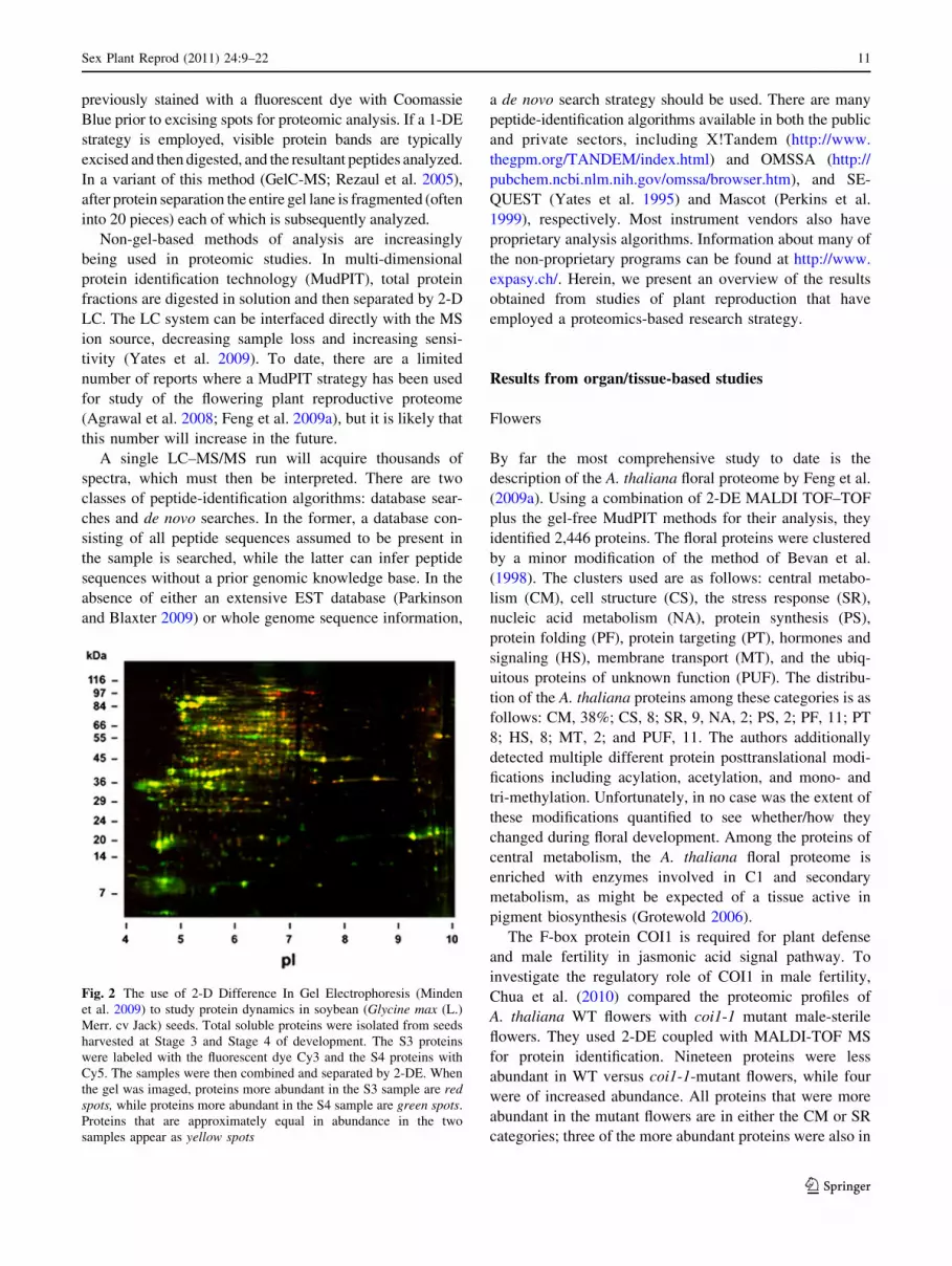
resis (DIGE, Minden et al. 2009) (Fig. 2). While fluorescent
dyes are excellent for quantifying proteins separated by 2-DE,
they are often ‘‘too sensitive’’ for down-stream protein iden-
tification, and it is not unusual to have to over-stain gels
Fig. 1 Proteomic analysis strategy. After isolation, proteins can be
analyzed using either a ‘‘gel-based’’ or ‘‘gel-free’’ approach. In the
gel-based strategy, proteins are separated by either 1- or 2-DE, bands/
spots are excised from the gel and digested with trypsin. The resultant
peptides are analyzed by MS and the data were used for protein
identification. In the gel-free approach, the isolated proteins are
digested in solution with trypsin. In this approach, the resultant
peptides are separated by LC and analyzed by MS. In either case, the
acquired MS data are used to search databases using one or more
bioinformatics algorithms to identify the parent proteins
10 Sex Plant Reprod (2011) 24:9–22
123
previously stained with a fluorescent dye with Coomassie
Blue prior to excising spots for proteomic analysis. If a 1-DE
strategy is employed, visible protein bands are typically
excised and then digested, and the resultant peptides analyzed.
In a variant of this method (GelC-MS; Rezaul et al. 2005),
after protein separation the entire gel lane is fragmented (often
into 20 pieces) each of which is subsequently analyzed.
Non-gel-based methods of analysis are increasingly
being used in proteomic studies. In multi-dimensional
protein identification technology (MudPIT), total protein
fractions are digested in solution and then separated by 2-D
LC. The LC system can be interfaced directly with the MS
ion source, decreasing sample loss and increasing sensi-
tivity (Yates et al. 2009). To date, there are a limited
number of reports where a MudPIT strategy has been used
for study of the flowering plant reproductive proteome
(Agrawal et al. 2008; Feng et al. 2009a), but it is likely that
this number will increase in the future.
A single LC–MS/MS run will acquire thousands of
spectra, which must then be interpreted. There are two
classes of peptide-identification algorithms: database sear-
ches and de novo searches. In the former, a database con-
sisting of all peptide sequences assumed to be present in
the sample is searched, while the latter can infer peptide
sequences without a prior genomic knowledge base. In the
absence of either an extensive EST database (Parkinson
and Blaxter 2009) or whole genome sequence information,
a de novo search strategy should be used. There are many
peptide-identification algorithms available in both the public
and private sectors, including X!Tandem (http://www.
thegpm.org/TANDEM/index.html) and OMSSA (http://
pubchem.ncbi.nlm.nih.gov/omssa/browser.htm), and SE-
QUEST (Yates et al. 1995) and Mascot (Perkins et al.
1999), respectively. Most instrument vendors also have
proprietary analysis algorithms. Information about many of
the non-proprietary programs can be found at http://www.
expasy.ch/. Herein, we present an overview of the results
obtained from studies of plant reproduction that have
employed a proteomics-based research strategy.
Results from organ/tissue-based studies
Flowers
By far the most comprehensive study to date is the
description of the A. thaliana floral proteome by Feng et al.
(2009a). Using a combination of 2-DE MALDI TOF–TOF
plus the gel-free MudPIT methods for their analysis, they
identified 2,446 proteins. The floral proteins were clustered
by a minor modification of the method of Bevan et al.
(1998). The clusters used are as follows: central metabo-
lism (CM), cell structure (CS), the stress response (SR),
nucleic acid metabolism (NA), protein synthesis (PS),
protein folding (PF), protein targeting (PT), hormones and
signaling (HS), membrane transport (MT), and the ubiq-
uitous proteins of unknown function (PUF). The distribu-
tion of the A. thaliana proteins among these categories is as
follows: CM, 38%; CS, 8; SR, 9, NA, 2; PS, 2; PF, 11; PT
8; HS, 8; MT, 2; and PUF, 11. The authors additionally
detected multiple different protein posttranslational modi-
fications including acylation, acetylation, and mono- and
tri-methylation. Unfortunately, in no case was the extent of
these modifications quantified to see whether/how they
changed during floral development. Among the proteins of
central metabolism, the A. thaliana floral proteome is
enriched with enzymes involved in C1 and secondary
metabolism, as might be expected of a tissue active in
pigment biosynthesis (Grotewold 2006).
The F-box protein COI1 is required for plant defense
and male fertility in jasmonic acid signal pathway. To
investigate the regulatory role of COI1 in male fertility,
Chua et al. (2010) compared the proteomic profiles of
A. thaliana WT flowers with coi1-1 mutant male-sterile
flowers. They used 2-DE coupled with MALDI-TOF MS
for protein identification. Nineteen proteins were less
abundant in WT versus coi1-1-mutant flowers, while four
were of increased abundance. All proteins that were more
abundant in the mutant flowers are in either the CM or SR
categories; three of the more abundant proteins were also in
Fig. 2 The use of 2-D Difference In Gel Electrophoresis (Minden
et al. 2009) to study protein dynamics in soybean (Glycine max (L.)
Merr. cv Jack) seeds. Total soluble proteins were isolated from seeds
harvested at Stage 3 and Stage 4 of development. The S3 proteins
were labeled with the fluorescent dye Cy3 and the S4 proteins with
Cy5. The samples were then combined and separated by 2-DE. When
the gel was imaged, proteins more abundant in the S3 sample are redspots, while proteins more abundant in the S4 sample are green spots.
Proteins that are approximately equal in abundance in the two
samples appear as yellow spots
Sex Plant Reprod (2011) 24:9–22 11
123

CM, the fourth being the GroEL molecular chaperone. The
results by themselves fail to provide any significant insight
into flower development or regulation of gene expression.
Watson et al. (2003) used 2-DE coupled with MALDI-TOF
PMF plus LC–MS/MS to identify 304 proteins from barrel
medic (Medicago truncatula) flowers. The distribution
among functional groups was nearly identical to that of the
A. thaliana proteins (Feng et al. 2009a).
Raharjo et al. (2004) and Ahsan and Komatsu (2010)
used the 2-DE MALDI-TOF PMF approach to compare the
leaf and flower proteomes of Cannabis sativa L. cv Purple
Haze and Glycine max L. cv Enrei, respectively. Not
surprisingly, the proteomes of the different organs are
dissimilar, reflecting their specialized biological roles.
Unfortunately, in neither case was there any true flower-
specific information.
Using a 2-DE LC–MS/MS experimental strategy,
Dafny-Yelin et al. (2005) and Bai et al. (2010) analyzed the
onset of petal senescence in rose (Rosa hybrida cv. Fra-
grant Cloud) and petunia (Petunia x hybrida Mitchell
Diploid), respectively. In the two studies, a total of *100
proteins were identified, and patterns of increased or
decreased abundance were quantified. However, none of
the identified proteins was clearly flower- or senescence
associated or specific. Bar-Akiva et al. (2010), as part of a
comprehensive analysis of Brunfelsia calycina petals after
flower opening, reported using LC–MS/MS to identify
seven proteins. In contrast to the results of others, all seven
were enzymes involved in metabolism of either pigment or
scent compounds.
The male gametophyte
Temporal and spatial regulation of gene expression during
male gametophyte development in flowering plants has
been described (Suzuki 2009; Wilson and Zhang 2009);
however, only Kerim et al. (2003) have reported high-
quality 2-DE maps of developing male gametophytes (rice
(Oryza sativa L. cv. Doongara) at the pollen mother cell,
tetrad, early young microspore, early binucleate, late
binucleate, and heading stages. Using MALDI-TOF MS
and PMF analysis, they were able to identify 33 non-
redundant proteins but none of them appeared to have any
stage-specific or anther-specific roles.
There have been extensive ([100 proteins identified)
gel-based proteomic analyses of mature pollen from
A. thaliana (Noir et al. 2005; Holmes-Davis et al. 2005;
Sheoran et al. 2006), tomato (Lycopersicon esculentum)
(Sheoran et al. 2007), and rice (O. sativa L. ssp japonica;
Dai et al. 2006). Proteins were identified by LC–MS/MS.
In each study, the proportion of identified protein assigned
to each cluster was very similar; the majority of the pro-
teins were distributed among the CM, CS, SR, and PUF
clusters. The results of the meta-analysis (3 species, 654
proteins) can be seen in Fig. 3.
Pollen grains are rich in triacylglycerols and other
storage compounds (Piffanelli et al. 2003) that are used to
support the huge demand for energy and biosynthetic
intermediates during postgerminative pollen-tube growth
(Taylor and Hepler 1997). It is then not surprising that the
most populous class of pollen proteins includes the many
enzymes of CM (glycolysis, the Krebs cycle, and mito-
chondrial respiration). The next most abundant pollen
proteins are those assigned to CS and PF, followed by the
SR and HS clusters.
Sheoran et al. (2009a) employed 2-DE, DIGE, and
MALDI-TOF/TOF MS to identify 130 proteins from ger-
minating canola (Brassica napus) pollen. All of the pro-
teins identified were present in both mature pollen and
germinated grains. Similarly, Dai et al. (2006, 2007a, b)
used a combination of MALDI-TOF MS plus ESI Q-TOF
MS/MS to identify 120 non-redundant proteins from ger-
minating rice (O. sativa L. ssp. japonica cv. Zhonghua 10)
pollen. However, contrary to the claim apparently made in
the titles, they failed to identify any proteins specifically
associated with germination of pollen grains.
There are a handful of publications that present data
from more specialized pollen analyses. For example, Pertl
et al. (2009) isolated subcellular fractions (endoplasmic
reticulum, Golgi, mitochondria, and plasma membrane)
and then used LC–MS/MS to characterize the membrane
proteome. Unfortunately, in the absence of any other such
study in pollen, the only comparisons that can be made are
to the results from analyses of other organs.
The protein allergens present in pollen have been
extensively studied (Puc 2003; Mohapatra et al. 2008).
Virtually without exception, however, these publications
address the biomedical consequences of the allergens
rather than any aspect of their role(s) in plant reproductive
biology, and they will not be further addressed herein.
Fig. 3 The functional distribution of 654 pollen proteins (L. escu-lentum, A. thaliana, and O. sativa) (Noir et al. 2005; Holmes-Davis
et al. 2005; Sheoran et al. 2007; Dai et al. 2006). Percent: CS 12, NA3, PS 4, PF 9, PT 7, CM 37, MT 4, SR 10, HS 8, PUF 7
12 Sex Plant Reprod (2011) 24:9–22
123

The female gametophyte
Egg cells are differentiated for fertilization and subsequent
embryogenesis. The signals and events that underlie the
differentiation of egg cells remain obscure despite their
importance in the plant life cycle (Hiscock and Allen
2008). Hopefully, description of the egg cell proteome
will contribute to a better understanding of the mecha-
nisms of female gametogenesis, fertilization, and early
embryogenesis.
The very limited amount of starting material is a major
obstacle in attempting to characterize the egg cell prote-
ome, and only a few proteins have been identified thus far.
Rice egg cells were isolated from unpollinated ovaries by
making a transverse incision followed by applying pressure
with a glass needle (Uchiumi et al., 2007). The isolated egg
cells were washed and then lysed by direct transfer into
SDS–PAGE sample buffer. Using a GeLC–MS/MS strat-
egy, Uchiumi et al. (2007) identified four proteins: the
cytoplasmic glycolytic enzyme glyceraldehyde-3-phos-
phate dehydrogenase, histone H4, cytoplasmic ascorbate
peroxidase, and a member of the Hsp90 family of molec-
ular chaperones (Hsp82). The same research group isolated
and analyzed maize egg cell proteins (Okamoto et al. 2004)
by 2-DE plus LC–MS/MS. The identified maize egg
cell proteins are three cytoplasmic glycolytic enzymes,
GAPDH, 3-phosphoglycerate kinase, and triosephosphate
isomerase, mitochondrial ATP synthase ß-subunit, a
mitochondrial adenine nucleotide transporter, and annexin
p35 (Okamoto et al. 2004). All of the identified proteins are
known to be relatively abundant in plant cells, which
facilitated identification. Hopefully, technical improve-
ments in egg cell isolation, such as those described by
Hoshino et al. 2006), will lead to larger amounts of starting
material and a more comprehensive description of the egg
cell proteome.
Can any significance be attributed to the nine different
egg cell proteins identified from maize and rice? The
glycolytic pathway is common to all living cells, and the
individual glycolytic enzymes are well known to be
abundant cellular proteins (Plaxton 1996), so it seems
unlikely that the identification of GAPDH, PGK, and TPI is
of any egg cell-specific significance. However, it is also
well known that many of the glycolytic enzymes have
additional, non-catalytic functions (Sirover 1997; Pancholi
2001), so the possibility of egg cell-specific functions
should be at least considered. It has been recently reported
that the mitochondria of rice egg cells have an unusual
morphology (Takanashi et al. 2010). Perhaps then a change
in the abundance or biological role for the ATP synthase
ß-subunit and/or adenine nucleotide transporter is also
possible? The annexins are abundant multifunctional cal-
cium-dependent phospholipid-binding proteins (Talukdar
et al. 2009). Are there egg cell-specific roles for annexins?
Perhaps they are abundant because of the increase in pro-
tein and polysaccharide secretion that is stimulated by the
fertilization-induced increase in Ca2? levels in the zygote
(Dumas and Rogowsky 2008). Are these and the other egg-
cell proteins specifically important, or are they simply
abundant and easily identified? The answer to this awaits
the results from additional studies of the egg-cell proteome.
It is noteworthy that similar types of proteomic analyses
of animal reproduction have led to the identification and
quantification of dozens of ‘‘spermatogenesis’’, ‘‘zygote-
specific’’, and ‘‘gamete-associated’’ proteins (Karr 2007;
Huang et al. 2008; Roux et al. 2008). Presumably, similar
advancements in the understanding of plant reproductive
proteomics await development of better methods and
instrumentation, and more attention from plant biologists.
Changes after fertilization
Vyetrogon et al. (2007) used 2-DE with Sypro Ruby
staining to quantify changes in the wild potato (Solanum
chacoense) ovary proteome 30, 36, 42, and 48 h after
pollination. Proteins were identified by LC–MS/MS anal-
ysis of tryptic peptides using a hybrid quadrupole-TOF
instrument. Of the [600 proteins quantified from 2-DE
maps, 38 showed a significant change postfertilization.
Most of the proteins decreased in abundance over the 48 h,
and unfortunately the six proteins that increased in abun-
dance all belong to the PUF group.
In the same study, the authors also quantified changes in
the P-proteome, using three methods in parallel. The spots
separated by 2-DE were either stained with the phospho-
protein-specific fluorescent dye ProQ Diamond (Agrawal
and Thelen 2009) or transferred from gels onto membranes
and then probed with antibodies to P-Ser, P-Thr, or P-Tyr
(Sefton and Shenolikar 2001). The third method of detec-
tion involved metabolic labeling with 32Pi. In toto, 262
(42%) of the 619 Sypro Ruby staining proteins were
detected as P-proteins. Among these, use of P-amino acid
antibodies detected 184 proteins, of which 78 were also
detected with at least one of the other two methods.
Staining with Pro-Q Diamond detected 111 proteins, of
which 76 were also detected with one of the other two
methods. The 32P in vivo labeling method detected 90
spots, of which 78 were also detected with one of the other
two methods. Comparison of before and after fertilization
profiles, 38 P-proteins showed a reproducible change in
their abundance. Most of the identified P-proteins corre-
spond only to entries in potato EST libraries that them-
selves correspond to members of the PUF group. The
relatively small number of changes in the S. chacoense
ovary proteome after fertilization was unexpected, since
the results of previous transcript profiling analyses led to
Sex Plant Reprod (2011) 24:9–22 13
123

the conclusion that this system is highly dynamic at the
mRNA level (Germain et al. 2005; Vyetrogon et al. 2007).
It is, however, necessary to keep in mind that proteomic
analyses target only the more abundant components of the
proteome and furthermore that in most biological systems,
there is a high degree of discordance between transcript
and protein levels (c.f., Hajduch et al. 2010).
Self-incompatibility
Self-incompatibility is a genetically controlled mechanism
to prevent inbreeding. Feng et al. (2006) used 2-DE
LC–MS/MS strategy to compare pistil-protein differences
between self- and cross-pollination of a self-incompatible
apricot (Prunus armeniaca) but were able to identify only
four pistil proteins that were increased in abundance after
cross-pollination (actin-12, enolase, a MYB transcription-
factor-like protein, and Hsp70), and three proteins were
detected only in self-pollinated pistils (actin-7, actin-8, and
a fructose 1,6-bisphosphate aldolase–like protein). More
recently, the same group (Feng et al. 2009b) used the same
strategy to compare compatible and self-incompatible
apricot cultivars. Nine proteins, including a receptor-like
protein kinase, were detected only in the compatible pistils,
and another 9 proteins, including actin-7, a putative protein
Ser/Thr kinase, and an S-RNase, were detected only in the
self-incompatible pistils.
Cytoplasmic male sterility is not addressed herein
because it is unimportant, but rather because of the paucity
of publications that have employed a proteomics-base
approach for analysis. The recent paper by Sheoran et al.
(2009b) indicates that this oversight has been recognized.
Seed abortion
Seed abortion is one of the mechanisms by which plants
can respond to extremes in environmental conditions (c.f.,
Fang et al. 2010). Liu et al. (2010) used a 2-DE MALDI-
TOF/TOF strategy to study seed abortion in Dimocarpus
longan. They identified more than 40 proteins from aborted
seeds, most of which belong to the CM, PF, and PUF
categories. Among the identified proteins are three Cys-
proteases that the authors suggest might be linked to pro-
grammed cell death. No other studies of seed abortion have
used a proteomics approach, so testing this suggestion
awaits the results of future experiments.
Seed development
Post fertilization, the various tissues of the female game-
tophyte continue to develop and differentiate, becoming
the embryo and then, after further specialization, the
developing seed (embryo, endosperm, seed coat; Weber
et al. 2005; Le et al. 2007). Certain aspects of seed biology
simplify study; all cell division is completed within the first
few days after fertilization. This marks the line of demar-
cation between embryogenesis and seed development. All
subsequent cellular specialization takes place in the
absence of cell division (Chandler 2008). The bulk of
metabolic activity during seed development is directed
specifically at the synthesis and accumulation of storage
polymers (oils, polysaccharides, and proteins) (Gallardo
et al. 2003; Hills 2004). These compounds and the sub-
cellular structures that contain them are inert depots
awaiting the activities responsible for mobilizing them to
provide biosynthetic intermediates necessary until the
developing seedling becomes autotrophic (Le et al. 2007;
Gallardo et al. 2008). All seeds contain one or more groups
of proteins that are present in high amounts acting as
depots for reduced nitrogen that will subsequently be used
during germination and seedling growth, the seed storage
proteins (SSP, Shewry et al. 1995). While SSP can account
for as much as 60% of total seed protein, most of them are
otherwise inert, so they have been manually subtracted
from all of the analyses presented herein.
Endosperm development
The endosperm, a tissue unique to flowering plants, is
composed of a few specialized cell types that produce large
quantities of storage polymers. The amount of endosperm
in a mature seed is variable. For example, it comprises a
large portion of the whole seed mass in cereals, such as rice
(O. sativa), maize (Zea mays), and wheat (Triticum aes-
tivum), and it is prominent in the seeds of a few dicotyle-
dons, such as castor (Ricinus communis L). In other
species, for example A. thaliana, the endosperm is almost
completely absent in the mature seed. Whether the endo-
sperm persists or not, it is vital for embryo development in
much the same way the placenta is essential for mamma-
lian embryos (Wang et al. 2009).
Castor is an unusual example of an oil-rich endosperm-
dominant seed; most accumulate starch rather than oil.
Using a 2-DE plus LC–MS/MS strategy, Houston et al.
(2009) identified 522 proteins from developing castor
endosperm. Discounting the SSP, the 20 most abundant
castor proteins were involved in CM, PF, SR, and CS. The
total distribution of the castor proteins was very similar to
that of pollen proteins (Fig. 3). This similarity could reflect
that both are oil-rich organs and therefore have relatively
high levels of proteins involved in both biosynthesis and
catabolism of the oil.
Similar patterns of identified protein distribution were
seen among the endosperm-dominant starch-rich seeds of
wheat, barley, rice, and maize. Using a 2-DE plus either
MALDI-TOF PMF (Finnie et al. 2002; Vensel et al. 2005;
14 Sex Plant Reprod (2011) 24:9–22
123

Finnie and Svensson 2009) or LC–MS/MS (Mechin et al.
2004; Mak et al. 2006; Xu et al. 2008; Kim et al. 2009)
experimental design, a total of 1,496 proteins were iden-
tified. When the proteins are separated into the same 10
functional classes that have been used throughout, the
distribution is as follows: CM, 34%; CS, 12; SR, 5; NA, 2;
PS, 2; PF, 5; PT, 7; HS, 2; MT, 2; and PUF, 29.
Larre et al. (2010) used a 2DE LC–MS/MS strategy to
study SSP of the ‘‘model cereal’’ Brachypodium distach-
yon. Electrophoresis of a urea extract of mature seeds
resolved 120 spots, 65 of which were excised for MS
analysis. Twenty-three well-resolved spots were identified
as members of the 11S storage protein family, encoded by
five genes. Two sets of minor spots were identified as 7S
globulins. In addition to the SSP, a xylanase inhibitor was
identified.
The results from separate proteomic analyses of devel-
oping wheat (Vensel et al. 2005) and rice (Xu et al. 2008)
endosperm, and wheat (Mak et al. 2006) and rice embryos
(Woo et al. 2002; Wang et al. 2008) highlight the differ-
ences in these two seed organs (Table 1). The CM proteins
were the most abundant in all instances; however, members
of the PF category were relatively more abundant in
endosperm, while proteins included in the SR and HS
categories were more abundant in embryos. The higher
proportion of proteins involved in signaling might reflect
the role(s) of the embryo in controlling metabolism in the
endosperm (c.f., Perata et al., 1997). Proportionally, there
are far more PUF proteins in endosperm than in embryos.
Amyloplasts are non-green plastids specialized for the
synthesis and accumulation of starch (Neuhaus and Emes
2000). Balmer et al. (2006) describe the isolation of amy-
loplasts from developing wheat endosperm and the use of a
2-DE LC–MS/MS strategy to identify 289 proteins. In
addition to all of the enzymes necessary for starch bio-
synthesis, they were also able to identify many proteins
involved in nitrogen and sulfur assimilation, and amino
acid and lipid biosynthesis. Overall, the composition of the
wheat endosperm amyloplast proteome is more similar to
that of castor endosperm leucoplasts (Campos et al. 2010)
or even green plastids (van Wijk 2004; van Wijk et al.
2007) than to the wheat endosperm cell cytoplasm.
Embryo development
Embryonic axes generally are removed before proteomic
analysis of embryo-dominant seeds. Alternatively, the
contributions of the axes are simply ignored as very minor
components. As a result, proteomic descriptions of
embryo-dominant seeds correspond almost entirely to the
cotyledonary proteome. Typically, embryo-dominant seeds
contain starch plus SSP as the storage polymers (peas,
beans, and lentils) or oil plus SSP (mouse-eared cress,
canola, and soybean/barrel medic/lotus). To date, the
cotyledons of legume seeds have received the most atten-
tion from ‘‘omics biologists,’’ in part due to their agro-
nomic importance (Weber et al. 2005; Le et al. 2007;
Gallardo et al. 2008; Thompson et al. 2009).
There have been extensive proteomic studies of devel-
oping G. max (Hajduch et al. 2005; Agrawal et al. 2008),
M. truncatula (Gallardo et al. 2003, 2007), and Lotus
japonicus (Dam et al. 2009) seeds. Experimental strategies
included 2-DE plus MALDI-TOF, 2-DE plus LC–MS/MS,
or GeLC–MS analysis of tryptic peptides. Seed proteome
data for G. max, M. truncatula, and L. japonicus have been
collected and analyzed and can be retrieved from: http://
bioinfoserver.rsbs.anu.edu.au/utils/PathExpress/pathexpress
4legumes.php. A total of 1,723 proteins were identified.
Manual subtraction of 316 SSP gave 1,407 identified pro-
teins from the three legume species. When these proteins
were separated into 10 categories, the percent distribution
was CM, 49%; CS, 19; NA, 2; PS, 3; PF, 5; PT, 3; HS, 3;
MT, 4; SR, 3; and PUF, 10. In all three species, a relatively
large number of the late embryo abundant proteins were
identified.
Similar studies of A. thaliana (Hajduch et al. 2010) and
B. napus (Hajduch et al. 2006; Agrawal et al. 2008) seeds
yielded similar results. A total of 1,290 proteins were
identified by either 2-DE plus LC–MS/MS or MudPIT.
After manual removal of 241 SSP entries, this leaves 1,049
identified proteins. Once again, the CM category was by far
the most populous (36%), followed by CS (19%) and PUF
(17%).
Agrawal et al. (2006) used the phospho-protein-specific
fluorescent dye ProQ Diamond as a probe for analysis of
developing B. napus seeds. They were able to detect 234
Table 1 Percent distribution of wheat and rice seed proteins; endo-
sperm versus embryo
Wheat Rice
Functional group Endosperm Embryo Endosperm Embryo
Central metabolism 27 26 34 27
Cell structure 0 2 12 2
Stress response 9 22 5 19
Nucleic acid 0 8 2 10
Protein synthesis 9 10 5 9
Protein folding 15 4 9 2
Protein targeting 7 3 5 5
Hormones/signaling 9 21 2 17
Membrane transport 1 2 2 1
Proteins of unknown
function
23 2 29 8
Total proteins identified: wheat endosperm, 256 (Vensel et al. 2005);
wheat embryo, 347 (Mak et al. 2006); rice endosperm 274 (Xu et al.
2008) and; rice embryo 132 (Woo et al. 2002; Wang et al. 2008)
Sex Plant Reprod (2011) 24:9–22 15
123

phospho-protein spots from 2-D gels, 103 of which were
identified by LC–MS/MS. Not surprisingly, most of the
identified proteins were in the CM and HS categories. It
was, however, somewhat surprising that many of the cru-
ciferin SSP subunits gave a positive phospho-protein stain
since these proteins had not been previously described as
phospho-proteins.
Jain et al. (2008) used MudPIT to identify 80 proteins
from plastids purified from developing B. napus embryos.
The plastid proteins complement was enriched with
enzymes of the CM grouping and was mid-way between
the non-green plastids from wheat and castor endosperm
(Balmer et al. 2006; Campos et al. 2010) and the chloro-
plasts isolated from green organs (van Wijk 2004; van
Wijk et al. 2007).
In contrast to the oil plus SSP embryo-dominant seeds,
the main starch plus SSP seeds that have been character-
ized are pea, Pisum sativum (Bourgeois et al. 2009), and
lentil, Lens culinaris (Scippa et al. 2010). A combination of
MALDI-TOF PMF and LC–MS/MS was used to identify
122 proteins from mature L. culinaris seeds. Manual sub-
traction of the SSP reduced this number to 25. Of these, 6
were grouped in CM, 4 each in CS and PT, and 3 each in
NA and SR. Similar results were obtained when Bourgeois
et al. (2009) used MALDI-TOF PMF to identify 156 pro-
teins from mature pea seeds. Manual subtraction of the SSP
left 39, mainly distributed among CM (16), PUF (8), CS
(7), and PF (4). A major problem with analysis of the starch
plus SSP legume seeds is the lack of good genomic or EST
resources.
Seed germination
Mature quiescent seeds are dispersed at low (5–15%)
moisture content and with metabolic activity at a standstill.
For germination, quiescent seeds need only be hydrated at
a suitable temperature in the presence of O2. Germination
begins with water uptake by the seed (imbibition), con-
tinues through the elongation the embryonic axis inside the
seed, and is visibly manifest by protrusion of the radicle
through the seed coat (Bove et al. 2003). The sum of these
events represents the transition of the quiescent embryo to
an autotrophic plant. Germination involves many cellular
and metabolic events, coordinated by complex regulatory
networks.
Herein, seed dormancy will be treated as a specialized
variant of quiescence (Finkelstein et al. 2008). Dormancy
is a process whereby germination is delayed in order to
avoid conditions adverse for seedling survival by opti-
mizing the timing of germination. Release from dormancy
is controlled by perception of a combination of environ-
mental signals (Bove et al. 2003; Finkelstein et al. 2008;
Holdsworth et al. 2008; Pawłowski 2010). Temperature,
light quality, and hormonal balance (abscisic acid and
gibberellins) all play key roles in release from dormancy.
The period between germination and assumption of
autotrophy is broadly referred to as postgerminative
growth. It is during this period that the seed storage poly-
mers (oil, polysaccharides, and SSP) are mobilized to
provide biosynthetic intermediates. In general, the enzymes
necessary for polymer degradation are synthesized de novo
during postgerminative growth. Thus, SSP proteases (endo-
and exo-), lipases and the glyoxylate cycle enzymes, and
starch-degrading enzymes can serve as markers to define
the period of postgerminative growth.
Endosperm-dominant seeds
Total endosperm proteins from germinating castor seeds
were analyzed using a 2-DE plus MALDI-TOF/TOF MS
strategy, and nearly 400 total proteins were identified
(Campos et al. 2010). Essentially, all of these proteins are
the same as were previously described from analysis of
developing castor endosperm (Houston et al. 2009). The
extremely high ‘‘background’’ of SSP and CM proteins
precluded identification of any proteins that could be spe-
cifically attributed to germination/postgerminative growth.
Castor endosperm plastidial and mitochondrial fractions
were also prepared and subjected to proteomic analysis
(Campos et al. 2010). The proteins identified are the same
as have been previously described for these organelles
from other plant species and organs (van Wijk 2004; Lee
et al. 2008; Huang et al. 2009). Maltman et al. (2007) used
2-DE, DIGE, MALDI-TOF PMF, and LC–MS/MS to show
that more than 100 proteins are differentially abundant
when comparing the endoplasmic reticulum (ER) from
developing castor endosperm with that from germinated
seeds. With the exception of a few contaminants from other
organelles (RuBisCO, plastids; malate synthase, and gly-
oxysomes) and PUF proteins, those identified were all
either SSP or members of the PF and PT groups.
A proteomics strategy employing 2-DE and MALDI-
TOF PMF analysis was used to identify nearly 200 proteins
from germinating barley seeds (Ostergaard et al. 2004).
Nearly all of the proteins identified are included in the CM,
PF, CS, and SR categories. By 3 days after imbibitions,
there were large increases in the enzymes involved in
starch breakdown and mobilization, such as the a- and
b-amylases.
Because barley is used extensively in the food and
brewing industries, there have been detailed analyses of a
few specific proteins: a-amylase, peroxidases, and thiore-
doxins (Finnie and Svensson 2009). Hynek et al. (2009)
used LC–MS/MS to analyze a plasma membrane-enriched
fraction isolated from germinating barley. Many of the
proteins identified appear to be contaminants from other
16 Sex Plant Reprod (2011) 24:9–22
123

subcellular organelles, demonstrating how difficult it is to
obtain purified subcellular components. Despite the diffi-
culties, they also identified several bona fide plasma
membrane proteins including an H?-ATPase, a pyrophos-
phatase, and a voltage-dependent anion channel (Hynek
et al. 2009).
Analysis of germinating rice endosperm using a 2-DE
plus MALDI-TOF MS strategy allowed Yang et al. (2007)
to study proteins that changed in abundance. They found
that the SSP, along with members of the CM and CS
groups, decreased in abundance during germination, while
different members of the CM group increased, some of
which are involved in starch breakdown. Results similar to
those found with germinating rice and barley endosperm
proteins were also found during studies of wheat and
maize.
Evidence is accumulating that indicates the cellular
environment is increasingly oxidizing as seeds dehydrate
and approach quiescence. Formation of disulfide bonds is
one of the manifestations of the changing redox environ-
ment. Subsequently, in response to imbibition, the thiore-
doxin system, composed of NADPH, thioredoxin h, and
NADP-thioredoxin reductase, is activated (Hagglund et al.
2008). This system reduces disulfide bonds and in doing so
increases protein solubility, the rate of proteolysis, and
ultimately the extent of nitrogen and carbon mobilization
(Yano and Kuroda 2006). The endosperm-based thiore-
doxin paradigm was subsequently extended to include
dicotyledon seeds (Alkhalfioui et al. 2007).
Cotyledon-dominant seeds
Essentially all reported proteomic analyses of ‘‘germinat-
ing seeds’’ are actually from studies of postgerminative
growth. Inevitably, 2-DE-based LC–MS/MS analyses of
the postgerminative growth of cotyledon-dominant seeds
yield datasets identical to those of mature seeds and are
dominated by the presence of SSP (Fu et al. 2005; Sheoran
et al. 2005; Pawłowski 2007, 2009; Sghaier-Hammami
et al. 2009; Yang et al. 2009). If proteins are quantified,
then the major theme is the decrease in levels of SSP. In
some instances, this overall decrease is accompanied by
transient accumulation of SSP degradation intermediates.
In contrast to most ‘‘shotgun’’ proteomic analyses of
seeds, Muller et al. (2010) reported the results from anal-
ysis of a specific tissue, the endosperm cap, during ger-
mination of Lepisium sativum seeds. The cap is a specific
part of the endosperm surrounding the embryo radicle.
Using a 2-DE plus LC–MS/M strategy, 140 proteins were
identified. The largest group of proteins was the CM
cluster, followed by SR and PF. The endosperm cap pro-
teome was both qualitatively and quantitatively different
from the rest of the endosperm, but no proteins were
identified, which might be responsible for modification of
the endosperm structure in response to radicle protrusion.
Apomixis
Asexual reproduction of plants by female syngamy is
referred to as apomixis. Embryos that are genetically
identical to the maternal parent are produced without
meiosis or egg cell fertilization (Chen 2007). There are two
forms of apomixis: sporophytic and gametophytic. Apo-
mixis allows formation of clonal embryos, which has
recently stimulated interest among plant breeders because
it suggests the ability to fix heterosis.
Genetic mapping has been successfully used in a wide
variety of apomictic taxa to explore the genetics bases of
the trait. Employing a proteomic approach to analysis of
apomixis allows researchers to additionally consider epi-
genetic contributions, as well as the roles of a myriad of
protein posttranslational modifications. To date, there has
been only a single publication that focuses on the proteo-
mics of apomixis, a comparative study of interspecific
hybrids between diploid Beta vulgaris and tetraploid
B. corolliflora (Zhu et al. 2008). From this cross, the
monosomic addition line M14 was selected based upon an
apomictic phenotype.
Using a 2-DE plus MALDI-TOF MS strategy, a total of
27 protein spots that varied in abundance among lines were
identified. These included five protein spots that were
detected only in M14 and two protein spots found only in
B. vulgaris. Among the identified proteins, 13 were more
abundant in M14 than in B. vulgaris and seven were less
abundant.
The identified proteins could be separated into eight
clusters, the largest of which was once again CM. While no
mechanistic conclusions can be reached based upon a
sample size of 27, the results will contribute to a better
understanding of the genetic mechanisms underlying apo-
mixis and how they might be exploited.
Prospectus
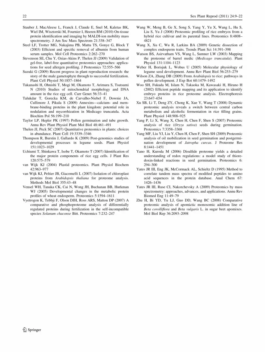
To date, there have been relatively few studies where a
proteomics-based strategy has been applied to the study of
angiosperm reproduction. Of the studies that have been
conducted, a large majority have addressed seeds; either
developing or ‘‘germinating’’ (Fig. 4). One goal of this
review was to identify areas where there is a need for
increased application of proteomics-based methods.
Clearly, these include all aspects of floral biology, study of
the female gametophytes, self-incompatibility, environ-
mental and genetic contributions to seed abortion, and
study of the proteome changes that accompany seed after-
ripening and dormancy. There is additionally a need for
Sex Plant Reprod (2011) 24:9–22 17
123
greater comparative analysis for all aspects of plant
reproductive biology, although in many cases this will
require parallel development of better genomic/EST
resources to facilitate protein identification.
Proteomics as a research area is increasingly moving
from gel-based to gel-free approaches for protein analysis.
Without question the latter strategy allows identification of
more proteins and is amenable to use with automated, high-
throughput platforms (Schulze and Usadel 2010). At the
same time, gel-free analyses require application of more
robust standards of statistical analysis (Nie et al. 2008). At
least in the foreseeable future, there will remain applica-
tions for gel-based proteomics, especially in targeted-pro-
teomic studies such as those involving DIGE. In all
likelihood, a combination of gel-based and gel-free meth-
ods will comprise the state-of-the-art for years to come
(c.f., Insenser et al. 2010).
The need for a transition from qualitative to quantitative
proteomics is without debate. In contrast, how to best
achieve this is virtually without agreement! There are
essentially two schools of thought: methods involving
protein labeling and label-free methods (Thelen and Peck
2007). Not surprisingly, there are a plethora of methods
proposed to achieve both ends (Schulze and Usadel 2010).
While this debate is likely to continue into the future, a
label-free method gaining some traction is spectral count-
ing (c.f., Stevenson et al. 2009; Huang et al. 2010;
Lundgren et al. 2010). Regardless of the final evolution of
analysis methods, it is clear that any future proteomics-
based studies of plant reproduction would benefit from
quantitative approaches.
One aspect of biology that cannot be addressed in any
other large-scale survey (e.g., transcript profiling) is protein
PTM, and much of the breadth of the proteome results from
a profusion of protein PTM (Farley and Link 2009). In
addition to the nature of the PTM, it is also important to
determine the specific residue that is modified and the
result of the modification (Møller et al. 2007). Do the PTM
affect protein turnover, activity, and interactions? In
addition to using bottom-up analysis strategies, it is
important that top–down proteomic analyses are included
in analysis of PTM (Siuti and Kelleher 2007). In only a
very few instances have analyses of plant reproduction
included systematic analysis of PTM, and these have only
addressed protein phosphorylation (Agrawal et al. 2006;
Agrawal and Thelen 2009). In addition to identification of
the nature of PTM, it is important to determine their stoi-
chiometry in the proteome. Changes in PTM stoichiometry
likely indicate a specific functional change, while increase
in protein amount with a parallel increase in the amount of
PTM means that the stoichiometry has not changed, and
there is unlikely to be an extensive functional change.
The other major aspect of biology that cannot be
addressed in any sort of high-throughput or computational
context is that of protein interactions (e.g., the interac-
tome). There are no extant publications addressing
MS-based analysis of protein interactions during angio-
sperm reproduction. There should be! The way that a
protein behaves in dilute solution after purification is
unlikely to be the same as it behaves in vivo. The next
wave of understanding of how proteins function involves
analysis of protein interactions in a cellular or subcellular
context (Gache et al. 2010; Sharon 2010).
Essentially all of contemporary proteomic analyses are
MS based. Any discipline that is largely or exclusively
instrument based will always be searching for improve-
ments (c.f., Olsen et al. 2009). The development of
instruments that are faster and more precise will be bene-
ficial for not only study of angiosperm reproduction but
also all proteomics research.
An exciting and relatively new application of MS in
proteomics studies is at the tissue- and sometimes even
cellular level of analysis; imaging MS or MALDI-imaging
(Heeren et al. 2009; Stauber et al. 2010). By gating the
detector to a specified mass (or range), it is possible to
detect the location of a protein in whole tissue/organ
mounts. Especially in conjunction with high-sensitivity
instruments, this method could have significant applica-
tions to analysis of either small floral structures or larger
structures with low protein concentrations (MacAleese
et al. 2009).
From the very beginning, analyses have been plagued by
the enormous dynamic range of proteins in biological
samples. The archetype of this problem has been human
serum. Until simple, inexpensive, and efficient methods
were developed to remove albumin and IgG proteins,
meaningful proteomic analyses of serum were impossible
(Steel et al. 2003). The situation is much the same in plant
leaves (RuBisCO) and seeds (SSP). Multiple strategies for
removal of abundant proteins are available, including
0
10
20
30L
iter
atu
re r
epo
rts
Fig. 4 Numerical analysis of literature reports describing use of a
proteomics-based strategy, as a function of reproductive tissue/organ/
stage
18 Sex Plant Reprod (2011) 24:9–22
123

differential solubility, affinity chromatography, and
immuno-removal (Miernyk and Johnston, 2006; Li et al.
2008; Krishnan et al. 2009). Application of these methods,
further refinements, and ingenious experimental designs
will allow a much greater depth of analysis to studies of
plant reproduction and help us to better understand this
fascinating but extremely complex process.
Acknowledgments This investigation was supported by Seventh
Framework Program of the European Union—International Reinte-
gration Grant (MIRG-CT-2007-200165), COST Action FAO903,
Bilateral project CSIC-SAS (2007 SK 0001), Project APVV-0115-07,
VEGA 2/0005/08 (Study of the cell events in course of embryo for-
mation in situ and in vitro conditions in Arabidopsis and maize) and is
a joint publication within the action COST FA 0903, ‘‘Harnessing of
Plant Reproduction for Crop improvement,’’ a bilateral Spanish-
Slovak cooperation (2007–2009) and the Spanish MEC Project
(BFU2006-09876/BFI). Support for JAM was in part from the
National Scholarship Program of the Slovak Republic, administered
by the Slovak Academic Information Agency. The authors thank
B.A. McClure, J.J. Thelen, and two anonymous reviewers for their
constructive comments.
References
Agrawal GK, Thelen JJ (2009) A high-resolution two dimensional
Gel- and Pro-Q DPS-based proteomics workflow for phospho-
protein identification and quantitative profiling. Meth Mol Biol
527:3–19
Agrawal GK, Hajduch M, Graham K, Thelen JJ (2008) In-depth
investigation of the soybean seed-filling proteome and comparison
with a parallel study of rapeseed. Plant Physiol 148:504–518
Ahn NG, Shabb JB, Old WM, Resing KA (2007) Achieving in-depth
proteomics profiling by mass spectrometry. ACS Chem Biol
2:39–52
Ahsan N, Komatsu S (2010) Comparative analyses of the proteomes
of leaves and flowers at various stages of development reveal
organ specific functional differentiation of proteins in soybean.
Proteomics 9:4889–4907
Alkhalfioui F, Renard M, Vensel WH, Wong J, Tanaka CK, Hurkman
WJ, Buchanan BB, Montrichard F (2007) Thioredoxin-linked
proteins are reduced during germination of Medicago truncatulaseeds. Plant Physiol 44:1559–1579
Bai S, Willard B, Chapin LJ, Kinter MT, Francis DM, Stead AD,
Jones ML (2010) Proteomic analysis of pollination-induced
corolla senescence in petunia. J Exp Bot 61:1089–1109
Balmer Y, Vensel WH, DuPont FM, Buchanan BB, Hurkman WJ
(2006) Proteome of amyloplasts isolated from developing wheat
endosperm presents evidence of broad metabolic activity. J Exp
Bot 57:1591–1602
Bar-Akiva A, Ovadia R, Rogachev I, Bar-Or C, Bar E, Freiman Z,
Nissim-Levi A, Gollop N, Lewinsohn E, Aharoni A, Weiss D,
Koltai H, Oren-Shamir M (2010) Metabolic networking in
Brunfelsia calycina petals after flower opening. J Exp Bot
61:1393–1403
Bevan M, Bancroft I, Bent E, Love K, Goodman H, Dean C,
Bergkamp R, Dirkse W, Van Staveren M, Stiekema W et al
(1998) Analysis of 19 Mb of contiguous sequence from chro-
mosome 4 of Arabidopsis thaliana. Nature 391:485–488
Borges F, Gomes G, Gardner R, Moreno N, McCormick S, Feijo JA,
Becker JD (2008) Comparative transcriptomics of Arabidopsissperm cells. Plant Physiol 148:1168–1181
Bourgeois M, Jacquin F, Savois V, Sommerer N, Labas V, Henry C,
Burstin J (2009) Dissecting the proteome of pea mature seeds
reveals the phenotypic plasticity of seed protein composition.
Proteomics 9:254–271
Bove J, Jullien M, Grappin P (2003) Functional genomics in the study of
seed germination. Genome Biol 3:reviews1002.1–reviews1002.5
Campos FAP, Nogueira FCS, Cardoso FC, Costa GCL, Del Bem
LEV, Domont GB, Da Silva MJ, Moreira RC, Soares AA, Juca
TL (2010) Proteome analysis of castor bean seeds. Pure Appl
Chem 82:259–267
Chandler JW (2008) Cotyledon organogenesis. J Exp Bot
59:2917–2931
Chen ZJ (2007) Genetic and epigenetic mechanisms for gene
expression and phenotypic variation in plant polyploids. Annu
Rev Plant Biol 58:377–406
Chua L, Shan X, Wang J, Peng W, Zhang G, Xie D (2010) Proteomics
study of COI1-regulated proteins in Arabidopsis flower. J Integr
Plant Biol 52:410–419
Dafny-Yelin M, Guterman I, Menda N, Ovadis M, Shalit M,
Pichersky E, Zamir D, Lewinsohn E, Adam Z, Weiss D,
Vainstein A (2005) Flower proteome: changes in protein
spectrum during the advanced stages of rose petal development.
Planta 222:37–46
Dai S, Li L, Chen T, Chong K, Xue Y, Wang T (2006) Proteomic
analyses of Oryza sativa mature pollen reveal novel proteins
associated with pollen germination and tube growth. Proteomics
6:2504–2529
Dai S, Chen T, Chong K, Xue Y, Liu S, Wang T (2007a) Proteomics
identification of differentially expressed proteins associated with
pollen germination and tube growth reveals characteristics of
germinated Oryza sativa pollen. Mol Cell Proteomics 6:207–230
Dai S, Wang T, Yan X, Chen S (2007b) Proteomics of pollen
development and germination. J Proteome Res 6:4556–4563
Dam S, Laursen BS, Ornfelt JH, Jochimsen B, Staerfeldt HH, Friis C,
Nielsen K, Goffard N, Besenbacher S, Krusell L, Sato S, Tabata
S, Thøgersen IB, Enghild JJ, Stougaard J (2009) The proteome
of seed development in the model legume Lotus japonicus. Plant
Physiol 149:1325–1340
Dumas C, Rogowsky P (2008) Fertilization and early seed formation.
C R Biol 331:715–725
Fang X, Turner NC, Yan G, Li F, Siddique KH (2010) Flower
numbers, pod production, pollen viability, and pistil function are
reduced and flower and pod abortion increased in chickpea
(Cicer arietinum L.) under terminal drought. J Exp Bot 61:
335–345
Farley AR, Link AJ (2009) Identification and quantification of protein
posttranslational modifications. Meth Enzymol 463:725–763
Feng J, Chen X, Yuan Z, He T, Zhang L, Wu Y, Liu W, Liang Q
(2006) Proteome comparison following self- and across-pollina-
tion in self-incompatible apricot (Prunus armeniaca L.). Protein
J 25:328–335
Feng B, Li L, Zhou X, Stanley B, Ma H (2009a) Analysis of the
Arabidopsis floral proteome: detection of over 2,000 proteins
and evidence for posttranslational modifications. J Integr Plant
Biol 51:207–223
Feng JR, Chen XS, Yuan ZH, Zhang LJ, Ci J, Liu XL, Zhang CY
(2009b) Primary molecular features of self-incompatible and
self-compatible F1 seedling from apricot (Prunus armeniaca L.)
Katy 9 Xinshiji. Mol Biol Rep 36:263–272
Finkelstein R, Reeves W, Ariizumi T, Steber C (2008) Molecular
aspects of seed dormancy. Annu Rev Plant Biol 59:387–415
Finnie C, Svensson B (2009) Barley seed proteomics from spots to
structures. J Proteomics 72:315–324
Finnie C, Melchior S, Roepstorff P, Svensson B (2002) Proteome
analysis of grain filling and seed maturation in barley. Plant
Physiol 129:1308–1319
Sex Plant Reprod (2011) 24:9–22 19
123

Friedman DB, Hoving S, Westermeier R (2009) Isoelectric focusing
and two-dimensional gel electrophoresis. Meth Enzymol
463:515–540
Fu Q, Wang BC, Jin X, Li HB, Han P, Wei KH, Zhang XM, Zhu YX
(2005) Proteomic analysis and extensive protein identification
from dry, germinating Arabidopsis seeds and young seedlings.
J Biochem Mol Biol 38:650–660
Gache V, Waridel P, Winter C, Juhem A, Schroeder M, Shevchenko
A, Popov AV (2010) Xenopus meiotic microtubule-associated
interactome. PLoS One 5:e9248
Gallagher S, Chakavarti D (2008) Staining proteins in gels. J Vis Exp
pii:760
Gallardo K, Le Signor C, Vandekerckhove J, Thompson RD, Burstin
J (2003) Proteomics of Medicago truncatula seed development
establishes the time frame of diverse metabolic processes related
to reserve accumulation. Plant Physiol 133:664–682
Gallardo K, Firnhaber C, Zuber H, Hericher D, Belghazi M, Henry C,
Kuster H, Thompson RD (2007) A combined proteome and
transcriptome analysis of developing Medicago truncatula seeds.
Mol Cell Proteomics 6:2165–2179
Gallardo K, Thompson R, Burstin J (2008) Reserve accumulation in
legume seeds. C R Biol 331:755–762
Germain H, Rudd S, Zotti C, Caron S, O’Brien M, Chantha SC,
Lagace M, Major F, Matton DP (2005) A 6374 unigene set
corresponding to low abundance transcripts expressed following
fertilization in Solanum chacoense Bitt, and characterization of
30 receptor-like kinases. Plant Mol Biol 59:515–532
Grotewold E (2006) The genetics and biochemistry of floral pigments.
Annu Rev Plant Biol 57:761–780
Gundry RL, White MY, Murray CI, Kane LA, Fu Q, Stanley BA, Van
Eyk JE (2009) Preparation of proteins and peptides for mass
spectrometry analysis in a bottom-up proteomics workflow. Curr
Protoc Mol Biol 10:10–25
Hagglund P, Bunkenborg J, Maeda K, Svensson B (2008) Identifi-
cation of thioredoxin disulfide targets using a quantitative
proteomics approach based on isotope-coded affinity tags.
J Proteome Res 7:5270–5276
Hajduch M, Ganapathy A, Stein JW, Thelen JJ (2005) A systematic
proteomic study of seed filling in soybean. Establishment of
high-resolution two-dimensional reference maps, expression
profiles, and an interactive proteome database. Plant Physiol
137:1397–1419
Hajduch M, Casteel JE, Hurrelmeyer KE, Song Z, Agrawal GK,
Thelen JJ (2006) Proteomic analysis of seed filling in Brassicanapus. Developmental characterization of metabolic isozymes
using high-resolution two-dimensional gel electrophoresis. Plant
Physiol 141:32–46
Hajduch M, Hearne LB, Miernyk JA, Casteel JE, Joshi T, Agrawal
GK, Song Z, Zhou M, Xu D, Thelen JJ (2010) Systems analysis
of seed filling in Arabidopsis thaliana: Using general linear
modeling to assess concordance of transcript and protein
expression. Plant Physiol 152:2078–2087
Heeren RM, Smith DF, Stauber J, Kukrer-Kaletas B, MacAleese L
(2009) Imaging mass spectrometry: hype or hope? J Am Soc
Mass Spectrom 20:1006–1014
Hills MJ (2004) Control of storage-product synthesis in seeds. Curr
Opin Plant Biol 7:302–308
Hiscock SJ, Allen AM (2008) Diverse cell signalling pathways
regulate pollen-stigma interactions: the search for consensus.
New Phytol 179:286–317
Hochholdinger F, Sauer M, Dembinsky D, Hoecker N, Muthreich N,
Saleem M, Liu Y (2006) Proteomic dissection of plant devel-
opment. Proteomics 6:4076–4083
Holdsworth MJ, Bentsink L, Soppe WJ (2008) Molecular networks
regulating Arabidopsis seed maturation, after-ripening, dor-
mancy and germination. New Phytol 179:33–54
Holmes-Davis R, Tanaka CK, Vensel WH, Hurkman WJ, McCormick
S (2005) Proteome mapping of mature pollen of Arabidopsisthaliana. Proteomics 5:4864–4884
Hoshino Y, Murata N, Shinoda K (2006) Isolation of individual egg cells
and zygotes in alstroemeria followed by manual selection with a
microcapillary-connected micropump. Ann Bot 97:1139–1144
Houston NL, Hajduch M, Thelen JJ (2009) Quantitative proteomics of
seed filling in castor: comparison with soybean and rapeseed
reveals differences between photosynthetic and nonphotosyn-
thetic seed metabolism. Plant Physiol 151:857–868
Huang XY, Guo XJ, Shen J, Wang YF, Chen L, Xie J, Wang NL,
Wang FQ, Zhao C, Huo R, Lin M, Wang X, Zhou ZM, Sha JH
(2008) Construction of a proteome profile and functional
analysis of the proteins involved in the initiation of mouse
spermatogenesis. J Proteome Res 7:3435–3446
Huang S, Taylor NL, Narsai R, Eubel H, Whelan J, Millar AH (2009)
Experimental analysis of the rice mitochondrial proteome, its
biogenesis, and heterogeneity. Plant Physiol 149:719–734
Huang Y, Houston NL, Tovar-Mendez A, Stevenson SE, Miernyk JA,Randall DD, Thelen JJ (2010) A quantitative mass spectrometry-
based approach for identifying protein kinase-clients and quan-
tifying kinase activity. Anal Biochem 402:69–76
Hynek R, Svensson B, Jensen ON, Barkholt V, Finnie C (2009) The
plasma membrane proteome of germinating barley embryos.
Proteomics 9:3787–3794
Insenser MR, Hernaez ML, Nombela C, Molina M, Molero G, Gil C
(2010) Gel and gel-free proteomics to identify Saccharomycescerevisiae cell surface proteins. J Proteomics 73:1183–1195
Jain R, Katavic V, Agrawal GK, Guzov VM, Thelen JJ (2008)
Purification and proteomic characterization of plastids from
Brassica napus developing embryos. Proteomics 8:3397–3405
Karr TL (2007) Fruit flies and the sperm proteome. Hum Mol Genet
16:R124–R133
Kerim T, Imin N, Weinman JJ, Rolfe BG (2003) Proteome analysis of
male gametophytes development rice anthers. Proteomics
3:738–751
Kim ST, Wang Y, Kang SY, Kim SG, Rakwal R, Kim YC, Kang KY
(2009) Developing rice embryo proteomics reveals essential role
for embryonic proteins in regulation of seed germination.
J Proteome Res 8:3598–3605
Krishnan HB, Oehrle NW, Natarajan SS (2009) A rapid and simple
procedure for the depletion of abundant storage proteins from
legume seeds to advance proteome analysis: a case study using
Glycine max. Proteomics 9:3174–3188
Larre C, Penninck S, Bouchet B, Lollier V, Tranquet O, Denery-
Papini S, Guillon F, Rogniaux H (2010) Brachypodium distach-yon grain: identification and subcellular localization of storage
proteins. J Exp Bot 61:1771–1783
Le BH, Wagmaister JA, Kawashima T, Bui AQ, Harada JJ, Goldberg
RB (2007) Using genomics to study legume seed development.
Plant Physiol 144:562–574
Lee CP, Eubel H, O’Toole N, Millar AH (2008) Heterogeneity of the
mitochondrial proteome for photosynthetic and non-photosyn-
thetic Arabidopsis metabolism. Mol Cell Proteomics 7:1297–1316
Li G, Nallamilli BRR, Tan F, Peng Z (2008) Removal of high-
abundance proteins for nuclear subproteome studies in rice
(Oryza sativa) endosperm. Electrophoresis 29:604–617
Liu H, Liu Y-z, Zheng S-q, Jiang J-m, Wang P, Chen W (2010)
Comparative proteomic analysis of longan (Dimocarpus longanLour.) seed abortion. Planta 231:847–860
Lundgren DH, Hwang SI, Wu L, Han DK (2010) Role of spectral
counting in quantitative proteomics. Expert Rev Proteomics
7:39–53
MacAleese L, Stauber J, Heeren RM (2009) Perspectives for imaging
mass spectrometry in the proteomics landscape. Proteomics
9:819–834
20 Sex Plant Reprod (2011) 24:9–22
123

Mak Y, Skylas DJ, Willows R, Connolly A, Cordwell SJ, Wrigley
CW, Sharp PJ, Copeland L (2006) A proteomic approach to the
identification and characterisation of protein composition in
wheat germ. Funct Integr Genomics 6:322–337
Maltman DJ, Gadd SM, Simon WJ, Slabas AR (2007) Differential
proteomic analysis of the endoplasmic reticulum from develop-
ing and germinating seeds of castor (Ricinus communis)
identifies seed protein precursors as significant components of
the endoplasmic reticulum. Proteomics 7:1513–1528
Mechin V, Balliau T, Chateau-Joubert S, Davanture M, Langella O,
Negroni L, Prioul JL, Thevenot C, Zivy M, Damerval C (2004)
A two-dimensional proteome map of maize endosperm. Phyto-
chemistry 65:1609–1618
Miernyk JA, Johnston ML (2006) Chemical cross-linking immobi-
lized-concanavalin a for use in proteomic analyses. Prep
Biochem Biotech 36:203–213
Minden JS, Dowd SR, Meyer HE, Stuhler K (2009) Difference gel
electrophoresis. Electrophoresis Suppl 1:S156–S161
Mohapatra SS, Lockey RF, Polo F (2008) Weed pollen allergens. Clin
Allergy Immunol 21:127–139
Møller IM, Jensen PE, Hansson A (2007) Oxidative modifications to
cellular components in plants. Annu Rev Plant Biol 58:459–481
Muller K, Job C, Belghazi M, Job D, Leubner-Metzger G (2010)
Proteomics reveal tissue-specific features of the cress (Lepidiumsativum L.) endosperm cap proteome and its hormone-induced
changes during seed germination. Proteomics 10:406–416
Neuhaus HE, Emes MJ (2000) Nonphotosynthetic metabolism in
plastids. Annu Rev Plant Physiol Plant Mol Biol 51:111–140
Nie L, Wu G, Zhang W (2008) Statistical application and challenges
in global gel-free proteomic analysis by mass spectrometry. Crit
Rev Biotechnol 28:297–307
Noir S, Brautigam A, Colby T, Schmidt J, Panstruga R (2005) A
reference map of the Arabidopsis thaliana mature pollen
proteome. Biochem Biophys Res Commun 337:1257–1266
Okamoto T, Higuchi K, Shinkawa T, Isobe T, Lorz H, Koshiba T,
Kranz E (2004) Identification of major proteins in maize egg
cells. Plant Cell Physiol 45:1406–1412
Olsen JV, Schwartz JC, Griep-Raming J, Nielsen ML, Damoc E,
Denisov E, Lange O, Remes P, Taylor D, Splendore M, Wouters
ER, Senko M, Makarov A, Mann M, Horning S (2009) A dual
pressure linear ion trap orbitrap instrument with very high
sequencing speed. Mol Cell Proteomics 8:2759–2769
Ostergaard O, Finnie C, Laugesen S, Roepstorff P, Svennson B (2004)
Proteome analysis of barley seeds: identification of major proteins
from two-dimensional gels (pI 4–7). Proteomics 4:2437–2447
Pancholi V (2001) Multifunctional alpha-enolase: its role in diseases.
Cell Mol Life Sci 58:902–920
Parkinson J, Blaxter M (2009) Expressed sequence tags: an overview.
Meth Mol Biol 533:1–12
Pawłowski TA (2007) Proteomics of European beech (Fagus sylvatica
L.) seed dormancy breaking: influence of abscisic and gibberellic
acids. Proteomics 7:2246–2257
Pawłowski TA (2009) Proteome analysis of Norway maple (Acer
platanoides L.) seeds dormancy breaking and germination:
influence of abscisic and gibberellic acids. BMC Plant Biol 9:48
Pawłowski TA (2010) Proteomic approach to analyze dormancy
breaking of tree seeds. Plant Mol Biol 73:15–25
Perata P, Matsukura C, Vernieri P, Yamaguchi J (1997) Sugar
repression of a gibberellin-dependent signaling pathway in
barley embryos. Plant Cell 9:2197–2208
Perkins DN, Pappin DJ, Creasy DM, Cottrell JS (1999) Probability-
based protein identification by searching sequence databases
using mass spectrometry data. Electrophoresis 20:3551–3567
Pertl H, Schulze WX, Obermeyer G (2009) The pollen organelle
membrane proteome reveals highly spatial-temporal dynamics
during germination and tube growth of lily pollen. J Proteome
Res 8:5142–5152
Piffanelli P, Ross JHE, Murphy DJ (2003) Intra- and extracellular
lipid composition and associated gene expression patterns during
pollen development in Brassica napus. Plant Journal 11:549–562
Pink M, Verma N, Rettenmeier AW, Schmitz-Spanke S (2010) CBB
staining protocol with higher sensitivity and mass spectrometric
compatibility. Electrophoresis 31:593–598
Plaxton WC (1996) The organization and regulation of plant
glycolysis. Annu Rev Plant Physiol Plant Mol Biol 47:185–214
Puc M (2003) Characterisation of pollen allergens. Ann Agric
Environ Med 10:143–149
Raharjo TJ, Widjaja I, Roytrakul S, Verpoorte R (2004) Comparative
proteomics of Cannabis sativa plant tissues. J Biomol Tech
15:97–106
Rezaul K, Wu L, Mayya V, Hwang SI, Han D (2005) A systematic
characterization of mitochondrial proteome from human T
leukemia cells. Mol Cell Proteomics 4:169–181
Roux MM, Radeke MJ, Goel M, Mushegian A, Foltz KR (2008) 2DE
identification of proteins exhibiting turnover and phosphoryla-
tion dynamics during sea urchin egg activation. Dev Biol
313:630–647
Sabelli PA, Larkins BA (2009) The development of endosperm in
grasses. Plant Physiol 149:14–26
Schulze WX, Usadel B (2010) Quantitation in mass-spectrometry-
based proteomics. Annu Rev Plant Biol [Epub ahead of print]
Scippa GS, Rocco M, Ialicicco M, Trupiano D, Viscosi V, Di Michele
M, Arena S, Chiatante D, Scaloni A (2010) The proteome of
lentil (Lens culinaris Medik.) seeds: discriminating between
landraces. Electrophoresis 31:497–506
Sefton BM, Shenolikar S (2001) Overview of protein phosphoryla-
tion. Curr Protoc Protein Sci 13:Unit13.1
Sghaier-Hammami B, Valledor L, Drira N, Jorrin-Novo JV (2009)
Proteomic analysis of the development and germination of date
palm (Phoenix dactylifera L.) zygotic embryos. Proteomics
9:2543–2554
Sharon M (2010) How far can we go with structural mass
spectrometry of protein complexes? J Am Soc Mass Spec
21:487–500
Sheoran IS, Olson DJ, Ross AR, Sawhney VK (2005) Proteome
analysis of embryo and endosperm from germinating tomato
seeds. Proteomics 5:3752–3764
Sheoran IS, Sproule KA, Olson DJH, Ross ARS, Sawhney VK (2006)
Proteome profile and functional classification of proteins in
Arabidopsis thaliana (Landsberg erecta) mature pollen. Sexual
Plant Reprod 19:185–196
Sheoran IS, Ross ARS, Olson DJH, Sawhney VK (2007) Proteomic
analysis of tomato (Lyropersicon esculentum) pollen. J Exp Bot
58:3525–3535
Sheoran IS, Pedersen EJ, Ross AR, Sawhney VK (2009a) Dynamics
of protein expression during pollen germination in canola
(Brassica napus). Planta 230:779–793
Sheoran IS, Ross ARS, Olson DJH, Sawhney VK (2009b) Compat-
ibility of plant protein extraction methods with mass spectrom-
etry for proteome analysis. Plant Sci 176:99–104
Shevchenko A, Wilm M, Vorm O, Mann M (1996) Mass spectrometric
sequencing of proteins from silver-stained polyacrylamide gels.
Anal Chem 68:850–858
Shewry PR, Napier JA, Tatham AS (1995) Seed storage proteins:
structures and biosynthesis. Plant Cell 7:945–956
Sirover MA (1997) Role of the glycolytic protein, glyceraldehyde-3-
phosphate dehydrogenase, in normal cell function and in cell
pathology. J Cell Biochem 66:133–140
Siuti N, Kelleher NL (2007) Decoding protein modifications using
top-down mass spectrometry. Nature Meth 4:817–821
Sex Plant Reprod (2011) 24:9–22 21
123
Stauber J, MacAleese L, Franck J, Claude E, Snel M, Kaletas BK,
Wiel IM, Wisztorski M, Fournier I, Heeren RM (2010) On-tissue
protein identification and imaging by MALDI-ion mobility mass
spectrometry. J Am Soc Mass Spectrom 21:338–347
Steel LF, Trotter MG, Nakajima PB, Mattu TS, Gonye G, Block T
(2003) Efficient and specific removal of albumin from human
serum samples. Mol Cell Proteomics 2:262–270
Stevenson SE, Chu Y, Ozias-Akins P, Thelen JJ (2009) Validation of
gel-free, label-free quantitative proteomics approaches: applica-
tions for seed allergen profiling. J Proteomics 72:555–566
Suzuki G (2009) Recent progress in plant reproduction research: the
story of the male gametophyte through to successful fertilization.
Plant Cell Physiol 50:1857–1864
Takanashi H, Ohnishi T, Mogi M, Okamoto T, Arimura S, Tsutsumi
N (2010) Studies of mitochondrial morphology and DNA
amount in the rice egg cell. Curr Genet 56:33–41
Talukdar T, Gorecka KM, de Carvalho-Niebel F, Downie JA,
Cullimore J, Pikula S (2009) Annexins—calcium- and mem-
brane-binding proteins in the plant kingdom: potential role in
nodulation and mycorrhization in Medicago truncatula. Acta
Biochim Pol 56:199–210
Taylor LP, Hepler PK (1997) Pollen germination and tube growth.
Annu Rev Plant Physiol Plant Mol Biol 48:461–491
Thelen JJ, Peck SC (2007) Quantitative proteomics in plants: choices
in abundance. Plant Cell 19:3339–3346
Thompson R, Burstin J, Gallardo K (2009) Post-genomics studies of
developmental processes in legume seeds. Plant Physiol
151:1023–1029
Uchiumi T, Shinkawa T, Isobe T, Okamoto T (2007) Identification of
the major protein components of rice egg cells. J Plant Res
120:575–579
van Wijk KJ (2004) Plastid proteomics. Plant Physiol Biochem
42:963–977
van Wijk KJ, Peltier JB, Giacomelli L (2007) Isolation of chloroplast
proteins from Arabidopsis thaliana for proteome analysis.
Methods Mol Biol 355:43–48
Vensel WH, Tanaka CK, Cai N, Wong JH, Buchanan BB, Hurkman
WJ (2005) Developmental changes in the metabolic protein
profiles of wheat endosperm. Proteomics 5:1594–1611
Vyetrogon K, Tebbji F, Olson DJH, Ross ARS, Matton DP (2007) A
comparative and phosphoproteome analysis of differentially
regulated proteins during fertilization in the self-incompatible
species Solanum chaconse Bitt. Proteomics 7:232–247
Wang W, Meng B, Ge X, Song S, Yang Y, Yu X, Wang L, Hu S,
Liu S, Yu J (2008) Proteomic profiling of rice embryos from a
hybrid rice cultivar and its parental lines. Proteomics 8:4808–
4821
Wang X, Xu C, Wu R, Larkins BA (2009) Genetic dissection of
complex endosperm traits. Trends Plant Sci 14:391–398
Watson BS, Asirvatham VS, Wang L, Sumner LW (2003) Mapping
the proteome of barrel medic (Medicago truncatula). Plant
Physiol 131:1104–1123
Weber H, Borisjuk L, Wobus U (2005) Molecular physiology of
legume seed development. Annu Rev Plant Biol 56:253–279
Wilson ZA, Zhang DB (2009) From Arabidopsis to rice: pathways in
pollen development. J Exp Bot 60:1479–1492
Woo SH, Fukuda M, Islam N, Takaoka M, Kawasaki H, Hirano H
(2002) Efficient peptide mapping and its application to identify
embryo proteins in rice proteome analysis. Electrophoresis
23:647–654
Xu SB, Li T, Deng ZY, Chong K, Xue Y, Wang T (2008) Dynamic
proteomic analysis reveals a switch between central carbon
metabolism and alcoholic fermentation in rice filling grains.
Plant Physiol 148:908–925
Yang P, Li X, Wang X, Chen H, Chen F, Shen S (2007) Proteomic
analysis of rice (Oryza sativa) seeds during germination.
Proteomics 7:3358–3368
Yang MF, Liu YJ, Liu Y, Chen H, Chen F, Shen SH (2009) Proteomic
analysis of oil mobilization in seed germination and postgermi-
nation development of Jatropha curcas. J Proteome Res
8:1441–1451
Yano H, Kuroda M (2006) Disulfide proteome yields a detailed
understanding of redox regulations: a model study of thiore-
doxin-linked reactions in seed germination. Proteomics 6:
294–300
Yates JR III, Eng JK, McCormack AL, Schieltz D (1995) Method to
correlate tandem mass spectra of modified peptides to amino
acid sequences in the protein database. Anal Chem 67:
1426–1436
Yates JR III, Ruse CI, Nakorchevsky A (2009) Proteomics by mass
spectrometry: approaches, advances, and applications. Annu Rev
Biomed Eng 11:49–79
Zhu H, Bi YD, Yu LJ, Guo DD, Wang BC (2008) Comparative
proteomic analysis of apomictic monosomic addition line of
Beta corolliflora and Beta vulgaris L. in sugar beet apomixes.
Mol Biol Rep 36:2093–2098
22 Sex Plant Reprod (2011) 24:9–22
123


