Embed Size (px)
Citation preview
1
Respiratory System
Exercises 36 and 37

2
Respiration• Entire process of exchanging gases
between the atmosphere and body cells is called respiration.
• Breathing or ventilation – moving air in and out of the lungs
• External respiration – exchange of gases between the air in the lungs and the blood.
• Internal respiration – exchange of gases between the blood and body cells.
• Cellular respiration – use of O2and production of CO2 by cells of the body
3
Organs of the Respiratory System:
• Two parts or tracts:
• Upper respiratory tract (URT) – nose, nasal cavity, sinuses and pharynx
• Lower respiratory tract – larynx, trachea, bronchial tree, and lungs

4

5

6
7
Pharynx• Muscular tube lined by mucous membrane
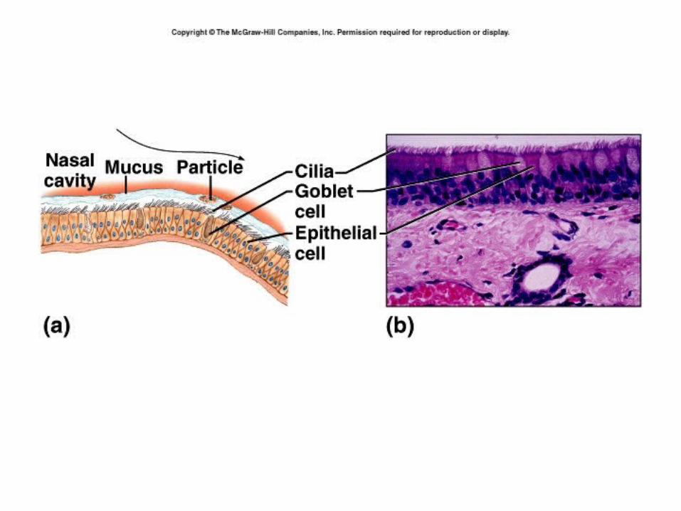
• Nasopharynx –– Respiration – lined with pseudostratified ciliated
columnar epithelium
• Oropharynx and Laryngopharynx– digestion and respiration– Nonkeratinized stratified squamous epithelium

8
Larynx
• Connects pharynx and trachea
• Passage of air in and out of lungs
• Prevents foreign objects entering trachea
• Contains vocal cord – speech
• Muscle and 9 cartilages connected by elastic tissue

9

10
Thyroid cartilage
• “Adam’s Apple”
• In female more rounded – shorter front to back – shorter vocal cords
• In male more “V” shaped – longer front to back – longer vocal cords
11
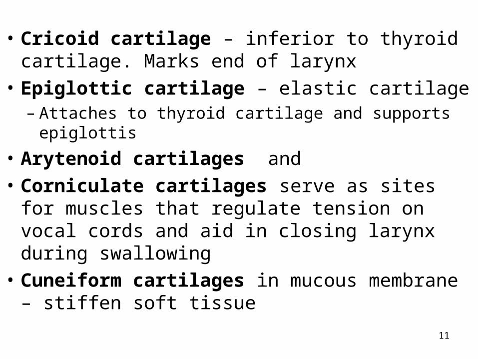
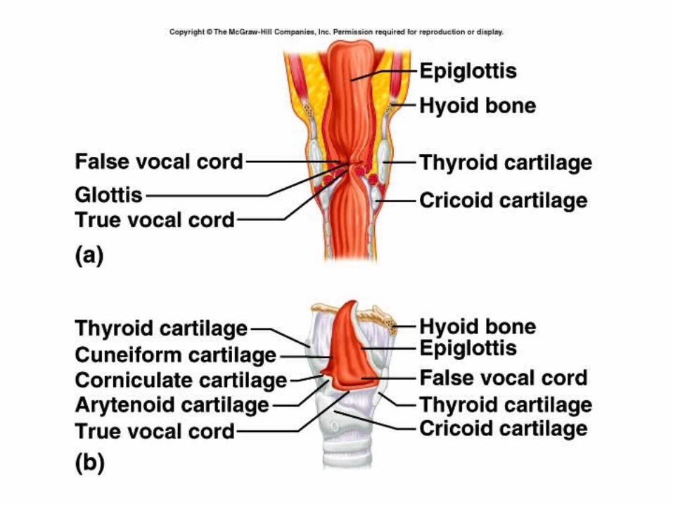
• Cricoid cartilage – inferior to thyroid cartilage. Marks end of larynx
• Epiglottic cartilage – elastic cartilage – Attaches to thyroid cartilage and supports epiglottis
• Arytenoid cartilages and
• Corniculate cartilages serve as sites for muscles that regulate tension on vocal cords and aid in closing larynx during swallowing
• Cuneiform cartilages in mucous membrane – stiffen soft tissue
12
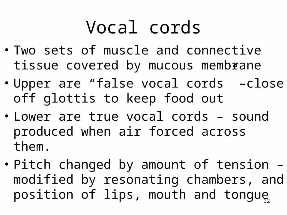
Vocal cords• Two sets of muscle and connective tissue
covered by mucous membrane
• Upper are “false vocal cords” –close off glottis to keep food out
• Lower are true vocal cords – sound produced when air forced across them.
• Pitch changed by amount of tension – modified by resonating chambers, and position of lips, mouth and tongue
13

14

15
Trachea• “windpipe”
• Extends from larynx to primary bronchi (T5)
• Smooth muscle – trachealis muscle
• 20 – “C” shaped rings of hyaline cartilage
• Open end of C posterior to allow esophagus to expand in
• Lined with pseudostratifed ciliated columnar epithelium
• Ridge – carina – most sensitive areas for triggering cough reflex

16

17

18

19
20

21
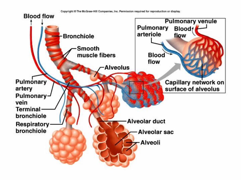
Bronchial Tree• Right and left primary bronchi
– Right bronchus shorter, wider, more vertical
• Secondary or lobar bronchi – 3 branches on right ; 2 on left
• Tertiary or segmental bronchi– 10 branches on right; 8 on left
• Bronchi lined with pseudostratified ciliated columnar epithelium
• Held open by complete rings of cartilage
22
23

24
Lungs
• Separated by mediastinum
• Pleural membranes
• Right has 3 lobes and two fissures, shorter
• Left has 2 lobes and one fissure, cardiac notch
• Apex, base, hilum

25
26
27
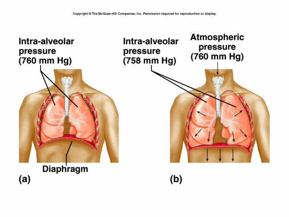
Pulmonary ventilation
• Air moves depending on a pressure gradient
• Before inspiration, pressure in lungs = pressure of atmosphere = 760 mm Hg
• Inspiration increases the size of the thoracic cavity and ↓ pressure .
• Diaphragm is the most important muscle of inspiration.- 75 % of air

28
29
30

31

32
• Diaphragm is innervated by the phrenic nerve.
• Decreases pressure 1-3 mm Hg
• Moves 500 ml of air
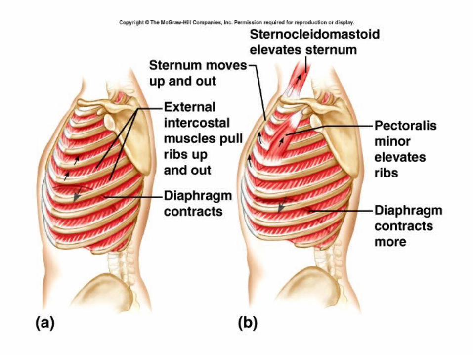
• External intercostals also contract – move ribs up and sternum forward
• Strenuous breathing – sternocleidomastoid, scalenes, and pectoralis minor
33
Expiration
• Passive process – the diaphragm and external intercostals relax. Elastic recoil of stretched elastic fibers and surface tension of the liquid in the lungs decreases the lung volume and increases the pressure, forcing air out of the lungs.
• Forced expiration involves contraction of the internal intercostal and abdominal muscles.

34
35
Lung volumes
• Clinically, respiration = one inhalation and one exhalation – respiratory cycle
• Adult breathes about 12 X/ min.
• Air volumes can be measured with a spirometer.

36
37
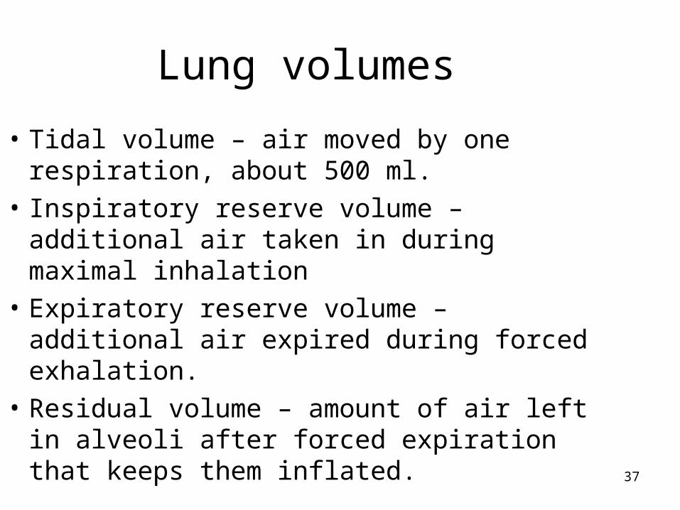
Lung volumes
• Tidal volume – air moved by one respiration, about 500 ml.
• Inspiratory reserve volume – additional air taken in during maximal inhalation
• Expiratory reserve volume – additional air expired during forced exhalation.
• Residual volume – amount of air left in alveoli after forced expiration that keeps them inflated.

38

39
Respiratory capacities
• Inspirational capacity – tidal vol. + inspiratory reserve volume
• Functional residual – residual volume + expir. res. vol.
• Vital capacity - inspir. Res. + tidal vol.+ expir. Res.
• Total lung capacity – sum of all volumes
40
Other lung volumes
• Anatomic dead space – due to conducting parts of system

41
Regulation of the Respiratory Center
• Respirations can be modified by factors from inside and outside the brain.
• Chemical influences:– Central chemoreceptors in medulla oblongata
– Sensitive to changes in conc. of CO2 and pH
– CO2 + H2O ↔ H2CO3 ↔ H+ + HCO3-
carbon + water carbonic hydrogen bicarbonate dioxide acid ion ion
42
Peripheral Chemoreceptors
• Carotid bodies and aortic bodies
• Stimulated by oxygen concentration decrease
• Send impulses to respiratory centers, and breathing increases
• Not triggered until O2 is very low (50 mm Hg)