Embed Size (px)
Citation preview
Methods to Enhance Digital Fundus Image for Diabetic Retinopathy Detection
Husna Ab Rahim, Ahmad Syahir Ibrahim, W Mimi Diyana W Zaki, Aini Hussain Department of Electric, Electronic and Systems Engineering
Faculty of Engineering and Built Environment UniversitiKebangsaan Malaysia
{husna.rahim, Syahir.Ibrahim}@siswa.ukm.edu.my, {wmdiyana, aini}@eng.ukm.my
Abstract— Digital fundus images (DFIs) are crucial in detecting pathological phenomenon that would lead to various diseases. However, DFI has multiple contrast and illumination problems which makes enhancement a necessity. Consequently, DFI must be enhanced to allow for better visualization in order to facilitate ophthalmologists to carry out their diagnosis.In this work, an investigation of three enhancement methods namely, Histogram Equalization(HE), Contrast Limited Adaptive Histogram Equalization(CLAHE) and Mahalanobis Distance (MD), were conducted on the digital fundus images and the results are qualitatively presented using histogram representation and image product quality. The result shows that the MD is the best algorithm for the application of blood vessels image enhancement.
IndexTerms—Fundus Image enhancement, blood vessel detection, Diabetic Retinopathy detection.
I. INTRODUCTION Digital fundus images (DFIs) are images obtained through
fundus photography, capturing the retina, optic disc, macular regions and the posterior surface of an eye. These regions are used by ophthalmologists during diabetic eye screening and diabetic retinopathy (DR) grading [1]. DR is an eye condition complications faced by diabetic patient which may contribute to blindness. In few cases, pathological effects such as blood vessel raptures may present in patient’s retina. There are a few characteristics in fundus images being used to detect the DR grades such asexudates, micro aneurysms, hemorrhage and the blood vessels [2]. Regular diabetic eye screening is an important step in detecting DR. Patients with sight-threatening DR might be identified during the screening process so that necessary treatment to prevent blindness could be given [3].
The best method to obtain perfect contrast in analyzing the fundus surface is using the images obtained from Fluorescein Angiography (FA). However, FA is an invasive method since it is obtained by injecting a yellow dye (fluorescein) into the patient’s body to enhance the RV and choroid during photography and has its side effects which include physiological problems such as Urticaria, severe seizure attack, myocardial infarction and anaphylactic attacks [4]. According to [5], the DFI method does not need such invasive procedure but the contrast is much lower than those of FA.
DFI is known to have very low contrast between the retinal vasculature and the background and it varies within the image which makes visualization and analysis of small retinal vasculatures difficult [6]. The illumination is very frequently uneven or non-uniform which causes the presence of local luminosity and contrast variability in the images that may lead to difficulty to a human observer to visualize and diagnose lesions in certain areas. This in turn can seriously affect the diagnostic process and its product [7]. Therefore, to guarantee visualization of the retinal blood vessels is at its best, image enhancement is required. Normalization method for DFIs is depending on the frequency domain and space [8]. In [9], they used vessel central light removal and background equalization to enhance the images. Both methods were successful to remove brightness and standardize the intensity. Meanwhile, V.Saravanan et al. applied background subtraction after converting the fundus images to green channel and subtracted by median filtered gray scale image [10]. In addition, they also used adaptive histogram equalization to enhance the DFIs contrast. The above methods are considered as intensity normalization in the preprocessing stage.
This research focuses on DFI enhancement and in this work, three different methods are considered. It is initially anticipated that the enhanced DFI can facilitate ophthalmologists to perform manual DR detection and grading and thus, reducing the need for FA. Additionally, this enhancement is a necessary pre-processing step for further processing techniques and it is important that any significant details in medical images to be preserved while being enhanced.
II. METHODOLOGY For the enhancement tests, 40 images obtained from the
DRIVE database were used. Each image was captured using 8 bits per color plane at 768 by 584 pixels using the Canon CR5 non-mydriatic 3CCD camera with a 45 degree field of view (FOV) and it is circular in form with a diameter of approximately 540 pixels [11]. Each of the images in this database has been cropped around the FOV area and was given a mask image to delineate the FOV. As mentioned previously, the three enhancement methods considered are 1) Histogram Equalization (HE), 2) Mahalanobis Distance (MD) and 3)
2014 IEEE 10th International Colloquium on Signal Processing & its Applications (CSPA2014), 7 - 9 Mac. 2014, Kuala Lumpur, Malaysia
978-1-4799-3091-3/14/$31.00 ©2014 IEEE 221
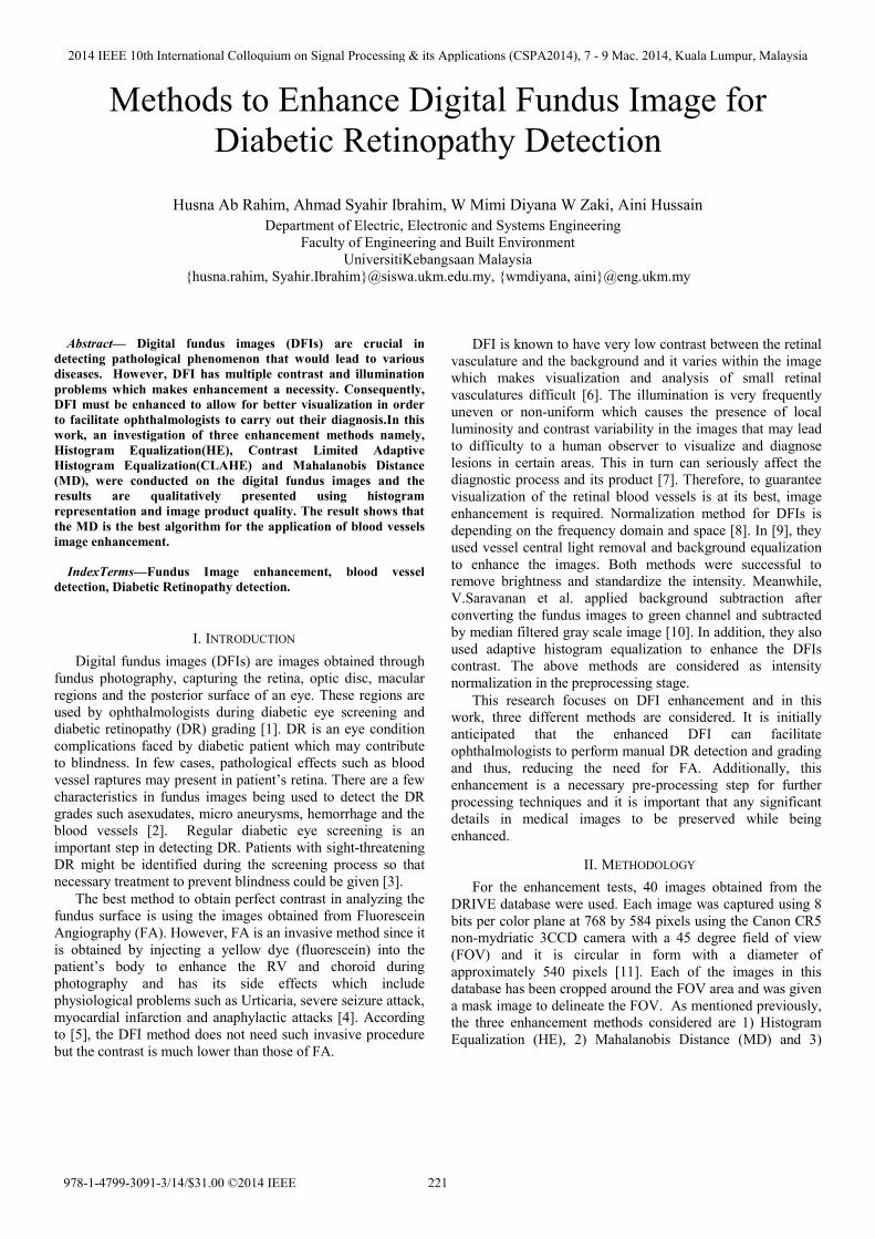
Contrast Limited Adaptive Histogram Equalization (CLAHE). Step by step procedures for this experimental work are as shown in Figure 1.
In RGB DFI, the green channel typically shows the best contrast between the background and vessels whereas the other two channels produce more noise [12]. As such, the gray images from the green channel are used since the retinal blood vessels in these images are more visible. Upon extraction, the images are processed using the three methods mentioned by the application of the respective algorithms.
Fig. 1 Step-by step procedures of digital fundus image enhancement
A. Mahalanobis Distance (MD) Image enhancement using the MD method is carried out by
identifying the background image pixels and eliminating them, leaving only the foreground image. It is based on the assumption that in image neighborhood , the background pixels has significantly different intensity value than those of the foreground pixels [13].
For each pixel , in the image, the mean μ , and the standard deviation σ , of the statistical distribution of intensities in are estimated. The sample mean; μ̂ is used as the estimator for μ , and the sample standard deviation;σ ̂ is the estimator for σ , . If the intensity of pixel , is close to the mean intensity in ,it is considered to belong to the background set β. As defined mathematically in Eq. 1, the expression implies that pixel , belongs to β if the stated condition is satisfied.
, μ̂σ ̂
(1) Those images would later be combined to evaluate the MD image, which can be segmented using the threshold to identify the background pixels.
B. Histogram Equalization (HE) HE is an operation that is based on histogram specification
or modification to obtain new images. The objective of this contrast enhancement technique is to obtain a new enhanced image that has a uniform histogram that simply plots the frequency at which each gray-level happens from 0 (black) to 255 (white) [14]. Each histogram represents the frequency of occurrence of all gray-level in the image, which also tell us how the distribution of the values of individual pixel in an image. The histogram equation is given as in Eq. 2:
/ (2)
Where , is the intensity level and is the number of
pixels in image with intensity. HE is to re-assign the intensity values of the pixels to create a uniform intensity distribution to reach the utmost value [15]. It is noted that HE can enhance the contrast of an image but has the tendency to its brightness. This means that HE technique is a global operation so it does not preserve the brightness of image. [16]
C. Contrast Limited Adaptive Histogram Equalization CLAHE Adaptive histogram equalization (AHE) transforms each
pixel in a gray-scale image using a transformation function that is derived from a neighborhood region. Simply, each pixel is transformed based on the histogram of a square surrounding the pixel. The transformation function derived from the histograms is similar to those of the ordinary HE, where the transformation function is proportional to the pixel values cumulative distribution function (CDF) in the neighborhood. AHE enables information with various intensities to be analyzed simultaneously [17]. This method is also automated and reproducible. However the results of AHE are image dependent and limited to images with low contrast variation only.
CLAHE uses RGB images directly and as such, the noise content of an image is not excessively enhanced in the resulting image, nevertheless visualization of the structures within the image is made by the sufficient contrast enhancement. This is achieved by limiting the contrast enhancement of AHE when the contrast amplification around a given pixel value is obtained according to the slope of the transformation function [18]. Thus, limiting the slope of the CDF is done by CLAHE when it limits the amplification by clipping the histogram at a predefined value before computing the CDF. The clip limit depends on the size of the neighborhood region and the normalization of the histogram. Images tend to appear more natural when processed with and can facilitate the comparison of different areas of the image [19]. However, the ability of an observer to detect the presence of some significant gray-scale contrast may be hindered because of the reduced contrast enhancement of CLAHE [20].
2014 IEEE 10th International Colloquium on Signal Processing & its Applications (CSPA2014), 7 - 9 Mac. 2014, Kuala Lumpur, Malaysia
222
III. RESULTS AND ANALYSIS This study is implemented in MATLAB R2012b. The
algorithm is applied on the database of 40 images with both normal and abnormal criteria. Performance of three algorithms namely HE, CLAHE, and MD are analyzed in the preliminary study for enhancement of DFI before progressing to the next stage that deals with detection of blood vessels. A comparison was made and focused on the histogram of the enhanced images upon implementation of the three algorithms.
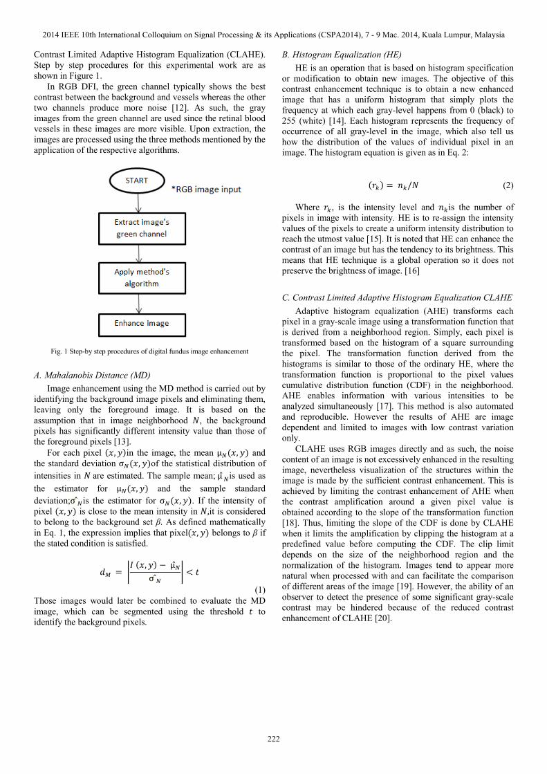
Fig. 2 Original Green component and its histogram
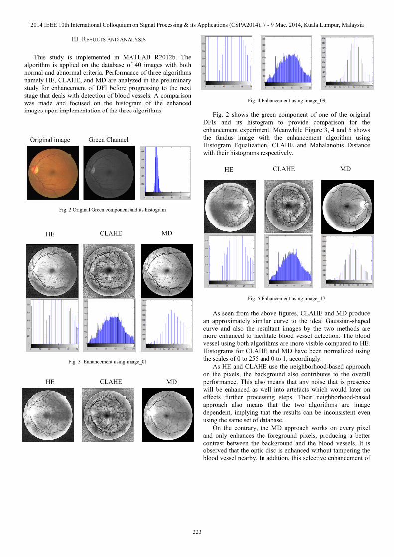
Fig. 3 Enhancement using image_01
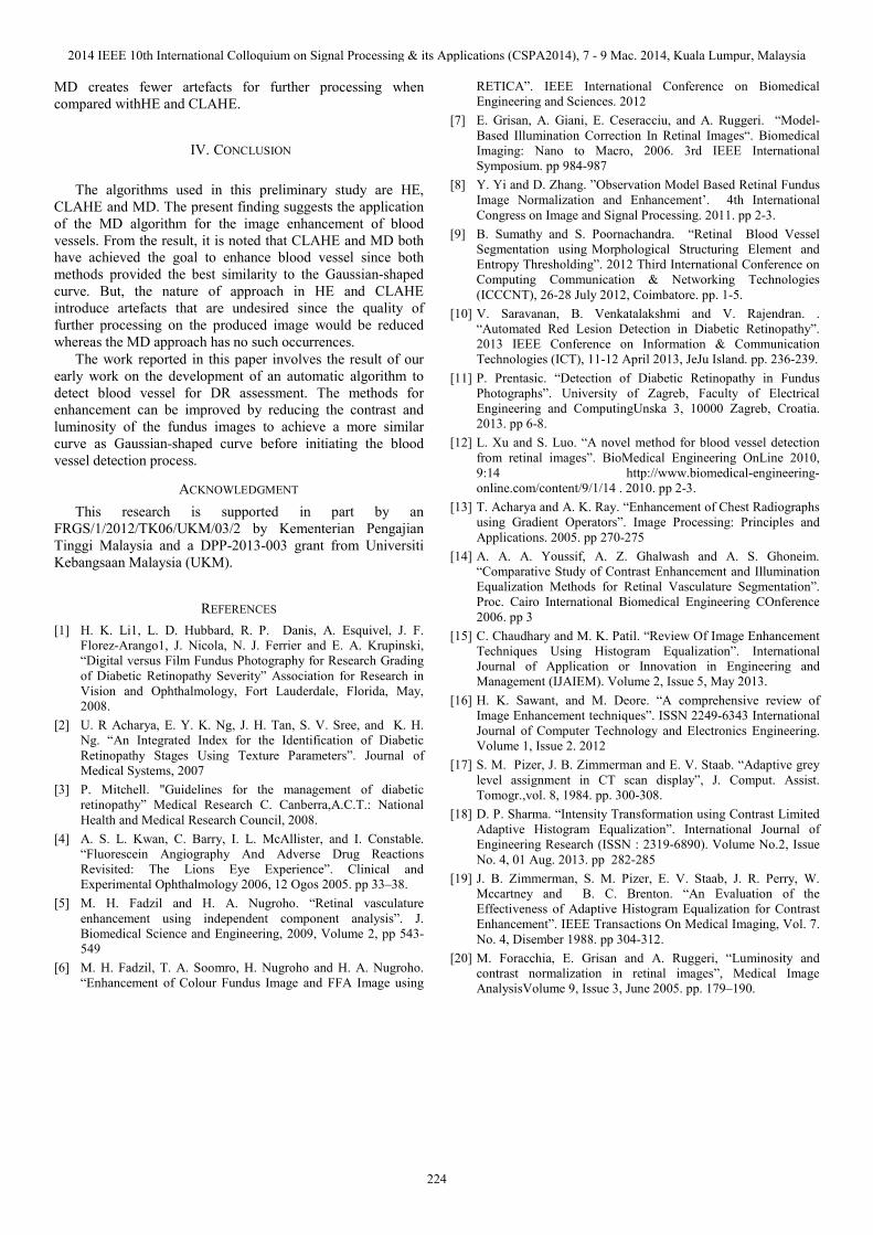
Fig. 4 Enhancement using image_09
Fig. 2 shows the green component of one of the original DFIs and its histogram to provide comparison for the enhancement experiment. Meanwhile Figure 3, 4 and 5 shows the fundus image with the enhancement algorithm using Histogram Equalization, CLAHE and Mahalanobis Distance with their histograms respectively.
Fig. 5 Enhancement using image_17
As seen from the above figures, CLAHE and MD produce an approximately similar curve to the ideal Gaussian-shaped curve and also the resultant images by the two methods are more enhanced to facilitate blood vessel detection. The blood vessel using both algorithms are more visible compared to HE. Histograms for CLAHE and MD have been normalized using the scales of 0 to 255 and 0 to 1, accordingly.
As HE and CLAHE use the neighborhood-based approach on the pixels, the background also contributes to the overall performance. This also means that any noise that is presence will be enhanced as well into artefacts which would later on effects further processing steps. Their neighborhood-based approach also means that the two algorithms are image dependent, implying that the results can be inconsistent even using the same set of database.
On the contrary, the MD approach works on every pixel and only enhances the foreground pixels, producing a better contrast between the background and the blood vessels. It is observed that the optic disc is enhanced without tampering the blood vessel nearby. In addition, this selective enhancement of
Original image Green Channel
HE CLAHE MD
HE CLAHE MD
CLAHE MD HE
2014 IEEE 10th International Colloquium on Signal Processing & its Applications (CSPA2014), 7 - 9 Mac. 2014, Kuala Lumpur, Malaysia
223
MD creates fewer artefacts for further processing when compared withHE and CLAHE.
IV. CONCLUSION The algorithms used in this preliminary study are HE,
CLAHE and MD. The present finding suggests the application of the MD algorithm for the image enhancement of blood vessels. From the result, it is noted that CLAHE and MD both have achieved the goal to enhance blood vessel since both methods provided the best similarity to the Gaussian-shaped curve. But, the nature of approach in HE and CLAHE introduce artefacts that are undesired since the quality of further processing on the produced image would be reduced whereas the MD approach has no such occurrences.
The work reported in this paper involves the result of our early work on the development of an automatic algorithm to detect blood vessel for DR assessment. The methods for enhancement can be improved by reducing the contrast and luminosity of the fundus images to achieve a more similar curve as Gaussian-shaped curve before initiating the blood vessel detection process.
ACKNOWLEDGMENT This research is supported in part by an
FRGS/1/2012/TK06/UKM/03/2 by Kementerian Pengajian Tinggi Malaysia and a DPP-2013-003 grant from Universiti Kebangsaan Malaysia (UKM).
REFERENCES [1] H. K. Li1, L. D. Hubbard, R. P. Danis, A. Esquivel, J. F.
Florez-Arango1, J. Nicola, N. J. Ferrier and E. A. Krupinski, “Digital versus Film Fundus Photography for Research Grading of Diabetic Retinopathy Severity” Association for Research in Vision and Ophthalmology, Fort Lauderdale, Florida, May, 2008.
[2] U. R Acharya, E. Y. K. Ng, J. H. Tan, S. V. Sree, and K. H. Ng. “An Integrated Index for the Identification of Diabetic Retinopathy Stages Using Texture Parameters”. Journal of Medical Systems, 2007
[3] P. Mitchell. "Guidelines for the management of diabetic retinopathy” Medical Research C. Canberra,A.C.T.: National Health and Medical Research Council, 2008.
[4] A. S. L. Kwan, C. Barry, I. L. McAllister, and I. Constable. “Fluorescein Angiography And Adverse Drug Reactions Revisited: The Lions Eye Experience”. Clinical and Experimental Ophthalmology 2006, 12 Ogos 2005. pp 33–38.
[5] M. H. Fadzil and H. A. Nugroho. “Retinal vasculature enhancement using independent component analysis”. J. Biomedical Science and Engineering, 2009, Volume 2, pp 543-549
[6] M. H. Fadzil, T. A. Soomro, H. Nugroho and H. A. Nugroho. “Enhancement of Colour Fundus Image and FFA Image using
RETICA”. IEEE International Conference on Biomedical Engineering and Sciences. 2012
[7] E. Grisan, A. Giani, E. Ceseracciu, and A. Ruggeri. “Model-Based Illumination Correction In Retinal Images“. Biomedical Imaging: Nano to Macro, 2006. 3rd IEEE International Symposium. pp 984-987
[8] Y. Yi and D. Zhang. ”Observation Model Based Retinal Fundus Image Normalization and Enhancement’. 4th International Congress on Image and Signal Processing. 2011. pp 2-3.
[9] B. Sumathy and S. Poornachandra. “Retinal Blood Vessel Segmentation using Morphological Structuring Element and Entropy Thresholding”. 2012 Third International Conference on Computing Communication & Networking Technologies (ICCCNT), 26-28 July 2012, Coimbatore. pp. 1-5.
[10] V. Saravanan, B. Venkatalakshmi and V. Rajendran. . “Automated Red Lesion Detection in Diabetic Retinopathy”. 2013 IEEE Conference on Information & Communication Technologies (ICT), 11-12 April 2013, JeJu Island. pp. 236-239.
[11] P. Prentasic. “Detection of Diabetic Retinopathy in Fundus Photographs”. University of Zagreb, Faculty of Electrical Engineering and ComputingUnska 3, 10000 Zagreb, Croatia. 2013. pp 6-8.
[12] L. Xu and S. Luo. “A novel method for blood vessel detection from retinal images”. BioMedical Engineering OnLine 2010, 9:14 http://www.biomedical-engineering-online.com/content/9/1/14 . 2010. pp 2-3.
[13] T. Acharya and A. K. Ray. “Enhancement of Chest Radiographs using Gradient Operators”. Image Processing: Principles and Applications. 2005. pp 270-275
[14] A. A. A. Youssif, A. Z. Ghalwash and A. S. Ghoneim. “Comparative Study of Contrast Enhancement and Illumination Equalization Methods for Retinal Vasculature Segmentation”. Proc. Cairo International Biomedical Engineering COnference 2006. pp 3
[15] C. Chaudhary and M. K. Patil. “Review Of Image Enhancement Techniques Using Histogram Equalization”. International Journal of Application or Innovation in Engineering and Management (IJAIEM). Volume 2, Issue 5, May 2013.
[16] H. K. Sawant, and M. Deore. “A comprehensive review of Image Enhancement techniques”. ISSN 2249-6343 International Journal of Computer Technology and Electronics Engineering. Volume 1, Issue 2. 2012
[17] S. M. Pizer, J. B. Zimmerman and E. V. Staab. “Adaptive grey level assignment in CT scan display”, J. Comput. Assist. Tomogr.,vol. 8, 1984. pp. 300-308.
[18] D. P. Sharma. “Intensity Transformation using Contrast Limited Adaptive Histogram Equalization”. International Journal of Engineering Research (ISSN : 2319-6890). Volume No.2, Issue No. 4, 01 Aug. 2013. pp 282-285
[19] J. B. Zimmerman, S. M. Pizer, E. V. Staab, J. R. Perry, W. Mccartney and B. C. Brenton. “An Evaluation of the Effectiveness of Adaptive Histogram Equalization for Contrast Enhancement”. IEEE Transactions On Medical Imaging, Vol. 7. No. 4, Disember 1988. pp 304-312.
[20] M. Foracchia, E. Grisan and A. Ruggeri, “Luminosity and contrast normalization in retinal images”, Medical Image AnalysisVolume 9, Issue 3, June 2005. pp. 179–190.
2014 IEEE 10th International Colloquium on Signal Processing & its Applications (CSPA2014), 7 - 9 Mac. 2014, Kuala Lumpur, Malaysia
224