Embed Size (px)
Citation preview
SHORT COMMUNICATION
A new subtype of Entamoeba gingivalis: BE. gingivalis ST2,kamaktli variant^
Gabriela García1 & Fernando Ramos1 & Fernando Martínez-Hernández2 & Lilian Hernández1 & Jorge Yáñez3 &
Paul Gaytán3
Received: 10 November 2017 /Accepted: 31 January 2018 /Published online: 10 February 2018# The Author(s) 2018. This article is an open access publication
AbstractEntamoeba gingivalis is a protozoan that resides in the oral cavity. Using molecular biology techniques, we identified a novelorganism that shares the same ecological niche as E. gingivalis. To differentiate this organism from E. gingivalis, we named itBkamaktli variant.^ By sequencing the 18S-ITS1-5.8S-ITS2 rRNA region, we demonstrated that kamaktli variant is 89%identical to E. gingivalis. To elucidate the relationship between kamaktli variant and E. gingivalis, we performed a phylogeneticanalysis. Both taxa clustered in the same clade with high support, indicating that the amoebas are closely related (98/99/1.00,maximum parsimony/maximum likelihood/MrBayes, respectively). Given this information, we propose that these moleculardifferences between kamaktli variant and E. gingivalis ST1 are sufficient to distinguish them as independent subtypes, and wename the new subtype BE. gingivalis ST2, kamaktli variant.^
Keywords Kamaktli variant . Oral microbiota . Genetic diversity . Entamoeba species . Molecular identification . Entamoebagingivalis ST2
Introduction
Entamoeba gingivalis is primarily found in the human oralcavity; it has also been found in the genitourinary tract(Clark and Diamond 1997; McNeill and de Morales-Ruehsen 1978; Foda and El-Malky 2012). To date, difficultyof cultivating this amoeba has precluded its completecharacterization.
The prevalence of E. gingivalis in people with a healthyperiodontium is highly variable, ranging from undetectable
levels (Lucht et al. 1998; Trim et al. 2011) to a high prevalence(30–80%) in people with oral cavity problems such as gingi-vitis or periodontal disease (Trim et al. 2011; Bonner et al.2014) and in people with systemic diseases such as diabetes(Chomicz et al. 2004).
The study and characterization of amoebas in the oral cav-ity have been ongoing for more than a century. Technologicaladvancements have made the molecular identification ofamoebas more feasible and have provided more detailedknowledge of their molecular characteristics (Clark et al.
* Gabriela Garcí[email protected]
Fernando [email protected]
Fernando Martínez-Herná[email protected]
Lilian Herná[email protected]
Jorge Yáñ[email protected]
Paul Gaytá[email protected]
1 Departamento de Microbiología y Parasitología, Facultad deMedicina, Universidad Nacional Autónoma de México, Av.Universidad 3000, Cd. Universitaria Coyoacán, CP.04510 Ciudad deMéxico, Mexico
2 Departamento de Ecología de Agentes Patógenos, Hospital GeneralDr. Manuel Gea González, Calzada de Tlalpan 4800, Tlalpan,CP.14080 Ciudad de México, Mexico
3 Unidad de Síntesis y Secuenciación de DNA, Instituto deBiotecnología, UNAM, Av. Universidad 2001,CP.14000 Cuernavaca, Morelos, Mexico
Parasitology Research (2018) 117:1277–1284https://doi.org/10.1007/s00436-018-5798-6
2006; Stensvold et al. 2011; García et al. 2014; Jacob et al.2016). Our research group has focused on the molecular char-acterization of the microbiota of the oral cavity, with a partic-ular interest in Entamoeba parasites.
Preliminary data from our lab suggested that some of thepartial nucleotide sequences of the 18S rRNA gene ofEntamoeba strains from the oral cavity are highly divergentfrom the E. gingivalis sequences reported previously inGenBank (D28490, KF250433-36). For the purposes of thispaper, we refer to the previously reported strains asBE. gingivalis ST1^ and use Bkamaktli variant^ to refer tothe amoeba that we identified; Bkamaktli^ is a Náhuatl wordmeaning Bmouth^ (Rodriguez-Villegas 2009). The aim of thisstudy was to determine if the sequences of the 18S-ITS1-5.8S-ITS2 rRNA region are sufficiently divergent betweenE. gingivalis ST1 (D28490, KF250433-36) and the kamaktlivariant to define this variant as a new subtype (BE. gingivalisST2, kamaktli variant^).
Material and methods
Samples and DNA extraction
The Ethics and Scientific research committees of theMedicineSchool of the University of Mexico (UNAM) approved theprotocol for this study (projects 042-2011, 151/2014 and 008/2016). The collected samples came from patients that attendedthe BClinic of Periodontology and Oral Implantology^ and theBClinic of Orthodontics^ in the Postgrad Division of theOdontology School at the University of Mexico (UNAM).Before the collecting procedures started, the patients wereinvited to participate in this study and they received a breveexplanation of the objectives and clinical procedure of thestudy. The patients who accepted to participate in the studysigned an informed consent and if they wanted, they couldanswer a clinical history questionnaire. A trained specialistdentist performed the patients’ examinations, gathered theclinical histories, and collected the obtained samples. Patientwith periodontal pockets greater than 6 mmwas sampled witha sterile curet in the affected site. Patients without periodontaldisease were sampled from gingival sulcus and surroundinggingival tissue on inferior molars with sterile brush. Eachsample obtained was deposited in a 2-ml vial with 100 μl ofRNAlater™ Qiagen® solution and transported to the labora-tory and store at − 20 °C until their processing.
As previously mentioned, some positive samples gave par-tial nucleotide sequences of the 18S rRNA gene different tothe E. gingivalis reported. Therefore, we selected five samplesto obtain the complete sequences of the 18S rRNA gene andalso the ITS1, 5.8S, and ITS2 regions. Three samples camefrom patients with periodontal disease and two from healthypatients. Two out of three samples from periodontal disease
patient were similar to the partial E. gingivalis sequences pre-viously reported. The other three samples showed some dif-ferences with respect to E. gingivalis. Buccal sample DNAwas extracted with the QIAamp DNA minikit (Qiagen®,Hilden, Germany) following the manufacturer’s instructions.
PCR, sequence assembly, and sequence analyses
The primers used in this study were synthesized at the DNAsequencing unit of the Biotechnology Institute at theUniversidad Nacional Autónoma de México (UNAM).These primers were chosen from previous reports: RD5′-RD3′ (Clark and Diamond 1997), Entam1-Entam2 (Verweijet al., 2001), P1-P2 (Som et al., 2000), GEI18SF (García et al.2014) and GE18SR (5′-GTACAAAGGGCAGGGACGTA-3′), and GEg3F (5′-GTAATTCCACCTCCAATAGTRT-3′)and GEg3R (5′AACTAAGAACGGCCATGCAC-3′). Theprimers GE18SR, GEg3F, and GEg3R were designed for thisstudy. Amplification consisted of 35 cycles at 94 °C for 30 s,55 °C for 30 s, and 72 °C for 30 s. All amplifications werepreceded by an initial 2-min cycle at 94 °C and ended with a 3-min cycle at 72 °C. These primers gave amplicons of around2000 bp for RD5′-RD3′, 600 bp for Entam1-Entam2, 400 bpfor P1-P2, and 700 bp for GEg3F-GEg3R. The PCR productswere separated by electrophoresis on a 1.2% agarose gel inTris/Borate/EDTA (TBE), stained with ethidium bromide, andobserved on a UV transilluminator. Bands of the expected sizewere cut and purified with a commercial kit (QIAquick DNAgel extraction kit, Qiagen®) following the manufacturer’s in-structions. The amplicons were then sequenced in both direc-tions using the BigDye Terminator-V.3.1 sequencing kit sys-tem (Applied Biosystems™, Foster City, CA, USA) followingthe manufacturer’s instructions and using a 3130xl GeneticAnalyzer (Applied Biosystems™). Sequences were visualizedusing 4Peaks software (4Peaks 2004).
To assemble the obtained sequences, we used the multiplealignment program MultAlin (Corpet 1988). The assembledsequences were compared with BLAST against sequences re-ported in GenBank.
Maximum similarity analysis
The Needleman-Wunsch algorithmwas used to align the iden-tified sequences; this algorithm determines the best possiblealignment to obtain the maximum similarity between two mo-lecular sequences (Needleman and Wunsch 1970). For thisanalysis, each pair of sequences to be compared was aligned.The 18S rRNA region of kamaktli variant (KX027294) wasconsidered the prototype and was compared to otherEntamoeba strains. The species and sequence accession num-bers are provided in Table 1. Similarly, E. gingivalis ST1(KX027297) (obtained in this study) was compared with otherE. gingivalis ST1 strains and with E. suis. Additionally, the
1278 Parasitol Res (2018) 117:1277–1284
18S, 18S-ITS1-5.8S-ITS2, and ITS1-5.8S-ITS2 rRNA re-gions were each compared between E. dispar andE. histolytica as well as between kamaktli variant andE. gingivalis ST1 to determine the region that best differenti-ated closely related species.
Phylogeny
The multiple alignments needed for the phylogenetic analyseswere performed by using ClustalW and MUSCLE in MEGAsoftware version 7 (Tamura et al. 2011) with manual adjust-ment to delete ambiguities. The best-fit model of nucleotidesubstitution was determined using the Akaike InformationCriterion in Modeltest version 3.7 software and applying theGeneral Time Reversible model of evolution specifying agamma distribution and invariable sites. The algorithms usedto obtain the phylogenetic analyses were maximum parsimo-ny (MP), maximum likelihood (ML), and Bayesian algo-rithms (BA). MP and ML were run with 1000 bootstrap rep-licas under the General Time Reversible model of evolution,and the algorithms were implemented in MEGA7 (Yamamotoet al. 1995). For the BA reconstruction, MrBayes version 3.4(Huelsenbeck and Ronquist 2001) was used, and the analysiswas performed with 3 × 10⁶ generations, sampling trees every100 generations. Trees with a score lower than those at thestationary phase (burn-in) were discarded, and trees that
reached the stationary phase were collected and used to buildconsensus trees.
A phylogenetic tree was generated using 53 sequences,which included all of the E. gingivalis reported sequences inGenBank and the sequences obtained in the present study.Selection of the sequences was performed to obtain a repre-sentative balanced tree of all of the species reported. Takinginto account the different sequences of E. gingivalis, we ana-lyzed whether kamaktli variant differs from previously knownEntamoeba species.
Results
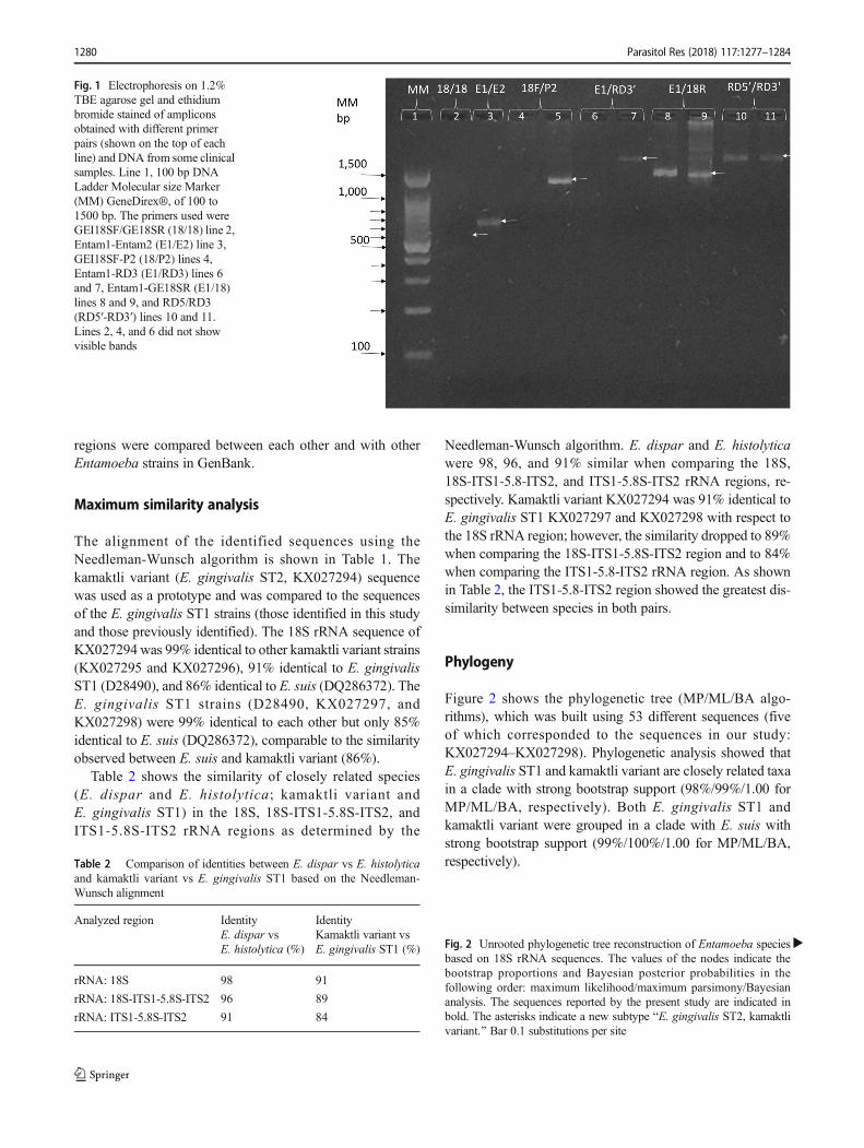
The amplicons generated with the different primers were iden-tified by electrophoresis on agarose gels stained with ethidiumbromide. Figure 1 shows an electrophoresis of someamplicons obtained with different primers and as it can beseen the amplicons had different size that depended on theused primers. The bands with the expected size were cut,purified and sequenced as mentioned before. The sequencesobtained with the different primers from each patient wereassembled as described in the methods.
To determine how similar kamaktli variant andE. gingivalis ST1 are, their 18S-ITS1-5.8S-ITS2 rRNA
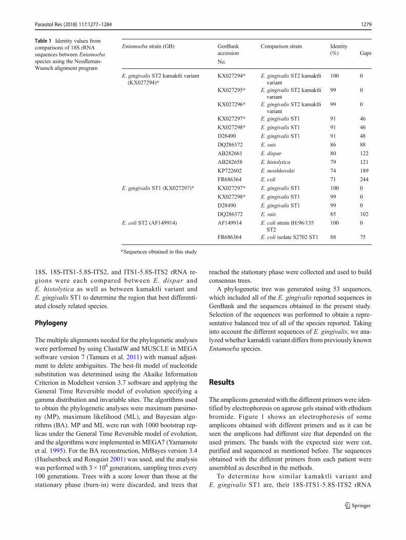
Table 1 Identity values fromcomparisons of 18S rRNAsequences between Entamoebaspecies using the Needleman-Wunsch alignment program
Entamoeba strain (GB) GenBankaccession
No.
Comparison strain Identity(%) Gaps
E. gingivalis ST2 kamaktli variant(KX027294)*
KX027294* E. gingivalis ST2 kamaktlivariant
100 0
KX027295* E. gingivalis ST2 kamaktlivariant
99 0
KX027296* E. gingivalis ST2 kamaktlivariant
99 0
KX027297* E. gingivalis ST1 91 46
KX027298* E. gingivalis ST1 91 46
D28490 E. gingivalis ST1 91 48
DQ286372 E. suis 86 88
AB282661 E. dispar 80 122
AB282658 E. histolytica 79 121
KP722602 E. moshkovskii 74 189
FR686364 E. coli 71 244
E. gingivalis ST1 (KX027297)* KX027297* E. gingivalis ST1 100 0
KX027298* E. gingivalis ST1 99 0
D28490 E. gingivalis ST1 99 0
DQ286372 E. suis 85 102
E. coli ST2 (AF149914) AF149914 E. coli strain IH:96/135ST2
100 0
FR686364 E. coli isolate S2702 ST1 88 75
*Sequences obtained in this study
Parasitol Res (2018) 117:1277–1284 1279
regions were compared between each other and with otherEntamoeba strains in GenBank.
Maximum similarity analysis
The alignment of the identified sequences using theNeedleman-Wunsch algorithm is shown in Table 1. Thekamaktli variant (E. gingivalis ST2, KX027294) sequencewas used as a prototype and was compared to the sequencesof the E. gingivalis ST1 strains (those identified in this studyand those previously identified). The 18S rRNA sequence ofKX027294was 99% identical to other kamaktli variant strains(KX027295 and KX027296), 91% identical to E. gingivalisST1 (D28490), and 86% identical to E. suis (DQ286372). TheE. gingivalis ST1 strains (D28490, KX027297, andKX027298) were 99% identical to each other but only 85%identical to E. suis (DQ286372), comparable to the similarityobserved between E. suis and kamaktli variant (86%).
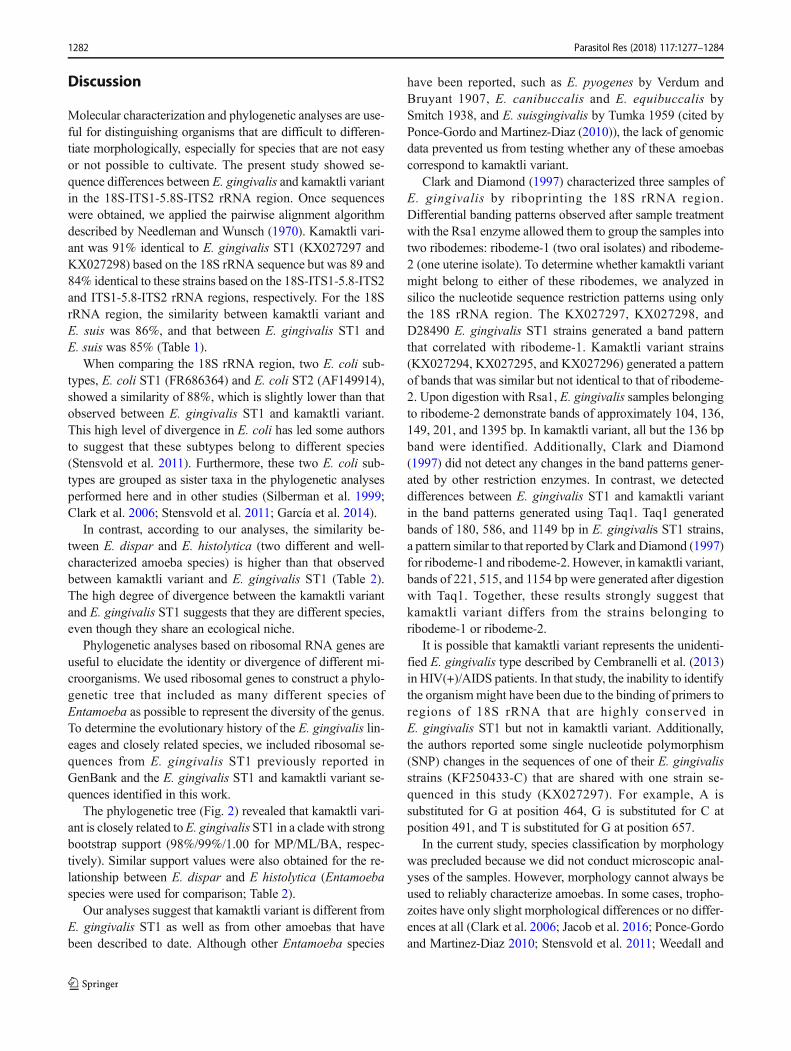
Table 2 shows the similarity of closely related species(E. dispar and E. histolytica; kamaktli variant andE. gingivalis ST1) in the 18S, 18S-ITS1-5.8S-ITS2, andITS1-5.8S-ITS2 rRNA regions as determined by the
Needleman-Wunsch algorithm. E. dispar and E. histolyticawere 98, 96, and 91% similar when comparing the 18S,18S-ITS1-5.8-ITS2, and ITS1-5.8S-ITS2 rRNA regions, re-spectively. Kamaktli variant KX027294 was 91% identical toE. gingivalis ST1 KX027297 and KX027298 with respect tothe 18S rRNA region; however, the similarity dropped to 89%when comparing the 18S-ITS1-5.8S-ITS2 region and to 84%when comparing the ITS1-5.8-ITS2 rRNA region. As shownin Table 2, the ITS1-5.8-ITS2 region showed the greatest dis-similarity between species in both pairs.
Phylogeny
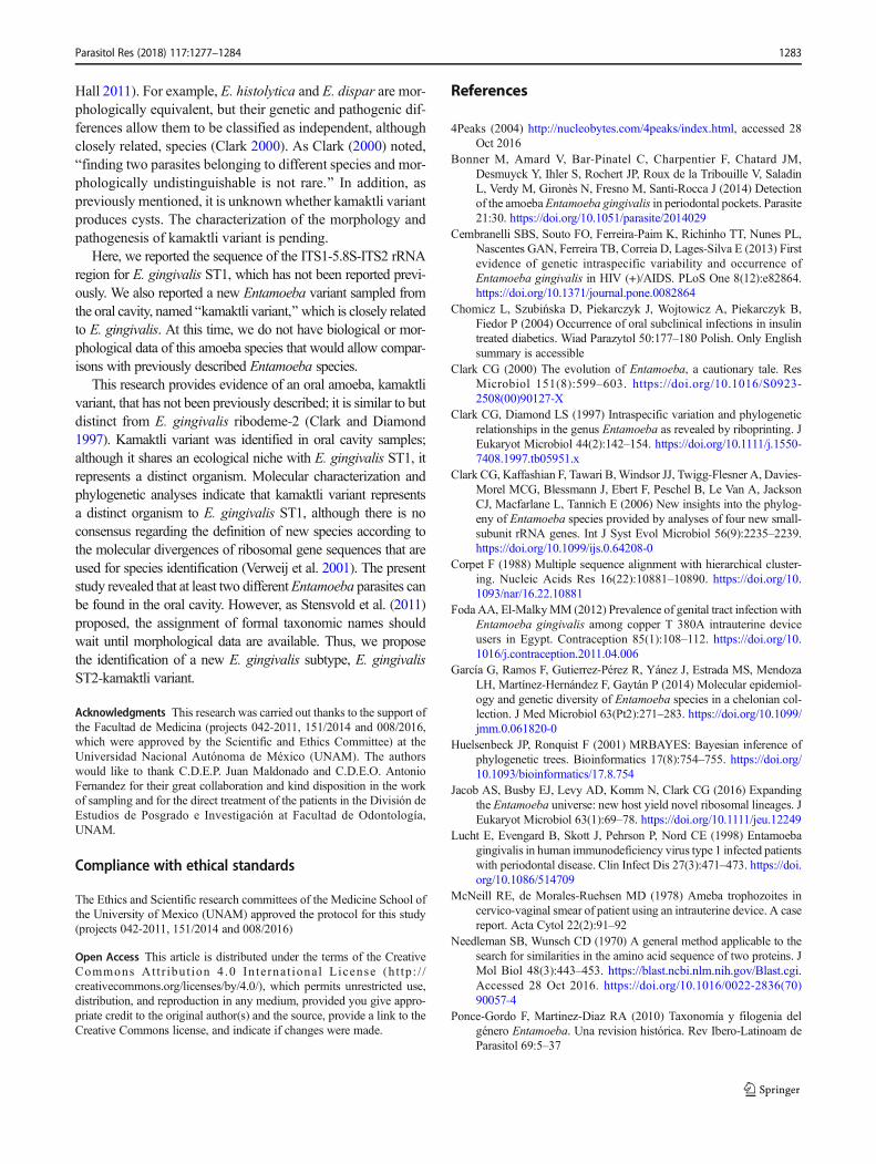
Figure 2 shows the phylogenetic tree (MP/ML/BA algo-rithms), which was built using 53 different sequences (fiveof which corresponded to the sequences in our study:KX027294–KX027298). Phylogenetic analysis showed thatE. gingivalis ST1 and kamaktli variant are closely related taxain a clade with strong bootstrap support (98%/99%/1.00 forMP/ML/BA, respectively). Both E. gingivalis ST1 andkamaktli variant were grouped in a clade with E. suis withstrong bootstrap support (99%/100%/1.00 for MP/ML/BA,respectively).
Fig. 1 Electrophoresis on 1.2%TBE agarose gel and ethidiumbromide stained of ampliconsobtained with different primerpairs (shown on the top of eachline) and DNA from some clinicalsamples. Line 1, 100 bp DNALadder Molecular size Marker(MM) GeneDirex®, of 100 to1500 bp. The primers used wereGEI18SF/GE18SR (18/18) line 2,Entam1-Entam2 (E1/E2) line 3,GEI18SF-P2 (18/P2) lines 4,Entam1-RD3 (E1/RD3) lines 6and 7, Entam1-GE18SR (E1/18)lines 8 and 9, and RD5/RD3(RD5′-RD3′) lines 10 and 11.Lines 2, 4, and 6 did not showvisible bands
Table 2 Comparison of identities between E. dispar vs E. histolyticaand kamaktli variant vs E. gingivalis ST1 based on the Needleman-Wunsch alignment
Analyzed region IdentityE. dispar vsE. histolytica (%)
IdentityKamaktli variant vsE. gingivalis ST1 (%)
rRNA: 18S 98 91
rRNA: 18S-ITS1-5.8S-ITS2 96 89
rRNA: ITS1-5.8S-ITS2 91 84
�Fig. 2 Unrooted phylogenetic tree reconstruction of Entamoeba speciesbased on 18S rRNA sequences. The values of the nodes indicate thebootstrap proportions and Bayesian posterior probabilities in thefollowing order: maximum likelihood/maximum parsimony/Bayesiananalysis. The sequences reported by the present study are indicated inbold. The asterisks indicate a new subtype BE. gingivalis ST2, kamaktlivariant.^ Bar 0.1 substitutions per site
1280 Parasitol Res (2018) 117:1277–1284

Parasitol Res (2018) 117:1277–1284 1281
Discussion
Molecular characterization and phylogenetic analyses are use-ful for distinguishing organisms that are difficult to differen-tiate morphologically, especially for species that are not easyor not possible to cultivate. The present study showed se-quence differences between E. gingivalis and kamaktli variantin the 18S-ITS1-5.8S-ITS2 rRNA region. Once sequenceswere obtained, we applied the pairwise alignment algorithmdescribed by Needleman and Wunsch (1970). Kamaktli vari-ant was 91% identical to E. gingivalis ST1 (KX027297 andKX027298) based on the 18S rRNA sequence but was 89 and84% identical to these strains based on the 18S-ITS1-5.8-ITS2and ITS1-5.8-ITS2 rRNA regions, respectively. For the 18SrRNA region, the similarity between kamaktli variant andE. suis was 86%, and that between E. gingivalis ST1 andE. suis was 85% (Table 1).
When comparing the 18S rRNA region, two E. coli sub-types, E. coli ST1 (FR686364) and E. coli ST2 (AF149914),showed a similarity of 88%, which is slightly lower than thatobserved between E. gingivalis ST1 and kamaktli variant.This high level of divergence in E. coli has led some authorsto suggest that these subtypes belong to different species(Stensvold et al. 2011). Furthermore, these two E. coli sub-types are grouped as sister taxa in the phylogenetic analysesperformed here and in other studies (Silberman et al. 1999;Clark et al. 2006; Stensvold et al. 2011; García et al. 2014).
In contrast, according to our analyses, the similarity be-tween E. dispar and E. histolytica (two different and well-characterized amoeba species) is higher than that observedbetween kamaktli variant and E. gingivalis ST1 (Table 2).The high degree of divergence between the kamaktli variantand E. gingivalis ST1 suggests that they are different species,even though they share an ecological niche.
Phylogenetic analyses based on ribosomal RNA genes areuseful to elucidate the identity or divergence of different mi-croorganisms. We used ribosomal genes to construct a phylo-genetic tree that included as many different species ofEntamoeba as possible to represent the diversity of the genus.To determine the evolutionary history of the E. gingivalis lin-eages and closely related species, we included ribosomal se-quences from E. gingivalis ST1 previously reported inGenBank and the E. gingivalis ST1 and kamaktli variant se-quences identified in this work.
The phylogenetic tree (Fig. 2) revealed that kamaktli vari-ant is closely related toE. gingivalis ST1 in a clade with strongbootstrap support (98%/99%/1.00 for MP/ML/BA, respec-tively). Similar support values were also obtained for the re-lationship between E. dispar and E histolytica (Entamoebaspecies were used for comparison; Table 2).
Our analyses suggest that kamaktli variant is different fromE. gingivalis ST1 as well as from other amoebas that havebeen described to date. Although other Entamoeba species
have been reported, such as E. pyogenes by Verdum andBruyant 1907, E. canibuccalis and E. equibuccalis bySmitch 1938, and E. suisgingivalis by Tumka 1959 (cited byPonce-Gordo and Martinez-Diaz (2010)), the lack of genomicdata prevented us from testing whether any of these amoebascorrespond to kamaktli variant.
Clark and Diamond (1997) characterized three samples ofE. gingivalis by riboprinting the 18S rRNA region.Differential banding patterns observed after sample treatmentwith the Rsa1 enzyme allowed them to group the samples intotwo ribodemes: ribodeme-1 (two oral isolates) and ribodeme-2 (one uterine isolate). To determine whether kamaktli variantmight belong to either of these ribodemes, we analyzed insilico the nucleotide sequence restriction patterns using onlythe 18S rRNA region. The KX027297, KX027298, andD28490 E. gingivalis ST1 strains generated a band patternthat correlated with ribodeme-1. Kamaktli variant strains(KX027294, KX027295, and KX027296) generated a patternof bands that was similar but not identical to that of ribodeme-2. Upon digestion with Rsa1, E. gingivalis samples belongingto ribodeme-2 demonstrate bands of approximately 104, 136,149, 201, and 1395 bp. In kamaktli variant, all but the 136 bpband were identified. Additionally, Clark and Diamond(1997) did not detect any changes in the band patterns gener-ated by other restriction enzymes. In contrast, we detecteddifferences between E. gingivalis ST1 and kamaktli variantin the band patterns generated using Taq1. Taq1 generatedbands of 180, 586, and 1149 bp in E. gingivalis ST1 strains,a pattern similar to that reported by Clark and Diamond (1997)for ribodeme-1 and ribodeme-2. However, in kamaktli variant,bands of 221, 515, and 1154 bp were generated after digestionwith Taq1. Together, these results strongly suggest thatkamaktli variant differs from the strains belonging toribodeme-1 or ribodeme-2.
It is possible that kamaktli variant represents the unidenti-fied E. gingivalis type described by Cembranelli et al. (2013)in HIV(+)/AIDS patients. In that study, the inability to identifythe organismmight have been due to the binding of primers toregions of 18S rRNA that are highly conserved inE. gingivalis ST1 but not in kamaktli variant. Additionally,the authors reported some single nucleotide polymorphism(SNP) changes in the sequences of one of their E. gingivalisstrains (KF250433-C) that are shared with one strain se-quenced in this study (KX027297). For example, A issubstituted for G at position 464, G is substituted for C atposition 491, and T is substituted for G at position 657.
In the current study, species classification by morphologywas precluded because we did not conduct microscopic anal-yses of the samples. However, morphology cannot always beused to reliably characterize amoebas. In some cases, tropho-zoites have only slight morphological differences or no differ-ences at all (Clark et al. 2006; Jacob et al. 2016; Ponce-Gordoand Martinez-Diaz 2010; Stensvold et al. 2011; Weedall and
1282 Parasitol Res (2018) 117:1277–1284
Hall 2011). For example, E. histolytica and E. dispar are mor-phologically equivalent, but their genetic and pathogenic dif-ferences allow them to be classified as independent, althoughclosely related, species (Clark 2000). As Clark (2000) noted,Bfinding two parasites belonging to different species and mor-phologically undistinguishable is not rare.^ In addition, aspreviously mentioned, it is unknown whether kamaktli variantproduces cysts. The characterization of the morphology andpathogenesis of kamaktli variant is pending.
Here, we reported the sequence of the ITS1-5.8S-ITS2 rRNAregion for E. gingivalis ST1, which has not been reported previ-ously. We also reported a new Entamoeba variant sampled fromthe oral cavity, named Bkamaktli variant,̂ which is closely relatedto E. gingivalis. At this time, we do not have biological or mor-phological data of this amoeba species that would allow compar-isons with previously described Entamoeba species.
This research provides evidence of an oral amoeba, kamaktlivariant, that has not been previously described; it is similar to butdistinct from E. gingivalis ribodeme-2 (Clark and Diamond1997). Kamaktli variant was identified in oral cavity samples;although it shares an ecological niche with E. gingivalis ST1, itrepresents a distinct organism. Molecular characterization andphylogenetic analyses indicate that kamaktli variant representsa distinct organism to E. gingivalis ST1, although there is noconsensus regarding the definition of new species according tothe molecular divergences of ribosomal gene sequences that areused for species identification (Verweij et al. 2001). The presentstudy revealed that at least two differentEntamoeba parasites canbe found in the oral cavity. However, as Stensvold et al. (2011)proposed, the assignment of formal taxonomic names shouldwait until morphological data are available. Thus, we proposethe identification of a new E. gingivalis subtype, E. gingivalisST2-kamaktli variant.
Acknowledgments This research was carried out thanks to the support ofthe Facultad de Medicina (projects 042-2011, 151/2014 and 008/2016,which were approved by the Scientific and Ethics Committee) at theUniversidad Nacional Autónoma de México (UNAM). The authorswould like to thank C.D.E.P. Juan Maldonado and C.D.E.O. AntonioFernandez for their great collaboration and kind disposition in the workof sampling and for the direct treatment of the patients in the División deEstudios de Posgrado e Investigación at Facultad de Odontología,UNAM.
Compliance with ethical standards
The Ethics and Scientific research committees of the Medicine School ofthe University of Mexico (UNAM) approved the protocol for this study(projects 042-2011, 151/2014 and 008/2016)
Open Access This article is distributed under the terms of the CreativeCommons At t r ibut ion 4 .0 In te rna t ional License (h t tp : / /creativecommons.org/licenses/by/4.0/), which permits unrestricted use,distribution, and reproduction in any medium, provided you give appro-priate credit to the original author(s) and the source, provide a link to theCreative Commons license, and indicate if changes were made.
References
4Peaks (2004) http://nucleobytes.com/4peaks/index.html, accessed 28Oct 2016
Bonner M, Amard V, Bar-Pinatel C, Charpentier F, Chatard JM,Desmuyck Y, Ihler S, Rochert JP, Roux de la Tribouille V, SaladinL, Verdy M, Gironès N, Fresno M, Santi-Rocca J (2014) Detectionof the amoebaEntamoeba gingivalis in periodontal pockets. Parasite21:30. https://doi.org/10.1051/parasite/2014029
Cembranelli SBS, Souto FO, Ferreira-Paim K, Richinho TT, Nunes PL,Nascentes GAN, Ferreira TB, Correia D, Lages-Silva E (2013) Firstevidence of genetic intraspecific variability and occurrence ofEntamoeba gingivalis in HIV (+)/AIDS. PLoS One 8(12):e82864.https://doi.org/10.1371/journal.pone.0082864
Chomicz L, Szubińska D, Piekarczyk J, Wojtowicz A, Piekarczyk B,Fiedor P (2004) Occurrence of oral subclinical infections in insulintreated diabetics. Wiad Parazytol 50:177–180 Polish. Only Englishsummary is accessible
Clark CG (2000) The evolution of Entamoeba, a cautionary tale. ResMicrobiol 151(8):599–603. https://doi.org/10.1016/S0923-2508(00)90127-X
Clark CG, Diamond LS (1997) Intraspecific variation and phylogeneticrelationships in the genus Entamoeba as revealed by riboprinting. JEukaryot Microbiol 44(2):142–154. https://doi.org/10.1111/j.1550-7408.1997.tb05951.x
Clark CG, Kaffashian F, Tawari B,Windsor JJ, Twigg-Flesner A, Davies-Morel MCG, Blessmann J, Ebert F, Peschel B, Le Van A, JacksonCJ, Macfarlane L, Tannich E (2006) New insights into the phylog-eny of Entamoeba species provided by analyses of four new small-subunit rRNA genes. Int J Syst Evol Microbiol 56(9):2235–2239.https://doi.org/10.1099/ijs.0.64208-0
Corpet F (1988) Multiple sequence alignment with hierarchical cluster-ing. Nucleic Acids Res 16(22):10881–10890. https://doi.org/10.1093/nar/16.22.10881
Foda AA, El-MalkyMM (2012) Prevalence of genital tract infection withEntamoeba gingivalis among copper T 380A intrauterine deviceusers in Egypt. Contraception 85(1):108–112. https://doi.org/10.1016/j.contraception.2011.04.006
García G, Ramos F, Gutierrez-Pérez R, Yánez J, Estrada MS, MendozaLH, Martínez-Hernández F, Gaytán P (2014) Molecular epidemiol-ogy and genetic diversity of Entamoeba species in a chelonian col-lection. J Med Microbiol 63(Pt2):271–283. https://doi.org/10.1099/jmm.0.061820-0
Huelsenbeck JP, Ronquist F (2001) MRBAYES: Bayesian inference ofphylogenetic trees. Bioinformatics 17(8):754–755. https://doi.org/10.1093/bioinformatics/17.8.754
Jacob AS, Busby EJ, Levy AD, Komm N, Clark CG (2016) Expandingthe Entamoeba universe: new host yield novel ribosomal lineages. JEukaryot Microbiol 63(1):69–78. https://doi.org/10.1111/jeu.12249
Lucht E, Evengard B, Skott J, Pehrson P, Nord CE (1998) Entamoebagingivalis in human immunodeficiency virus type 1 infected patientswith periodontal disease. Clin Infect Dis 27(3):471–473. https://doi.org/10.1086/514709
McNeill RE, de Morales-Ruehsen MD (1978) Ameba trophozoites incervico-vaginal smear of patient using an intrauterine device. A casereport. Acta Cytol 22(2):91–92
Needleman SB, Wunsch CD (1970) A general method applicable to thesearch for similarities in the amino acid sequence of two proteins. JMol Biol 48(3):443–453. https://blast.ncbi.nlm.nih.gov/Blast.cgi.Accessed 28 Oct 2016. https://doi.org/10.1016/0022-2836(70)90057-4
Ponce-Gordo F, Martinez-Diaz RA (2010) Taxonomía y filogenia delgénero Entamoeba. Una revision histórica. Rev Ibero-Latinoam deParasitol 69:5–37
Parasitol Res (2018) 117:1277–1284 1283
Rodriguez-Villegas (2009) On-line Aulex Vocabularic compiled. http://aulex.org/es-nah/, accessed 28 Oct 2016
Silberman JD, Clark CG, Diamond LS, Sogin ML (1999) Phylogeny ofthe genera Entamoeba and Endolimax as deduced from small-subunit ribosomal RNA sequences. Mol Biol Evol 16(12):1740–1751
Som I, Azam A, Bhattacharya A, Bhattacharya S (2000) Inter- and intra-strain variation in the 5.8S ribosomal RNA and internal transcribedspacer sequences of Entamoeba histolytica and comparison withEntamoeba dispar, Entamoeba moshkovskii and Entamoebainvadens. Int J Parasitol 30:723–728
Stensvold CR, Lebbad M, Victory EL, Verweij JJ, Tannich E, AlfellaniM, Legarraga P, Clark CG (2011) Increased sampling reveals novellineages of Entamoeba: consequences of genetic diversity and hostspecificity for taxonomy and molecular detection. Protist 162(3):525–541. https://doi.org/10.1016/j.protis.2010.11.002
Tamura K, Peterson D, Peterson N, Stecher G, Nei M, Kumar S (2011)MEGA5: molecular evolutionary genetics analysis using maximumlikelihood evolutionary distance and maximum parsimony methods.
Mol Biol Evol 28:2731–2739. https://doi.org/10.1093/molbev/msr121
Trim RD, Skinner MA, Farone MB, DuBois JD, Newsome AL (2011)Use of PCR to detect Entamoeba gingivalis in diseased gingivalpockets and demonstrate its absence in healthy gingival sites.Parasitol Res 109(3):857–864. https://doi.org/10.1007/s00436-011-2312-9
Verweij JJ, Polderman AM, Clark CG (2001) Genetic variation amonghuman isolates of uninucleated cyst-producing Entamoeba species. JClin Microbiol 39(4):1644–1646. https://doi.org/10.1128/JCM.39.4.1644-1646.2001
Weedall GD, Hall N (2011) Evolutionary genomics Entamoeba. ResMicrobiol 162(6):637–645. https://doi.org/10.1016/j.resmic.2011.01.007
Yamamoto A, Kikuta N, Hashimoto T, Oyaizu H, Goto N (1995)Nucleotide sequence of the SrRNA gene of Entamoeba gingivalis:applications for construction of a species-specific DNA probe andphylogenetic analysis. Microbiol Immunol 39(3):185–192. https://doi.org/10.1111/j.1348-0421.1995.tb02187.x
1284 Parasitol Res (2018) 117:1277–1284