Embed Size (px)
Citation preview

ANATOMY AND PHYSIOLOGY OF SUPPORTING AND LIMITING STRUCTURES OF COMPLETELY EDENTULOUS ARCH
DR Roma Goswami Prof & Head Prosthodontics and Crown & Bridge Subharti Dental College & Hospital Swami Vivekanand Subharti University Meerut UP

CONTENTS
Introduction
Biological considerations of maxillary impressions • macroscopic anatomy- supporting structures limiting structures
• microscopic anatomy- supporting structures limiting structures Related anatomic structures
Conclusion

INTRODUCTION
Complete dentures are artificial substitutes for living tissues that have been lost.
The dentures must replace the form of the living tissues as closely as possible.
The denture must function in harmony with the remaining tissues that both support & surround them.
For this harmony of living tissues & dentures to coexist ,the dentist must fully understand both the macroscopic & microscopic anatomy of the supporting & limiting structures.

BIOLOGIC CONSIDERATIONS OF MAXILLARY IMPRESSIONS
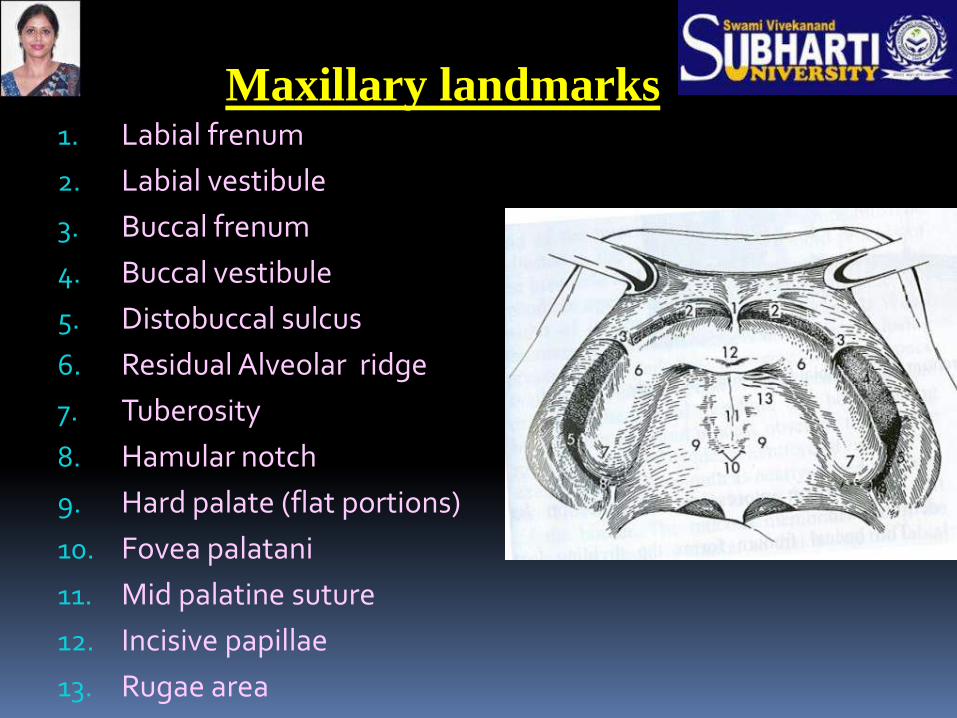
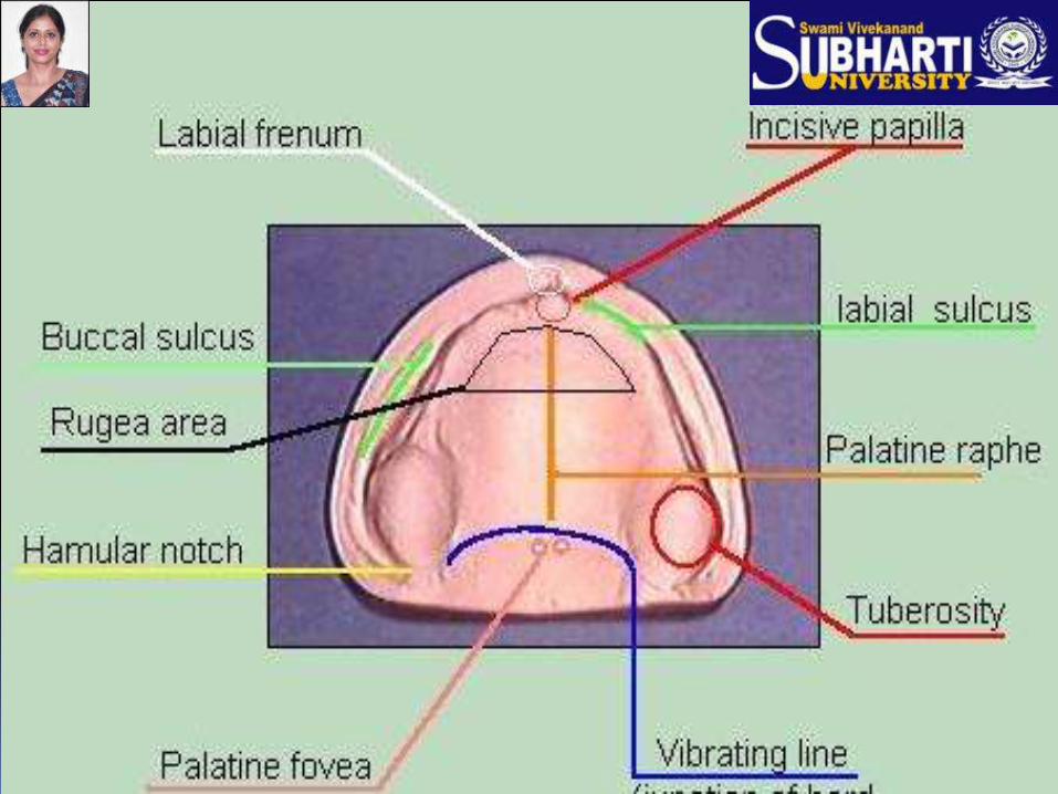
Maxillary landmarks 1. Labial frenum
2. Labial vestibule
3. Buccal frenum
4. Buccal vestibule
5. Distobuccal sulcus
6. Residual Alveolar ridge
7. Tuberosity
8. Hamular notch
9. Hard palate (flat portions)
10. Fovea palatani
11. Mid palatine suture
12. Incisive papillae
13. Rugae area

Foundation surface of maxillary
denture primarily consists of:
Stress bearing or supporting area
Relief areas
Peripheral or limiting area
MAXILLARY DENTURE SUPPORTING STRUCTURES INCLUDE
•RESIDUAL ALVEOLAR RIDGE •RUGAE •BONE OF THE BASAL SEAT: -INCISIVE PAPILLA -ZYGOMATIC PROCESS -MAXILLARY TUBEROSITY -SHARP SPINY PROCESSES - TORUS PALATINUS
ANATOMY OF SUPPORTING STUCTURES
Foundation of maxillary denture is made up of:
Bone of hard palate
Residual alveolar ridge
The denture base actually rests on mucous membrane
which serves as a cushion between denture base and
the supporting bone

HARD PALATE
The palatine processes of maxilla & the palatine bone forms the foundation of hard palate & provides considerable support for dentures & also support the soft tissue which increases the surface area for the basal seat.

PALATINE PROCESS
Arise as broad horizontal plates from the body of
the maxillae.
The two horizontal plates are united in the midline by the midpalatal suture.
The horizontal palatine processes of the maxillary bones appear to resist resorption .
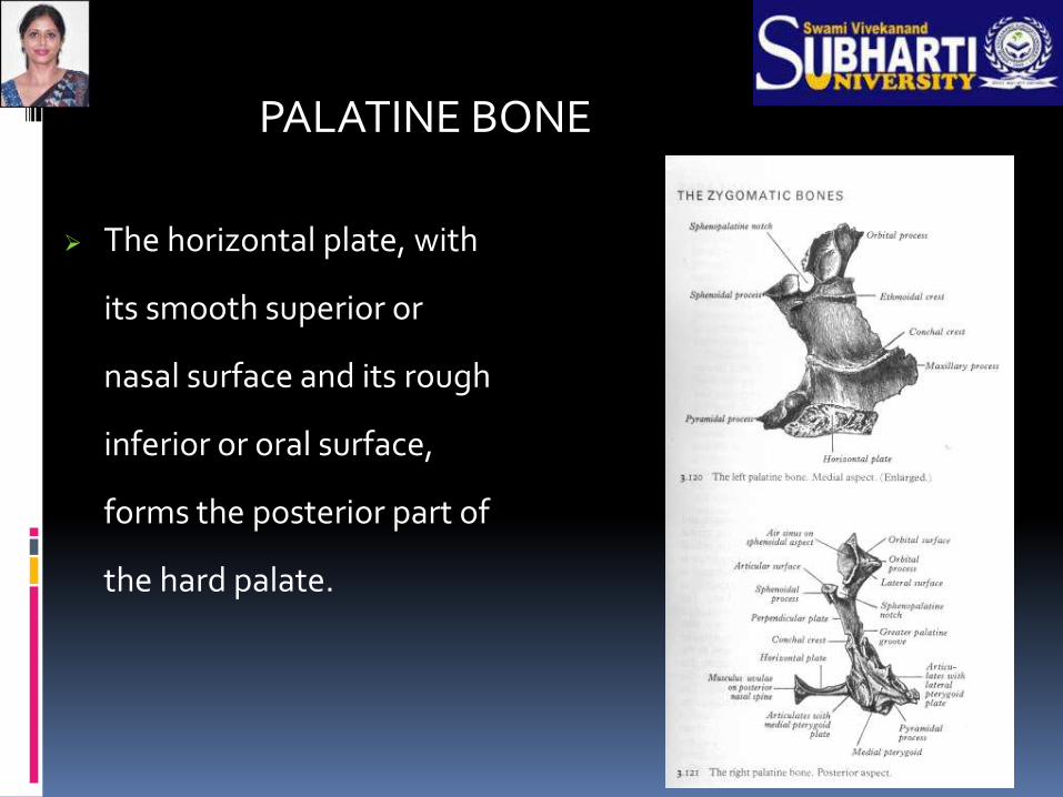
The horizontal plate, with
its smooth superior or
nasal surface and its rough
inferior or oral surface,
forms the posterior part of
the hard palate.
PALATINE BONE

The horizontal plates of the palatine bones articulate with the posterior rough border of the horizontal palatal processes of the maxillae .
Posterior border of the horizontal plates of the palatine bones unite in the midline to form the sharp posterior nasal spine.
Posterior margins of the hard palate serve as the anterior attachment for the aponeurosis of the soft palate.

Hard Palate
Median Palatine Raphe (midline palatine suture)
A bony midline structure
May require relief when covered by a denture

So as the ridge resorbs, pressure increases over
the palate and when it becomes prominent in
the mid palatal region it acts as a fulcrum
point around which the denture will rotate
So, no stress to be placed on this region
otherwise soreness , pain can occur and the
denture tends to rock over the center of palate
with this suture acting as fulcrum
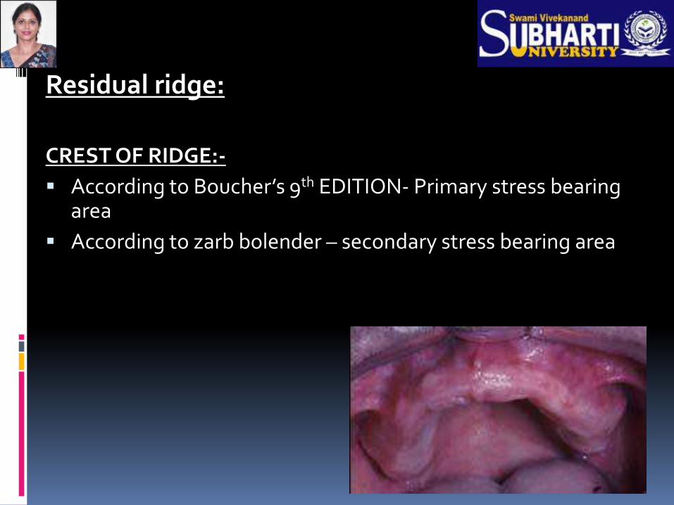
Residual ridge:
CREST OF RIDGE:-
According to Boucher’s 9th EDITION- Primary stress bearing area
According to zarb bolender – secondary stress bearing area

CREST OF THE RESIDUAL RIDGE
covered with thick fibrous connective tissue
Its mucous membrane is firmly attached to the
periosteum of bone by connective tissue
Stratified squamous epithelium is highly keratinized
Submucosa contains dense collagenous fibers that are contiguous with the lamina propria
Composed of Compact bone

• Since the crest of the ridge is subjected to resorption,there is lack of smooth cortical bone,so it is considered to be secondary supporting area

Rugae – Irregularly shaped rolls in the anterior part of the palate.
This area resists anterior displacement of the denture and is a secondary Stress bearing area.(palate is at an angle to the residual ridge).
They serve no function.
While making impressions they should not be distorted,since rebounding tissue tends to unseat the denture.

INCISIVE PAPILLA
The incisive fossa is located in the midline of the palate,posterior to the maxillary central incisors(in edentulous mouth,slightly to the palatal side of the anterior palatal alveolar plate.
Nasopalatine nerves & vessels exit to the palate at right angles to the margins of this bony fossa.

In case of increased resorption of residual alveolar ridge ,the incisive foramen is nearer crest of ridge .
The location of the incisive papilla(covering incisive foramen) in relation to crest of ridge gives an indication as to the amount of resorption of the residual ridge & thus is an aid in determining vertical dimension & the proper position of the teeth.
CLINICAL SIGNIFICANCE
Considered as a relief area
If not relieved, patient besides complaining of pain may also complain of a burning sensation in anterior palate

Hence,even though the fossa is covered with a protective pad of fibrous conn. tissue called the incisive papilla,denture base should be relieved over this area.
Incisive papilla can be used as a guide in arranging anterior teeth.
Ant. Maxillary central incisors should be placed anterior to the incisive papilla regardless of the relation of the papilla to the existing residual ridge.

BONE OF THE BASAL SEAT
Important components of the bone of the basal seat for the maxillary denture are:
Incisive foramen
Zygomatic process
Maxillary tuberosity
Sharp spiny processes
Torus palatinus

Incisive foramen location with resorption. . . relief area. determining vertical dimension.
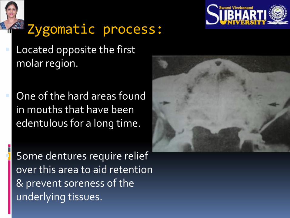
Zygomatic process:
Located opposite the first molar region.
One of the hard areas found in mouths that have been edentulous for a long time.
Some dentures require relief over this area to aid retention & prevent soreness of the underlying tissues.

Maxillary Tuberosity -
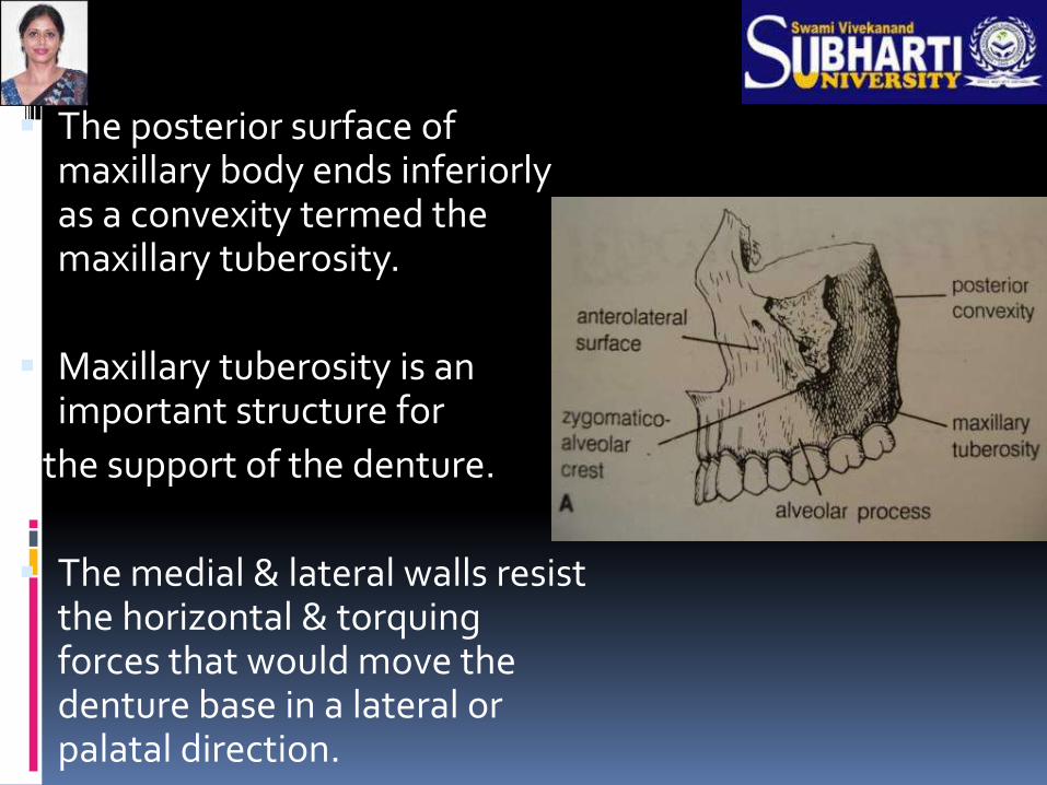
The posterior surface of maxillary body ends inferiorly as a convexity termed the maxillary tuberosity.
Maxillary tuberosity is an important structure for
the support of the denture.
The medial & lateral walls resist the horizontal & torquing forces that would move the denture base in a lateral or palatal direction.

The posterior wall will resist movement in an anterior direction.
To take advantage of this resistance to movement ,the maxillary denture base should cover the tuberosities & fill the hamular notches.

Overextension at the hamular notches will not be tolerated because of pressure on the pterygoid hamulus & interference with the pterygomandibular raphe .
When mouth is opened wide ,raphe is pulled forward.if denture is too long here,mucous memb. covering the raphe will be injured.
Underextension hampers the stability & retention of the denture.(when distal end fits into the notch ,it prevents anteroposterior movement of the denture).
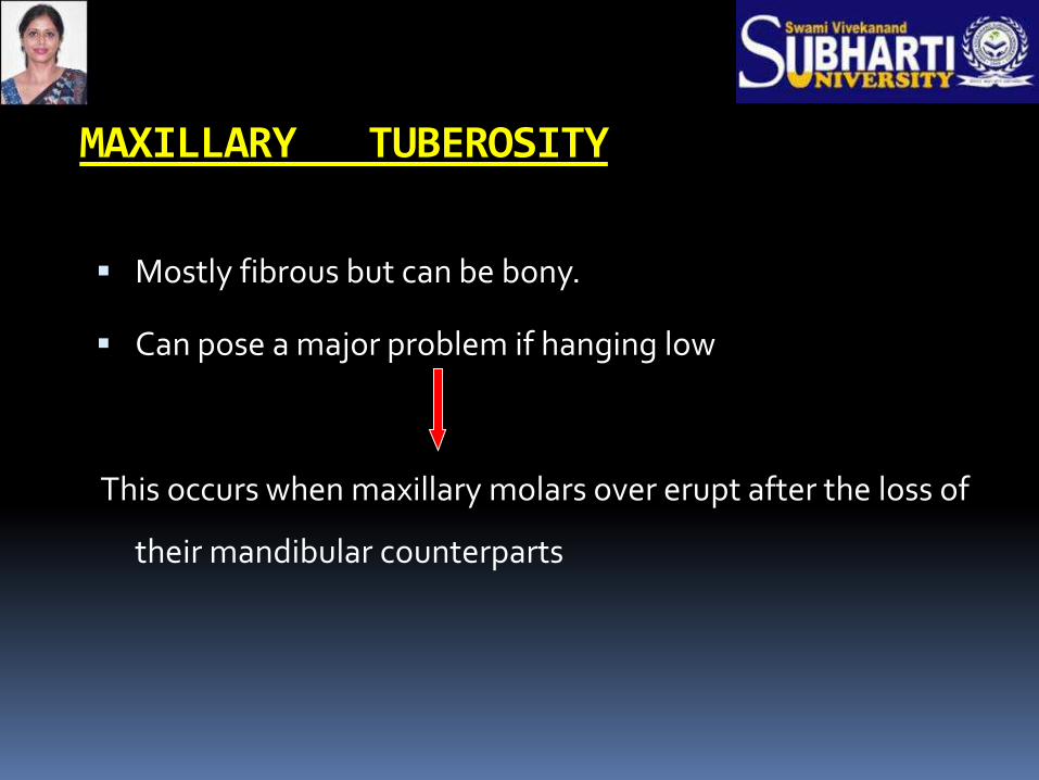
MAXILLARY TUBEROSITY
Mostly fibrous but can be bony.
Can pose a major problem if hanging low
This occurs when maxillary molars over erupt after the loss of
their mandibular counterparts

Fibrous tuberosity
Bony overhanging tuberosity
When maxillary prosthesis
against mandibular natural
teeth
Seen after extraction of maxillary molars that had supra erupted due to loss of antagonists
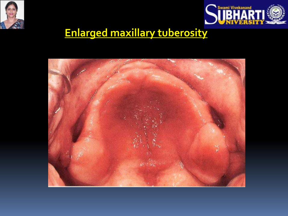
Enlarged maxillary tuberosity

SHARP SPINY SPICULES
Frequently encountered on maxillary or
palatine bones
Usually cause no problem except in
individuals with considerable
resorption(irritate soft tissues left
between them & denture base).
When pronounced spicules are present
at the ridge, definitive treatment is
ALVEOLOPLASTY
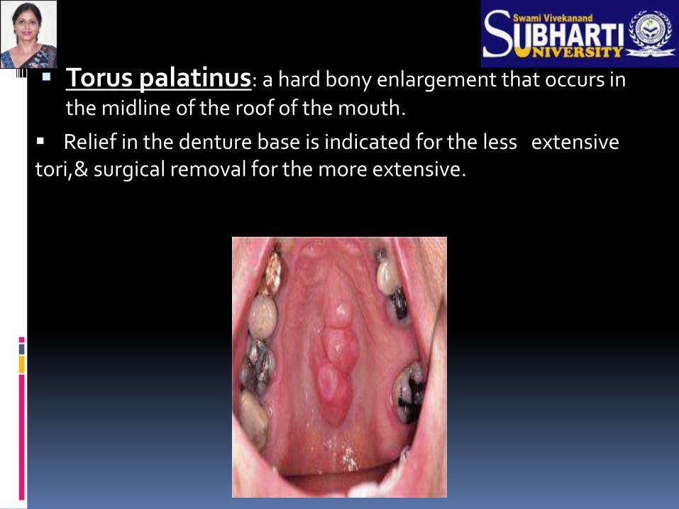
Torus palatinus: a hard bony enlargement that occurs in
the midline of the roof of the mouth.
Relief in the denture base is indicated for the less extensive tori,& surgical removal for the more extensive.

MICROSCOPIC ANATOMY OF SUPPORTING STRUCTURES

RESIDUAL RIDGE ( MAXILLA )–
EPITHELIUM – stratified squamous,thick , keratinized
SUBMUCOSA – dense collagenous fibres & by this the mucous membrane is firmly attached to the periosteum of bone, devoid of fat or glandular cells
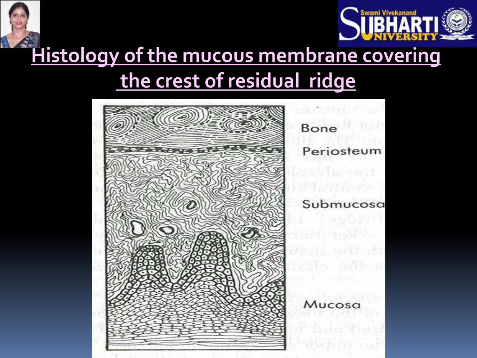
Histology of the mucous membrane covering the crest of residual ridge

SLOPE OF RIDGE:
Loosely attached mucous membrane.
Epithelium - non keratinized.
Submucosa – has loose connective tissue so cannot withstand the forces of mastication as well as crest of the ridge.
Less stress is placed on the movable tissue of the slope of the ridge during the making of final impression.

HARD PALATE
EPITHELIUM – thick , keratinized
LAMINA PROPRIA – long papillae , thick dense collagenous tissue
SUBMUCOSA – very thin , anterolaterally the submucosa contains adipose tissue and posterolaterally contains glandular tissue
which is covered by the denture to aid in retention.(but it should not be expected to provide support for the denture)

Histology of the mucosa in the anterolateral and posterolateral part of the hard palate
INCISIVE PAPILLA –
Thin stratified squamous epithelium,keratinized.
Submucosa contains nasopalatine vessels and nerves
So forms the primary Relieving area
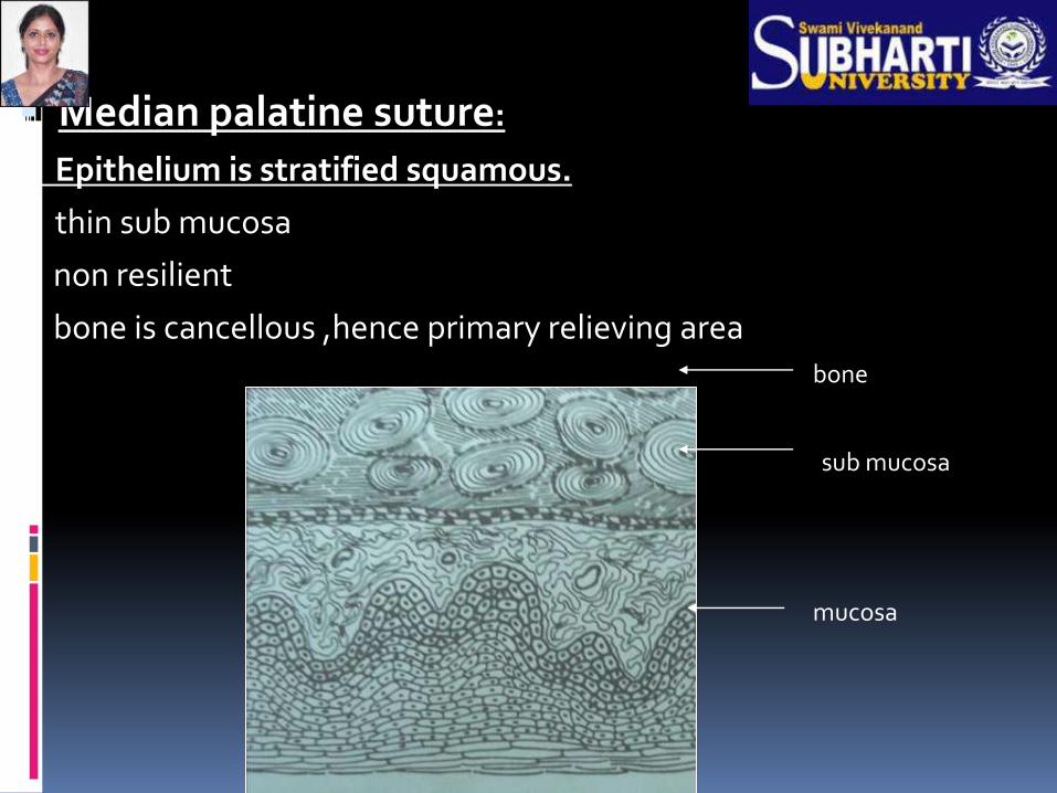
Median palatine suture:
Epithelium is stratified squamous.
thin sub mucosa
non resilient
bone is cancellous ,hence primary relieving area bone
sub mucosa
mucosa

DENTURE LIMITING STRUCTURES INCLUDE
LABIAL FRENUM
BUCCAL FRENUM
LABIAL VESTIBULE
BUCCAL VESTIBULE
PTERYGOMAXILLARY NOTCH
FOVEA PALATINAE
VIBRATING LINES
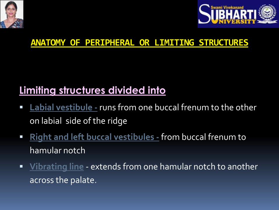
ANATOMY OF PERIPHERAL OR LIMITING STRUCTURES
Limiting structures divided into
Labial vestibule - runs from one buccal frenum to the other
on labial side of the ridge
Right and left buccal vestibules - from buccal frenum to
hamular notch
Vibrating line - extends from one hamular notch to another
across the palate.
LABIAL FRENUM
Fan or ‘V’ shaped fold of mucosa in the midline
Has No muscle itself
Starts superiorly from the inner aspect of the lip in a fan
shaped manner and converges near its attachment to
the labial side of the ridge
It has no action of its own.

It is a relief area and recorded as labial notch in the
impression failure to which can lead to irritation
When the attachment is close to the crest of the
ridge ,frenectomy may be indicated
Clinical implications
LABIAL VESTIBULE
Divided into left and right labial vestibule by labial frenum
The main muscle of lip that forms the outer surface of labial
vestibule is Orbicularis Oris
( it rests upon the labial flange & teeth of denture)
The fibers of this muscle run horizontally through the lip
and anastomose with fibers of buccinator

As the fibers run horizontally this muscle
has only an indirect effect on extent of impression
and on denture
BUCCAL FRENUM
Fold of mucous membrane found on the buccal side .
It may be seen in
1. Single fold
2. Double fold
3. Broad and fan shaped
It is related to 3 muscles
1. Canninus {LAO} is attached below
the frenum and affect its position
2. Orbicularis oris pulls the frenum forward
Buccinator pulls it backward
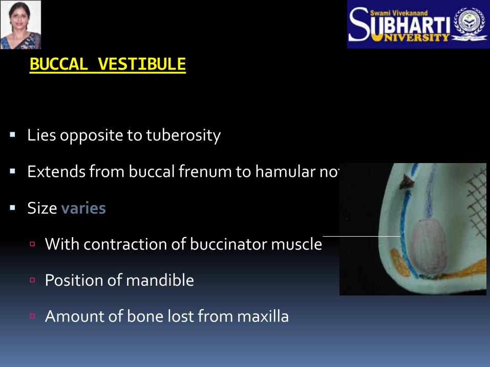
BUCCAL VESTIBULE
Lies opposite to tuberosity
Extends from buccal frenum to hamular notch
Size varies
With contraction of buccinator muscle
Position of mandible
Amount of bone lost from maxilla
Distal end of buccal flange of denture has to be adjusted,
according to ramus and coronoid process of mandible and
masseter muscle as they function during impression making
procedure (to make the denture stable during various
mandibular movements)
When masseter contracts under heavy closing
pressure,it also reduces the size of space available
for the distal end of the buccal flange.
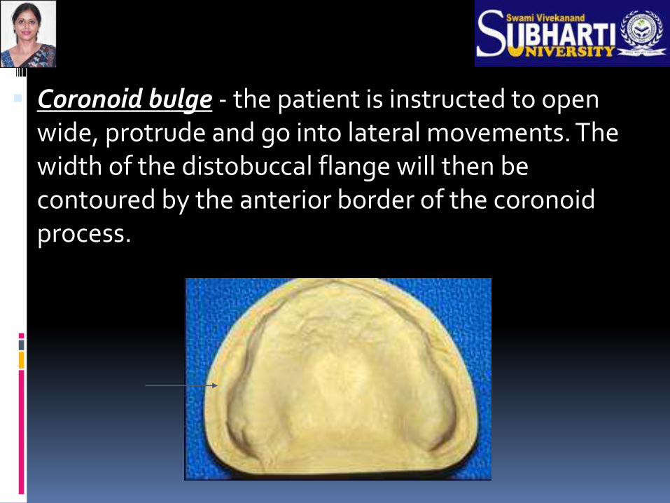
Coronoid bulge - the patient is instructed to open wide, protrude and go into lateral movements. The width of the distobuccal flange will then be contoured by the anterior border of the coronoid process.
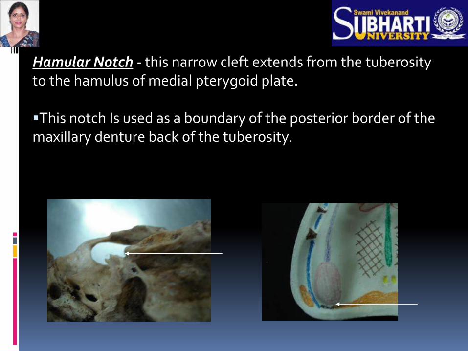
Hamular Notch - this narrow cleft extends from the tuberosity to the hamulus of medial pterygoid plate. This notch Is used as a boundary of the posterior border of the maxillary denture back of the tuberosity.

The posterior palatal seal is placed through the center of the deep part of the hamular notch,since no muscle or ligament is present at a level to prevent the placement of extra pressure.

These are 2 small indentations near the midline
of the posterior palate that are formed by
joining together of several mucous gland ducts.
The opening are close to the vibrating line and
are always in the soft tissue, which make them an
ideal guide for the location of posterior border
of the denture
FOVEA PALATINE

ANTERIOR AND POSTERIOR VIBRATING LINES
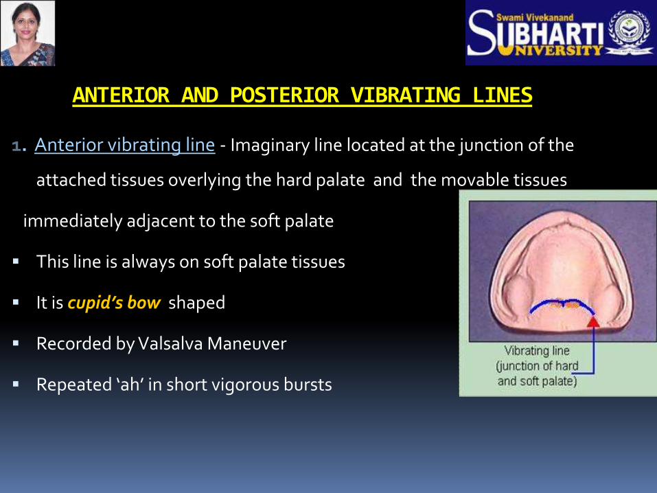
1. Anterior vibrating line - Imaginary line located at the junction of the
attached tissues overlying the hard palate and the movable tissues
immediately adjacent to the soft palate
This line is always on soft palate tissues
It is cupid’s bow shaped
Recorded by Valsalva Maneuver
Repeated ‘ah’ in short vigorous bursts
POSTERIOR VIBRATING LINE
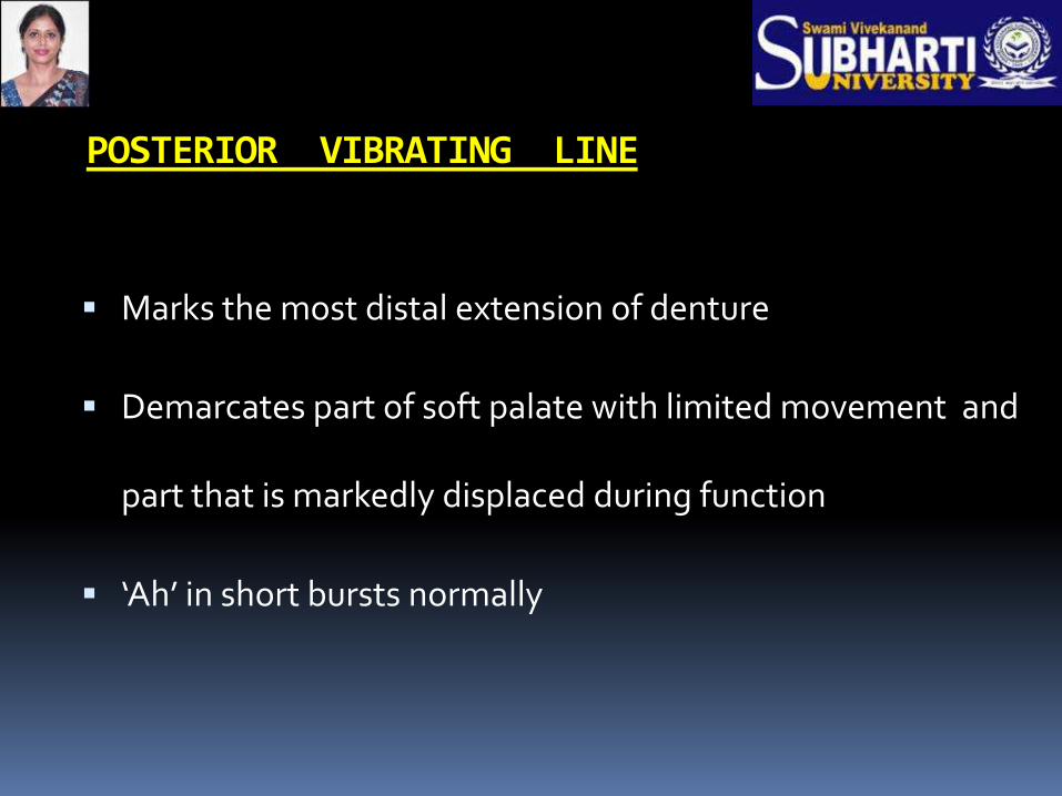
Marks the most distal extension of denture
Demarcates part of soft palate with limited movement and
part that is markedly displaced during function
‘Ah’ in short bursts normally

VIBRATING LINE:
The direction of the vibrating line usually varies according to the shape of the palate.
The higher the vault ,the more abrupt & forward the vibrating line.
In a mouth with a flat vault,the vibrating line is usually farther posterior or has a gradual curvature,affording a broader pps area.


Limiting structures: (microscopic anatomy)
Vestibular spaces:
Thin epithelium,nonkeratinized.
Thick sub mucosa containing large
amounts of areolar tissue & elastic fibres.
The nature of the submucosa makes this tissue easily movable,so the labial or buccal flanges of upper impression can easily be overextended or underextended.

HAMULAR NOTCHES Submucosa is thick made up
of loose areolar tissue.
Additional pressure can be placed
on this tissue at the centre of the notch to complete the posterior palatal seal.
No space provided in the final impression tray in this region.
Thus the loose areolar tissue in submucosal can be displaced without trauma by the complete denture to improve PPS.
AREOLARCONN TISSUE

SUBMUCOSA IN REGION OF VIBRATING LINES
Contains glandular tissue similar to posterolateral
part of hard palate
As the soft palate does not rest directly on
bone,these tissues can be repositioned in a controlled
manner in the impression procedure to improve
posterior palatal seal

Ideal maxillary ridge:
Abundant keratinized attached tissue
Square arch
Palate U-shaped in cross-section
Moderate palatal vault
Absence of undercuts
High frenum attachments
Well-defined hamular notches

CONCLUSION
Scientific knowledge of denture supporting and influencing structures helps in accurate recording of these tissues , & meticulous fabrication of
satisfactory dentures .