Embed Size (px)
Citation preview
&ANATOMY PHYSIOLOGYLab Manual
Customized for Morton University
John Smith
231
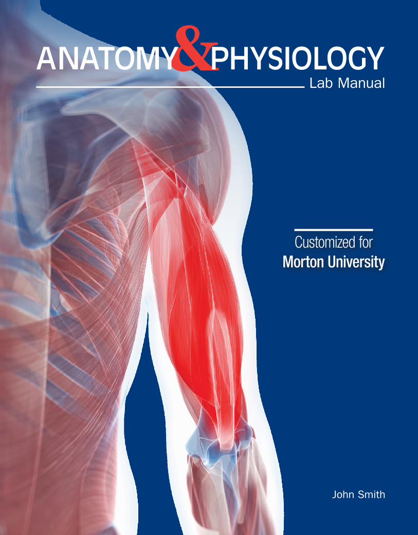
Senses and Sheep Eyeball Dissection
LAB
99Learning Outcomes
◗◗ Students will be able to measure bone length and height using the metric system.
◗◗ Students will use anatomical position terminology correctly.
◗◗ Students will correctly identify the major organ systems in the human body.
IntroductionIn this lab you will examine the senses—sight, hearing, taste, touch, and smell. You will be able to determine if your sensory pathways are in working order. You will dissect a sheep eye and learn the major anatomical features of the eye.
Pre-lab Exercise: SightEye AnatomyExtrinsic Muscles of the Eye
1. Label Figure 9-1 with the following:
extrinsic muscles inferior oblique inferior rectus lateral rectus medial rectus superior oblique superior rectus
trochlea (a piece of cartilage that acts as a pulley for the superior oblique)
FIGURE 9-1 Extrinsic muscles of the eye.
232 Anatomy and Physiology Lab Manual School of Morton
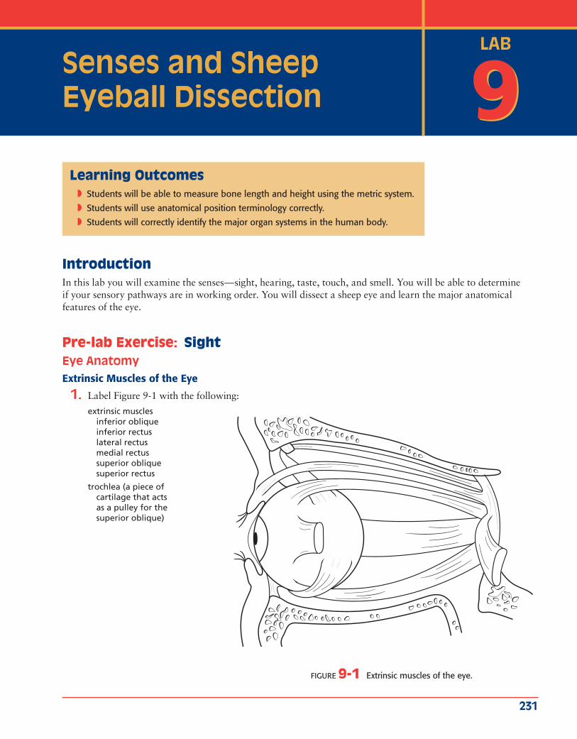
Exercise: Anatomy of the EyeSensation is broadly defined as the detection of changes in the inter-nal and external environments. Sensation may be conscious or sub-conscious, depending on the destination of the sensory information. For example, certain blood vessels have receptors that detect blood pressure. This information is taken to the brainstem, which makes changes as necessary to ensure blood pressure remains relatively con-stant. This information never makes it to the cerebral cortex, so you are not consciously aware of it.
The following exercises ask you to examine the anatomy and physiology of the special senses: vision, hearing and equilibrium, taste, and smell. You also will examine the general senses in this unit, which include touch, pain, and temperature.
The eye is a complex organ consisting of three components:
1. External structures, such as the eyelids (Figure 9.3),
2. Accessory structures, such as the lacrimal (LAK-rim-ul) gland (Figure 9.3), and
3. The eyeball (Figure 9.4).
Many of the external and accessory structures of the eye protect the delicate eyeball. Anteriorly, the eye is covered by the accessory structures known as the palpebrae (pal-PEE-bray), or eye-
lids. The internal surface of the eyelids and much of the anterior eyeball are covered with a thin mucous membrane called the conjunctiva (kahn-junk-TEE-vuh). Another accessory structure of the eye is the lacrimal apparatus, which produces and drains tears. The lacrimal apparatus consists of the lacrimal gland, located in the superolateral orbit, and the ducts that drain the tears it produces. The other major accessory structures are the extraocular muscles, which move the eyeball.
Lacrimal gland
Lacrimal canals
Lacrimal caruncle
Lacrimal sac
Nasolacrimal duct
Lateral canthus
Medial canthus
Superior palpebra
Inferior palpebra
FIGURE 9.3 External and accessory structures of the eye
◗◗ Eye models
◗◗ Preserved eyeballs
◗◗ Dissection equipment
◗◗ Dissection trays
◗◗ Snellen vision chart
◗◗ Dark green or blue paper
◗◗ Ruler
Materials
LAB 9 General and Special Senses 233
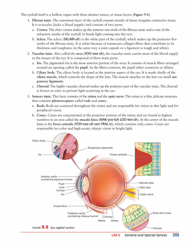
The eyeball itself is a hollow organ with three distinct tunics, or tissue layers (Figure 9.4):
1. Fibrous tunic. The outermost layer of the eyeball consists mostly of dense irregular connective tissue. It is avascular (lacks a blood supply) and consists of two parts:a. Cornea. The clear cornea makes up the anterior one-sixth of the fibrous tunic and is one of the
refractory media of the eyeball (it bends light coming into the eye). b. Sclera. The sclera (SKLAIR-uh) is the white part of the eyeball, which makes up the posterior five-
sixths of the fibrous tunic. It is white because of numerous collagen fibers that contribute to its thickness and toughness (in the same way a joint capsule or a ligament is tough and white).
2. Vascular tunic. Also called the uvea (YOO-vee-uh), the vascular tunic carries most of the blood supply to the tissues of the eye. It is composed of three main parts:a. Iris. The pigmented iris is the most anterior portion of the uvea. It consists of muscle fibers arranged
around an opening called the pupil. As the fibers contract, the pupil either constricts or dilates.b. Ciliary body. The ciliary body is located at the anterior aspect of the eye. It is made chiefly of the
ciliary muscle, which controls the shape of the lens. The muscle attaches to the lens via small sus-pensory ligaments.
c. Choroid. The highly vascular choroid makes up the posterior part of the vascular tunic. The choroid is brown in color to prevent light scattering in the eye.
3. Sensory tunic. This layer consists of the retina and the optic nerve. The retina is a thin, delicate structure that contains photoreceptors called rods and cones. a. Rods. Rods are scattered throughout the retina and are responsible for vision in dim light and for
peripheral vision. b. Cones. Cones are concentrated at the posterior portion of the retina and are found in highest
numbers in an area called the macula lutea (MAK-yoo-luh LOO-tee-uh). At the center of the macula lutea is the fovea centralis (FOH-vee-uh sen-TRAL-iz), which contains only cones. Cones are responsible for color and high-acuity (sharp) vision in bright light.
Anterior cavity(containing aqueous humor)
Optic nerve
Conjunctiva
Optic disc
Rods and cones
Choroid
Posterior cavity(containing vitreous humor)
Retina
Choroid
Sclera
Pupil
Ciliary body
Suspensory ligaments
Fovea centralisIris
LensCornea
Iris
Macula lutea
FIGURE 9.4 Eye sagittal section
234 Anatomy and Physiology Lab Manual School of Morton
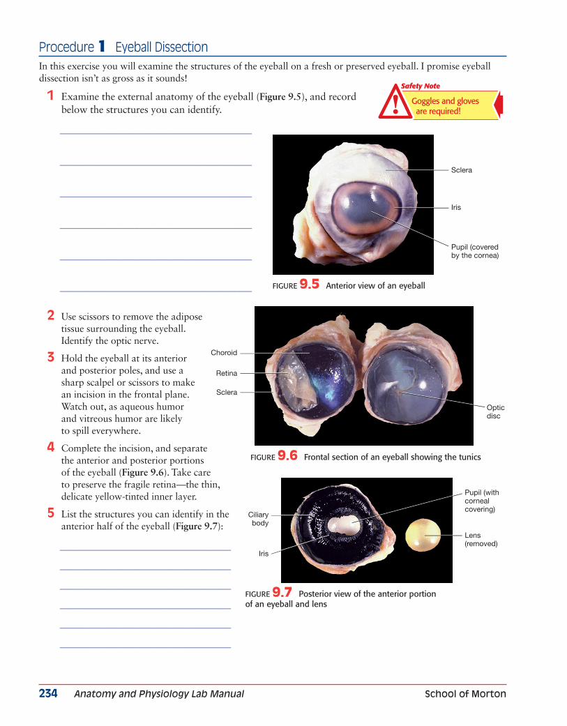
Procedure 1 Eyeball DissectionIn this exercise you will examine the structures of the eyeball on a fresh or preserved eyeball. I promise eyeball dissection isn’t as gross as it sounds!
1 Examine the external anatomy of the eyeball (Figure 9.5), and record below the structures you can identify.
______________________________________________
______________________________________________
______________________________________________
______________________________________________
______________________________________________
______________________________________________
2 Use scissors to remove the adipose tissue surrounding the eyeball. Identify the optic nerve.
3 Hold the eyeball at its anterior and posterior poles, and use a sharp scalpel or scissors to make an incision in the frontal plane. Watch out, as aqueous humor and vitreous humor are likely to spill everywhere.
4 Complete the incision, and separate the anterior and posterior portions of the eyeball (Figure 9.6). Take care to preserve the fragile retina—the thin, delicate yellow-tinted inner layer.
5 List the structures you can identify in the anterior half of the eyeball (Figure 9.7):
_________________________________________
_________________________________________
_________________________________________
_________________________________________
_________________________________________
_________________________________________
Pupil (coveredby the cornea)
Sclera
Iris
Opticdisc
Choroid
Retina
Sclera
Ciliarybody
Iris
Pupil (with cornealcovering)
Lens(removed)
FIGURE 9.5 Anterior view of an eyeball
FIGURE 9.6 Frontal section of an eyeball showing the tunics
FIGURE 9.7 Posterior view of the anterior portion of an eyeball and lens
Goggles and gloves are required!
Safety Note
LAB 9 General and Special Senses 235
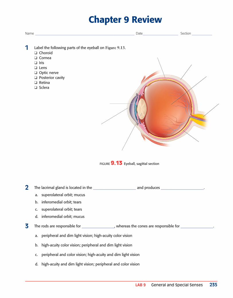
Chapter 9 ReviewName ___________________________________________________________ Date _____________________ Section ____________
1 Label the following parts of the eyeball on Figure 9.13. ❑ Choroid ❑ Cornea ❑ Iris ❑ Lens ❑ Optic nerve ❑ Posterior cavity ❑ Retina ❑ Sclera
2 The lacrimal gland is located in the _________________________ and produces _________________________.
a. superolateral orbit; mucus
b. inferomedial orbit; tears
c. superolateral orbit; tears
d. inferomedial orbit; mucus
3 The rods are responsible for ___________________, whereas the cones are responsible for ___________________.
a. peripheral and dim light vision; high-acuity color vision
b. high-acuity color vision; peripheral and dim light vision
c. peripheral and color vision; high-acuity and dim light vision
d. high-acuity and dim light vision; peripheral and color vision
FIGURE 9.13 Eyeball, sagittal section

236 Anatomy and Physiology Lab Manual School of Morton