Embed Size (px)
Citation preview
Bio& 241 Unit 1 / Lecture 5
Glandular Epithelium
• Gland:– a single cell or a mass of epithelial cells adapted
for secretion– derived from epithelial cells that sank below the
surface during development
• Endocrine glands are ductless. They secrete their products into ducts that empty at the surface of covering and lining epithelium or directly onto a free surface .
Glandular Epithelium
• Exocrine glands– cells that secrete---sweat, ear wax, saliva, digestive
enzymes onto free surface of epithelial layer– connected to the surface by tubes (ducts)– unicellular glands or multicellular glands
• Endocrine glands– secrete hormones into the bloodstream– hormones help maintain homeostasis
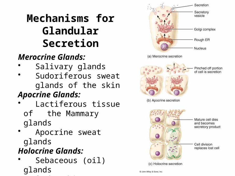
Mechanisms for Glandular Secretion
Merocrine Glands:• Salivary glands• Sudoriferous sweat
glands of the skinApocrine Glands:• Lactiferous tissue of
the Mammary glands• Apocrine sweat glandsHolocrine Glands:• Sebaceous (oil) glands
of the skin
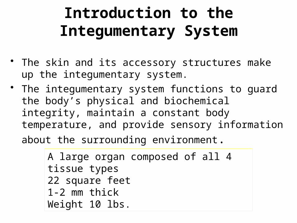
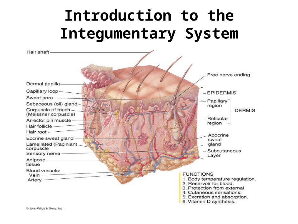
Introduction to the Integumentary System
• The skin and its accessory structures make up the integumentary system.
• The integumentary system functions to guard the body’s physical and biochemical integrity, maintain a constant body temperature, and provide sensory information about the
surrounding environment.
A large organ composed of all 4 tissue types22 square feet 1-2 mm thickWeight 10 lbs.
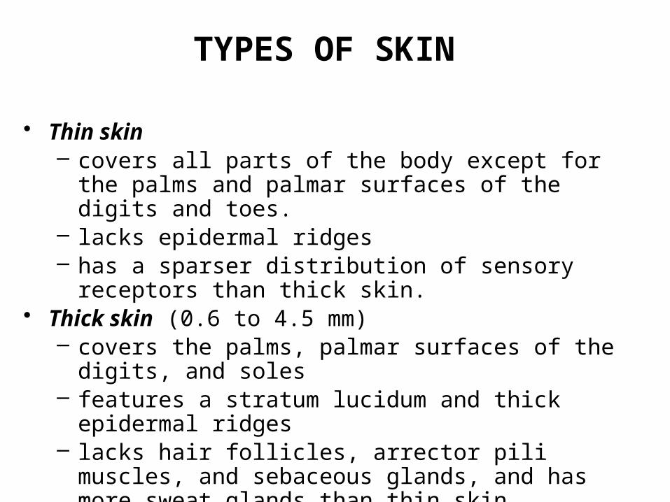
TYPES OF SKIN
• Thin skin – covers all parts of the body except for the palms and
palmar surfaces of the digits and toes.– lacks epidermal ridges– has a sparser distribution of sensory receptors than thick
skin.• Thick skin (0.6 to 4.5 mm)
– covers the palms, palmar surfaces of the digits, and soles– features a stratum lucidum and thick epidermal ridges– lacks hair follicles, arrector pili muscles, and sebaceous
glands, and has more sweat glands than thin skin.
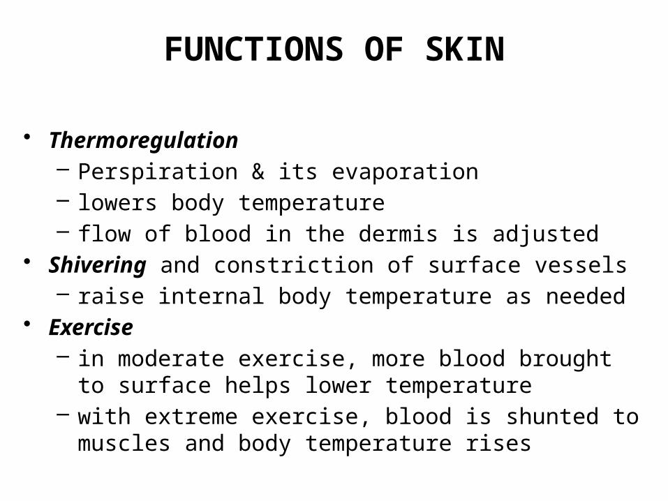
FUNCTIONS OF SKIN
• Thermoregulation– Perspiration & its evaporation – lowers body temperature– flow of blood in the dermis is adjusted
• Shivering and constriction of surface vessels– raise internal body temperature as needed
• Exercise– in moderate exercise, more blood brought to surface
helps lower temperature– with extreme exercise, blood is shunted to muscles and
body temperature rises
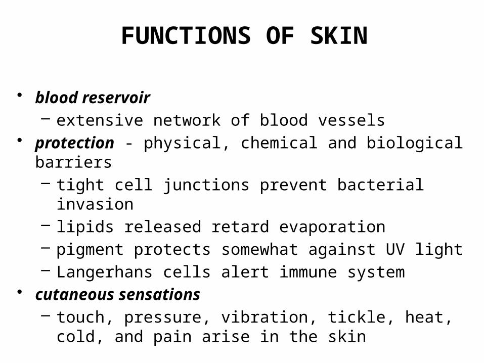
FUNCTIONS OF SKIN
• blood reservoir – extensive network of blood vessels
• protection - physical, chemical and biological barriers– tight cell junctions prevent bacterial invasion– lipids released retard evaporation– pigment protects somewhat against UV light– Langerhans cells alert immune system
• cutaneous sensations – touch, pressure, vibration, tickle, heat, cold, and pain
arise in the skin
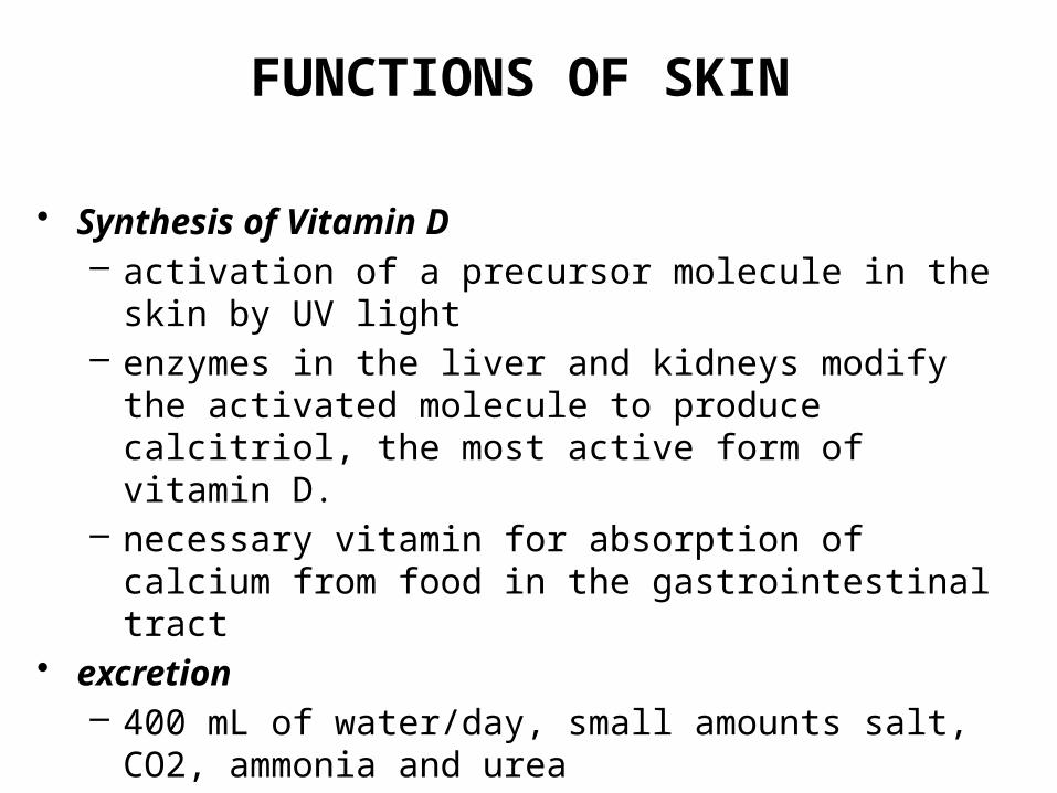
FUNCTIONS OF SKIN
• Synthesis of Vitamin D – activation of a precursor molecule in the skin by UV light– enzymes in the liver and kidneys modify the activated
molecule to produce calcitriol, the most active form of vitamin D.
– necessary vitamin for absorption of calcium from food in the gastrointestinal tract
• excretion– 400 mL of water/day, small amounts salt, CO2, ammonia
and urea
Introduction to the Integumentary System
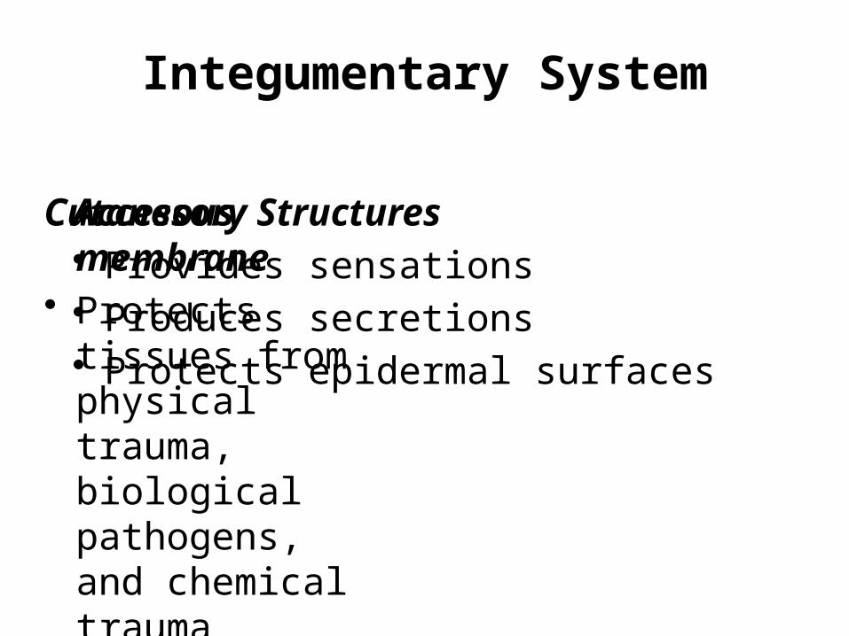
Integumentary System
Cutaneous membrane
• Protects tissues from physical trauma, biological pathogens, and chemical trauma
• Provides sensations
Accessory Structures• Provides sensations• Produces secretions• Protects epidermal surfaces
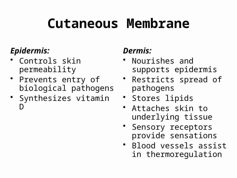
Cutaneous Membrane
Epidermis:• Controls skin permeability• Prevents entry of biological
pathogens• Synthesizes vitamin D
Dermis:• Nourishes and supports
epidermis• Restricts spread of
pathogens• Stores lipids• Attaches skin to underlying
tissue• Sensory receptors provide
sensations• Blood vessels assist in
thermoregulation
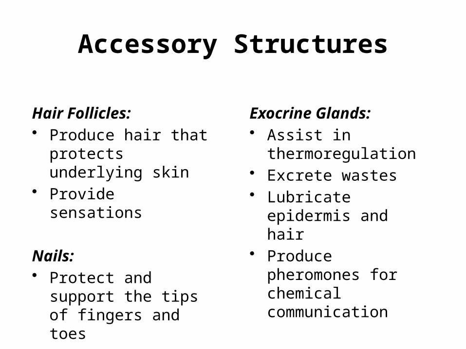
Accessory Structures
Hair Follicles:• Produce hair that
protects underlying skin• Provide sensations
Nails:• Protect and support the
tips of fingers and toes
Exocrine Glands:• Assist in
thermoregulation• Excrete wastes• Lubricate epidermis and
hair• Produce pheromones
for chemical communication
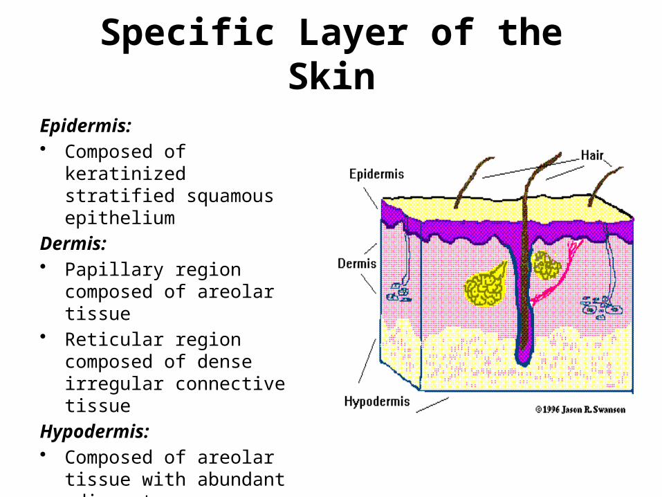
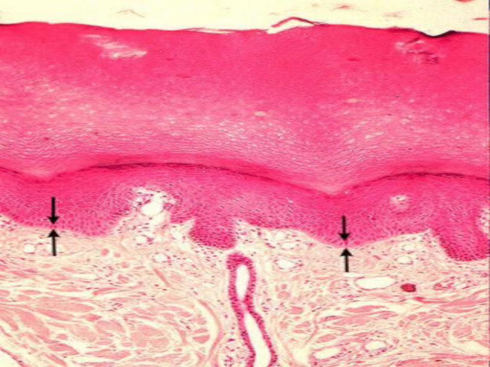
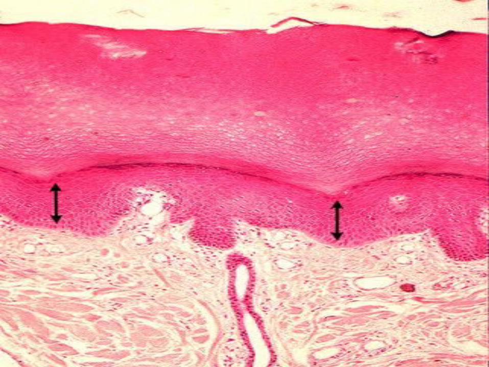
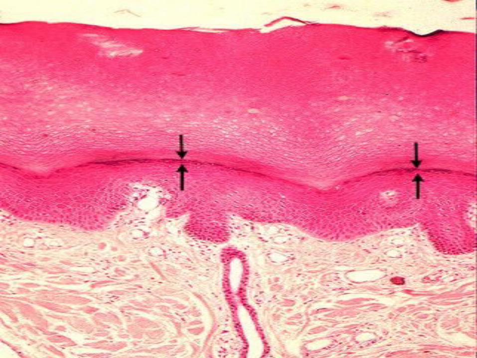
Specific Layer of the Skin
Epidermis:• Composed of keratinized
stratified squamous epithelium
Dermis:• Papillary region composed
of areolar tissue• Reticular region composed
of dense irregular connective tissue
Hypodermis:• Composed of areolar tissue
with abundant adipocytes
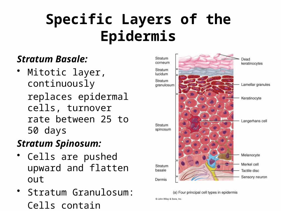
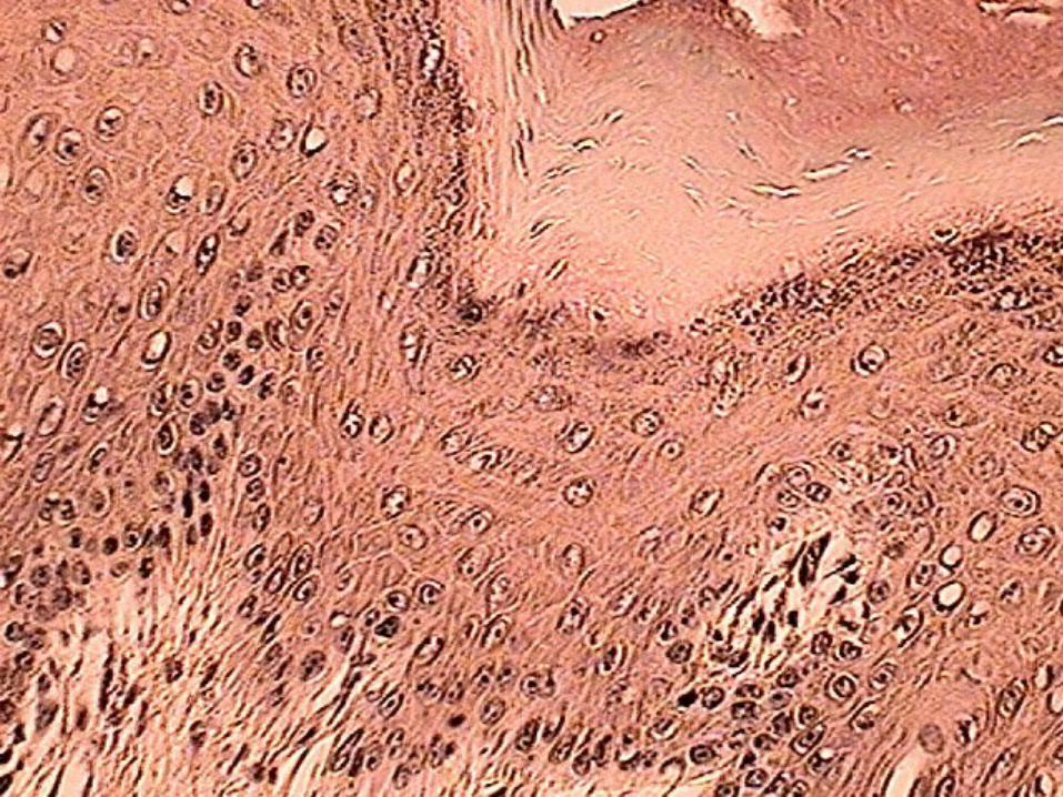
Specific Layers of the Epidermis
Stratum Basale:• Mitotic layer, continuously
replaces epidermal cells, turnover rate between 25 to 50 days
Stratum Spinosum:• Cells are pushed upward
and flatten out• Stratum Granulosum:
Cells contain granules of Keratin
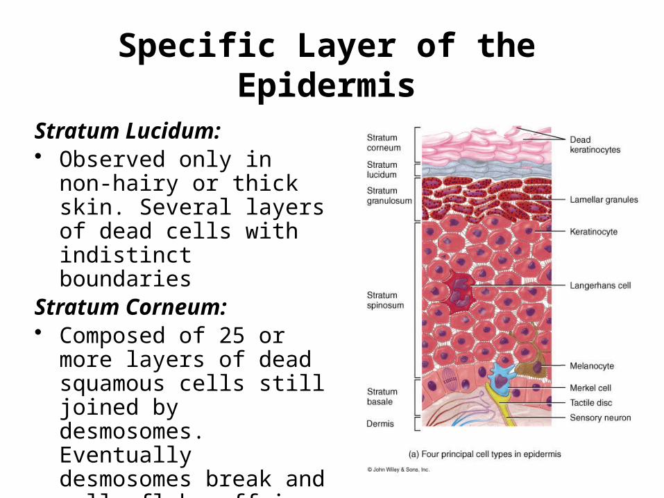
Specific Layer of the Epidermis
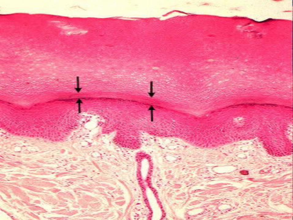
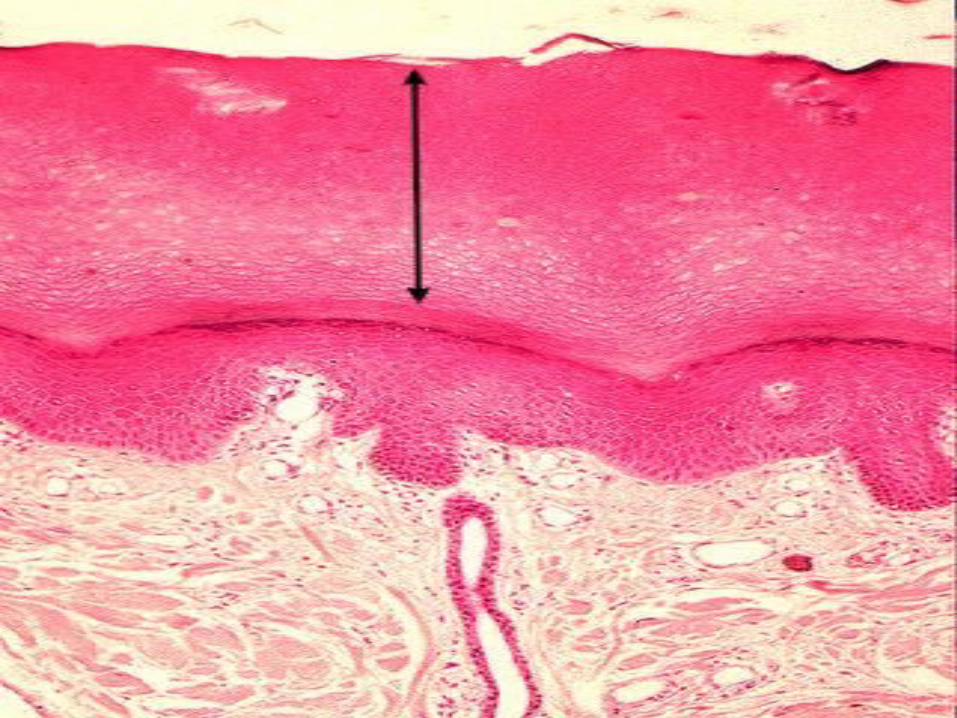
Stratum Lucidum:• Observed only in non-hairy
or thick skin. Several layers of dead cells with indistinct boundaries
Stratum Corneum:• Composed of 25 or more
layers of dead squamous cells still joined by desmosomes. Eventually desmosomes break and cells flake off in a process called desquamation

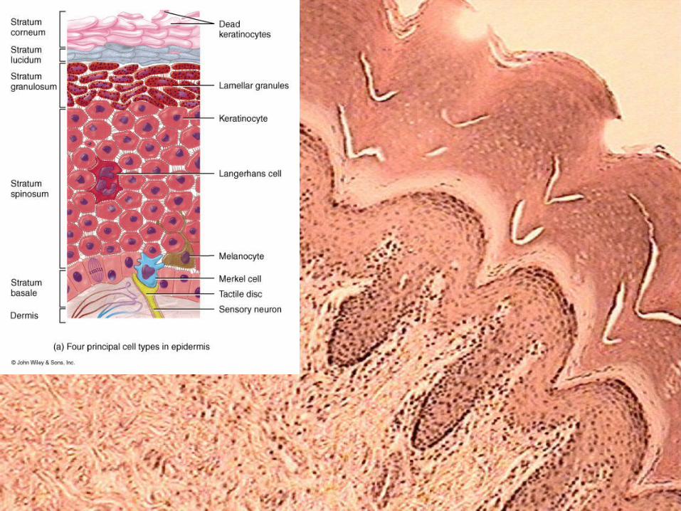
Specialized Cells of the EpidermisKeratinocytes:
Most common cells of the epidermis. Provides protection and waterproofing sealant
Melanocytes:
Produces and transfer the protein melanin to Keratinocytes. Melanin is a brown/black pigment that absorbs UV-light.
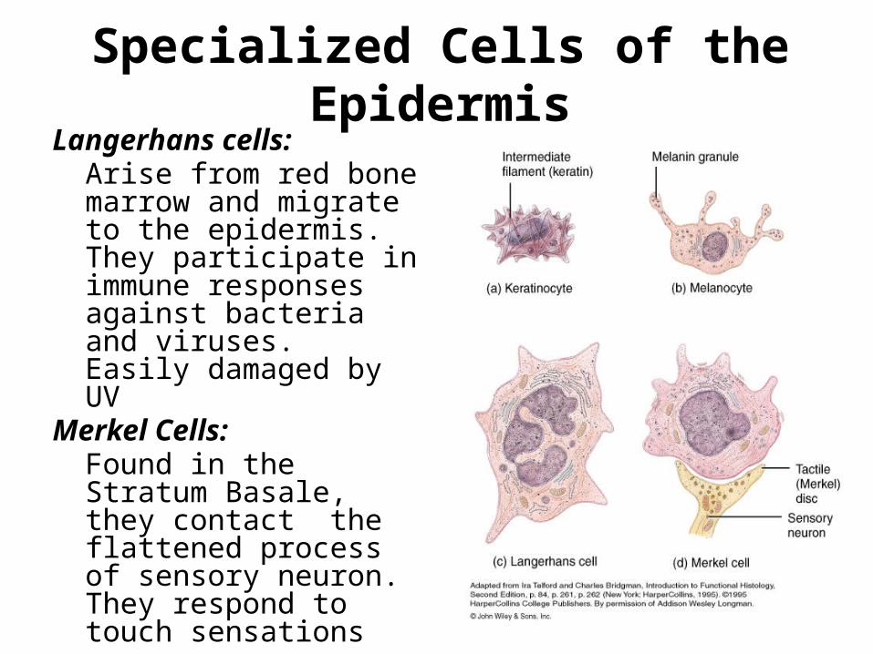
Specialized Cells of the EpidermisLangerhans cells:
Arise from red bone marrow and migrate to the epidermis. They participate in immune responses against bacteria and viruses. Easily damaged by UV
Merkel Cells:Found in the Stratum Basale, they contact the flattened process of sensory neuron. They respond to touch sensations
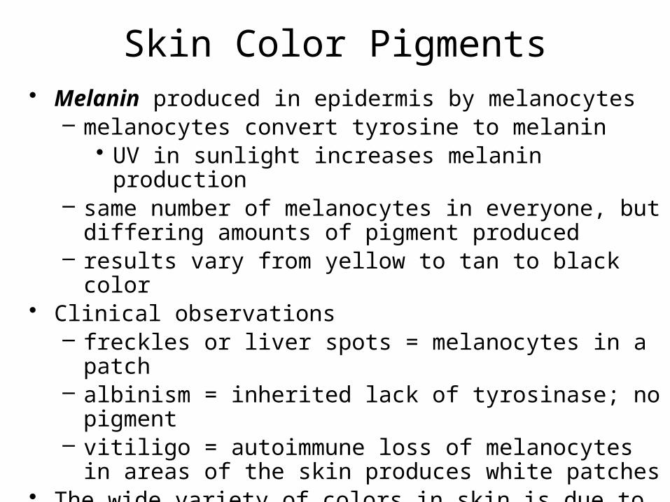
Skin Color Pigments• Melanin produced in epidermis by melanocytes
– melanocytes convert tyrosine to melanin• UV in sunlight increases melanin production
– same number of melanocytes in everyone, but differing amounts of pigment produced
– results vary from yellow to tan to black color• Clinical observations
– freckles or liver spots = melanocytes in a patch– albinism = inherited lack of tyrosinase; no pigment– vitiligo = autoimmune loss of melanocytes in areas of the
skin produces white patches• The wide variety of colors in skin is due to three pigments -
melanin, carotene, and hemoglobin (in blood in capillaries) - in the dermis.
Skin Color Pigments
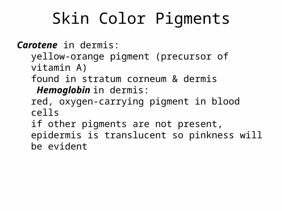
Carotene in dermis:yellow-orange pigment (precursor of vitamin A)found in stratum corneum & dermis Hemoglobin in dermis:red, oxygen-carrying pigment in blood cellsif other pigments are not present, epidermis is translucent so pinkness will be evident
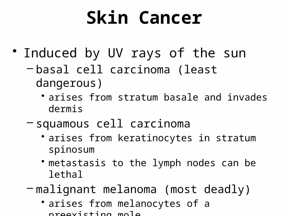
Skin Cancer
• Induced by UV rays of the sun– basal cell carcinoma (least dangerous)
• arises from stratum basale and invades dermis
– squamous cell carcinoma• arises from keratinocytes in stratum spinosum• metastasis to the lymph nodes can be lethal
– malignant melanoma (most deadly)• arises from melanocytes of a preexisting mole• ABCD--asymmetry, border irregular, color
mixed and diameter over 6 mm• Result of oncogene BRAF in men
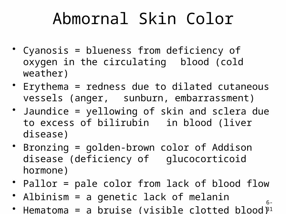
Abmornal Skin Color
6-31
• Cyanosis = blueness from deficiency of oxygen in the circulating blood (cold weather)
• Erythema = redness due to dilated cutaneous vessels (anger, sunburn, embarrassment)
• Jaundice = yellowing of skin and sclera due to excess of bilirubin in blood (liver disease)
• Bronzing = golden-brown color of Addison disease (deficiency of glucocorticoid hormone)
• Pallor = pale color from lack of blood flow• Albinism = a genetic lack of melanin• Hematoma = a bruise (visible clotted blood)
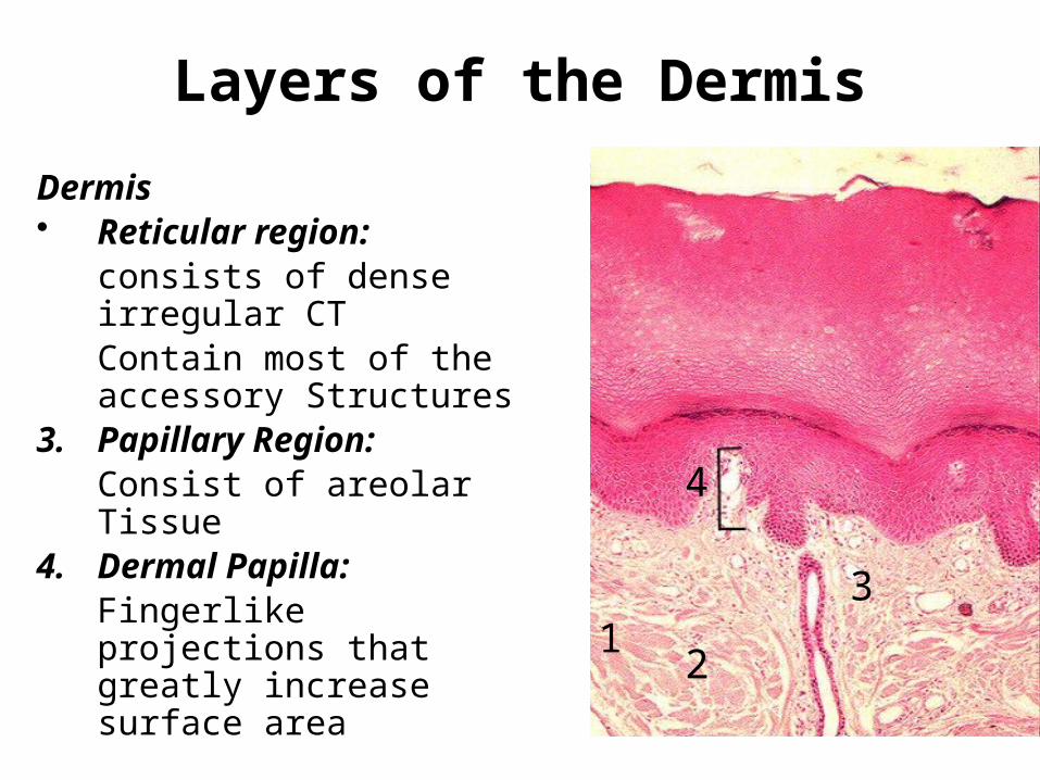
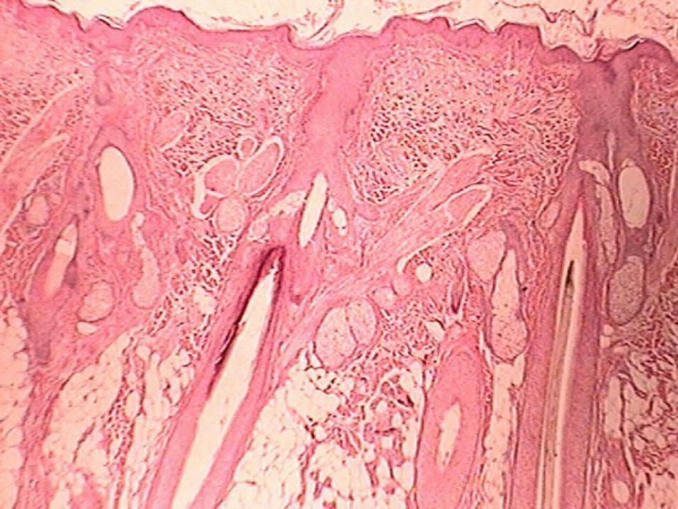
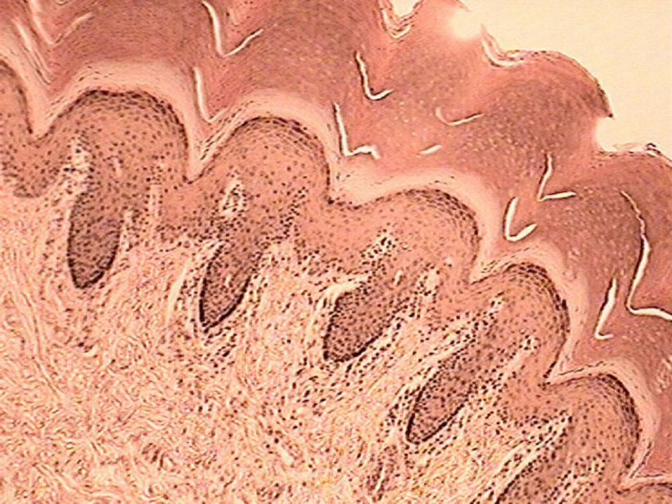
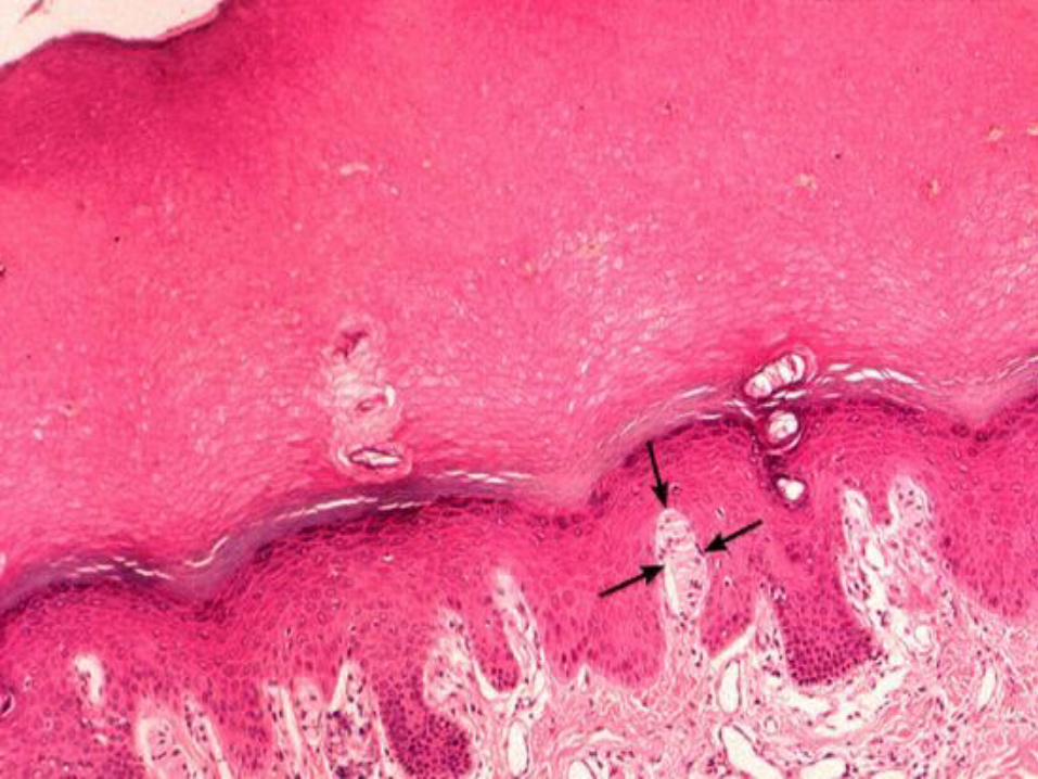
Layers of the Dermis
Dermis• Reticular region:
consists of dense irregular CTContain most of the accessory Structures
3. Papillary Region:Consist of areolar Tissue
4. Dermal Papilla:Fingerlike projections that greatly increase surface area
12
3
4
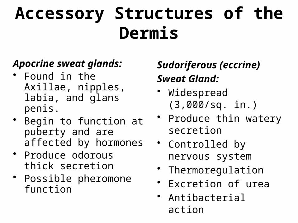
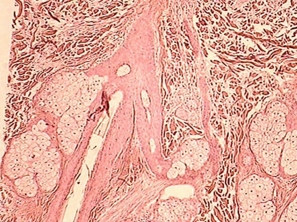
Accessory Structures of the Dermis
Apocrine sweat glands:• Found in the Axillae,
nipples, labia, and glans penis.
• Begin to function at puberty and are affected by hormones
• Produce odorous thick secretion
• Possible pheromone function
Sudoriferous (eccrine)
Sweat Gland:• Widespread (3,000/sq. in.)• Produce thin watery
secretion• Controlled by nervous
system• Thermoregulation• Excretion of urea• Antibacterial action
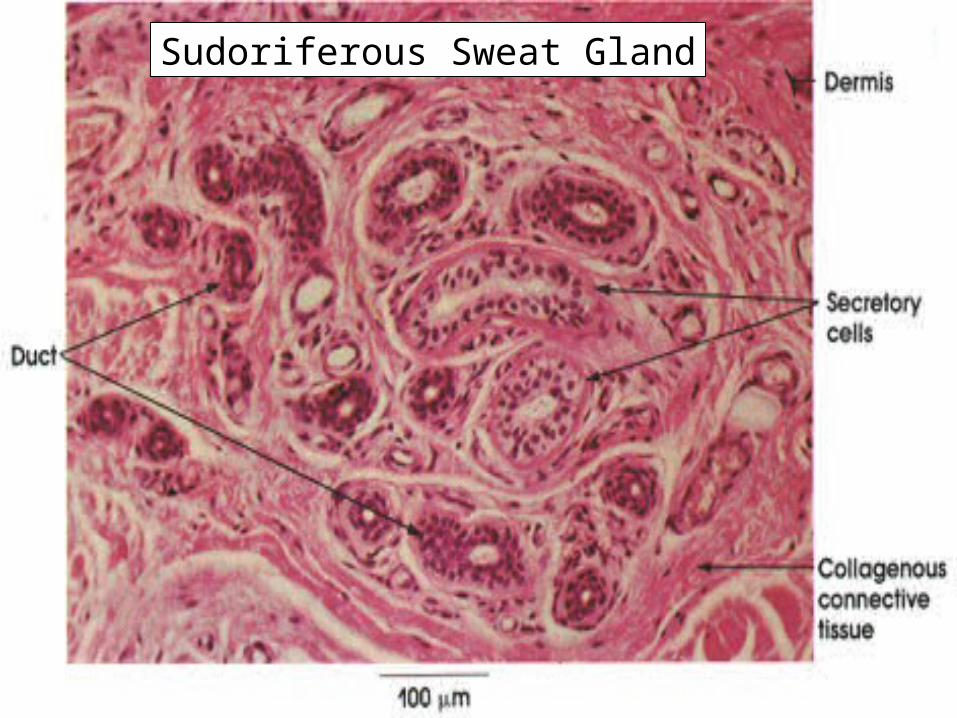
Sudoriferous Sweat Gland
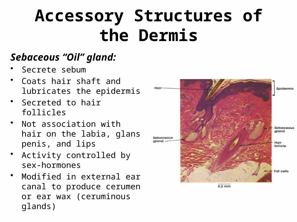
Accessory Structures of the Dermis
Sebaceous “Oil” gland:• Secrete sebum• Coats hair shaft and lubricates
the epidermis• Secreted to hair follicles• Not association with hair on the
labia, glans penis, and lips• Activity controlled by sex-
hormones• Modified in external ear canal to
produce cerumen or ear wax (ceruminous glands)
Accessory Structures of the Dermis
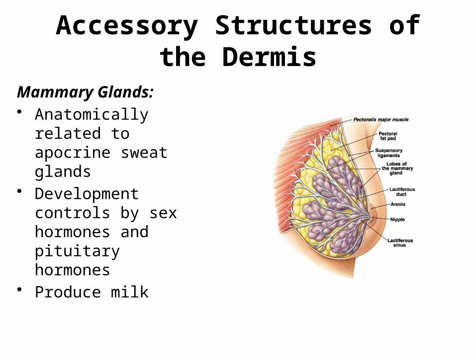
Mammary Glands:• Anatomically related to
apocrine sweat glands• Development controls
by sex hormones and pituitary hormones
• Produce milk
Accessory Structures of the Dermis
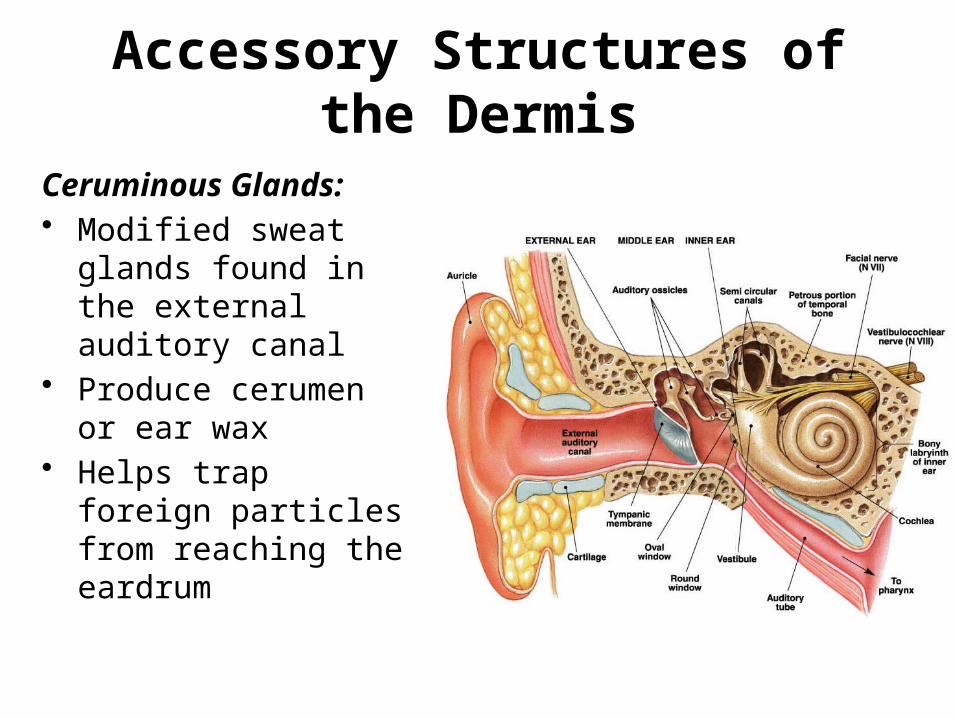
Ceruminous Glands:• Modified sweat glands
found in the external auditory canal
• Produce cerumen or ear wax
• Helps trap foreign particles from reaching the eardrum
Accessory Structures of the Dermis
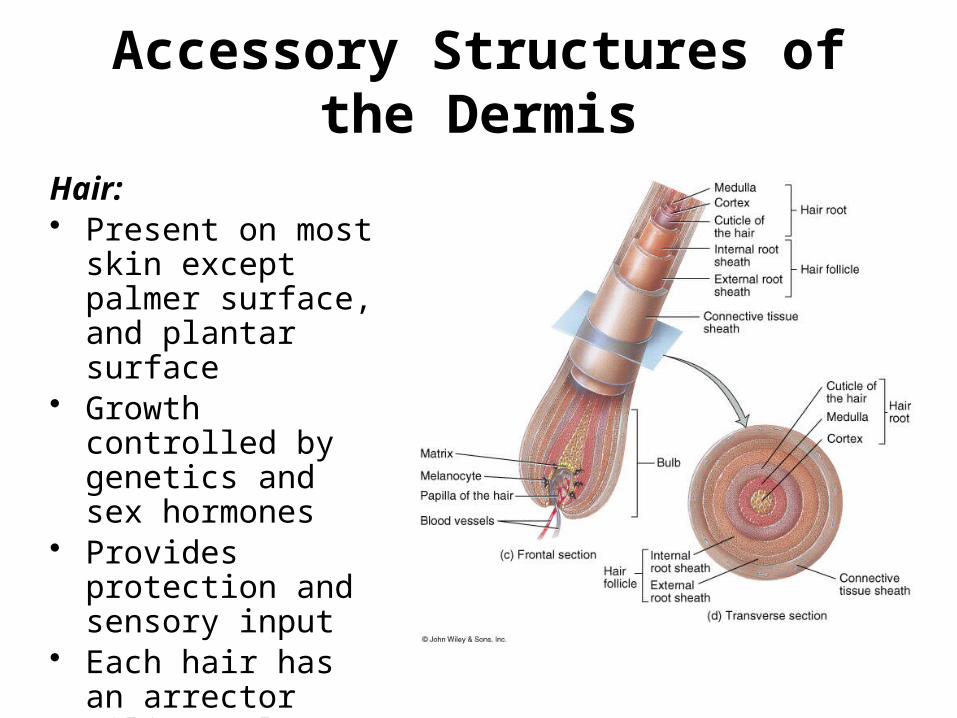
Hair:• Present on most
skin except palmer surface, and plantar surface
• Growth controlled by genetics and sex hormones
• Provides protection and sensory input
• Each hair has an arrector pili muscle
Hair Growth
• The hair growth cycle consists of a growing stage and a resting stage.– Growth cycle = growth stage & resting stage
• Growth stage – lasts for 2 to 6 years– matrix cells at base of hair root producing length
• Resting stage– lasts for 3 months– matrix cells inactive & follicle atrophies
– Old hair falls out as growth stage begins again• normal hair loss is 70 to 100 hairs per day
• Both rate of growth and the replacement cycle can be altered by illness, diet, high fever, surgery, blood loss, severe emotional stress, and gender.
• Chemotherapeutic agents affect the rapidly dividing matrix hair cells resulting in hair loss.
Hair Color
• Hair color is due primarily to the amount and type of melanin.
• Graying of hair occurs because of a progressive decline in tyrosinase.– Dark hair contains true melanin– Blond and red hair contain melanin with iron and sulfur
added– Graying hair is result of decline in melanin production– White hair has air bubbles in the medullary shaft
• Hormones influence the growth and loss of hair (Clinical applications).

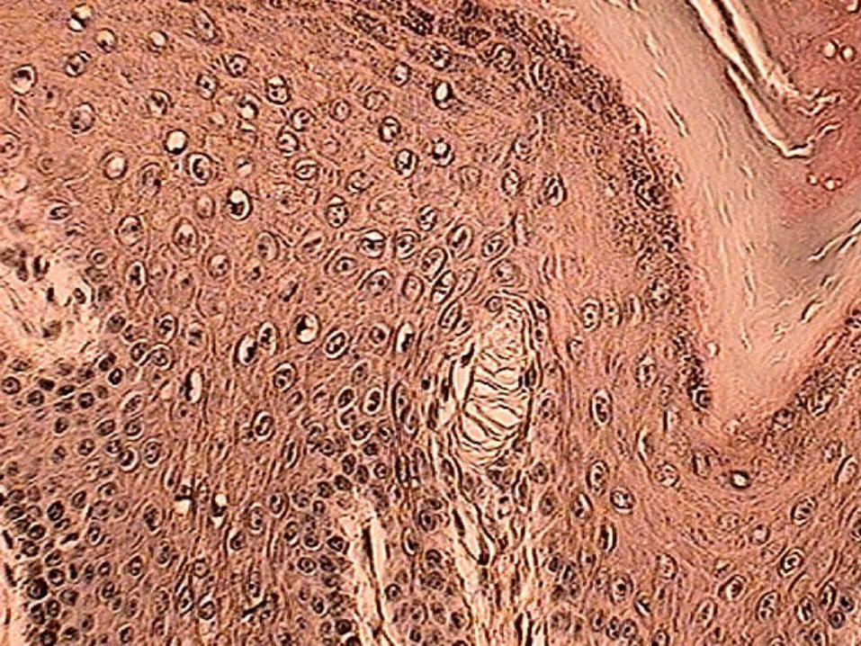
Special Sensory apparatus of the dermis
Meissner’s Corpuscles:• Present in dermal papilla• Specialized sensory neuron nerve endings• Respond to touch• Most numerous in thick or non-hairy skin of the
palmar and plantar surfaces
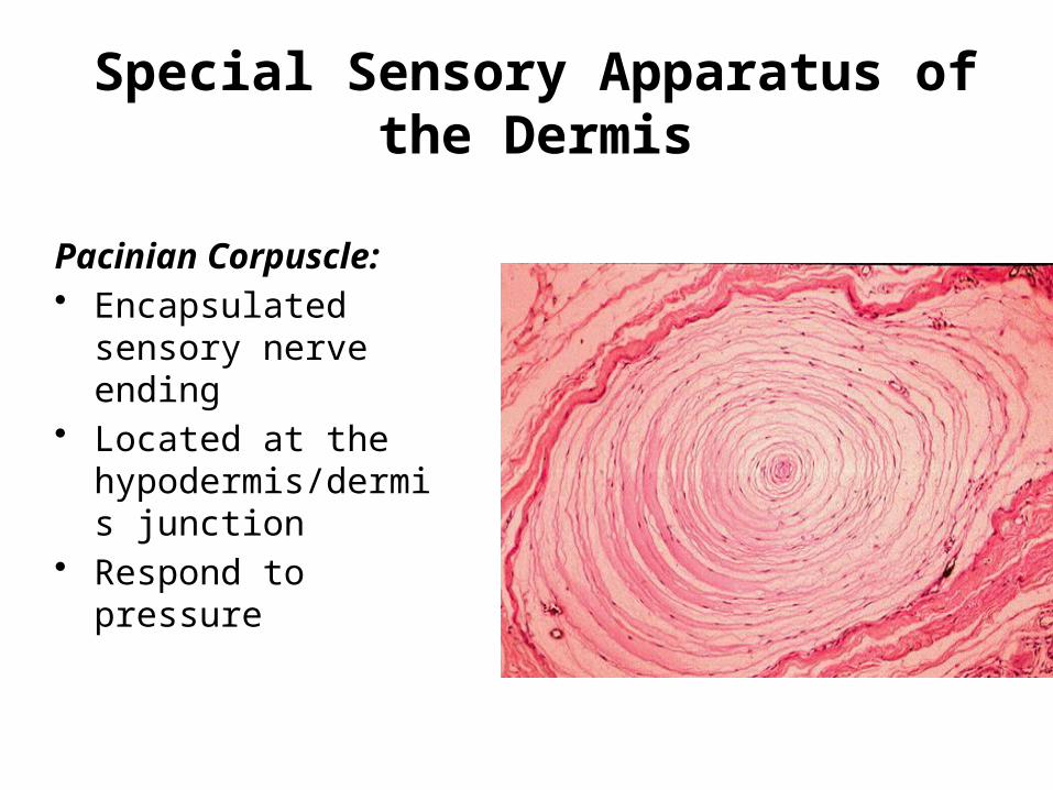
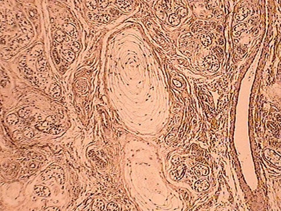
Special Sensory Apparatus of the Dermis
Pacinian Corpuscle:• Encapsulated sensory
nerve ending• Located at the
hypodermis/dermis junction
• Respond to pressure
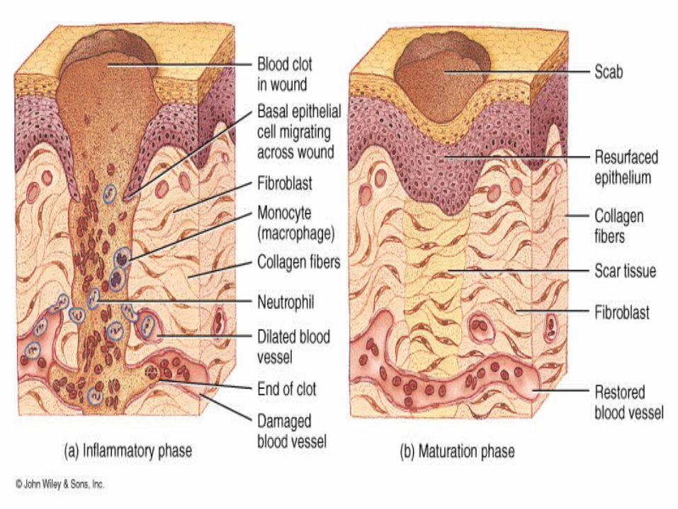
Deep Wound Healing
• When an injury extends to tissues deep to the epidermis, the repair process is more complex than epidermal healing, and scar formation results.
• Healing occurs in 4 phases– inflammatory phase has clot unite wound edges and WBCs arrive from
dilated and more permeable blood vessels– migratory phase begins the regrowth of epithelial cells and the
formation of scar tissue by the fibroblasts– proliferative phase is a completion of tissue formation– maturation phase sees the scab fall off
• Scar formation– hypertrophic scar remains within the boundaries of the original wound– keloid scar extends into previously normal tissue
• collagen fibers are very dense and fewer blood vessels are present so the tissue is lighter in color
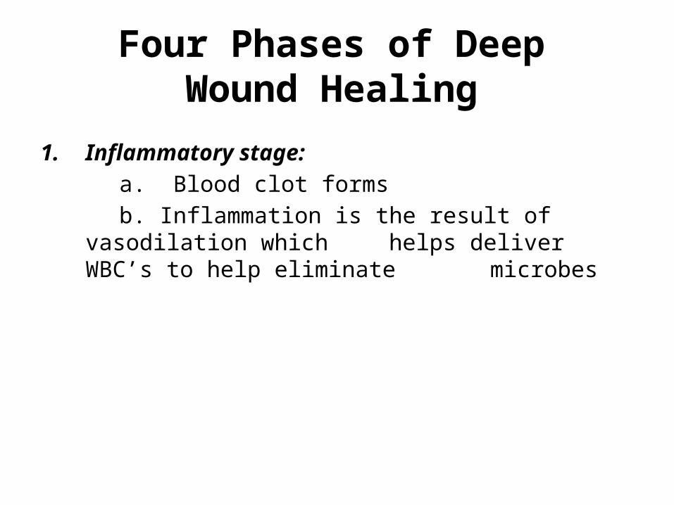
Four Phases of Deep Wound Healing
1. Inflammatory stage:
a. Blood clot forms
b. Inflammation is the result of vasodilation which helps deliver WBC’s to help eliminate
microbes
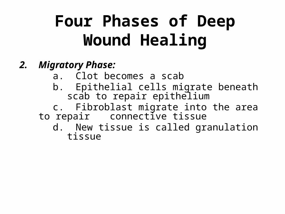
Four Phases of Deep Wound Healing
2. Migratory Phase:a. Clot becomes a scabb. Epithelial cells migrate beneath scab to repair epitheliumc. Fibroblast migrate into the area to repair connective tissued. New tissue is called granulation tissue
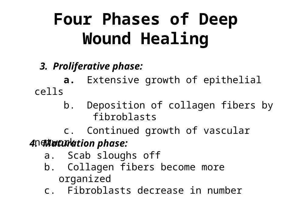
Four Phases of Deep Wound Healing
3. Proliferative phase:
a. Extensive growth of epithelial cells
b. Deposition of collagen fibers by fibroblasts
c. Continued growth of vascular network
4. Maturation phase:a. Scab sloughs offb. Collagen fibers become more
organizedc. Fibroblasts decrease in number
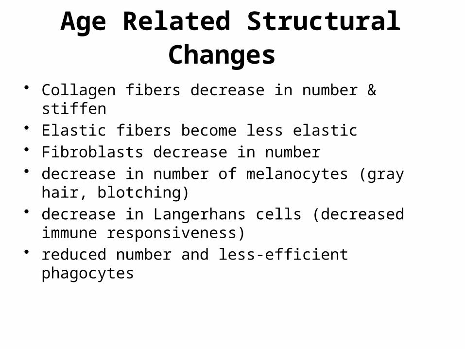
Age Related Structural Changes
• Collagen fibers decrease in number & stiffen • Elastic fibers become less elastic• Fibroblasts decrease in number• decrease in number of melanocytes (gray hair, blotching)• decrease in Langerhans cells (decreased immune
responsiveness)• reduced number and less-efficient phagocytes
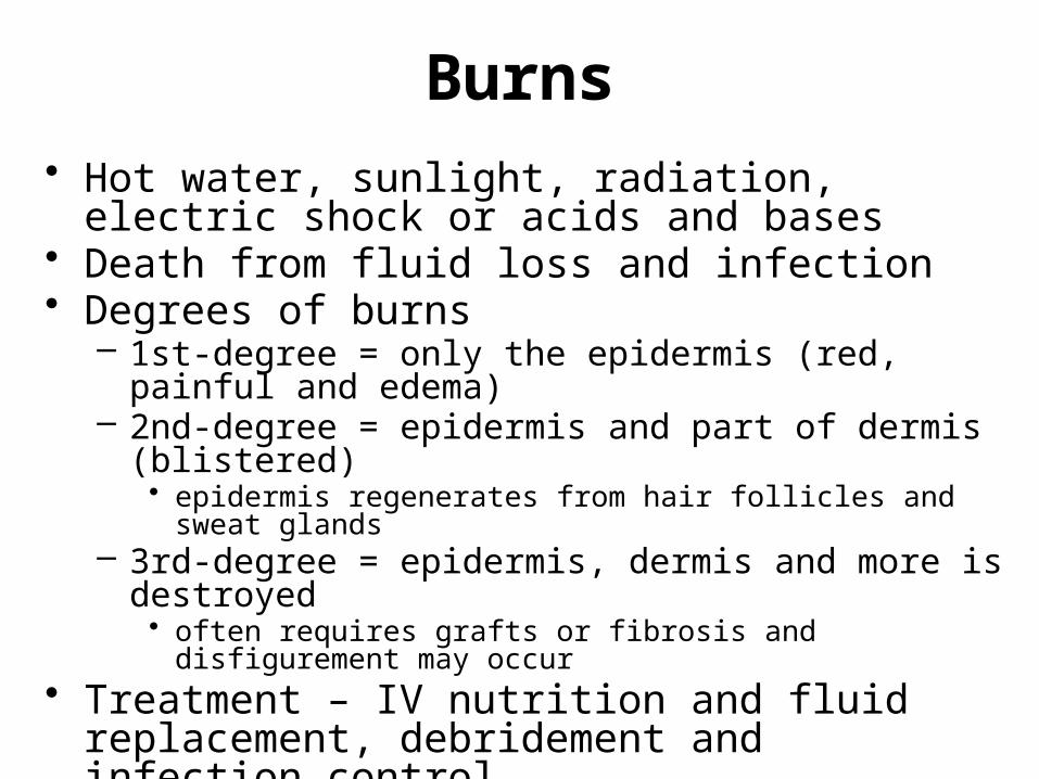
Burns• Hot water, sunlight, radiation, electric shock or
acids and bases• Death from fluid loss and infection• Degrees of burns
– 1st-degree = only the epidermis (red, painful and edema)– 2nd-degree = epidermis and part of dermis (blistered)
• epidermis regenerates from hair follicles and sweat glands– 3rd-degree = epidermis, dermis and more is destroyed
• often requires grafts or fibrosis and disfigurement may occur• Treatment – IV nutrition and fluid replacement,
debridement and infection control
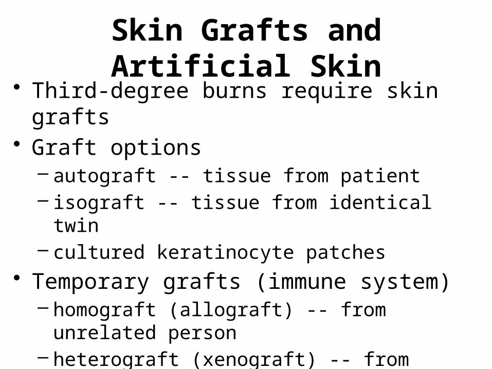
Skin Grafts and Artificial Skin
• Third-degree burns require skin grafts• Graft options
– autograft -- tissue from patient– isograft -- tissue from identical twin– cultured keratinocyte patches
• Temporary grafts (immune system)– homograft (allograft) -- from unrelated person– heterograft (xenograft) -- from another species– amnion from afterbirth– artificial skin from silicone and collagen