Embed Size (px)
Citation preview
Cellular/Molecular
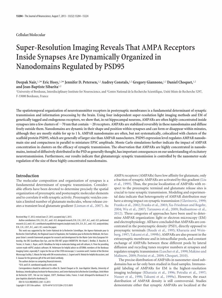
Super-Resolution Imaging Reveals That AMPA ReceptorsInside Synapses Are Dynamically Organized inNanodomains Regulated by PSD95
Deepak Nair,1,2* Eric Hosy,1,2* Jennifer D. Petersen,1,2 Audrey Constals,1,2 Gregory Giannone,1,2 Daniel Choquet,1,2
and Jean-Baptiste Sibarita1,2
1University of Bordeaux, Interdisciplinary Institute for Neuroscience, and 2Centre National de la Recherche Scientifique, Unite Mixte de Recherche 5297,F-33000 Bordeaux, France
The spatiotemporal organization of neurotransmitter receptors in postsynaptic membranes is a fundamental determinant of synaptictransmission and information processing by the brain. Using four independent super-resolution light imaging methods and EM ofgenetically tagged and endogenous receptors, we show that, in rat hippocampal neurons, AMPARs are often highly concentrated insidesynapses into a few clusters of �70 nm that contain �20 receptors. AMPARs are stabilized reversibly in these nanodomains and diffusefreely outside them. Nanodomains are dynamic in their shape and position within synapses and can form or disappear within minutes,although they are mostly stable for up to 1 h. AMPAR nanodomains are often, but not systematically, colocalized with clusters of thescaffold protein PSD95, which are generally of larger size than AMPAR nanoclusters. PSD95 expression level regulates AMPAR nanodo-main size and compactness in parallel to miniature EPSC amplitude. Monte Carlo simulations further indicate the impact of AMPARconcentration in clusters on the efficacy of synaptic transmission. The observation that AMPARs are highly concentrated in nanodo-mains, instead of diffusively distributed in the PSD as generally thought, has important consequences on our understanding of excitatoryneurotransmission. Furthermore, our results indicate that glutamatergic synaptic transmission is controlled by the nanometer-scaleregulation of the size of these highly concentrated nanodomains.
IntroductionThe molecular composition and organization of synapses is afundamental determinant of synaptic transmission. Consider-able efforts have been devoted to determine precisely the spatialorganization of presynaptic and postsynaptic molecular elementsand to correlate it with synaptic activity. Presynaptic vesicles con-tain a limited number of glutamate molecules, whose release cre-ates a transient local glutamate gradient (Lisman et al., 2007). As
AMPA receptors (AMPARs) have low affinity for glutamate, onlya fraction of synaptic AMPARs are activated by this gradient (Liuet al., 1999). Thus, the precise localization of AMPARs with re-spect to the presynaptic terminal and glutamate release sites iscrucial to tune synaptic transmission. Modeling and experimen-tal data indicate that heterogeneity of AMPAR distribution mayhave a strong impact on synaptic transmission (Glavinovic, 1999;Franks et al., 2002; Franks et al., 2003; Xu-Friedman and Regehr,2004; Wu et al., 2007; Tarusawa et al., 2009; Budisantoso et al.,2012). Three categories of approaches have been used to deter-mine AMPAR organization: light or electron microscopy (EM)and electrophysiology. All have concluded that AMPARs are con-centrated in the postsynaptic density (PSD), directly opposed topresynaptic terminals (Baude et al., 1995; Kharazia and Wein-berg, 1997; Takumi et al., 1999b). AMPARs are also present in theextrasynaptic membrane and in intracellular pools, and constantexchange of AMPARs between these different pools by lateraldiffusion and recycling tunes receptor numbers at synapses andregulates synaptic transmission (Luscher et al., 1999; Makino andMalinow, 2009; Petrini et al., 2009; Choquet, 2010).
The precise distribution of AMPARs in nanometer-sized sub-domains has so far only been studied in fixed samples. Immuno-gold labeling of AMPARs for EM is the highest-resolutionimaging technique (Kharazia et al., 1996; Petralia et al., 1997;Nusser et al., 1998; Takumi et al., 1999a). However, the exactdistribution of AMPAR density is still controversial. Studiesdemonstrate either that synaptic AMPARs are localized at the
Received May 17, 2012; revised June 27, 2013; accepted July 5, 2013.Author contributions: D.N., E.H., D.C., and J.-B.S. designed research; D.N., E.H., J.D.P., A.C., and J.-B.S. performed
research; G.G. and J.-B.S. contributed unpublished reagents/analytic tools; D.N., E.H., D.C., and J.-B.S. analyzed data;D.N., E.H., J.D.P., D.C., and J.-B.S. wrote the paper.
This work was supported by the Centre National de la Recherche Scientifique, the Agence Nationale pour laRecherche (StimTrafPark), the Regional Council of Aquitaine, the Fondation pour la Recherche Medicale, the Euro-pean Union’s seventh framework program for research and development for the Marie-Curie-Intra European Fel-lowship, the ERC GrantNano-Dyn-Syn, and the ERA-NET project MODIFSYN. We thank C. Breillat, D. Bouchet, B.Tessier, A. Frouin, L. Royer, and N. Retailleau for help in molecular biology and cell cultures; A. Penn for providingcustom-made mEPSC analysis software; M. Heilemann for helpful advice on dSTORM; members of the BordeauxImaging Center, especially P. Legros for the use of the STED microscope; S. Lacomme and E. Gontier of the ElectronMicroscopy Facility; S. Marais for help with EM reconstructions; L. Cognet and N. Rebola for helpful discussions; andE. Gouaux for the generous gift of the anti-GluA2 antibody.
The authors declare no competing financial interests.*D.N. and E.H. contributed equally to this work.Correspondence should be addressed to either Dr. Daniel Choquet or Dr. Jean-Baptiste Sibarita, University of
Bordeaux, Interdisciplinary Institute for Neuroscience, and Centre National de la Recherche Scientifique, Unite Mixtede Recherche 5297, 146 rue Leo Saignat, 33077, Bordeaux Cedex, France. E-mail: [email protected] [email protected].
DOI:10.1523/JNEUROSCI.2381-12.2013Copyright © 2013 the authors 0270-6474/13/3313204-21$15.00/0
13204 • The Journal of Neuroscience, August 7, 2013 • 33(32):13204 –13224
PSD periphery (Kharazia et al., 1996; Matsubara et al., 1996; Ber-nard et al., 1997; Kharazia and Weinberg, 1997; Chen et al., 2008)or randomly distributed (Nusser et al., 1994; Masugi-Tokita andShigemoto, 2007; Masugi-Tokita et al., 2007). Pre-embeddingimmunogold EM and replica-based labeling indicate the exis-tence of AMPAR clusters on the membrane (Masugi-Tokita et al.,2007; Tarusawa et al., 2009; Tao-Cheng et al., 2011; Budisantosoet al., 2012). Complementary to EM, super-resolution light mi-croscopy of immunofluorescently labeled samples, which allowsbreaking the diffraction barrier to �250 nm, has revealed themolecular organization of postsynaptic molecules at nanometricresolution (Dani et al., 2010). In many synapses, AMPARs wereorganized centrally, whereas in others they were organized later-ally to the PSD. We recently performed mathematical analysis oflive super-resolution tracking of AMPAR movements and foundthat AMPARs move to attracting interaction potential wells at thesubdiffraction level in hippocampal dendrites (Hoze et al., 2012).
Here, we performed a detailed analysis of AMPAR and PSD95localization and mobility at synapses using a combination ofhigh-resolution imaging approaches. We demonstrate thatAMPARs are strongly nonuniformly organized in synapses.
Materials and MethodsConstructs. mEos2::GluA1 (noted Eos::GluA1), GluA2::tdEos (notedEos::GluA2), and Homer1c::Cerulean were subcloned from SEP::GluA1,GluA2::GFP, Homer1C::Dsred, and Homer1c::GFP, which were de-scribed previously (Saglietti et al., 2007; Opazo et al., 2010).Eos::TEV::GluA1 was modified from mEos2::GluA1 by inserting a To-bacco Etch Virus Protease recognition site comprising a seven aminoacid sequence (Glu-Asn-Leu-Tyr-Phe-Gln-Gly, cleaving between Glnand Gly), for removing Eos tag from surface-expressed Eos::GluA1 of liveneurons. PSD95::mEos2 (noted Eos::PSD95) was subcloned fromPSD95::EGFP (used for overexpression experiments), which was de-scribed previously (Mondin et al., 2011). The GFP-tagged shRNA con-struct designed to silence expression of PSD95 (SH::PSD95) and thePSD95 rescue construct (Rescue::PSD95), which expresses shRNAagainst PSD95 simultaneously with an shRNA-resistant form of GFP-tagged PSD95, were characterized previously (Schluter et al., 2006; Mon-din et al., 2011). A generic scrambled RNA (SH::Scramble) vectorcontaining a noneffective mammalian protein coexpressed either withGFP (SH::Scramble::GFP) or Homer1c::GFP (SH::Scramble::Homer1c)was used as a negative control to compare the effect of SH::PSD95::GFP.The compensatory mutant for stagazin and PSD95 were described andcharacterized previously (Schnell et al., 2002; Bats et al., 2007). The Star-gazin/PSD95 compensatory mutants fused with GFP were obtained bymodifying the �2 position threonine of Stargazin to phenylalanine(StgT231F) and the 225 position histidine of PSD-95 to valine(PSD95H225V).
Cell culture and transfection. Preparation of cultured neurons was per-formed as previously described (Heine et al., 2008). Hippocampal neu-rons from 18-d-old rat embryos of either sex were cultured on glasscoverslips following the Banker protocol (Kaech and Banker, 2006).Neurons were transfected using Effectene at 9 –11 d in vitro (DIV) or byNucleofection at the time of plating (Nucleofector II Device, LonzaCologne) with mEos2::GluA1, Eos::TEV::GluA1, GluA2::tdEos, andPSD95::mEos2, alone or in combination with Homer1c::Cerulean,HA::GluA1, or Homer1c::GFP, and experiments were performed at14 –20 DIV.
Single particle tracking photoactivation localization microscopy (spt-PALM). Cells were imaged at 37°C in an open chamber (Ludin chamber,Life Imaging Services) mounted on an inverted motorized microscope(Nikon) equipped with a 100� 1.49 NA PL-APO objective and a perfectfocus system, allowing long acquisition in oblique illumination mode.Imaging was performed for �20 min unless otherwise stated in an extra-cellular solution as in Petrini et al. (2009). Cells expressing Eos::GluA1 orEos::GluA2 were photoactivated using a 405 nm laser (Omicron), and theresulting photoconverted single molecule fluorescence was excited with a
561 nm laser (Cobolt). Both lasers illuminated the sample simultane-ously. Their respective power was adjusted to keep the number of thestochastically activated molecules constant and well separated duringthe acquisition. Laser intensities were tuned to leave the single moleculefluorescent during multiple frames before bleaching. The fluorescencewas collected by the combination of a dichroic and emission filters(D101-R561 and F39 – 617, respectively, Chroma) and a sensitiveEMCCD camera (Evolve, Photometric). The acquisition was steered byMetaMorph software (Molecular Devices) in streaming mode at 50frames per second (20 ms exposure time) using a 200 � 200 pixels regionof interest. The native nonactivated fluorescence form of Eos moleculeswas excited using a conventional GFP filter cube (ET470/40, T495LPXR,ET525/50, Chroma). Homer1c::Cerulean fluorescent protein was ob-served using a CFP filter (ET436/20, T455LP, ET480/40, Chroma). Mul-ticolor fluorescent micro-beads (Tetraspeck, Invitrogen) were used asfiduciary markers to register long-term acquisitions and correct for lat-eral drifts.
Universal point accumulation in nanoscale topography (uPAINT). uPA-INT was applied as reported previously (Giannone et al., 2010). Disso-ciated neurons were nucleofected at the time of plating withHomer1c::GFP, allowing synapse identification. Experiments took placeat 13–15 DIV. Coverslips were mounted on a Ludin chamber filled with600 �l of extracellular solution at 37°C as in Petrini et al. (2009). A lowconcentration of ATTO-647 nm-coupled anti-GluA2 antibodies wasthen added to the chamber. Stochastic labeling of endogenous GluA2-containing AMPA receptors by dye-coupled antibodies allowed us torecord thousands of trajectories lasting longer than 1 s. The amount ofnonspecific binding of antibodies was evaluated using hippocampal cul-tures derived from GluA2 knock-out mice and was found to be �15%.Potential effects of the antibody binding on receptor dynamics was testedby comparing two different GluA2 antibodies and results were found tobe similar (see Fig. 6 A, B). Multicolor fluorescent micro-beads (Tetras-peck, Invitrogen) were used as fiduciary markers to register long-termacquisitions and correct for lateral drifts.
Single molecule localization and tracking. A typical single-cell sptPALMor uPAINT experiment acquired with the microscope setup and protocoldescribed above produced a set of 20,000 images that were analyzed toextract molecule localization and dynamics. Single molecule fluorescentspots were localized in each image frame and tracked over time using acombination of wavelet segmentation (Izeddin et al., 2012) and simu-lated annealing (Racine et al., 2006, 2007) algorithms. Under the exper-imental conditions described above, the image resolution of theexperimental setup was quantified to 46.6 nm for sptPALM. The local-ization accuracy, which depends on the image signal-to-noise ratio andthe segmentation algorithm (Kubitscheck et al., 2000; Cheezum et al.,2001; Izeddin et al., 2012), was determined experimentally using fixedsamples expressing Eos-tagged proteins. We analyzed 133 2D distribu-tions of single molecule positions belonging to long trajectories (�50frames) by bidimensional Gaussian fitting, and the resolution was deter-mined to be 2.3�. The software package used to derive quantitative dataon protein localization and dynamics is custom written as a plug-inrunning within the MetaMorph software environment. For the trajectoryanalysis, synapses were identified by wavelet image segmentation of theHomer1c postsynaptic marker. The corresponding binary mask was thenused to sort single particle data analyses to specific synaptic regions.Dendrites were identified as subcellular regions lacking the synapticmarker.
Live-cell surface staining and stimulated emission depletion microscopy(STED). The monocolonal antibody against an extracellular epitope ofGluA2 was prepared as described previously (Giannone et al., 2010). Pri-mary neuronal cultures transfected with Homer1c::GFP were incubatedwith the monoclonal anti-GluA2 antibody (3 �g/ml) in the neuronal growthmedium for 4–6 min at 37°C, and then fixed in 4% paraformaldehyde and4% sucrose in PBS. Fixed samples were rinsed in PBS and then blocked inPBS containing 1% BSA. The primary antibodies were then revealed byincubating ATTO647N (ATTO-tec) coupled anti-mouse IgG secondary an-tibodies for 30 min at room temperature. Finally, samples were rinsed in PBSand mounted in Vectashield mounting medium. We used a commercialSTED inverted microscope (DMI6000 TCS SP5 AOBS, Leica) to obtain
Nair, Hosy et al. • AMPA Receptors Are Organized in Nanodomains J. Neurosci., August 7, 2013 • 33(32):13204 –13224 • 13205
super-resolved images of GluA2-ATTO647N-labeled neurons expressingHomer1c::GFP as a postsynaptic marker. The Homer1c::GFP images wererecorded using the regular confocal mode on the same microscope. A spatialresolution of 63.7 � 1.2 nm was measured using 40 nm crimson beads.
Direct stochastic optical reconstruction microscopy (dSTORM). Primaryneuronal cultures were incubated with rabbit-anti-GluA1 antibody (tar-geted to the extracellular region RTSDSRDHTRVDWKRC, Agro-Bio)for 20 –30 min. They were then fixed using 4% paraformaldehyde and4% sucrose in PBS, and washed with PBS. They were incubated withNH4Cl 50 mM for 30 min before permeabilization. They were permeabil-ized using 0.1% Triton X-100 and incubated with PBS containing 1%BSA for 30 min. They were then incubated with mouse-anti-PSD95 an-tibody (MA1– 046, Thermo Fisher Scientific) for 30 min and washedseveral times with PBS containing 1% BSA. The primary antibodies werethen revealed by incubating Alexa-647-coupled anti-rabbit IgG secondary(A21245, Invitrogen) and RhodamineRed coupled anti-mouse secondaryantibodies (715-295-151, Jackson ImmunoResearch Laboratories) for 30min at room temperature. For single-color dSTORM experiments, primaryneuronal cultures were incubated with mouse-anti-GluA2 antibody (Gian-none et al., 2010) for 5–7 min. They were then fixed, as described previously.The primary antibodies were then revealed by incubating Alexa-647 coupledanti-mouse IgG secondary (Invitrogen) for 30 min at room temperature.
The stained coverslips were imaged the next day at room temperaturein a closed chamber (Ludin Chamber, Life Imaging Services) mountedon an inverted motorized microscope equipped with a 100� 1.49 NAPL-APO objective and a Perfect Focus System (Nikon), allowing longacquisition in oblique illumination mode. Imaging was performed in anextracellular solution containing reducing agents and oxygen scavengers.For dSTORM, ensemble fluorescence of Rhodamine Red and Alexa-647was first converted in to dark state using a 561 nm laser or 640 nm laser(Coherent) at 30 –50 kw/cm 2 intensity. Once the ensemble fluorescencewas converted into the desired density of single molecules per frame, thelaser power was reduced to 7–15 kw/cm 2 and imaged continuously at 50fps for 20,000 frames. The level of single molecules per frame was con-trolled by using a 405 nm laser (Omicron). The dyes were sequentiallyimaged (Alexa-647 first and followed by Rhodamine) to collect the de-sired single molecule frames. The laser powers were adjusted to keep anoptimal level of stochastically activated molecules during the acquisition.Both the ensemble and single molecule fluorescence was collected by thecombination of a dichroic and emission filter (D101-R561 and F39-617,respectively, Chroma; and quad-band dichroic filter (Di01-R405/488/561/635, Semrock). The fluorescence was collected using a sensitiveEMCCD (Evolve, Photometric). Single molecule localization and re-construction were performed online using automatic feedback controlon the lasers, enabling optimal molecule density during the acquisition(Kechkar et al., 2013). The acquisition and localization sequences weredriven by MetaMorph software (Molecular Devices) in streaming modeat 50 frames per second (20 ms exposure time) using an area equal to or�256 � 256 pixels region of interest. We used multicolor fluorescentmicrobeads (Tetraspeck, Invitrogen) as fiduciary markers to registerlong-term acquisitions and correct for lateral drifts and chromatic shifts.A spatial resolution of 14 nm was measured using centroid determina-tion on 100 nm Tetraspeck beads acquired with similar signal-to-noiseratio than dSTORM single molecule images.
Synaptic surface area quantification. Epifluorescence images of the syn-aptic marker protein Homer1c (Homer1c::GFP or Homer1c::Cerulean)were used to differentiate between synapses and the rest of the dendrite.Epifluorescence images of Homer1c were first thresholded and seg-mented using the morphometric image analysis module of MetaMorphsoftware (Molecular Devices) for structures �0.02 �m 2. Morphologicalfeatures, such as area, length, and breadth of each segmented structure,were exported to calculate their respective distributions. The mean syn-aptic area of Homer1c labeled structures was measured to be 0.11 � 0.04�m 2, which is consistent with the surface area of the spine head. TheHomer1c segmented images were then used to define synapses area forsingle particle tracking experiments.
Super-resolution cluster analysis. AMPAR nanodomains and PSD95subclusters were identified from super-resolution images by custom soft-ware written as a plug-in running inside MetaMorph. Single-molecule-
based super-resolution images were reconstructed from the 20,000frames before being analyzed, whereas STED bulk images were analyzedraw. Nanodomains, which corresponded to clustered areas where thesignal density was higher, were first identified by wavelet segmentation.Nanodomain number and dimensions were then computed by 2D aniso-tropic Gaussian fitting, from which the principal and the auxiliary axeswere extracted as 2.3�long and 2.3�short, respectively. The shape factorwas calculated as a ratio between the auxiliary and the principal axes.
For the quantification of PSD95 cluster and nanodomain surface ar-eas, the single molecule-based super-resolution images were first seg-mented by the morphometric image analysis module of MetaMorphsoftware for structures �0.02 �m 2. Morphologic features, such as area,length, and breadth of each cluster, were exported to calculate their re-spective distribution. The dimensions of PSD95 subclusters were calcu-lated the same way as AMPAR nanodomains.
Proteolytic cleavage assay of extracellular Eos. The proportionEos::TEV::GluA1 that was localized on neuronal surface was assessedusing AcTEV Protease (Invitrogen) to cleave the Eos tag from surfaceexpressed receptors. sptPALM was used to measure fluorescence inten-sity of Eos::TEV::GluA1-expressing cells before and after incubation withAcTEV protease in extracellular medium (1:60 dilution in presence of 1mM DTT, for 4 –5 min, 37°C). Control neurons were incubated in AcTEVprotease, which was rendered inactive by boiling for 10 min.
Live cell nanogold immunolabeling and electron microscopy of culturedneurons. Coverslips with attached neurons (DIV 18 –21) were incubatedfor 3 min at 37°C in GluA2 monoclonal antibody diluted in culturemedium (6 �g/ml, gift from E. Gouaux). Neurons were removed fromantibody and fixed in freshly prepared, prewarmed EM-grade 4% para-formaldehyde (EMS) in 0.15 M Sorensen’s phosphate buffer (EMS) atroom temperature for 45 min. All subsequent steps were performed atroom temperature. Neurons were rinsed 3 times in 0.15 M Sorensen’s PB,once in 0.1 M Millonig’s PBS, and then blocked in 0.1 M Millonig’s PBSwith 2% BSA and 0.1% cold water fish skin gelatin (Aurion, EMS) for 60min. Next, neurons were incubated in FluoroNanogold anti mouse Fab�AlexaFluor-488 (Nanoprobes) diluted 1:100 in 0.1 M Millonig’s PBSblocking solution for 90 min, then rinsed once in blocking solution, oncein Sorensen’s PB, and placed in freshly prepared 2% glutaraldehyde(EMS) in Sorensen’s PB for 30 min. Afterward, neurons were stored inSorensen’s PB until silver intensification.
Processing and embedding for electron microscopy. FluoroNanogold la-bel was enhanced for 6 – 8 min using HQ Silver Reagent (Nanoprobes)according to manufacturer’s instructions and processed immediately forEM; all steps were performed at room temperature. After several rinses inSorensen’s PB, neurons were incubated in 0.2% OsO4 in Sorensen’s PBfor 30 min, and then rinsed 10 times in dH2O to remove all traces of PBbefore placing neurons in filtered 0.25% uranyl acetate dissolved indH2O for 30 min. After several water rinses, neurons were dehydrated by3 min incubations in a graded series of ethanol: 50%, 70%, 95%, and100%. No propylene oxide was used to prevent loss of immunogold label.Samples were infiltrated during 1–2 h steps in 70% Epon812/ethanolmixture followed by 2 exchanges of 100% freshly prepared Epon812(Taab Laboratory and Microscopy), and finally embedded in freshly pre-pared Epon812. To allow cutting of en face sections of neurons, coverslipswere placed cell side facing up on a glass slide and gelatin capsules filledwith Epon812 were inverted and placed on top of coverslip, and polym-erized at 60°C for 48 h. Coverslips were removed from polymerizedsamples by gentle heating over a flame while pulling slightly on the glassslide and gelatin capsules. Ultrathin sections (60 –70 nm) were cut usingan Ultra 35°diamond knife (Diatome) and a Leica Ultracut UCT M26(Leica Microsystems) and picked up on 2 mm slot grids with a 1% form-var support film (EMS). When cutting serial sections, a 50:50 mix ofrubber cement/xylene was applied to the top and bottom sides of thetrimmed block to prevent sections from separating. Sections were con-trasted with 3% aqueous uranyl acetate for 5 min, and then Reynolds’slead citrate for 7 min before imaging using a Hitachi H7650 transmissionelectron microscope operated at 80 kV. Images were captured using anOrius CCD (Gatan).
Analysis of the pattern and distribution of immunogold labeling on 2DEM sections. Micrographs were analyzed using MetaMorph software
13206 • J. Neurosci., August 7, 2013 • 33(32):13204 –13224 Nair, Hosy et al. • AMPA Receptors Are Organized in Nanodomains
(Molecular Devices). To quantify the density of immunolabel on differ-ent membrane domains, ROIs were drawn along membranes that werecategorized as follows: spine PSD membrane, extrasynaptic spine mem-brane, or dendritic shaft membrane. For each ROI, the surface was mea-sured and the number of immunolabels that were within 20 nm of themembrane was counted. The size of the clusters was measured using theIntegrated Morphometry Analysis module, after adjusting the thresholdto select label from the background. Immunolabel particles that wereoverlapping or separated by �20 nm were identified as a cluster.
3D-reconstruction of synaptic spines by serial section EM. Series of im-ages taken at 40,000 –50,000 magnification from 6 to 8 serial sections of70 nm thickness were used to generate reconstructions of individualimmunogold labeled spines. Stacks of images were aligned using theMetaMorph software auto-align module. Reconstructions were gener-ated from the aligned stacks using Imaris software (Bitplane). The mem-brane contours of the spine were traced in each section to create the spinevolume. Immunolabel was reconstructed using the Spots tool. The num-ber and size of nanodomains per spine were measured on each image ofthe serial sections as previously described. Label that appeared in thesame location in successive serial sections was considered to be the sameimmunolabel and counted only once.
Quantification of AMPAR content in nanodomains. The number ofAMPARs per nanodomain was estimated from dSTORM super-resolution reconstructions of endogenous GluA2 using a custom-madeanalysis module operating inside MetaMorph software. For each cell,single AMPARs were identified using wavelet segmentation and Gauss-ian fitting as isotropic and isolated objects of size �40 nm in diameter.Isolated single AMPARs could be differentiated from nanoclusters re-sulting from the high spatial resolution provided by the dSTORM imag-ing technique. Then, the histogram and median of the integratedintensity of each individual AMPAR per cell were computed. This corre-sponds to the intensity distribution of one tetrameric structure, in func-tion of the ratio of immunolabeled GluA2 with the other nonlabeledAMPAR subunits. Finally, synaptic nanodomains were detected usingwavelet segmentation similarly to the cluster analysis described in Mate-rials and Methods, and the number of receptors per cluster was estimatedby dividing the cluster’s total intensity by the median intensity of theidentified isolated AMPARs.
Electrophysiology. The extracellular recording solution contained inmM concentration as follows: 130 NaCl, 2.5 KCl, 2 MgCl2, 2 CaCl2, 10HEPES, and 10 D-glucose, pH 7.4. To block GABA-A receptors, 50 �M
picrotoxin was added to solution, and mini-EPSC recordings were per-formed in the presence of 1 �M TTX. The bath temperature was kept at34°C. Patch-clamp microelectrodes were pulled for a 4 – 6 M� resistance,from borosilicate on a P-97 model puller (Sutter Instruments). Minirecordings were performed with an EPC 10 double patch-clamp ampli-fier (HEKA Elektronik). Data analysis was performed with a homemadesoftware (Penn et al., 2012). Cells are clamped at �70 mV.
Statistics. Statistical values are given as medians � 25%/75% intervalor mean � SEM, except for Figure 10 where mean � SD is used. Statis-tical significances were performed using Sigmaplot software (SystatSoftware). Non-Gaussian distribution datasets were tested by Mann–Whitney U test, and Gaussian distributions were tested by t test.
Calculation of required sample size. The experiments were designed tocompare two conditions per sample, including a control condition. Theexperimental observations of interest were changes in the localizationand mobility of the nanoscale organization of synaptic moleculeson primary cultured hippocampal neurons. In these experimental condi-tions, the sample size was calculated with a power factor of 0.6 – 0.8and � of 0.2– 0.5, which required sample sizes of 5–12 cells per con-dition depending upon the SD of the sample. To account for variabilitybetween cultures, samples were chosen over 2– 4 independent cultures.These values were obtained from the power and sample size calculatorfrom statistical solutions. (Source: http://www.statisticalsolutions.net/pss_calc.php).
Modeling. AMPAR kinetics were modeled using Monte Carlo simula-tion essentially as described previously (Glavinovic and Rabie, 1998;Franks et al., 2002; Raghavachari and Lisman, 2004; Heine et al., 2008)using a 7 state model (see Fig. 10) and published values for kinetics rate
constants, adjusted to fit the experimentally recorded miniature EPSCs(mEPSCs) in our system. The basic model considered a 300 � 300 nmpostsynaptic membrane with 400 receptors disposed at varying distances.Glutamate (3000 molecules) was released in the presynaptic cell lying 15 nmaway from the postsynapse. Glutamate was allowed to diffuse at 0.1 �m2/ms(Nielsen et al., 2004; Budisantoso et al., 2012) as in the synaptic cleft (15 nmwide). Monte Carlo simulations were performed with a T 1 �s time step.For the kinetic constants, the values were modified from Heine et al. (2008):C1 to C2, K1 4,590,000 M
�1 s�1; C2 to C1, K1r 4260 s�1; C2 to C3,K2 28,400,000 M
�1 s�1; C3 to C2, K2r 3260 s�1; D7 to D6, K3 1,270,000 M
�1 s�1; D6 to D7, K3r 45.7 s�1; C3 to O4, � 1000 s�1; C2to D7, �1 2890 s�1; C3 to D6, �2 120 s�1; O4 to D5, �3 200 s�1; D6to D5, �4 16.8 s�1; 04 to C3, � 200 s�1; D7 to C2, �1 20 s�1; D6 toC3, �2 0.727 s�1; D5 to 04, �3 4 s�1; and D5 to D6, �4 190 s�1.
ResultsVisualization of AMPAR subunit organization by super-resolution light microscopy and electron microscopyWe applied four super-resolution imaging techniques to identifyAMPAR distribution and mobility at a resolution well below thediffraction limit of light microscopy. On living hippocampal neu-rons, we applied sptPALM (Manley et al., 2008) and uPAINT(Giannone et al., 2010) to obtain high-density maps of AMPARlocalization and movement on neuronal surfaces. These tech-niques are based on the localization of individual fluorescentmolecules, either by stochastic activation of isolated fluores-cent molecules (PALM) or by stochastic labeling (uPAINT) of themolecule of interest to provide high-density localization of iso-lated single molecule events (�1000 localizations/�m 2/min)over neuronal surfaces at a spatial resolution �45 nm. Third,STED microscopy (Hell, 2009) was used as an additional bulksuper-resolution imaging technique to investigate the nanoscaleAMPAR localization in fixed hippocampal neurons. Fourth,dSTORM (Heilemann et al., 2008; van de Linde et al., 2011), alsocalled Ground State Depletion followed by Individual Moleculereturn (Folling et al., 2008), was used as a dual-color singlemolecule-based super-resolution microscopy method to describethe respective nanoscale distribution of AMPARs and PSD95 ata resolution of �20 nm. Finally, we compared these super-resolution light microscopy data with AMPAR distribution ob-served by pre-embedding immunogold EM.
sptPALM revealed that AMPARs are confined in stablesubsynaptic clusterssptPALM experiments were performed on neurons trans-fected with GluA1 and GluA2 AMPAR subunits geneticallyfused with the variant of Eos photo-switchable protein, mEos2(referred to as Eos::GluA1 and Eos::GluA2). Homer1c fused tothe Cerulean fluorescent protein (Homer1c::Cerulean) wasused as a synaptic marker because it labels postsynaptic sitesand does not interfere with the mobility and localization ofAMPARs (Bats et al., 2007).
Diffraction limited epifluorescence images of Eos::GluA1displayed a relatively homogeneous distribution of GluA1proteins along the dendrites and enrichment in the spine head(Fig. 1A; Epifluorescence). In contrast, sptPALM super-resolved intensity images obtained from living neurons pre-sented a strongly nonuniform distribution of Eos::GluA1 indifferent subcellular regions (Fig. 1A; sptPALM-Intensity).Reconnecting the localization of individual Eos::GluA1 mole-cules between sequential images allowed us to compute the tra-jectories of individual GluA1 molecules along the neuronalsurface (Fig. 1A; sptPALM-Trajectories) and to map the instan-taneous diffusion coefficient distribution (Fig. 1A; sptPALM-
Nair, Hosy et al. • AMPA Receptors Are Organized in Nanodomains J. Neurosci., August 7, 2013 • 33(32):13204 –13224 • 13207
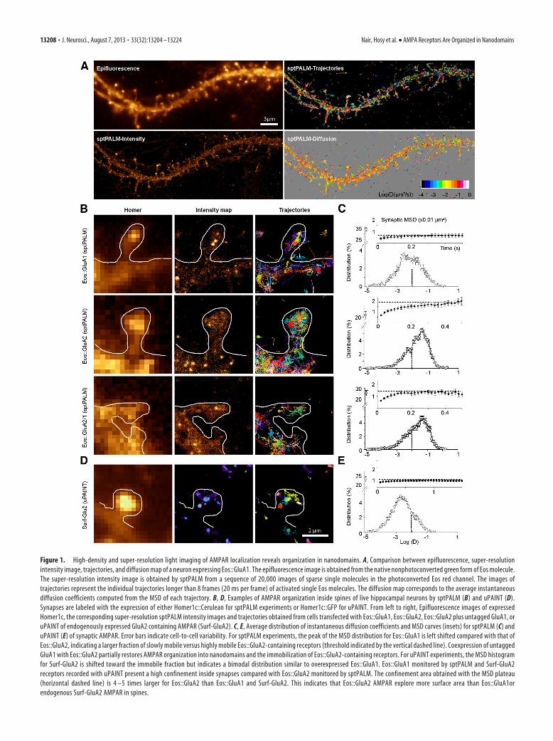
Figure 1. High-density and super-resolution light imaging of AMPAR localization reveals organization in nanodomains. A, Comparison between epifluorescence, super-resolutionintensity image, trajectories, and diffusion map of a neuron expressing Eos::GluA1. The epifluorescence image is obtained from the native nonphotoconverted green form of Eos molecule.The super-resolution intensity image is obtained by sptPALM from a sequence of 20,000 images of sparse single molecules in the photoconverted Eos red channel. The images oftrajectories represent the individual trajectories longer than 8 frames (20 ms per frame) of activated single Eos molecules. The diffusion map corresponds to the average instantaneousdiffusion coefficients computed from the MSD of each trajectory. B, D, Examples of AMPAR organization inside spines of live hippocampal neurons by sptPALM (B) and uPAINT (D).Synapses are labeled with the expression of either Homer1c::Cerulean for sptPALM experiments or Homer1c::GFP for uPAINT. From left to right, Epifluorescence images of expressedHomer1c, the corresponding super-resolution sptPALM intensity images and trajectories obtained from cells transfected with Eos::GluA1, Eos::GluA2, Eos::GluA2 plus untagged GluA1, oruPAINT of endogenously expressed GluA2 containing AMPAR (Surf-GluA2). C, E, Average distribution of instantaneous diffusion coefficients and MSD curves (insets) for sptPALM (C) anduPAINT (E) of synaptic AMPAR. Error bars indicate cell-to-cell variability. For sptPALM experiments, the peak of the MSD distribution for Eos::GluA1 is left shifted compared with that ofEos::GluA2, indicating a larger fraction of slowly mobile versus highly mobile Eos::GluA2-containing receptors (threshold indicated by the vertical dashed line). Coexpression of untaggedGluA1 with Eos::GluA2 partially restores AMPAR organization into nanodomains and the immobilization of Eos::GluA2-containing receptors. For uPAINT experiments, the MSD histogramfor Surf-GluA2 is shifted toward the immobile fraction but indicates a bimodal distribution similar to overexpressed Eos::GluA1. Eos::GluA1 monitored by sptPALM and Surf-GluA2receptors recorded with uPAINT present a high confinement inside synapses compared with Eos::GluA2 monitored by sptPALM. The confinement area obtained with the MSD plateau(horizontal dashed line) is 4 –5 times larger for Eos::GluA2 than Eos::GluA1 and Surf-GluA2. This indicates that Eos::GluA2 AMPAR explore more surface area than Eos::GluA1orendogenous Surf-GluA2 AMPAR in spines.
13208 • J. Neurosci., August 7, 2013 • 33(32):13204 –13224 Nair, Hosy et al. • AMPA Receptors Are Organized in Nanodomains
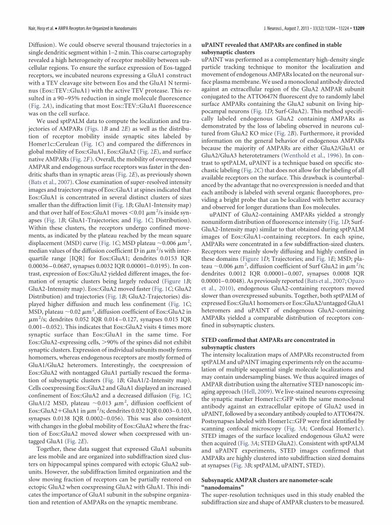
Diffusion). We could observe several thousand trajectories in asingle dendritic segment within 1–2 min. This coarse cartographyrevealed a high heterogeneity of receptor mobility between sub-cellular regions. To ensure the surface expression of Eos-taggedreceptors, we incubated neurons expressing a GluA1 constructwith a TEV cleavage site between Eos and the GluA1 N termi-nus (Eos::TEV::GluA1) with the active TEV protease. This re-sulted in a 90 –95% reduction in single molecule fluorescence(Fig. 2A), indicating that most Eos::TEV::GluA1 fluorescencewas on the cell surface.
We used sptPALM data to compute the localization and tra-jectories of AMPARs (Figs. 1B and 2E) as well as the distribu-tion of receptor mobility inside synaptic sites labeled byHomer1c::Cerulean (Fig. 1C) and compared the differences inglobal mobility of Eos::GluA1, Eos::GluA2 (Fig. 2E), and surfacenative AMPARs (Fig. 2F). Overall, the mobility of overexpressedAMPAR and endogenous surface receptors was faster in the den-dritic shafts than in synaptic areas (Fig. 2E), as previously shown(Bats et al., 2007). Close examination of super-resolved intensityimages and trajectory maps of Eos::GluA1 at spines indicated thatEos::GluA1 is concentrated in several distinct clusters of sizessmaller than the diffraction limit (Fig. 1B; GluA1-Intensity map)and that over half of Eos::GluA1 moves �0.01 �m 2/s inside syn-apses (Fig. 1B; GluA1-Trajectories; and Fig. 1C; Distribution).Within these clusters, the receptors undergo confined move-ments, as indicated by the plateau reached by the mean squaredisplacement (MSD) curve (Fig. 1C; MSD plateau �0.006 �m 2,median values of the diffusion coefficient D in �m 2/s with inter-quartile range [IQR] for Eos::GluA1; dendrites 0.0153 IQR0.00036 – 0.0687, synapses 0.0032 IQR 0.00001– 0.0195). In con-trast, expression of Eos::GluA2 yielded different images, the for-mation of synaptic clusters being largely reduced (Figure 1B;GluA2-Intensity map). Eos::GluA2 moved faster (Fig. 1C; GluA2Distribution) and trajectories (Fig. 1B; GluA2-Trajectories) dis-played higher diffusion and much less confinement (Fig. 1C;MSD, plateau �0.02 �m 2, diffusion coefficient of Eos::GluA2 in�m 2/s; dendrites 0.052 IQR 0.014 – 0.127, synapses 0.015 IQR0.001– 0.052). This indicates that Eos::GluA2 visits 4 times moresynaptic surface than Eos::GluA1 in the same time. ForEos::GluA2-expressing cells, �90% of the spines did not exhibitsynaptic clusters. Expression of individual subunits mostly formshomomers, whereas endogenous receptors are mostly formed ofGluA1/GluA2 heteromers. Interestingly, the coexpression ofEos::GluA2 with nontagged GluA1 partially rescued the forma-tion of subsynaptic clusters (Fig. 1B; GluA1/2-Intensity map).Cells coexpressing Eos::GluA2 and GluA1 displayed an increasedconfinement of Eos::GluA2 and a decreased diffusion (Fig. 1C;GluA1/2 MSD, plateau �0.013 �m 2, diffusion coefficient ofEos::GluA2�GluA1 in �m 2/s; dendrites 0.032 IQR 0.003– 0.103,synapses 0.0138 IQR 0.0002– 0.056). This was also consistentwith changes in the global mobility of Eos::GluA2 where the frac-tion of Eos::GluA2 moved slower when coexpressed with un-tagged GluA1 (Fig. 2E).
Together, these data suggest that expressed GluA1 subunitsare less mobile and are organized into subdiffraction sized clus-ters on hippocampal spines compared with ectopic GluA2 sub-units. However, the subdiffraction limited organization and theslow moving fraction of receptors can be partially restored onectopic GluA2 when coexpressing GluA2 with GluA1. This indi-cates the importance of GluA1 subunit in the subspine organiza-tion and retention of AMPARs on the synaptic membrane.
uPAINT revealed that AMPARs are confined in stablesubsynaptic clustersuPAINT was performed as a complementary high-density singleparticle tracking technique to monitor the localization andmovement of endogenous AMPARs located on the neuronal sur-face plasma membrane. We used a monoclonal antibody directedagainst an extracellular region of the GluA2 AMPAR subunitconjugated to the ATTO647N fluorescent dye to randomly labelsurface AMPARs containing the GluA2 subunit on living hip-pocampal neurons (Fig. 1D; Surf-GluA2). This method specifi-cally labeled endogenous GluA2 containing AMPARs asdemonstrated by the loss of labeling observed in neurons cul-tured from GluA2 KO mice (Fig. 2B). Furthermore, it providedinformation on the general behavior of endogenous AMPARsbecause the majority of AMPARs are either GluA2/GluA1 orGluA2/GluA3 heterotetramers (Wenthold et al., 1996). In con-trast to sptPALM, uPAINT is a technique based on specific sto-chastic labeling (Fig. 2C) that does not allow for the labeling of allavailable receptors on the surface. This drawback is counterbal-anced by the advantage that no overexpression is needed and thateach antibody is labeled with several organic fluorophores, pro-viding a bright probe that can be localized with better accuracyand observed for longer durations than Eos molecules.
uPAINT of GluA2-containing AMPARs yielded a stronglynonuniform distribution of fluorescence intensity (Fig. 1D; Surf-GluA2-Intensity map) similar to that obtained during sptPALMimages of Eos::GluA1-containing receptors. In each spine,AMPARs were concentrated in a few subdiffraction-sized clusters.Receptors were mainly slowly diffusing and highly confined inthese domains (Figure 1D; Trajectories; and Fig. 1E; MSD; pla-teau �0.006 �m 2, diffusion coefficient of Surf GluA2 in �m 2/s;dendrites 0.0012 IQR 0.00001– 0.007, synapses 0.0008 IQR0.00001– 0.0048). As previously reported (Bats et al., 2007; Opazoet al., 2010), endogenous GluA2-containing receptors movedslower than overexpressed subunits. Together, both sptPALM ofexpressed Eos::GluA1 homomers or Eos::GluA2/untagged GluA1heteromers and uPAINT of endogenous GluA2-containingAMPARs yielded a comparable distribution of receptors con-fined in subsynaptic clusters.
STED confirmed that AMPARs are concentrated insubsynaptic clustersThe intensity localization maps of AMPARs reconstructed fromsptPALM and uPAINT imaging experiments rely on the accumu-lation of multiple sequential single molecule localizations andmay contain undersampling biases. We thus acquired images ofAMPAR distribution using the alternative STED nanoscopic im-aging approach (Hell, 2009). We live-stained neurons expressingthe synaptic marker Homer1c::GFP with the same monoclonalantibody against an extracellular epitope of GluA2 used inuPAINT, followed by a secondary antibody coupled to ATTO647N.Postsynapses labeled with Homer1c::GFP were first identified byscanning confocal microscopy (Fig. 3A; Confocal Homer1c).STED images of the surface localized endogenous GluA2 werethen acquired (Fig. 3A; STED GluA2). Consistent with sptPALMand uPAINT experiments, STED images confirmed thatAMPARs are highly clustered into subdiffraction sized domainsat synapses (Fig. 3B; sptPALM, uPAINT, STED).
Subsynaptic AMPAR clusters are nanometer-scale“nanodomains”The super-resolution techniques used in this study enabled thesubdiffraction size and shape of AMPAR clusters to be measured.
Nair, Hosy et al. • AMPA Receptors Are Organized in Nanodomains J. Neurosci., August 7, 2013 • 33(32):13204 –13224 • 13209
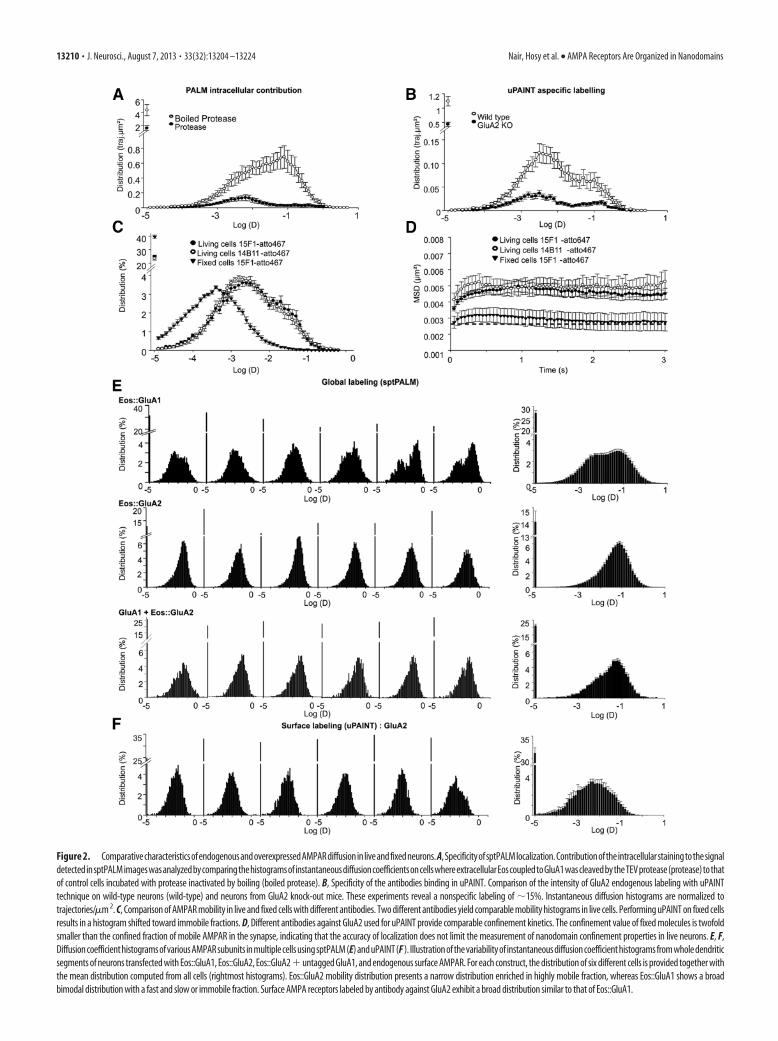
Figure 2. ComparativecharacteristicsofendogenousandoverexpressedAMPARdiffusioninliveandfixedneurons.A,SpecificityofsptPALMlocalization.ContributionoftheintracellularstainingtothesignaldetectedinsptPALMimageswasanalyzedbycomparingthehistogramsofinstantaneousdiffusioncoefficientsoncellswhereextracellularEoscoupledtoGluA1wascleavedbytheTEVprotease(protease)tothatof control cells incubated with protease inactivated by boiling (boiled protease). B, Specificity of the antibodies binding in uPAINT. Comparison of the intensity of GluA2 endogenous labeling with uPAINTtechnique on wild-type neurons (wild-type) and neurons from GluA2 knock-out mice. These experiments reveal a nonspecific labeling of �15%. Instantaneous diffusion histograms are normalized totrajectories/�m 2. C, Comparison of AMPAR mobility in live and fixed cells with different antibodies. Two different antibodies yield comparable mobility histograms in live cells. Performing uPAINT on fixed cellsresults in a histogram shifted toward immobile fractions. D, Different antibodies against GluA2 used for uPAINT provide comparable confinement kinetics. The confinement value of fixed molecules is twofoldsmaller than the confined fraction of mobile AMPAR in the synapse, indicating that the accuracy of localization does not limit the measurement of nanodomain confinement properties in live neurons. E, F,Diffusion coefficient histograms of various AMPAR subunits in multiple cells using sptPALM (E) and uPAINT (F ). Illustration of the variability of instantaneous diffusion coefficient histograms from whole dendriticsegments of neurons transfected with Eos::GluA1, Eos::GluA2, Eos::GluA2�untagged GluA1, and endogenous surface AMPAR. For each construct, the distribution of six different cells is provided together withthe mean distribution computed from all cells (rightmost histograms). Eos::GluA2 mobility distribution presents a narrow distribution enriched in highly mobile fraction, whereas Eos::GluA1 shows a broadbimodal distribution with a fast and slow or immobile fraction. Surface AMPA receptors labeled by antibody against GluA2 exhibit a broad distribution similar to that of Eos::GluA1.
13210 • J. Neurosci., August 7, 2013 • 33(32):13204 –13224 Nair, Hosy et al. • AMPA Receptors Are Organized in Nanodomains
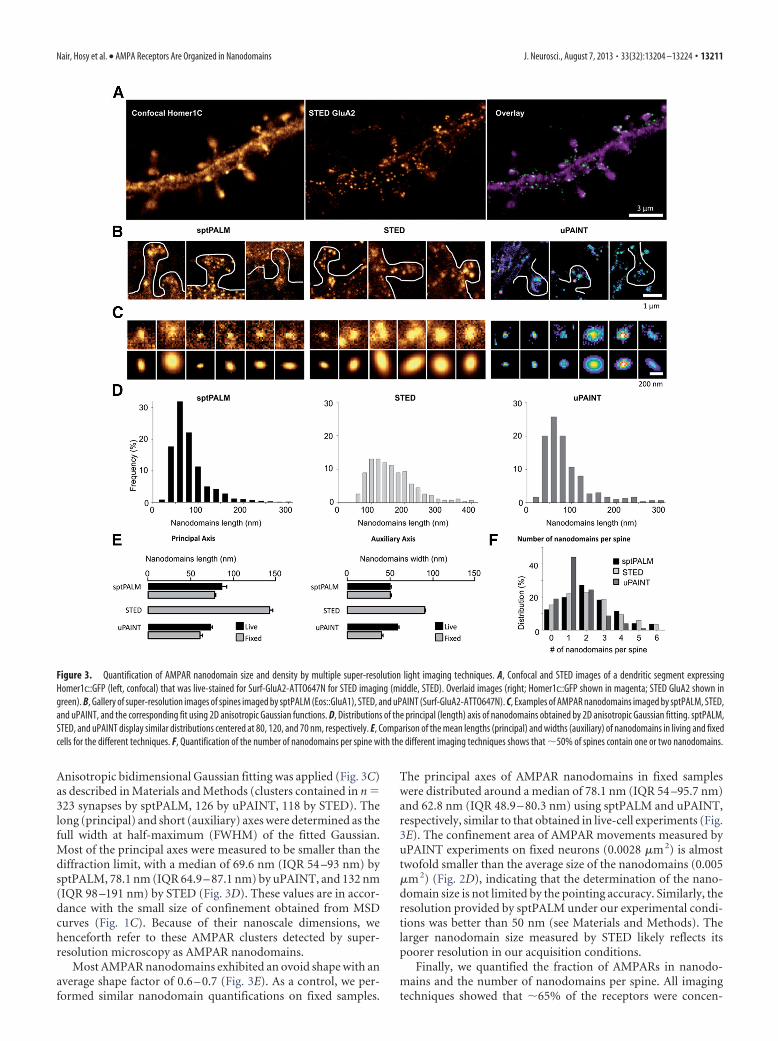
Anisotropic bidimensional Gaussian fitting was applied (Fig. 3C)as described in Materials and Methods (clusters contained in n 323 synapses by sptPALM, 126 by uPAINT, 118 by STED). Thelong (principal) and short (auxiliary) axes were determined as thefull width at half-maximum (FWHM) of the fitted Gaussian.Most of the principal axes were measured to be smaller than thediffraction limit, with a median of 69.6 nm (IQR 54 –93 nm) bysptPALM, 78.1 nm (IQR 64.9 – 87.1 nm) by uPAINT, and 132 nm(IQR 98 –191 nm) by STED (Fig. 3D). These values are in accor-dance with the small size of confinement obtained from MSDcurves (Fig. 1C). Because of their nanoscale dimensions, wehenceforth refer to these AMPAR clusters detected by super-resolution microscopy as AMPAR nanodomains.
Most AMPAR nanodomains exhibited an ovoid shape with anaverage shape factor of 0.6 – 0.7 (Fig. 3E). As a control, we per-formed similar nanodomain quantifications on fixed samples.
The principal axes of AMPAR nanodomains in fixed sampleswere distributed around a median of 78.1 nm (IQR 54 –95.7 nm)and 62.8 nm (IQR 48.9 – 80.3 nm) using sptPALM and uPAINT,respectively, similar to that obtained in live-cell experiments (Fig.3E). The confinement area of AMPAR movements measured byuPAINT experiments on fixed neurons (0.0028 �m 2) is almosttwofold smaller than the average size of the nanodomains (0.005�m 2) (Fig. 2D), indicating that the determination of the nano-domain size is not limited by the pointing accuracy. Similarly, theresolution provided by sptPALM under our experimental condi-tions was better than 50 nm (see Materials and Methods). Thelarger nanodomain size measured by STED likely reflects itspoorer resolution in our acquisition conditions.
Finally, we quantified the fraction of AMPARs in nanodo-mains and the number of nanodomains per spine. All imagingtechniques showed that �65% of the receptors were concen-
Figure 3. Quantification of AMPAR nanodomain size and density by multiple super-resolution light imaging techniques. A, Confocal and STED images of a dendritic segment expressingHomer1c::GFP (left, confocal) that was live-stained for Surf-GluA2-ATTO647N for STED imaging (middle, STED). Overlaid images (right; Homer1c::GFP shown in magenta; STED GluA2 shown ingreen). B, Gallery of super-resolution images of spines imaged by sptPALM (Eos::GluA1), STED, and uPAINT (Surf-GluA2-ATTO647N). C, Examples of AMPAR nanodomains imaged by sptPALM, STED,and uPAINT, and the corresponding fit using 2D anisotropic Gaussian functions. D, Distributions of the principal (length) axis of nanodomains obtained by 2D anisotropic Gaussian fitting. sptPALM,STED, and uPAINT display similar distributions centered at 80, 120, and 70 nm, respectively. E, Comparison of the mean lengths (principal) and widths (auxiliary) of nanodomains in living and fixedcells for the different techniques. F, Quantification of the number of nanodomains per spine with the different imaging techniques shows that �50% of spines contain one or two nanodomains.
Nair, Hosy et al. • AMPA Receptors Are Organized in Nanodomains J. Neurosci., August 7, 2013 • 33(32):13204 –13224 • 13211
trated in nanodomains and that �80% of spines contained atleast one nanodomain (88% by sptPALM, 82% by uPAINT, and85% by STED) (Fig. 3F). The average number of nanodomainsper spine was �2.5 (2.42 � 0.18, n 584 spines) when measuredwith sptPALM. STED yielded similar results (2.38 � 0.17, n 351 spines), whereas uPAINT led to a lower average of 1.4 � 0.1(n 299 spines), probably because of the intrinsic underlabelingof the technique (Fig. 3F). Spines without nanodomains usuallyexhibited a low level of diffuse AMPAR staining that was notfurther quantified.
AMPAR nanodomains detected by live-labeling andpre-embedding immunogold electron microscopyDespite the advent of super-resolution optical microscopy, im-munogold labeling by EM still provides the highest resolution forAMPAR localization and remains the “gold standard” (Kharaziaet al., 1996; Bernard et al., 1997; Kharazia and Weinberg, 1997;Nusser et al., 1998; Takumi et al., 1999b; Ganeshina et al., 2004),with the important drawback that it can only image fixed tissue.Most previous EM studies have used the postembedding immu-nolabeling method, which provides incomplete informationabout the distributions of receptors because of its relatively lowsensitivity (Nusser et al., 1998; Masugi-Tokita and Shigemoto,2007). Pre-embedding immunogold labeling (Bernard et al.,1997) allows mapping the distribution of surface AMPARs withincreased sensitivity and has been recently used to observe thedistribution of GluA2, the most prevalent subunit in AMPARs(Tao-Cheng et al., 2011). Here, we performed similar pre-embedding immunogold labeling, with two deviations. First, we
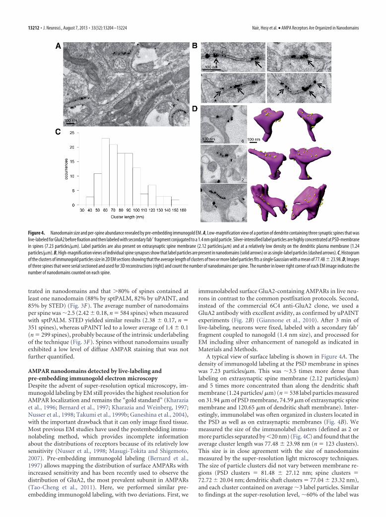
immunolabeled surface GluA2-containing AMPARs in live neu-rons in contrast to the common postfixation protocols. Second,instead of the commercial 6C4 anti-GluA2 clone, we used aGluA2 antibody with excellent avidity, as confirmed by uPAINTexperiments (Fig. 2B) (Giannone et al., 2010). After 3 min oflive-labeling, neurons were fixed, labeled with a secondary fab�fragment coupled to nanogold (1.4 nm size), and processed forEM including silver enhancement of nanogold as indicated inMaterials and Methods.
A typical view of surface labeling is shown in Figure 4A. Thedensity of immunogold labeling at the PSD membrane in spineswas 7.23 particles/�m. This was �3.5 times more dense thanlabeling on extrasynaptic spine membrane (2.12 particles/�m)and 5 times more concentrated than along the dendritic shaftmembrane (1.24 particles/ �m) (n 538 label particles measuredon 31.94 �m of PSD membrane, 74.59 �m of extrasynaptic spinemembrane and 120.65 �m of dendritic shaft membrane). Inter-estingly, immunolabel was often organized in clusters located inthe PSD as well as on extrasynaptic membranes (Fig. 4B). Wemeasured the size of the immunolabel clusters (defined as 2 ormore particles separated by �20 nm) (Fig. 4C) and found that theaverage cluster length was 77.48 � 23.98 nm (n 123 clusters).This size is in close agreement with the size of nanodomainsmeasured by the super-resolution light microscopy techniques.The size of particle clusters did not vary between membrane re-gions (PSD clusters 81.48 � 27.12 nm; spine clusters 72.72 � 20.04 nm; dendritic shaft clusters 77.04 � 23.32 nm),and each cluster contained on average �3 label particles. Similarto findings at the super-resolution level, �60% of the label was
Figure 4. Nanodomain size and per-spine abundance revealed by pre-embedding immunogold EM. A, Low-magnification view of a portion of dendrite containing three synaptic spines that waslive-labeled for GluA2 before fixation and then labeled with secondary fab� fragment conjugated to a 1.4 nm gold particle. Silver-intensified label particles are highly concentrated at PSD-membranein spines (7.23 particles/�m). Label particles are also present on extrasynaptic spine membrane (2.12 particles/�m) and at a relatively low density on the dendritic plasma membrane (1.24particles/�m). B, High-magnification views of individual spine synapses show that label particles are present in nanodomains (solid arrows) or as single-label particles (dashed arrows). C, Histogramof the clusters of immunogold particles size in 2D EM sections showing that the average length of clusters of two or more label particles fits a single Gaussian with a mean of 77.48 � 23.98. D, Imagesof three spines that were serial sectioned and used for 3D reconstructions (right) and count the number of nanodomains per spine. The number in lower right corner of each EM image indicates thenumber of nanodomains counted on each spine.
13212 • J. Neurosci., August 7, 2013 • 33(32):13204 –13224 Nair, Hosy et al. • AMPA Receptors Are Organized in Nanodomains
contained in a cluster, a value that was consistent between mem-brane regions (% of particles in cluster: PSD 55%, spine 60%,dendritic shaft 66%). Based on these similarities, we concludedthat the clustered immunolabel viewed at the EM level corre-sponds to nanodomains detected using super-resolution lightmicroscopy.
To count the number of nanodomains present on individualspines and specify their location in the PSD or extrasynaptic spinemembrane, seven spines were reconstructed by serial section EM(Fig. 4D). On average, there were 2.14 � 0.90 nanodomains in thePSD and 4.14 � 1.86 nanodomains on the spine extrasynapticmembrane.
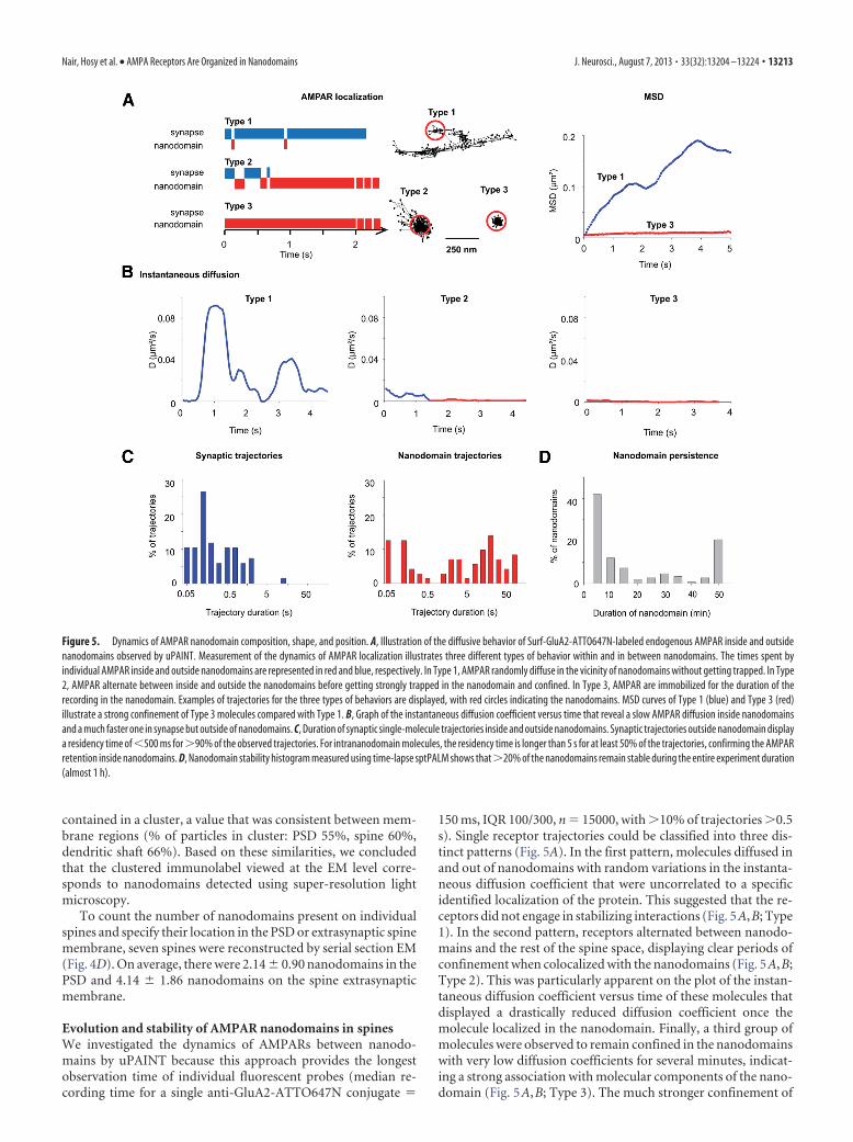
Evolution and stability of AMPAR nanodomains in spinesWe investigated the dynamics of AMPARs between nanodo-mains by uPAINT because this approach provides the longestobservation time of individual fluorescent probes (median re-cording time for a single anti-GluA2-ATTO647N conjugate
150 ms, IQR 100/300, n 15000, with �10% of trajectories �0.5s). Single receptor trajectories could be classified into three dis-tinct patterns (Fig. 5A). In the first pattern, molecules diffused inand out of nanodomains with random variations in the instanta-neous diffusion coefficient that were uncorrelated to a specificidentified localization of the protein. This suggested that the re-ceptors did not engage in stabilizing interactions (Fig. 5A,B; Type1). In the second pattern, receptors alternated between nanodo-mains and the rest of the spine space, displaying clear periods ofconfinement when colocalized with the nanodomains (Fig. 5A,B;Type 2). This was particularly apparent on the plot of the instan-taneous diffusion coefficient versus time of these molecules thatdisplayed a drastically reduced diffusion coefficient once themolecule localized in the nanodomain. Finally, a third group ofmolecules were observed to remain confined in the nanodomainswith very low diffusion coefficients for several minutes, indicat-ing a strong association with molecular components of the nano-domain (Fig. 5A,B; Type 3). The much stronger confinement of
Figure 5. Dynamics of AMPAR nanodomain composition, shape, and position. A, Illustration of the diffusive behavior of Surf-GluA2-ATTO647N-labeled endogenous AMPAR inside and outsidenanodomains observed by uPAINT. Measurement of the dynamics of AMPAR localization illustrates three different types of behavior within and in between nanodomains. The times spent byindividual AMPAR inside and outside nanodomains are represented in red and blue, respectively. In Type 1, AMPAR randomly diffuse in the vicinity of nanodomains without getting trapped. In Type2, AMPAR alternate between inside and outside the nanodomains before getting strongly trapped in the nanodomain and confined. In Type 3, AMPAR are immobilized for the duration of therecording in the nanodomain. Examples of trajectories for the three types of behaviors are displayed, with red circles indicating the nanodomains. MSD curves of Type 1 (blue) and Type 3 (red)illustrate a strong confinement of Type 3 molecules compared with Type 1. B, Graph of the instantaneous diffusion coefficient versus time that reveal a slow AMPAR diffusion inside nanodomainsand a much faster one in synapse but outside of nanodomains. C, Duration of synaptic single-molecule trajectories inside and outside nanodomains. Synaptic trajectories outside nanodomain displaya residency time of �500 ms for �90% of the observed trajectories. For intrananodomain molecules, the residency time is longer than 5 s for at least 50% of the trajectories, confirming the AMPARretention inside nanodomains. D, Nanodomain stability histogram measured using time-lapse sptPALM shows that �20% of the nanodomains remain stable during the entire experiment duration(almost 1 h).
Nair, Hosy et al. • AMPA Receptors Are Organized in Nanodomains J. Neurosci., August 7, 2013 • 33(32):13204 –13224 • 13213
receptors in nanodomains was also apparent when comparingthe average MSD for the type 1 and type 3 mobility modes (Fig.5A; MSD). These plots also indicate that, in between nanodo-mains, the receptors undergo rapid free diffusion. Based on thisobservation, synapses were divided into two functional zonesdiscriminated by receptor mobility: zones inside nanodomainsand zones outside nanodomains. Interestingly, the residencetime of receptors outside nanodomains was short while they ran-domly moved rapidly (median residence in extra-nanodomain 0.25 s, IQR 0.1/0.7, n 161; median total trajectory length 0.75 s,IQR 0.55/1.4, n 5000). In contrast, molecules were confinedinside nanodomains for much longer durations, going up to fewminutes (median residence inside nanodomains 2.25 s, IQR0.1/16.2, n 155; Fig. 5C). Together, these data indicate that,inside synapses, AMPARs diffuse freely between nanodomainsthen rapidly hop inside nanodomains where they are stronglyimmobilized and reside for long periods. These stabilization do-mains could be related to the attracting molecular interactionpotential wells we recently found by mathematical analysis per-formed on sptPALM data (Hoze et al., 2012).
Then, we investigated the temporal stability of nanodomainsusing time-lapse sptPALM. We performed 50 s periods of spt-PALM acquisitions every 5 min over a time period of 45 min. Wecould identify and track individual nanodomains in the spine forseveral tens of minutes (Fig. 6A,B). As quantified in Figure 5D,�20% of the spines (n 75) had a stable nanodomain during theentire acquisition time (45 min). Approximately 40% of thespines contained nanodomains, which could not be detected forlonger than 5 min of observation, indicating that a fraction of thenanodomains in the spine are either dynamic or transient,whereas others have a very stable localization and lifetime. Nano-domains in the remaining 60% of the spines were stable for �5min inside the synapses. More than 50% of spines had multiplenanodomains, bearing combinations of stable and nonstablenanodomains in the same synapse (Fig. 5D). Kymographs per-formed on highly stable nanodomains illustrate that nanodo-mains are visited by many molecules (Fig. 6A,B).
Finally, we analyzed the differences of mobility between inand out nanodomains, from single-molecule trajectories ofEos::GluA1 molecules obtained by sptPALM. Most of the immo-bilized molecules resided within the nanodomains, whereas mol-ecules that were either outside or moving in and out of thenanodomains were either diffusive or weakly confined (Fig. 6C).We categorized the trajectories into three families, based on thenanoscale spatial heterogeneity as follows: (1) exclusively insidethe nanodomain (In), (2) exclusively outside nanodomain (Out),and (3) those who visited the nanodomains for a minimum of10% and maximum of 80% of the duration of their observedtrajectories (In/Out). The cumulative distribution of the instan-taneous diffusion of the trajectories displayed a shift towardlower mobility within, and higher mobility outside the nanodo-mains (Fig. 6D). The instantaneous diffusion distributions ofIn/Out molecules indicated an intermediate shift of mobilitycompared with In and Out ones (Fig. 6D). We also plotted theaverage of mean square diffusion curves for each of the threefamilies of receptors (In, Out, and In/Out). The mean squarediffusion of the molecules within nanodomains (In) displayed astrong confinement indicating immobilization (Fig. 6E). Recep-tors outside nanodomains (Out) exhibited unhindered Brown-ian diffusion. The mean square diffusion of receptors temporarilyassociated with nanodomains (In/Out) displayed weak confine-ment, indicating the transient changes in the mobility betweenstrong confinement and unhindered diffusion. These data con-
firm that nanodomains are indeed zones of strong confinementwhere AMPA receptors are transiently trapped with the potentialfor receptor to exchange and diffuse freely into the membraneoutside the nanodomain.
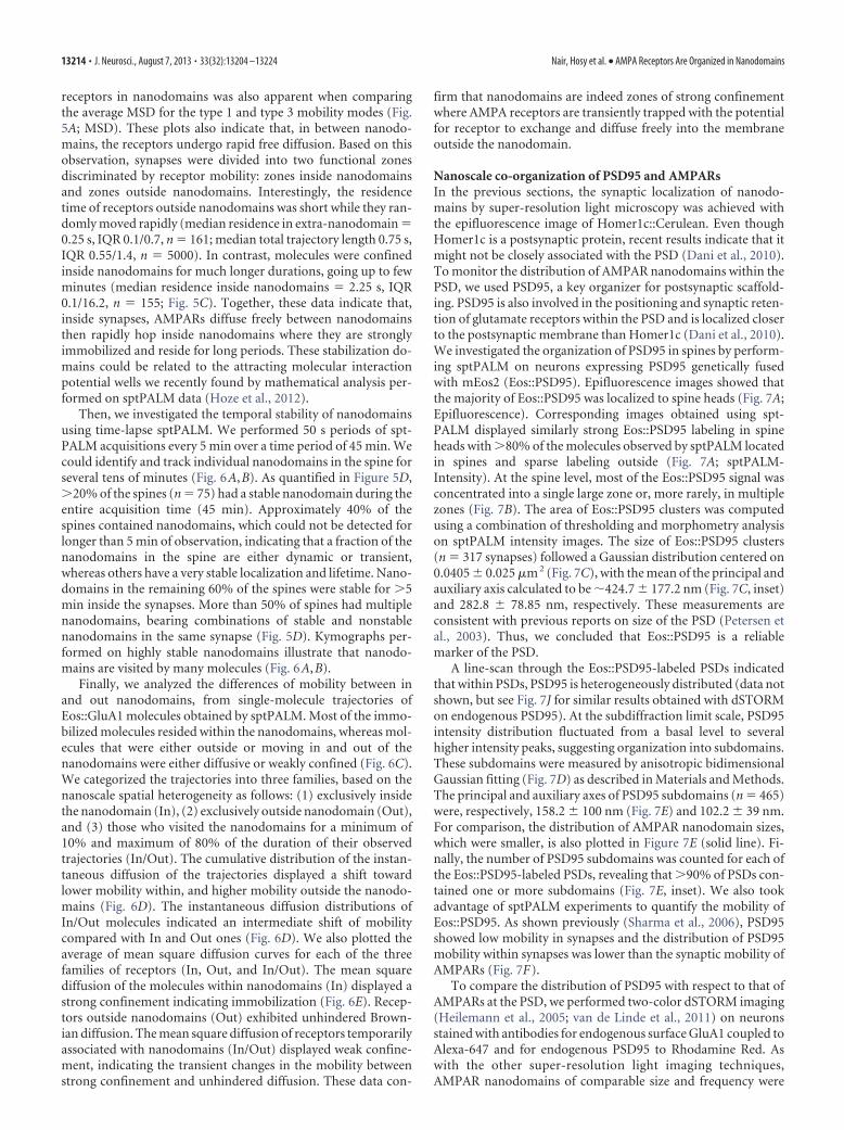
Nanoscale co-organization of PSD95 and AMPARsIn the previous sections, the synaptic localization of nanodo-mains by super-resolution light microscopy was achieved withthe epifluorescence image of Homer1c::Cerulean. Even thoughHomer1c is a postsynaptic protein, recent results indicate that itmight not be closely associated with the PSD (Dani et al., 2010).To monitor the distribution of AMPAR nanodomains within thePSD, we used PSD95, a key organizer for postsynaptic scaffold-ing. PSD95 is also involved in the positioning and synaptic reten-tion of glutamate receptors within the PSD and is localized closerto the postsynaptic membrane than Homer1c (Dani et al., 2010).We investigated the organization of PSD95 in spines by perform-ing sptPALM on neurons expressing PSD95 genetically fusedwith mEos2 (Eos::PSD95). Epifluorescence images showed thatthe majority of Eos::PSD95 was localized to spine heads (Fig. 7A;Epifluorescence). Corresponding images obtained using spt-PALM displayed similarly strong Eos::PSD95 labeling in spineheads with �80% of the molecules observed by sptPALM locatedin spines and sparse labeling outside (Fig. 7A; sptPALM-Intensity). At the spine level, most of the Eos::PSD95 signal wasconcentrated into a single large zone or, more rarely, in multiplezones (Fig. 7B). The area of Eos::PSD95 clusters was computedusing a combination of thresholding and morphometry analysison sptPALM intensity images. The size of Eos::PSD95 clusters(n 317 synapses) followed a Gaussian distribution centered on0.0405 � 0.025 �m 2 (Fig. 7C), with the mean of the principal andauxiliary axis calculated to be �424.7 � 177.2 nm (Fig. 7C, inset)and 282.8 � 78.85 nm, respectively. These measurements areconsistent with previous reports on size of the PSD (Petersen etal., 2003). Thus, we concluded that Eos::PSD95 is a reliablemarker of the PSD.
A line-scan through the Eos::PSD95-labeled PSDs indicatedthat within PSDs, PSD95 is heterogeneously distributed (data notshown, but see Fig. 7J for similar results obtained with dSTORMon endogenous PSD95). At the subdiffraction limit scale, PSD95intensity distribution fluctuated from a basal level to severalhigher intensity peaks, suggesting organization into subdomains.These subdomains were measured by anisotropic bidimensionalGaussian fitting (Fig. 7D) as described in Materials and Methods.The principal and auxiliary axes of PSD95 subdomains (n 465)were, respectively, 158.2 � 100 nm (Fig. 7E) and 102.2 � 39 nm.For comparison, the distribution of AMPAR nanodomain sizes,which were smaller, is also plotted in Figure 7E (solid line). Fi-nally, the number of PSD95 subdomains was counted for each ofthe Eos::PSD95-labeled PSDs, revealing that �90% of PSDs con-tained one or more subdomains (Fig. 7E, inset). We also tookadvantage of sptPALM experiments to quantify the mobility ofEos::PSD95. As shown previously (Sharma et al., 2006), PSD95showed low mobility in synapses and the distribution of PSD95mobility within synapses was lower than the synaptic mobility ofAMPARs (Fig. 7F).
To compare the distribution of PSD95 with respect to that ofAMPARs at the PSD, we performed two-color dSTORM imaging(Heilemann et al., 2005; van de Linde et al., 2011) on neuronsstained with antibodies for endogenous surface GluA1 coupled toAlexa-647 and for endogenous PSD95 to Rhodamine Red. Aswith the other super-resolution light imaging techniques,AMPAR nanodomains of comparable size and frequency were
13214 • J. Neurosci., August 7, 2013 • 33(32):13204 –13224 Nair, Hosy et al. • AMPA Receptors Are Organized in Nanodomains
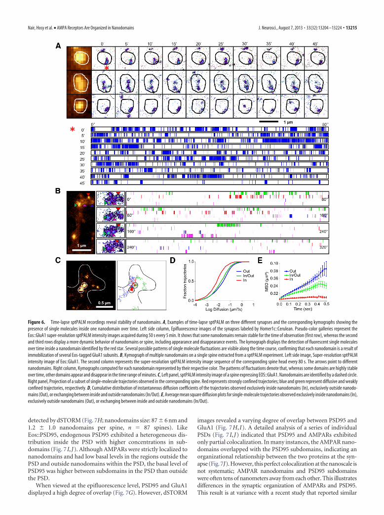
detected by dSTORM (Fig. 7H; nanodomains size: 87 � 6 nm and1.2 � 1.0 nanodomains per spine, n 87 spines). LikeEos::PSD95, endogenous PSD95 exhibited a heterogeneous dis-tribution inside the PSD with higher concentrations in sub-domains (Fig. 7 I, J). Although AMPARs were strictly localized tonanodomains and had low basal levels in the regions outside thePSD and outside nanodomains within the PSD, the basal level ofPSD95 was higher between subdomains in the PSD than outsidethe PSD.
When viewed at the epifluorescence level, PSD95 and GluA1displayed a high degree of overlap (Fig. 7G). However, dSTORM
images revealed a varying degree of overlap between PSD95 andGluA1 (Fig. 7H, I). A detailed analysis of a series of individualPSDs (Fig. 7 I, J) indicated that PSD95 and AMPARs exhibitedonly partial colocalization. In many instances, the AMPAR nano-domains overlapped with the PSD95 subdomains, indicating anorganizational relationship between the two proteins at the syn-apse (Fig. 7J). However, this perfect colocalization at the nanoscale isnot systematic; AMPAR nanodomains and PSD95 subdomainswere often tens of nanometers away from each other. This illustratesdifferences in the synaptic organization of AMPARs and PSD95.This result is at variance with a recent study that reported similar
Figure 6. Time-lapse sptPALM recordings reveal stability of nanodomains. A, Examples of time-lapse sptPALM on three different synapses and the corresponding kymographs showing thepresence of single molecules inside one nanodomain over time. Left side column, Epifluorescence images of the synapses labeled by Homer1c::Cerulean. Pseudo-color galleries represent theEos::GluA1 super-resolution sptPALM intensity images acquired during 50 s every 5 min. It shows that some nanodomains remain stable for the time of observation (first row), whereas the secondand third rows display a more dynamic behavior of nanodomains or spine, including appearance and disappearance events. The kymograph displays the detection of fluorescent single moleculesover time inside a nanodomain identified by the red star. Several possible patterns of single molecule fluctuations are visible along the time course, confirming that each nanodomain is a result ofimmobilization of several Eos-tagged GluA1 subunits. B, Kymograph of multiple nanodomains on a single spine extracted from a sptPALM experiment. Left side image, Super-resolution sptPALMintensity image of Eos::GluA1. The second column represents the super-resolution sptPALM intensity image sequence of the corresponding spine head every 80 s. The arrows point to differentnanodomains. Right column, Kymographs computed for each nanodomain represented by their respective color. The patterns of fluctuations denote that, whereas some domains are highly stableover time, other domains appear and disappear in the time range of minutes. C, Left panel, sptPALM intensity image of a spine expressing EOS::GluA1. Nanodomains are identified by a dashed circle.Right panel, Projection of a subset of single-molecule trajectories observed in the corresponding spine. Red represents strongly confined trajectories; blue and green represent diffusive and weaklyconfined trajectories, respectively. D, Cumulative distribution of instantaneous diffusion coefficients of the trajectories observed exclusively inside nanodomains (In), exclusively outside nanodo-mains (Out), or exchanging between inside and outside nanodomains (In/Out). E, Average mean square diffusion plots for single-molecule trajectories observed exclusively inside nanodomains (In),exclusively outside nanodomains (Out), or exchanging between inside and outside nanodomains (In/Out).
Nair, Hosy et al. • AMPA Receptors Are Organized in Nanodomains J. Neurosci., August 7, 2013 • 33(32):13204 –13224 • 13215
Figure 7. Subdiffraction quantification of PSD95 organization in spines and comparison with endogenous AMPARs. A, Comparison between epifluorescence and super-resolution intensity imageof a neuron expressing Eos::PSD95. B, Gallery of super-resolution images of spines imaged by sptPALM (Eos::PSD95). C, Distribution of the area of the clusters of PSD95 from the spine with insetrepresenting the distribution of the cluster lengths. D, Examples of PSD95 subclusters acquired by sptPALM and the corresponding fit performed using 2D anisotropic Gaussian functions. E, Theprincipal axis (length) of PSD95 subclusters obtained by 2D anisotropic Gaussian fitting. The continuous line represents the corresponding distribution of GluA1 subunit (Figure legend continues.)
13216 • J. Neurosci., August 7, 2013 • 33(32):13204 –13224 Nair, Hosy et al. • AMPA Receptors Are Organized in Nanodomains
nanodomains than in our work but founda high degree of colocalization betweenAMPAR and PSD95 (Macgillavry et al.,2013). This may arise from the fact that thisstudy used overexpressed PSD95 orAMPARs, manipulations that may alterthe normal reciprocal distribution ofboth elements.
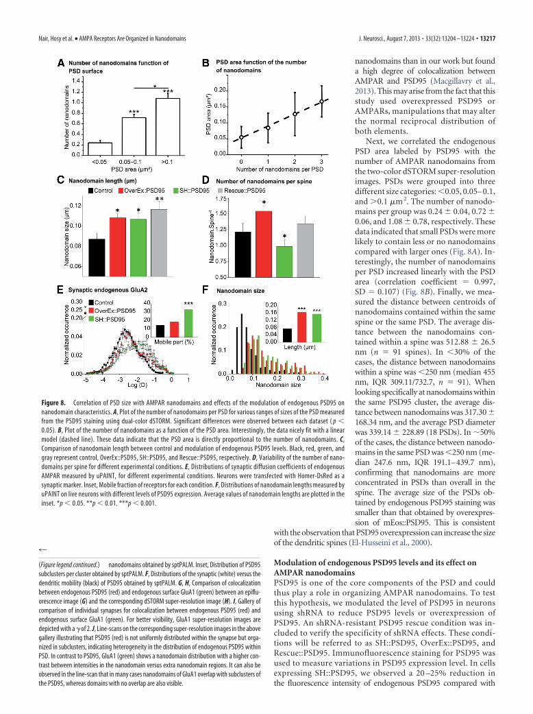
Next, we correlated the endogenousPSD area labeled by PSD95 with thenumber of AMPAR nanodomains fromthe two-color dSTORM super-resolutionimages. PSDs were grouped into threedifferent size categories: �0.05, 0.05– 0.1,and �0.1 �m 2. The number of nanodo-mains per group was 0.24 � 0.04, 0.72 �0.06, and 1.08 � 0.78, respectively. Thesedata indicated that small PSDs were morelikely to contain less or no nanodomainscompared with larger ones (Fig. 8A). In-terestingly, the number of nanodomainsper PSD increased linearly with the PSDarea (correlation coefficient 0.997,SD 0.107) (Fig. 8B). Finally, we mea-sured the distance between centroids ofnanodomains contained within the samespine or the same PSD. The average dis-tance between the nanodomains con-tained within a spine was 512.88 � 26.5nm (n 91 spines). In �30% of thecases, the distance between nanodomainswithin a spine was �250 nm (median 455nm, IQR 309.11/732.7, n 91). Whenlooking specifically at nanodomains withinthe same PSD95 cluster, the average dis-tance between nanodomains was 317.30 �168.34 nm, and the average PSD diameterwas 339.14 � 228.89 (18 PSDs). In �50%of the cases, the distance between nanodo-mains in the same PSD was �250 nm (me-dian 247.6 nm, IQR 191.1–439.7 nm),confirming that nanodomains are moreconcentrated in PSDs than overall in thespine. The average size of the PSDs ob-tained by endogenous PSD95 staining wassmaller than that obtained by overexpres-sion of mEos::PSD95. This is consistent
with the observation that PSD95 overexpression can increase the sizeof the dendritic spines (El-Husseini et al., 2000).
Modulation of endogenous PSD95 levels and its effect onAMPAR nanodomainsPSD95 is one of the core components of the PSD and couldthus play a role in organizing AMPAR nanodomains. To testthis hypothesis, we modulated the level of PSD95 in neuronsusing shRNA to reduce PSD95 levels or overexpression ofPSD95. An shRNA-resistant PSD95 rescue condition was in-cluded to verify the specificity of shRNA effects. These condi-tions will be referred to as SH::PSD95, OverEx::PSD95, andRescue::PSD95. Immunofluorescence staining for PSD95 wasused to measure variations in PSD95 expression level. In cellsexpressing SH::PSD95, we observed a 20 –25% reduction inthe fluorescence intensity of endogenous PSD95 compared with
Figure 8. Correlation of PSD size with AMPAR nanodomains and effects of the modulation of endogenous PSD95 onnanodomain characteristics. A, Plot of the number of nanodomains per PSD for various ranges of sizes of the PSD measuredfrom the PSD95 staining using dual-color dSTORM. Significant differences were observed between each dataset ( p �0.05). B, Plot of the number of nanodomains as a function of the PSD area. Interestingly, the data nicely fit with a linearmodel (dashed line). These data indicate that the PSD area is directly proportional to the number of nanodomains. C,Comparison of nanodomain length between control and modulation of endogenous PSD95 levels. Black, red, green, andgray represent control, OverEx::PSD95, SH::PSD95, and Rescue::PSD95, respectively. D, Variability of the number of nano-domains per spine for different experimental conditions. E, Distributions of synaptic diffusion coefficients of endogenousAMPAR measured by uPAINT, for different experimental conditions. Neurons were transfected with Homer-DsRed as asynaptic marker. Inset, Mobile fraction of receptors for each condition. F, Distributions of nanodomain lengths measured byuPAINT on live neurons with different levels of PSD95 expression. Average values of nanodomain lengths are plotted in theinset. *p � 0.05. **p � 0.01. ***p � 0.001.
4
(Figure legend continued.) nanodomains obtained by sptPALM. Inset, Distribution of PSD95subclusters per cluster obtained by sptPALM. F, Distributions of the synaptic (white) versus thedendritic mobility (black) of PSD95 obtained by sptPALM. G, H, Comparison of colocalizationbetween endogenous PSD95 (red) and endogenous surface GluA1 (green) between an epiflu-orescence image (G) and the corresponding dSTORM super-resolution image (H). I, Gallery ofcomparison of individual synapses for colocalization between endogenous PSD95 (red) andendogenous surface GluA1 (green). For better visibility, GluA1 super-resolution images aredepicted with a � of 2. J, Line-scans on the corresponding super-resolution images in the abovegallery illustrating that PSD95 (red) is not uniformly distributed within the synapse but orga-nized in subclusters, indicating heterogeneity in the distribution of endogenous PSD95 withinPSD. In contrast to PSD95, GluA1 (green) shows a nanodomain distribution with a higher con-trast between intensities in the nanodomain versus extra nanodomain regions. It can also beobserved in the line-scan that in many cases nanodomains of GluA1 overlap with subclusters ofthe PSD95, whereas domains with no overlap are also visible.
Nair, Hosy et al. • AMPA Receptors Are Organized in Nanodomains J. Neurosci., August 7, 2013 • 33(32):13204 –13224 • 13217
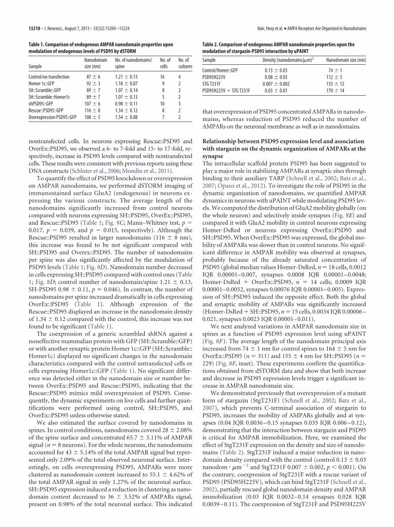
nontransfected cells. In neurons expressing Rescue::PSD95 andOverEx::PSD95, we observed a 6- to 7-fold and 15- to 17-fold, re-spectively, increase in PSD95 levels compared with nontransfectedcells. These results were consistent with previous reports using theseDNA constructs (Schluter et al., 2006; Mondin et al., 2011).
To quantify the effect of PSD95 knockdown or overexpressionon AMPAR nanodomains, we performed dSTORM imaging ofimmunostained surface GluA2 (endogenous) in neurons ex-pressing the various constructs. The average length of thenanodomains significantly increased from control neuronscompared with neurons expressing SH::PSD95, OverEx::PSD95,and Rescue::PSD95 (Table 1; Fig. 8C; Mann–Whitney test, p 0.017, p 0.039, and p 0.015, respectively). Although theRescue::PSD95 resulted in larger nanodomains (116 � 8 nm),this increase was found to be not significant compared withSH::PSD95 and Overex::PSD95. The number of nanodomainsper spine was also significantly affected by the modulation ofPSD95 levels (Table 1; Fig. 8D). Nanodomain number decreasedin cells expressing SH::PSD95 compared with control ones (Table1; Fig. 8D; control number of nanodomain/spine 1.21 � 0.13,SH-PSD95 0.98 � 0.11, p 0.046). In contrast, the number ofnanodomains per spine increased dramatically in cells expressingOverEx::PSD95 (Table 1). Although expression of theRescue::PSD95 displayed an increase in the nanodomain densityof 1.34 � 0.12 compared with the control, this increase was notfound to be significant (Table 1).
The coexpression of a generic scrambled shRNA against anoneffective mammalian protein with GFP (SH::Scramble::GFP)or with another synaptic protein Homer 1c::GFP (SH::Scramble::Homer1c) displayed no significant changes in the nanodomaincharacteristics compared with the control untransfected cells orcells expressing Homer1c::GFP (Table 1). No significant differ-ence was detected either in the nanodomain size or number be-tween OverEx::PSD95 and Rescue::PSD95, indicating that theRescue::PSD95 mimics mild overexpression of PSD95. Conse-quently, the dynamic experiments on live cells and further quan-tifications were performed using control, SH::PSD95, andOverEx::PSD95 unless otherwise stated.
We also estimated the surface covered by nanodomains inspines. In control conditions, nanodomains covered 28 � 2.08%of the spine surface and concentrated 65.7 � 3.11% of AMPARsignal (n 8 neurons). For the whole neurons, the nanodomainsaccounted for 43 � 5.14% of the total AMPAR signal but repre-sented only 2.09% of the total observed neuronal surface. Inter-estingly, on cells overexpressing PSD95, AMPARs were moreclustered as nanodomain content increased to 55.1 � 4.62% ofthe total AMPAR signal in only 1.27% of the neuronal surface.SH::PSD95 expression induced a reduction in clustering as nano-domain content decreased to 36 � 3.52% of AMPARs signal,present on 0.98% of the total neuronal surface. This indicated
that overexpression of PSD95 concentrated AMPARs in nanodo-mains, whereas reduction of PSD95 reduced the number ofAMPARs on the neuronal membrane as well as in nanodomains.
Relationship between PSD95 expression level and associationwith stargazin on the dynamic organization of AMPARs at thesynapseThe intracellular scaffold protein PSD95 has been suggested toplay a major role in stabilizing AMPARs at synaptic sites throughbinding to their auxiliary TARP (Schnell et al., 2002; Bats et al.,2007; Opazo et al., 2012). To investigate the role of PSD95 in thedynamic organization of nanodomains, we quantified AMPARdynamics in neurons with uPAINT while modulating PSD95 lev-els. We computed the distribution of GluA2 mobility globally (onthe whole neuron) and selectively inside synapses (Fig. 8E) andcompared it with GluA2 mobility in control neurons expressingHomer-DsRed or neurons expressing OverEx::PSD95 andSH::PSD95. When OverEx::PSD95 was expressed, the global mo-bility of AMPARs was slower than in control neurons. No signif-icant difference in AMPAR mobility was observed at synapses,probably because of the already saturated concentration ofPSD95 (global median values Homer-DsRed, n 18 cells, 0.0012IQR 0.00001– 0.007, synapses 0.0008 IQR 0.00001– 0.0048;Homer-DsRed � OverEx::PSD95, n 14 cells, 0.0009 IQR0.00001– 0.0052, synapses 0.00076 IQR 0.00001– 0.005). Expres-sion of SH::PSD95 induced the opposite effect. Both the globaland synaptic mobility of AMPARs was significantly increased(Homer-DsRed � SH::PSD95, n 15 cells, 0.0034 IQR 0.00006 –0.021, synapses 0.0023 IQR 0.00001– 0.011).
We next analyzed variations in AMPAR nanodomain size inspines as a function of PSD95 expression level using uPAINT(Fig. 8F). The average length of the nanodomain principal axisincreased from 74 � 1 nm for control spines to 164 � 5 nm forOverEx::PSD95 (n 311) and 155 � 4 nm for SH::PSD95 (n 229) (Fig. 8F, inset). These experiments confirm the quantifica-tions obtained from dSTORM data and show that both increaseand decrease in PSD95 expression levels trigger a significant in-crease in AMPAR nanodomain size.
We demonstrated previously that overexpression of a mutantform of stargazin (StgT231F) (Schnell et al., 2002; Bats et al.,2007), which prevents C-terminal association of stargazin toPSD95, increases the mobility of AMPARs globally and at syn-apses (0.04 IQR 0.0036 – 0.15 synapses 0.035 IQR 0.006 – 0.12),demonstrating that the interaction between stargazin and PSD95is critical for AMPAR immobilization. Here, we examined theeffect of StgT231F expression on the density and size of nanodo-mains (Table 2). StgT231F induced a major reduction in nano-domain density compared with the control (control 0.13 � 0.03nanodom � �m�2 and StgT231F 0.007 � 0.002, p � 0.001). Onthe contrary, coexpression of StgT231F with a rescue variant ofPSD95 (PSD95H225V), which can bind StgT231F (Schnell et al.,2002), partially rescued global nanodomain density and AMPARimmobilization (0.03 IQR 0.0032– 0.14 synapses 0.028 IQR0.0039 – 0.11). The coexpression of StgT231F and PSD95H225V
Table 1. Comparison of endogenous AMPAR nanodomain properties uponmodulation of endogenous levels of PSD95 by dSTORM
SampleNanodomainsize (nm)
No. of nanodomains/spine
No. ofcells
No. ofcultures
Control/no transfection 87 � 6 1.21 � 0.13 16 4Homer 1c::GFP 92 � 3 1.18 � 0.07 9 2SH::Scramble::GFP 89 � 7 1.07 � 0.14 8 2SH::Scramble::Homer1c 89 � 7 1.07 � 0.13 5 2shPSD95::GFP 107 � 6 0.98 � 0.11 10 3Rescue::PSD95::GFP 116 � 8 1.34 � 0.12 8 2Overexpression PSD95::GFP 108 � 5 1.54 � 0.08 7 2
Table 2. Comparison of endogenous AMPAR nanodomain properties upon themodulation of stargazin-PSD95 interaction by uPAINT
Sample Density (nanodomains/�m)2 Nanodomain size (nm)
Control/Homer::GFP 0.13 � 0.03 74 � 1PSD95H225V 0.08 � 0.03 112 � 5STG T231F 0.007 � 0.002 135 � 12PSD95H225V � STG T231F 0.03 � 0.01 170 � 14
13218 • J. Neurosci., August 7, 2013 • 33(32):13204 –13224 Nair, Hosy et al. • AMPA Receptors Are Organized in Nanodomains
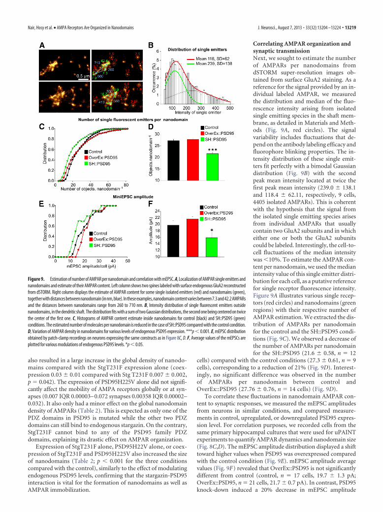
also resulted in a large increase in the global density of nanodo-mains compared with the StgT231F expression alone (coex-pression 0.03 � 0.01 compared with Stg T231F 0.007 � 0.002,p 0.042). The expression of PSD95H225V alone did not signifi-cantly affect the mobility of AMPA receptors globally or at syn-apses (0.007 IQR 0.00003– 0.072 synapses 0.00358 IQR 0.00002–0.032). It also only had a minor effect on the global nanodomaindensity of AMPARs (Table 2). This is expected as only one of thePDZ domains in PSD95 is mutated while the other two PDZdomains can still bind to endogenous stargazin. On the contrary,StgT231F cannot bind to any of the PSD95 family PDZdomains, explaining its drastic effect on AMPAR organization.
Expression of StgT231F alone, PSD95H22V alone, or coex-pression of StgT231F and PSD95H225V also increased the sizeof nanodomains (Table 2; p � 0.001 for the three conditionscompared with the control), similarly to the effect of modulatingendogenous PSD95 levels, confirming that the stargazin-PSD95interaction is vital for the formation of nanodomains as well asAMPAR immobilization.
Correlating AMPAR organization andsynaptic transmissionNext, we sought to estimate the numberof AMPARs per nanodomains fromdSTORM super-resolution images ob-tained from surface GluA2 staining. As areference for the signal provided by an in-dividual labeled AMPAR, we measuredthe distribution and median of the fluo-rescence intensity arising from isolatedsingle emitting species in the shaft mem-brane, as detailed in Materials and Meth-ods (Fig. 9A, red circles). The signalvariability includes fluctuations that de-pend on the antibody labeling efficacy andfluorophore blinking properties. The in-tensity distribution of these single emit-ters fit perfectly with a bimodal Gaussiandistribution (Fig. 9B) with the secondpeak mean intensity located at twice thefirst peak mean intensity (239.0 � 138.1and 118.4 � 62.11, respectively, 9 cells,4405 isolated AMPARs). This is coherentwith the hypothesis that the signal fromthe isolated single emitting species arisesfrom individual AMPARs that usuallycontain two GluA2 subunits and in whicheither one or both the GluA2 subunitscould be labeled. Interestingly, the cell-to-cell fluctuations of the median intensitywas �10%. To estimate the AMPAR con-tent per nanodomain, we used the medianintensity value of this single emitter distri-bution for each cell, as a putative referencefor single receptor fluorescence intensity.Figure 9A illustrates various single recep-tors (red circles) and nanodomains (greenregions) with their respective number ofAMPAR estimation. We extracted the dis-tribution of AMPARs per nanodomainfor the control and the SH::PSD95 condi-tions (Fig. 9C). We observed a decrease ofthe number of AMPARs per nanodomainfor the SH::PSD95 (21.6 � 0.58, n 12
cells) compared with the control conditions (27.3 � 0.61, n 9cells), corresponding to a reduction of 21% (Fig. 9D). Interest-ingly, no significant difference was observed in the numberof AMPARs per nanodomain between control andOverEx::PSD95 (27.76 � 0.76, n 14 cells) (Fig. 9D).
To correlate these fluctuations in nanodomain AMPAR con-tent to synaptic responses, we measured the mEPSC amplitudesfrom neurons in similar conditions, and compared measure-ments in control, upregulated, or downregulated PSD95 expres-sion level. For correlation purposes, we recorded cells from thesame primary hippocampal cultures that were used for uPAINTexperiments to quantify AMPAR dynamics and nanodomain size(Fig. 8C,D). The mEPSC amplitude distribution displayed a shifttoward higher values when PSD95 was overexpressed comparedwith the control condition (Fig. 9E). mEPSC amplitude averagevalues (Fig. 9F) revealed that OverEx::PSD95 is not significantlydifferent from control (control, n 17 cells, 19.7 � 1.3 pA;OverEx::PSD95, n 21 cells, 21.7 � 0.7 pA). In contrast, PSD95knock-down induced a 20% decrease in mEPSC amplitude
Figure 9. Estimation of number of AMPAR per nanodomain and correlation with mEPSC. A, Localization of AMPAR single emitters andnanodomains and estimate of their AMPAR content. Left column shows two spines labeled with surface endogenous GluA2 reconstructedfrom dSTORM. Right column displays the estimate of AMPAR content for some single isolated emitters (red) and nanodomains (green),togetherwithdistancesbetweennanodomain(innm,blue). Intheseexamples,nanodomaincontentvariesbetween7.3and42.2AMPARsand the distances between nanodomains range from 260 to 710 nm. B, Intensity distribution of single fluorescent emitters outsidenanodomains, in the dendritic shaft. The distribution fits with a sum of two Gaussian distributions, the second one being centered on twicethe center of the first one. C, Histograms of AMPAR content estimate inside nanodomains for control (black) and SH::PSD95 (green)conditions. The estimated number of molecules per nanodomain is reduced in the case of SH::PSD95 compared with the control condition.D, Variation of AMPAR density in nanodomains for various levels of endogenous PSD95 expression. ***p � 0.001. E, mEPSC distributionobtained by patch-clamp recordings on neurons expressing the same constructs as in Figure 8C, D. F, Average values of the mEPSCs areplotted for various modulations of endogenous PSD95 levels. *p � 0.05.
Nair, Hosy et al. • AMPA Receptors Are Organized in Nanodomains J. Neurosci., August 7, 2013 • 33(32):13204 –13224 • 13219
(SH::PSD95, n 14 cells, 15.8 � 1.5 pA). Together, these exper-iments demonstrate a positive correlation between nanodomainAMPAR content and mEPSC responses.
Modeling the impact of AMPAR organization onsynaptic transmissionTo estimate the potential impact of AMPAR nanoscale orga-nization on synaptic transmission, we performed Monte Carlosimulations as in Heine et al. (2008), but using variousAMPAR organizations (Fig. 10). We used previously establishedparameters of glutamate release in the synaptic cleft, AMPARactivation, and desensitization kinetics schemes (Fig. 10A)(Jonas et al., 1993; Franks et al., 2002; Lisman et al., 2007;Heine et al., 2008). The model perfectly mimicked the kineticsand amplitude of experimentally recorded mEPSCs (Fig.10C). We then systematically varied the localization (Fig.10D–F ) and density (Fig. 10G–I ) of an AMPAR nanodomaincontaining 25 receptors and also analyzed the crosstalk be-tween multiple nanodomains located at varying distances (Fig.10 J, K ). All parameters strongly impacted the amplitude ofmEPSCs. However, our simulations indicate that there is adegree of tolerance on the location of the glutamate release sitewith respect to the AMPAR nanodomain because the ampli-tude of the mEPSC only starts to decrease when glutamate isreleased �100 nm away from the nanodomain center (Fig.10D–F ). In contrast, the mEPSC amplitude was very sensitiveto AMPAR density because it started to decrease as soon as itwas lower than the maximal packaging we used of one receptorevery 20 nm (Fig. 10G–I ).
We then modeled the presence of two similar nanodomainsin the synapse. First, glutamate was released on top of onecluster and the second one positioned at varying distances.The second cluster had no more impact on the AMPAR re-sponse when it was distant of �300 nm (Fig. 10 J1,2), which isabout the mean value that we measured for intercluster dis-tances. Second, we fixed the intercluster distance at 300 nmand varied the location of glutamate release. Interestingly, theAMPAR response was markedly increased when glutamatewas released in between the two clusters (maximum 19.2 � 3.6activated AMPARs) compared with the condition with a singlecluster in the synapse (maximum 16.2 � 3.3 activated AMPARs,p � 0.002) (Fig. 10K1,2).
Together, our experimental data and these simulations indi-cate that AMPAR nanoscale organization as well as location of theglutamate release strongly impact mEPSC amplitude.
DiscussionUsing four independent super-resolution light imaging methodson both genetically tagged and endogenous receptors, as well aspre-embedding EM, we demonstrate that, in live and fixed hip-pocampal neurons, AMPARs are organized inside most synapsesinto a few highly concentrated domains of �70 nm, and nothomogeneously distributed as previously described. AMPARsare stabilized in nanodomains and freely randomly diffuse be-tween them. Nanodomains are dynamic in their compositionand overall structure. AMPARs can enter and exit nanodomainswithin seconds by lateral diffusion. Inside synapses, nanodo-mains can have two different behaviors: they can be stable forup to an hour or change shape and move and/or disappearwithin minutes, albeit at rather slow rates. Nanodomain esti-mated size is robust as measures of AMPAR distribution bysptPALM and uPAINT using both localization maps and con-finement of receptor movements from MSD curves yielded
comparable values. This was confirmed on fixed cells usingSTED, dSTORM, and EM. The observation of similar clustersin fixed and living cells reinforces that they are functionalreceptor domains. Live-cell single molecule imaging demon-strates that nanodomains are dynamic as AMPARs could enterand exit them frequently. Previous low-density single particletracking data showed the alternating trapping and free diffu-sion of AMPARs in the PSD (Groc et al., 2004), as well asconfinement in subspine compartments (Ehlers et al., 2007).However, these data could not provide enough spatial informationto map nanodomains inside synapses. A recent super-resolutionlight microscopy study using immunocytochemistry and STORMon fixed samples (Dani et al., 2010) has highlighted the mo-lecular organization of many postsynaptic molecules, includ-ing AMPARs. Even though an inhomogeneous AMPARdistribution was observed, with clusters apparent on the mi-crographs, the heterogeneity of receptor staining within thesynapses appeared lower compared with our study and theexistence of nanodomains was not reported.
Studies of AMPAR distribution at the nanometer level us-ing EM and either postembedding immunogold labeling(Kharazia et al., 1996; Petralia et al., 1997; Nusser et al., 1998;Takumi et al., 1999a) or pre-embedding labeling (Tao-Chenget al., 2011) or SDS-digested freeze fracture replica labeling(Masugi-Tokita et al., 2007; Tarusawa et al., 2009; Budisan-toso et al., 2012; Fukazawa and Shigemoto, 2012) have sug-gested a nonhomogeneous distribution of receptors within thePSD. SDS-digested freeze fracture replica labeling revealed amicrocluster intrasynaptic arrangement of AMPARs in corti-cogeniculate synapses and a more diffuse arrangement in reti-nogeniculate synapses (Tarusawa et al., 2009). Similarly,AMPARs are organized in intrasynaptic microclusters in par-allel fiber-Purkinje cell synapses, whereas they are homoge-neously distributed in climbing fiber-Purkinje cell synapses(Masugi-Tokita et al., 2007). AMPAR density in the clustersreported in these preparations ranged from �500 to 2000AMPAR/�m 2. We found that AMPAR density inside nanodo-mains reached �2500 AMPAR/�m 2, which is comparable.The number of nanodomains detected per synapse was slightlydifferent in between the different techniques, although in thesame range, with more domains detected by EM comparedwith dSTORM, sptPALM, and uPAINT. This can be explainedby the more efficient detection efficacy reached by EM.
A novel approach to immunogold labeling in which neu-rons were live-labeled with a highly avid GluA2 primary anti-body, followed by pre-embedding immunogold secondaryapplication and silver intensification, resulted in high-densitylabeling of surface GluA2 and the presence of AMPAR nano-domains. The underestimation of the degree of AMPAR clus-tering in earlier EM studies (Kharazia et al., 1996; Petralia etal., 1997; Nusser et al., 1998; Takumi et al., 1999a) could arisefrom undersampling of the epitopes with postembeddingmethods and use of different primary antibodies. Importantly,our super-resolution experiments in live cells indicate that theAMPAR organization into nanodomains does not result froma fixation artifact.
The stabilization of AMPARs at postsynaptic sites is regu-lated by the interaction of AMPAR subunits and auxiliaryproteins, such as TARPs, with intracellular and extracellularinteractors (Nicoll et al., 2006; Bats et al., 2007; Frischknechtet al., 2009). AMPAR nanodomains are relatively stable, asthey can last for at least an hour, and most likely originatefrom interactions of AMPAR complex components with intra-
13220 • J. Neurosci., August 7, 2013 • 33(32):13204 –13224 Nair, Hosy et al. • AMPA Receptors Are Organized in Nanodomains
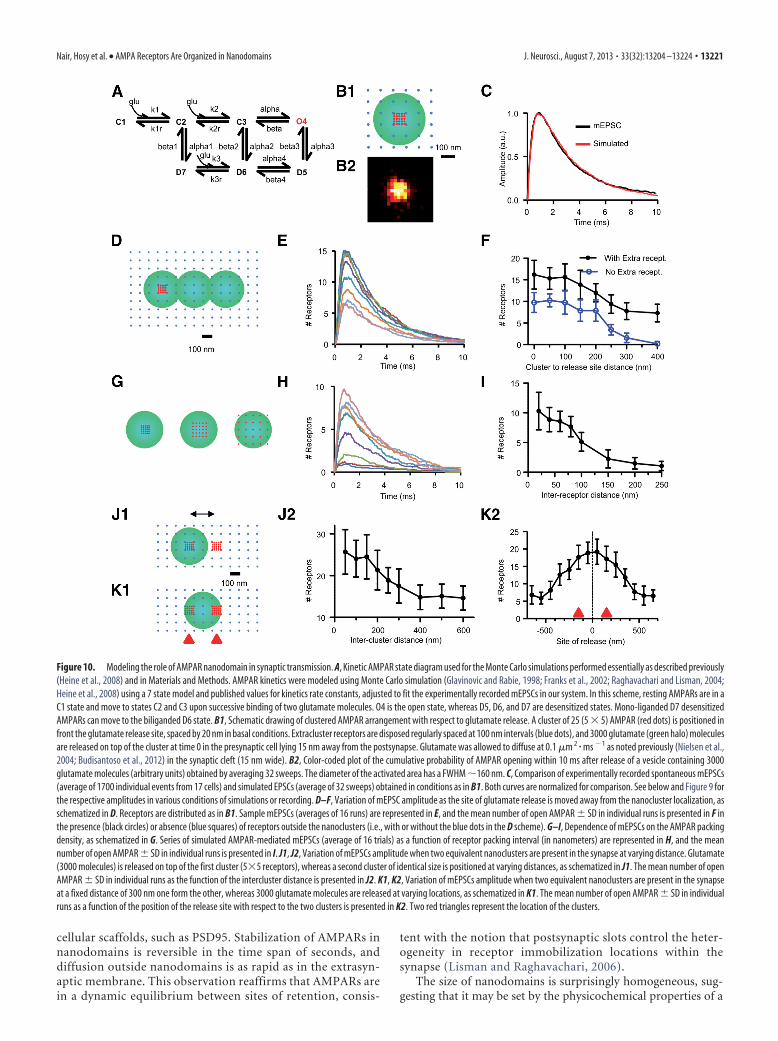
cellular scaffolds, such as PSD95. Stabilization of AMPARs innanodomains is reversible in the time span of seconds, anddiffusion outside nanodomains is as rapid as in the extrasyn-aptic membrane. This observation reaffirms that AMPARs arein a dynamic equilibrium between sites of retention, consis-
tent with the notion that postsynaptic slots control the heter-ogeneity in receptor immobilization locations within thesynapse (Lisman and Raghavachari, 2006).
The size of nanodomains is surprisingly homogeneous, sug-gesting that it may be set by the physicochemical properties of a
Figure 10. Modeling the role of AMPAR nanodomain in synaptic transmission. A, Kinetic AMPAR state diagram used for the Monte Carlo simulations performed essentially as described previously(Heine et al., 2008) and in Materials and Methods. AMPAR kinetics were modeled using Monte Carlo simulation (Glavinovic and Rabie, 1998; Franks et al., 2002; Raghavachari and Lisman, 2004;Heine et al., 2008) using a 7 state model and published values for kinetics rate constants, adjusted to fit the experimentally recorded mEPSCs in our system. In this scheme, resting AMPARs are in aC1 state and move to states C2 and C3 upon successive binding of two glutamate molecules. O4 is the open state, whereas D5, D6, and D7 are desensitized states. Mono-liganded D7 desensitizedAMPARs can move to the biliganded D6 state. B1, Schematic drawing of clustered AMPAR arrangement with respect to glutamate release. A cluster of 25 (5 � 5) AMPAR (red dots) is positioned infront the glutamate release site, spaced by 20 nm in basal conditions. Extracluster receptors are disposed regularly spaced at 100 nm intervals (blue dots), and 3000 glutamate (green halo) moleculesare released on top of the cluster at time 0 in the presynaptic cell lying 15 nm away from the postsynapse. Glutamate was allowed to diffuse at 0.1 �m 2 � ms �1 as noted previously (Nielsen et al.,2004; Budisantoso et al., 2012) in the synaptic cleft (15 nm wide). B2, Color-coded plot of the cumulative probability of AMPAR opening within 10 ms after release of a vesicle containing 3000glutamate molecules (arbitrary units) obtained by averaging 32 sweeps. The diameter of the activated area has a FWHM �160 nm. C, Comparison of experimentally recorded spontaneous mEPSCs(average of 1700 individual events from 17 cells) and simulated EPSCs (average of 32 sweeps) obtained in conditions as in B1. Both curves are normalized for comparison. See below and Figure 9 forthe respective amplitudes in various conditions of simulations or recording. D–F, Variation of mEPSC amplitude as the site of glutamate release is moved away from the nanocluster localization, asschematized in D. Receptors are distributed as in B1. Sample mEPSCs (averages of 16 runs) are represented in E, and the mean number of open AMPAR � SD in individual runs is presented in F inthe presence (black circles) or absence (blue squares) of receptors outside the nanoclusters (i.e., with or without the blue dots in the D scheme). G–I, Dependence of mEPSCs on the AMPAR packingdensity, as schematized in G. Series of simulated AMPAR-mediated mEPSCs (average of 16 trials) as a function of receptor packing interval (in nanometers) are represented in H, and the meannumber of open AMPAR � SD in individual runs is presented in I. J1, J2, Variation of mEPSCs amplitude when two equivalent nanoclusters are present in the synapse at varying distance. Glutamate(3000 molecules) is released on top of the first cluster (5�5 receptors), whereas a second cluster of identical size is positioned at varying distances, as schematized in J1. The mean number of openAMPAR � SD in individual runs as the function of the intercluster distance is presented in J2. K1, K2, Variation of mEPSCs amplitude when two equivalent nanoclusters are present in the synapseat a fixed distance of 300 nm one form the other, whereas 3000 glutamate molecules are released at varying locations, as schematized in K1. The mean number of open AMPAR � SD in individualruns as a function of the position of the release site with respect to the two clusters is presented in K2. Two red triangles represent the location of the clusters.
Nair, Hosy et al. • AMPA Receptors Are Organized in Nanodomains J. Neurosci., August 7, 2013 • 33(32):13204 –13224 • 13221
limited number of interacting building blocks. This subsynapticorganization is linked to the homeostasis of PSD95, as an increaseor decrease in PSD95 expression levels modifies the nanodomaincharacteristics. A decrease in endogenous PSD95 expression lev-els broadens the surface of nanodomains and decreases AMPARcontent and mEPSC amplitude. This is likely the result of analteration in the nanodomain structure and AMPAR destabiliza-tion. This is in agreement with the recent EM tomography obser-vation of RNA interference knockdown of PSD95 leading topatchy loss of PSD material. This loss correlates with the loss ofputative AMPAR-type structures in the PSD (Chen et al., 2011).In contrast, overexpression of PSD95 increases AMPAR nano-domain size, but not its total content (i.e., lowering AMPARdensity in the nanodomain). PSD95 overexpression does notsignificantly change the mEPSC amplitude, in agreement withprevious work (Ehrlich and Malinow, 2004). However, itshould be noted that PSD95 is most likely not the only coreorganizer of nanodomains, in particular as AMPAR andPSD95 do not perfectly colocalize, and as knocking downPSD95 (this study) or disrupting the PSD95-Stargazin inter-action (Sainlos et al., 2011) does not fully disrupt nanodomainorganization or synaptic transmission.
Together, we find it particularly interesting that mEPSC am-plitude is well correlated with AMPAR nanodomain total contentand not their size. Indeed, the amplitude of unitary synaptic re-sponses is determined largely by the number of receptors bindingneurotransmitters after presynaptic terminal release. This pa-rameter is directly linked to the q value, which is the size of aquantum of glutamate inside one vesicle. Previously, this quan-tum was thought to be sufficient to saturate all synaptic AMPARs.More recently, biophysical models of glutamatergic synaptictransmission have outlined that, because of the steep glutamategradient in the synaptic cleft after vesicle release, AMPARs canonly be activated in an area of �150 nm (Lisman et al., 2007; Wuet al., 2007; Budisantoso et al., 2012) around the release site. Asa consequence, organization of AMPARs inside the PSD, to-gether with their proximity with presynaptic release sites, tunethe strength and the variability of synaptic responses (Frankset al., 2002; Franks et al., 2003). Existing models of synaptictransmission rely mostly on a classical uniform distribution ofAMPARs at the PSD. It means that, even if the q value is notsufficient to saturate all the receptors of the synapse, the AM-PAR response will be directly correlated with the size of thequantum: the bigger the quantum, the more receptors areopened. The demonstration that AMPARs are highly concen-trated in nanodomains and that these domains are long lastingopens the new possibility that single vesicle release may acti-vate a nanodomain, and this could represent a postsynapticquantum. Synaptic transmission could then be powerfullyregulated by the composition and the position of the nanodo-mains relative to the presynaptic release site.
To investigate this concept further, we measured the distancebetween nanodomains inside single spines by STORM and spt-PALM and performed modeling. In PSDs bearing multiple nano-domains, only approximately half of them are closer one to theother by �250 nm. With respect to the area over which AMPARsare thought to be activated by glutamate (diameter of �200 nm)(Lisman et al., 2007), FWHM computed from our simulations156 � 15 nm (Fig. 10B2), this implies that a large fraction ofAMPAR nanodomains are functionally isolated one from theother with respect to single vesicle release (Fig. 10J1). UsingMonte Carlo simulations, we also found that the distance be-tween glutamate release site and an AMPAR nanodomain
strongly impacts the efficacy of transmission. The identity, loca-tion, and number of vesicle release sites at individual terminalsare still a matter of debate. Nevertheless, it is tempting to postu-late a correlation between release sites and AMPAR nanodo-mains, likely through trans-synaptic adhesion proteins, such asneurexin/neuroligins.
Finally, previous work demonstrated that fast AMPAR dif-fusion in and out release sites renews the pool of naive recep-tors that can be activated and removes desensitized ones,allowing the synapse to sustain high-frequency stimulation(Heine et al., 2008). We demonstrated that this effect ofAMPAR diffusion is possible only if AMPAR exchange takesplace on a small area (diameter of a couple of hundred nano-meters) or if AMPARs are moving rapidly. The concentrationof AMPARs in nanodomains, and their high mobility outsidethese, provides a framework for sufficiently fast exchange inAMPARs as well as for changes in receptor density in front ofrelease sites by fast aggregation and dissociation of nanodo-mains aided by lateral diffusion.
ReferencesBats C, Groc L, Choquet D (2007) The interaction between Stargazin and
PSD-95 regulates AMPA receptor surface trafficking. Neuron 53:719 –734. CrossRef Medline
Baude A, Nusser Z, Molnar E, McIlhinney RA, Somogyi P (1995) High-resolution immunogold localization of AMPA type glutamate receptorsubunits at synaptic and non-synaptic sites in rat hippocampus. Neuro-science 69:1031–1055. CrossRef Medline
Bernard V, Somogyi P, Bolam JP (1997) Cellular, subcellular, and subsyn-aptic distribution of AMPA-type glutamate receptor subunits in the neos-triatum of the rat. J Neurosci 17:819 – 833. Medline
Budisantoso T, Matsui K, Kamasawa N, Fukazawa Y, Shigemoto R (2012)Mechanisms underlying signal filtering at a multisynapse contact. J Neu-rosci 32:2357–2376. CrossRef Medline
Cheezum MK, Walker WF, Guilford WH (2001) Quantitative comparisonof algorithms for tracking single fluorescent particles. Biophys J 81:2378 –2388. CrossRef Medline
Chen X, Winters C, Azzam R, Li X, Galbraith JA, Leapman RD, Reese TS(2008) Organization of the core structure of the postsynaptic density.Proc Natl Acad Sci U S A 105:4453– 4458. CrossRef Medline
Chen X, Nelson CD, Li X, Winters CA, Azzam R, Sousa AA, Leapman RD,Gainer H, Sheng M, Reese TS (2011) PSD-95 is required to sustain themolecular organization of the postsynaptic density. J Neurosci 31:6329 –6338. CrossRef Medline
Choquet D (2010) Fast AMPAR trafficking for a high-frequency synaptictransmission. Eur J Neurosci 32:250 –260. CrossRef Medline
Dani A, Huang B, Bergan J, Dulac C, Zhuang X (2010) Superresolutionimaging of chemical synapses in the brain. Neuron 68:843– 856. CrossRefMedline
Ehlers MD, Heine M, Groc L, Lee MC, Choquet D (2007) Diffusional trap-ping of GluR1 AMPA receptors by input-specific synaptic activity. Neu-ron 54:447– 460. CrossRef Medline
Ehrlich I, Malinow R (2004) Postsynaptic density 95 controls AMPA recep-tor incorporation during long-term potentiation and experience-drivensynaptic plasticity. J Neurosci 24:916 –927. CrossRef Medline
El-Husseini AE, Schnell E, Chetkovich DM, Nicoll RA, Bredt DS (2000)PSD-95 involvement in maturation of excitatory synapses. Science 290:1364 –1368. Medline
Folling J, Bossi M, Bock H, Medda R, Wurm CA, Hein B, Jakobs S, Eggeling C,Hell SW (2008) Fluorescence nanoscopy by ground-state depletion andsingle-molecule return. Nat Methods 5:943–945. CrossRef Medline
Franks KM, Bartol TM Jr, Sejnowski TJ (2002) A Monte Carlo model revealsindependent signaling at central glutamatergic synapses. Biophys J 83:2333–2348. CrossRef Medline
Franks KM, Stevens CF, Sejnowski TJ (2003) Independent sources of quan-tal variability at single glutamatergic synapses. J Neurosci 23:3186 –3195.Medline
Frischknecht R, Heine M, Perrais D, Seidenbecher CI, Choquet D, Gun-delfinger ED (2009) Brain extracellular matrix affects AMPA recep-
13222 • J. Neurosci., August 7, 2013 • 33(32):13204 –13224 Nair, Hosy et al. • AMPA Receptors Are Organized in Nanodomains
tor lateral mobility and short-term synaptic plasticity. Nat Neurosci12:897–904. CrossRef Medline
Fukazawa Y, Shigemoto R (2012) Intra-synapse-type and inter-synapse-type relationships between synaptic size and AMPAR expression. CurrOpin Neurobiol 22:446 – 452. CrossRef Medline
Ganeshina O, Berry RW, Petralia RS, Nicholson DA, Geinisman Y (2004)Differences in the expression of AMPA and NMDA receptors betweenaxospinous perforated and nonperforated synapses are related to theconfiguration and size of postsynaptic densities. J Comp Neurol 468:86 –95. CrossRef Medline
Giannone G, Hosy E, Levet F, Constals A, Schulze K, Sobolevsky AI, RosconiMP, Gouaux E, Tampe R, Choquet D, Cognet L (2010) Dynamic super-resolution imaging of endogenous proteins on living cells at ultra-highdensity. Biophys J 99:1303–1310. CrossRef Medline
Glavinovíc MI (1999) Monte Carlo simulation of vesicular release, spa-tiotemporal distribution of glutamate in synaptic cleft and generationof postsynaptic currents. Pflugers Arch 437:462– 470. CrossRefMedline
Glavinovíc MI, Rabie HR (1998) Monte Carlo simulation of spontaneousminiature excitatory postsynaptic currents in rat hippocampal synapse inthe presence and absence of desensitization. Pflugers Arch 435:193–202.Medline
Groc L, Heine M, Cognet L, Brickley K, Stephenson FA, Lounis B, Choquet D(2004) Differential activity-dependent regulation of the lateral mobilitiesof AMPA and NMDA receptors. Nat Neurosci 7:695– 696. CrossRefMedline
Heilemann M, Margeat E, Kasper R, Sauer M, Tinnefeld P (2005) Carbo-cyanine dyes as efficient reversible single-molecule optical switch. J AmChem Soc 127:3801–3806. CrossRef Medline
Heilemann M, van de Linde S, Schuttpelz M, Kasper R, Seefeldt B, MukherjeeA, Tinnefeld P, Sauer M (2008) Subdiffraction-resolution fluorescenceimaging with conventional fluorescent probes. Angew Chem Int Ed Engl47:6172– 6176. CrossRef Medline
Heine M, Groc L, Frischknecht R, Beïque JC, Lounis B, Rumbaugh G,Huganir RL, Cognet L, Choquet D (2008) Surface mobility of postsyn-aptic AMPARs tunes synaptic transmission. Science 320:201–205.CrossRef Medline
Hell SW (2009) Microscopy and its focal switch. Nat Methods 6:24 –32.CrossRef Medline
Hoze N, Nair D, Hosy E, Sieben C, Manley S, Herrmann A, Sibarita JB,Choquet D, Holcman D (2012) Heterogeneity of AMPA receptor traf-ficking and molecular interactions revealed by superresolution analysis oflive cell imaging. Proc Natl Acad Sci U S A 109:17052–17057. CrossRefMedline
Izeddin I, Boulanger J, Racine V, Specht CG, Kechkar A, Nair D, Triller A,Choquet D, Dahan M, Sibarita JB (2012) Wavelet analysis for singlemolecule localization microscopy. Opt Express 20:2081–2095. CrossRefMedline
Jonas P, Major G, Sakmann B (1993) Quantal components of unitary EPSCsat the mossy fiber synapse on CA3 pyramidal cells of the rat hippocampus.J Physiol 472:615– 663. Medline
Kaech S, Banker G (2006) Culturing hippocampal neurons. Nat Protoc1:2406 –2415. CrossRef Medline
Kechkar A, Nair D, Heilemann M, Choquet D, Sibarita JB (2013) Real-timeanalysis and visualization for single-molecule based super-resolution mi-croscopy. PloS One 8:e62918. CrossRef Medline
Kharazia VN, Weinberg RJ (1997) Tangential synaptic distribution ofNMDA and AMPA receptors in rat neocortex. Neurosci Lett 238:41– 44.CrossRef Medline
Kharazia VN, Phend KD, Rustioni A, Weinberg RJ (1996) EM colocaliza-tion of AMPA and NMDA receptor subunits at synapses in rat cerebralcortex. Neurosci Lett 210:37– 40. CrossRef Medline
Kubitscheck U, Homann U, Thiel G (2000) Osmotically evoked shrinkingof guard-cell protoplasts causes vesicular retrieval of plasma membraneinto the cytoplasm. Planta 210:423– 431. CrossRef Medline
Lisman J, Raghavachari S (2006) A unified model of the presynaptic andpostsynaptic changes during LTP at CA1 synapses. Sci STKE 2006:re11.CrossRef Medline
Lisman JE, Raghavachari S, Tsien RW (2007) The sequence of events thatunderlie quantal transmission at central glutamatergic synapses. Nat RevNeurosci 8:597– 609. CrossRef Medline
Liu G, Choi S, Tsien RW (1999) Variability of neurotransmitter concentra-
tion and nonsaturation of postsynaptic AMPA receptors at synapses inhippocampal cultures and slices. Neuron 22:395– 409. CrossRef Medline
Luscher C, Xia H, Beattie EC, Carroll RC, von Zastrow M, Malenka RC, NicollRA (1999) Role of AMPA receptor cycling in synaptic transmission andplasticity. Neuron 24:649 – 658. CrossRef Medline
Macgillavry HD, Song Y, Raghavachari S, Blanpied TA (2013) Nanoscalescaffolding domains within the postsynaptic density concentrate synapticAMPA receptors. Neuron 78:615– 622. CrossRef Medline
Makino H, Malinow R (2009) AMPA receptor incorporation into synapsesduring LTP: the role of lateral movement and exocytosis. Neuron 64:381–390. CrossRef Medline
Manley S, Gillette JM, Patterson GH, Shroff H, Hess HF, Betzig E,Lippincott-Schwartz J (2008) High-density mapping of single-molecule trajectories with photoactivated localization microscopy.Nat Methods 5:155–157. CrossRef Medline
Masugi-Tokita M, Shigemoto R (2007) High-resolution quantitative visu-alization of glutamate and GABA receptors at central synapses. Curr OpinNeurobiol 17:387–393. CrossRef Medline
Masugi-Tokita M, Tarusawa E, Watanabe M, Molnar E, Fujimoto K, Shige-moto R (2007) Number and density of AMPA receptors in individualsynapses in the rat cerebellum as revealed by SDS-digested freeze-fracturereplica labeling. J Neurosci 27:2135–2144. CrossRef Medline
Matsubara A, Laake JH, Davanger S, Usami S, Ottersen OP (1996) Organi-zation of AMPA receptor subunits at a glutamate synapse: a quantitativeimmunogold analysis of hair cell synapses in the rat organ of Corti. J Neu-rosci 16:4457– 4467. Medline
Mondin M, Labrousse V, Hosy E, Heine M, Tessier B, Levet F, Poujol C,Blanchet C, Choquet D, Thoumine O (2011) Neurexin-neuroligin ad-hesions capture surface-diffusing AMPA receptors through PSD-95 scaf-folds. J Neurosci 31:13500 –13515. CrossRef Medline
Nicoll RA, Tomita S, Bredt DS (2006) Auxiliary subunits assist AMPA-typeglutamate receptors. Science 311:1253–1256. CrossRef Medline
Nielsen TA, DiGregorio DA, Silver RA (2004) Modulation of glutamatemobility reveals the mechanism underlying slow-rising AMPAREPSCs and the diffusion coefficient in the synaptic cleft. Neuron 42:757–771. CrossRef Medline
Nusser Z, Mulvihill E, Streit P, Somogyi P (1994) Subsynaptic segregation ofmetabotropic and ionotropic glutamate receptors as revealed by immu-nogold localization. Neuroscience 61:421– 427. CrossRef Medline
Nusser Z, Lujan R, Laube G, Roberts JD, Molnar E, Somogyi P (1998) Celltype and pathway dependence of synaptic AMPA receptor number andvariability in the hippocampus. Neuron 21:545–559. CrossRef Medline
Opazo P, Labrecque S, Tigaret CM, Frouin A, Wiseman PW, De KoninckP, Choquet D (2010) CaMKII triggers the diffusional trapping ofsurface AMPARs through phosphorylation of Stargazin. Neuron 67:239 –252. CrossRef Medline
Opazo P, Sainlos M, Choquet D (2012) Regulation of AMPA receptor sur-face diffusion by PSD-95 slots. Curr Opin Neurobiol 22:453– 460.CrossRef Medline
Penn AC, Balik A, Wozny C, Cais O, Greger IH (2012) Activity-mediatedAMPA receptor remodeling, driven by alternative splicing in the ligand-binding domain. Neuron 76:503–510. CrossRef Medline
Petersen JD, Chen X, Vinade L, Dosemeci A, Lisman JE, Reese TS (2003)Distribution of postsynaptic density (PSD)-95 and Ca 2�/calmodulin-dependent protein kinase II at the PSD. J Neurosci 23:11270 –11278.Medline
Petralia RS, Wang YX, Mayat E, Wenthold RJ (1997) Glutamate receptorsubunit 2-selective antibody shows a differential distribution of calcium-impermeable AMPA receptors among populations of neurons. J CompNeurol 385:456 – 476. CrossRef Medline
Petrini EM, Lu J, Cognet L, Lounis B, Ehlers MD, Choquet D (2009) Endo-cytic trafficking and recycling maintain a pool of mobile surface AMPAreceptors required for synaptic potentiation. Neuron 63:92–105.CrossRef Medline
Racine V, Hertzog A, Jouanneau J, Salamero J, Kervrann C, Sibarita JB(2006) Multiple-target tracking of 3D fluorescent objects based on sim-ulated annealing. IEEE International Symposium on Biomedical Imag-ing: Macro to nano. Vol 1–3:1020 –1023. CrossRef
Racine V, Sachse M, Salamero J, Fraisier V, Trubuil A, Sibarita JB (2007)Visualization and quantification of vesicle trafficking on a three-dimensional cytoskeleton network in living cells. J Microsc 225:214 –228.CrossRef Medline
Nair, Hosy et al. • AMPA Receptors Are Organized in Nanodomains J. Neurosci., August 7, 2013 • 33(32):13204 –13224 • 13223
Raghavachari S, Lisman JE (2004) Properties of quantal transmission atCA1 synapses. J Neurophysiol 92:2456 –2467. CrossRef Medline
Saglietti L, Dequidt C, Kamieniarz K, Rousset MC, Valnegri P, Thoumine O,Beretta F, Fagni L, Choquet D, Sala C, Sheng M, Passafaro M (2007)Extracellular interactions between GluR2 and N-cadherin in spine regu-lation. Neuron 54:461– 477. CrossRef Medline
Sainlos M, Tigaret C, Poujol C, Olivier NB, Bard L, Breillat C, Thiolon K,Choquet D, Imperiali B (2011) Biomimetic divalent ligands for theacute disruption of synaptic AMPAR stabilization. Nat Chem Biol 7:81–91. CrossRef Medline
Schluter OM, Xu W, Malenka RC (2006) Alternative N-terminal domainsof PSD-95 and SAP97 govern activity-dependent regulation of synapticAMPA receptor function. Neuron 51:99 –111. CrossRef Medline
Schnell E, Sizemore M, Karimzadegan S, Chen L, Bredt DS, Nicoll RA (2002)Direct interactions between PSD-95 and stargazin control synapticAMPA receptor number. Proc Natl Acad Sci U S A 99:13902–13907.CrossRef Medline
Sharma K, Fong DK, Craig AM (2006) Postsynaptic protein mobility indendritic spines: long-term regulation by synaptic NMDA receptor acti-vation. Mol Cell Neurosci 31:702–712. CrossRef Medline
Takumi Y, Matsubara A, Rinvik E, Ottersen OP (1999a) The arrange-ment of glutamate receptors in excitatory synapses. Ann N Y Acad Sci868:474 – 482. CrossRef Medline
Takumi Y, Ramírez-Leon V, Laake P, Rinvik E, Ottersen OP (1999b) Dif-ferent modes of expression of AMPA and NMDA receptors in hippocam-pal synapses. Nat Neurosci 2:618 – 624. CrossRef Medline
Tao-Cheng JH, Crocker VT, Winters CA, Azzam R, Chludzinski J, Reese TS(2011) Trafficking of AMPA receptors at plasma membranes of hip-pocampal neurons. J Neurosci 31:4834 – 4843. CrossRef Medline
Tarusawa E, Matsui K, Budisantoso T, Molnar E, Watanabe M, Matsui M,Fukazawa Y, Shigemoto R (2009) Input-specific intrasynaptic arrange-ments of ionotropic glutamate receptors and their impact on postsynapticresponses. J Neurosci 29:12896 –12908. CrossRef Medline
van de Linde S, Loschberger A, Klein T, Heidbreder M, Wolter S, Heile-mann M, Sauer M (2011) Direct stochastic optical reconstructionmicroscopy with standard fluorescent probes. Nat Protoc 6:991–1009. CrossRef Medline
Wenthold RJ, Petralia RS, Blahos J II, Niedzielski AS (1996) Evidence formultiple AMPA receptor complexes in hippocampal CA1/CA2 neurons.J Neurosci 16:1982–1989. Medline
Wu XS, Xue L, Mohan R, Paradiso K, Gillis KD, Wu LG (2007) The origin ofquantal size variation: vesicular glutamate concentration plays a signifi-cant role. J Neurosci 27:3046 –3056. CrossRef Medline
Xu-Friedman MA, Regehr WG (2004) Structural contributions to short-term synaptic plasticity. Physiol Rev 84:69 – 85. CrossRef Medline
13224 • J. Neurosci., August 7, 2013 • 33(32):13204 –13224 Nair, Hosy et al. • AMPA Receptors Are Organized in Nanodomains