Embed Size (px)
Citation preview
Circulating Activated Endothelial Cells in Pediatric Obesity
Aaron S. Kelly, PhD, Robert P. Hebbel, MD, Anna N. Solovey, MD, PhD, Sarah Jane Schwarzenberg, MD, Andrea M. Metzig, MA,
Antoinette Moran, MD, Alan R. Sinaiko, MD, David R. Jacobs, Jr., PhD, and Julia Steinberger, MD, MS
Objective We characterized the state of the vascular endothelium in pediatric obesity by comparing circulatingendothelial cell (CEC) number and activation phenotype in severely obese children to that of normal weight, over-weight, and obese children.Study design We used immunohistochemical examination of buffy-coat smears to enumerate CEC and immu-nofluorescence microscopy to quantify activated CEC in 107 children and adolescents. Normal weight (bodymass index [BMI] <85th percentile; n = 40), overweight (BMI 85th-<95th percentile; n = 17), and obese (BMI95th-<99th percentile; n = 23) participants were recruited from a longitudinal study. Severely obese (BMI $99th per-centile; n = 27) participants were recruited from a pediatric obesity clinic. Group means (adiposity; systolic bloodpressure [SBP] quartiles) were compared with general linear models, adjusted for sex, age, and race. With Pearsoncorrelations, we characterized relations of CEC with cardiovascular risk factors.Results Activated CEC increased across BMI groups (P < .002) and SBP quartiles (P < .05). CEC number and ac-tivated CEC were highest in the severely obese group. CEC number was significantly associated with SBP, diastolicblood pressure, and triglycerides level. Activated CEC were significantly associated with SBP and high-density li-poprotein cholesterol levels.Conclusions The vascular endothelium was activated in relation to excess adiposity, particularly in severelyobese children, and to elevated SBP in children and adolescents. (J Pediatr 2010;157:547-51).
See editorial, p 523 and relatedarticles, p 533 and p 540
One of the fastest growing obesity categories in children is severe obesity, defined as an age- and sex-specific body massindex (BMI) $ 99th percentile. Recent data indicate a 300% increase in severe obesity in the US pediatric populationsince 1976, with a reported prevalence of 3.8% between 1999 and 2004.1,2 There is a relative paucity of data in the most
extreme forms of obesity. Specifically, few studies have described the cardiovascular risk factor profile, and even fewer haveattempted to characterize the vascular status of severely obese children and adolescents.
Perturbation of the vascular endothelium is one of the earliest manifestations of atherosclerosis and is considered a seminalevent in its initiation.3 Whole blood circulating endothelial cells (CEC) have detached from the vascular wall and are thought toreflect structural damage and injury to the endothelial layer. Higher numbers may represent more advanced damage to thevascular endothelium.4 In addition to enumeration, CEC phenotype can be characterized by quantifying the surface expressionof endothelial biomarkers such as vascular cell adhesion molecule-1 (VCAM-1) to determine whether cells are activated (ac-tivated CEC).5 Increased numbers of CEC have been demonstrated in various vascular diseases and pathological conditionssuch as peripheral vascular disease,6 sickle cell anemia,7 acute myocardial infarction and angina pectoris,8 acute coronary syn-drome,5 Kawasaki disease,9 systemic inflammation,10 and pulmonary hypertension.11 CEC predict future cardiovascular eventsin individuals with cardiac disease, independent of conventional cardiovascular disease risk factors.12-14 Therefore, we evalu-ated CEC across a spectrum of adiposity in children and adolescents to describe the magnitude of endothelial activation in re-lation to obesity.
BMI
CEC
DBP
SBP
VCAM-1
Methods
From the Department of Pediatrics, University ofMinnesota Medical School, Minneapolis, MN (A.K., S.S.,A. Metzig, A. Moran, A. Sinaiko, J.S.); Vascular BiologyCenter, Division of Hematology, Oncology, andTransplantation, Department of Medicine, University ofMinnesota Medical School, Minneapolis, MN (R.H., A.Solovey); School of Public Health, University ofThis cross-sectional study included 107 children and adolescents (mean age, 13.1� 3.8; age range, 6-22 years; 68 male) who were categorized (after testing) in 4adiposity groups on the basis of age- and sex-specific BMI percentiles. Partici-
Minnesota, Minneapolis, MN (D.J.); and Department ofNutrition, University of Oslo, Oslo, Norway (D.J.)
Funding was provided in part by University of MinnesotaVikings Children’s Fund (A.K.), Minnesota MedicalFoundation (A.K.), National Institutes of Health (P01HL55552 to R.H.), National Institutes of Health(1RO1DK072124-01A3 to J.S.), and GCRC (M01-RR00400), General Clinical Research Center Program,NCRR/NIH. The authors declare no conflicts of interest.
0022-3476/$ - see front matter. Copyright � 2010 Mosby Inc.
All rights reserved. 10.1016/j.jpeds.2010.04.069
Body mass index
Circulating endothelial cells
Diastolic blood pressure
Systolic blood pressure
Vascular cell adhesion molecule-1
547
THE JOURNAL OF PEDIATRICS � www.jpeds.com Vol. 157, No. 4
pants in the normal weight (BMI <85th percentile; n = 40),overweight (BMI 85th-<95th percentile; n = 17), and obese(BMI 95th-<99th percentile; n = 23) groups were consecu-tively enrolled in a period of approximately 1 year from a lon-gitudinal cohort study investigating the early development ofobesity, insulin resistance, and other cardiovascular risk fac-tors. The severely obese (BMI $99th percentile; n = 27)group comprised children and adolescents initially enteringthe University of Minnesota Pediatric Weight ManagementClinic who were consecutively enrolled in the same period.No behavioral or drug therapies had yet been initiated inthese individuals. All subjects in this study were invited toparticipate (no exclusion criteria were used). The protocolwas approved by the University of Minnesota institutionalreview board, and consent/assent was obtained from par-ents/participants. Measures were obtained after participantshad been fasting $10 hours.
Height and weight were obtained with a standard stadiom-eter and electronic scale, respectively. Waist and hip circum-ferences were measured to the nearest 0.5 cm. Seated bloodpressure was obtained after 5 minutes of quiet rest, in theright arm with an automatic sphygmomanometer. Fastinglipid profile, glucose, and insulin assays were conductedwith standard procedures at the Fairview Diagnostic Labora-tories, Fairview-University Medical Center (Minneapolis,Minnesota), a Centers for Disease Control and Prevention-certified laboratory.
Blood samples were collected from patients in Vacutainertubes (BD Vacutainer Systems, Franklin Lakes, New Jersey)containing EDTA and were processed immediately for study.CEC analyses were performed in the University of MinnesotaVascular Biology Center, as previously described in detail.7,15
For CEC enumeration, we used immunohistochemical exam-ination of buffy-coat smears prepared with centrifugation of1 mL of whole blood placed on Histopague-1077 (Sigma-Aldrich, St. Louis, Missouri). The antibodies used for stainingwere specific mouse anti-endothelial P1H12 (anti-CD146),with secondary anti-mouse alkaline phosphatase conjugatedantibody (Jackson IRL, Westgrove, Pennsylvania), and visu-alized with alkaline phosphatase Fast Red substrate (Vector,Burlingame, California). Cells positive for P1H12 (CEC) onthe smear were counted under a light microscope. The resultswere expressed as the number of CEC per 1 mL of peripheralwhole blood.
The surface phenotyping was achieved by applying immu-nofluorescent staining to preparations of CEC. In this case,the isolation of CEC was performed by mixing whole bloodwith immunomagnetic beads (Dynal, Oslo, Norway) coatedwith P1H12 antibody. The beads with CEC attached werespun down with cytospin centrifuge. The panel of antibodiesused for double staining included mouse P1H12 (endothelialmarker), rabbit anti-VCAM-1 (Santa Cruz, California), anti-mouse FITC labeled, and anti-rabbit TRITC labeled (bothfrom Jackson IRL). Nuclei were counterstained with DAPI.
Slides were viewed under a fluorescent microscope, andthe results represented a percentage of VCAM-1 positive (ac-tivated) CEC in the total population of CEC.
548
Reproducibility was assessed for CEC enumeration andactivation phenotyping in a subset (n = 10) of samplesfrom this study. The coefficients of variation for CECnumber and percent activated CEC were 25% and 5%, re-spectively.
Means across BMI groups (normal weight, overweight,obese, severely obese) were compared with general linearmodel analysis (GLM procedure, SPSS version 16.0; SPSS,Inc., Chicago, Illinois), adjusted for sex, age, and race.The entire sample (n = 107) was grouped by SBP quar-tile, and means across groups were compared with gen-eral linear model or Pearson correlation analysis,adjusted for sex, age, race, and BMI. Multivariate linearregression analyses, adjusted for sex, age, and race, wereperformed to evaluate relations of CEC number and per-cent of activated cells with cardiovascular risk factors inthe entire sample. Statistical significance was consideredto be a P value <.05. Data are presented as means plusor minus SD.
Results
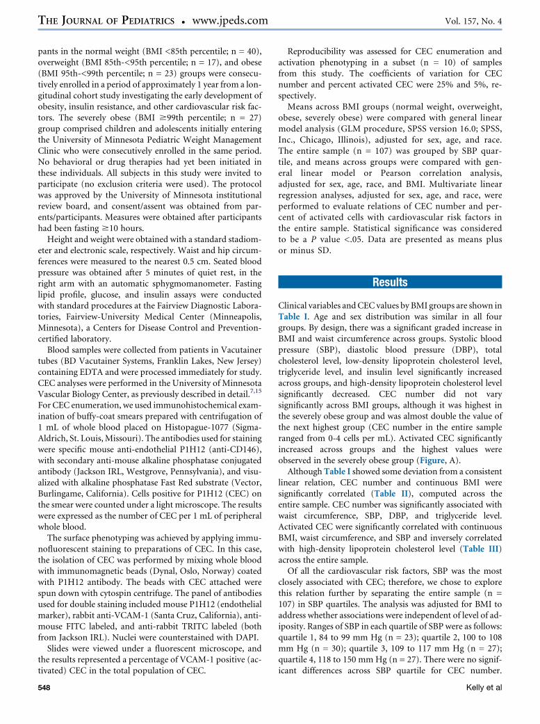
Clinical variables and CEC values by BMI groups are shown inTable I. Age and sex distribution was similar in all fourgroups. By design, there was a significant graded increase inBMI and waist circumference across groups. Systolic bloodpressure (SBP), diastolic blood pressure (DBP), totalcholesterol level, low-density lipoprotein cholesterol level,triglyceride level, and insulin level significantly increasedacross groups, and high-density lipoprotein cholesterol levelsignificantly decreased. CEC number did not varysignificantly across BMI groups, although it was highest inthe severely obese group and was almost double the value ofthe next highest group (CEC number in the entire sampleranged from 0-4 cells per mL). Activated CEC significantlyincreased across groups and the highest values wereobserved in the severely obese group (Figure, A).
Although Table I showed some deviation from a consistentlinear relation, CEC number and continuous BMI weresignificantly correlated (Table II), computed across theentire sample. CEC number was significantly associated withwaist circumference, SBP, DBP, and triglyceride level.Activated CEC were significantly correlated with continuousBMI, waist circumference, and SBP and inversely correlatedwith high-density lipoprotein cholesterol level (Table III)across the entire sample.
Of all the cardiovascular risk factors, SBP was the mostclosely associated with CEC; therefore, we chose to explorethis relation further by separating the entire sample (n =107) in SBP quartiles. The analysis was adjusted for BMI toaddress whether associations were independent of level of ad-iposity. Ranges of SBP in each quartile of SBP were as follows:quartile 1, 84 to 99 mm Hg (n = 23); quartile 2, 100 to 108mm Hg (n = 30); quartile 3, 109 to 117 mm Hg (n = 27);quartile 4, 118 to 150 mm Hg (n = 27). There were no signif-icant differences across SBP quartile for CEC number.
Kelly et al
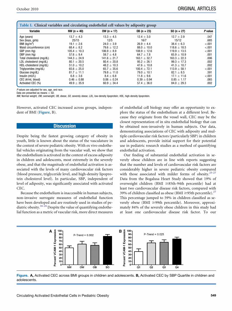
Table I. Clinical variables and circulating endothelial cell values by adiposity group
Variable NW (n = 40) OW (n = 17) OB (n = 23) SO (n = 27) P value
Age (years) 13.7 � 4.3 13.3 � 4.5 12.4 � 3.0 12.7 � 2.9 .547Sex (boys, girls) 25/15 12/5 16/7 15/12 .685BMI (kg/m2) 19.1 � 2.6 24.0 � 3.9 26.9 � 4.4 38.4 � 6.3 <.001Waist circumference (cm) 68.4 � 8.2 79.6 � 12.2 88.0 � 10.0 118.6 � 18.5 <.001SBP (mm Hg) 105.4 � 10.3 106.9 � 8.9 108.9 � 12.6 119.9 � 13.5 <.001DBP (mm Hg) 57.6 � 9.4 59.7 � 4.8 64.7 � 7.8 65.9 � 10.9 .001Total cholesterol (mg/dL) 144.3 � 24.9 141.6 � 21.7 163.1 � 32.7 163.3 � 22.3 .006LDL cholesterol (mg/dL) 80.1 � 20.5 80.4 � 20.8 95.2 � 26.1 99.3 � 17.3 .002HDL-cholesterol (mg/dL) 51.0 � 10.2 48.2 � 10.3 47.6 � 10.8 41.3 � 10.7 .002Triglycerides (mg/dL) 65.6 � 25.0 65.7 � 35.6 100.4 � 72.1 113.9 � 58.1 <.001Glucose (mg/dL) 81.7 � 11.1 79.8 � 11.0 79.0 � 10.1 82.1 � 8.5 .591Insulin (mU/L) 6.8 � 3.6 8.4 � 6.8 11.8 � 9.6 17.1 � 11.6 <.001CEC (#/mL blood) 0.46 � 0.88 0.06 � 0.24 0.39 � 0.94 0.85 � 1.17 .065Activated CEC (%) 49.9 � 35.9 60.9 � 34.4 57.4 � 36.0 84.0 � 29.3 .002
P values are adjusted for sex, age, and race.Data are presented as means � SD.NW, Normal weight; OW, overweight; OB, obese; SO, severely obese; LDL, low-density lipoprotein; HDL, high-density lipoprotein.
October 2010 ORIGINAL ARTICLES
However, activated CEC increased across groups, indepen-dent of BMI (Figure, B).
Discussion
Despite being the fastest growing category of obesity inyouth, little is known about the status of the vasculature inthe context of severe pediatric obesity. With ex vivo endothe-lial vehicles originating from the vascular wall, we show thatthe endothelium is activated in the context of excess adiposityin children and adolescents, most extremely in the severelyobese, and that the magnitude of endothelial activation is as-sociated with the levels of many cardiovascular risk factors(blood pressure, triglyceride level, and high-density lipopro-tein cholesterol level). In particular, SBP, independent oflevel of adiposity, was significantly associated with activatedCEC.
Because the endothelium is inaccessible in human subjects,non-invasive surrogate measures of endothelial functionhave been developed and are routinely used in studies of pe-diatric obesity.16-24 Despite the value of quantifying endothe-lial function as a metric of vascular risk, more direct measures
Figure. A, Activated CEC across BMI groups in children and adoadolescents.
Circulating Activated Endothelial Cells in Pediatric Obesity
of endothelial cell biology may offer an opportunity to ex-plore the status of the endothelium at a different level. Be-cause they originate from the vessel wall, CEC may be theclosest representation of in situ endothelial biology that canbe obtained non-invasively in human subjects. Our data,demonstrating associations of CEC with adiposity and mul-tiple cardiovascular risk factors (particularly SBP) in childrenand adolescents, provide initial support for their potentialuse in pediatric research studies as a method of quantifyingendothelial activation.
Our finding of substantial endothelial activation in se-verely obese children are in line with reports suggestingthat the number and levels of cardiovascular risk factors areconsiderably higher in severe pediatric obesity comparedwith those associated with milder forms of obesity.25-27
Data from the Bogalusa Heart Study showed that 19% ofoverweight children (BMI $85th-94th percentile) had atleast two cardiovascular disease risk factors, compared with39% of children classified as obese (BMI $95th percentile).27
This percentage jumped to 59% in children classified as se-verely obese (BMI $99th percentile). Moreover, approxi-mately 84% of the severely obese children in this study hadat least one cardiovascular disease risk factor. To our
lescents. B, Activated CEC by SBP Quartile in children and
549
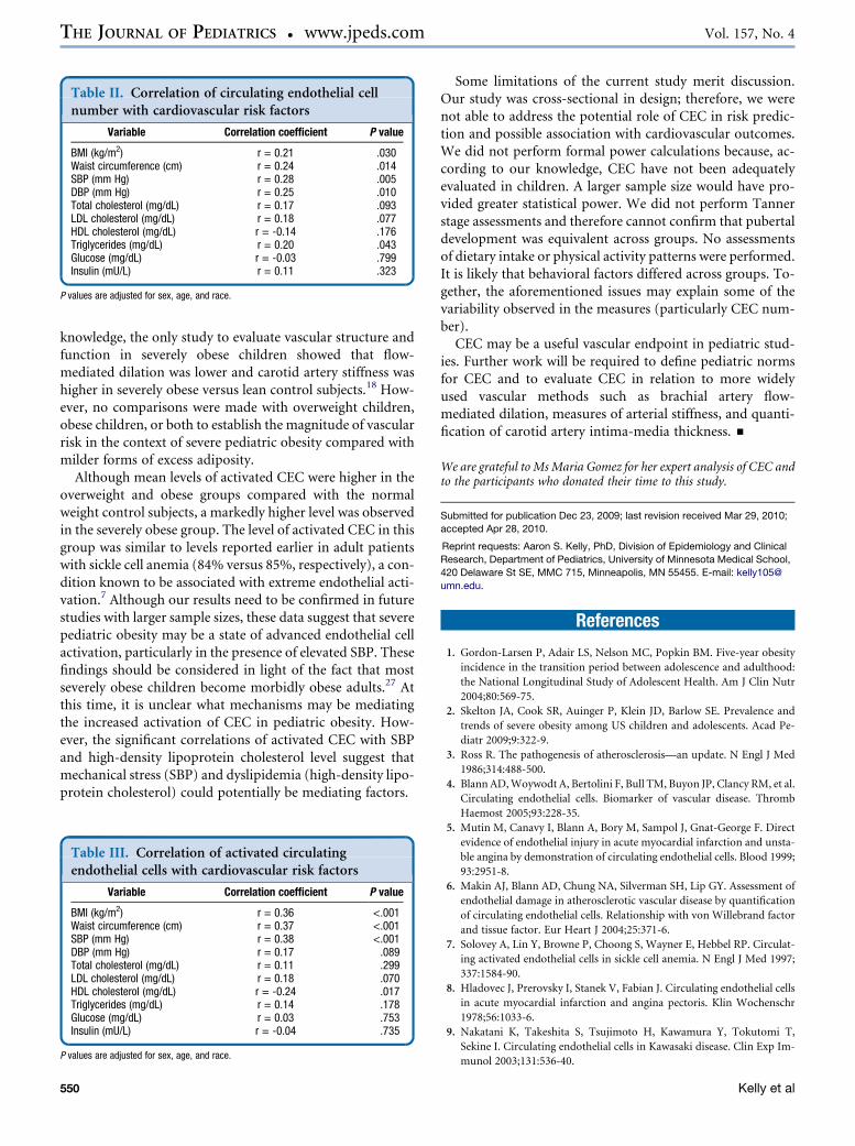
Table II. Correlation of circulating endothelial cellnumber with cardiovascular risk factors
Variable Correlation coefficient P value
BMI (kg/m2) r = 0.21 .030Waist circumference (cm) r = 0.24 .014SBP (mm Hg) r = 0.28 .005DBP (mm Hg) r = 0.25 .010Total cholesterol (mg/dL) r = 0.17 .093LDL cholesterol (mg/dL) r = 0.18 .077HDL cholesterol (mg/dL) r = -0.14 .176Triglycerides (mg/dL) r = 0.20 .043Glucose (mg/dL) r = -0.03 .799Insulin (mU/L) r = 0.11 .323
P values are adjusted for sex, age, and race.
THE JOURNAL OF PEDIATRICS � www.jpeds.com Vol. 157, No. 4
knowledge, the only study to evaluate vascular structure andfunction in severely obese children showed that flow-mediated dilation was lower and carotid artery stiffness washigher in severely obese versus lean control subjects.18 How-ever, no comparisons were made with overweight children,obese children, or both to establish the magnitude of vascularrisk in the context of severe pediatric obesity compared withmilder forms of excess adiposity.
Although mean levels of activated CEC were higher in theoverweight and obese groups compared with the normalweight control subjects, a markedly higher level was observedin the severely obese group. The level of activated CEC in thisgroup was similar to levels reported earlier in adult patientswith sickle cell anemia (84% versus 85%, respectively), a con-dition known to be associated with extreme endothelial acti-vation.7 Although our results need to be confirmed in futurestudies with larger sample sizes, these data suggest that severepediatric obesity may be a state of advanced endothelial cellactivation, particularly in the presence of elevated SBP. Thesefindings should be considered in light of the fact that mostseverely obese children become morbidly obese adults.27 Atthis time, it is unclear what mechanisms may be mediatingthe increased activation of CEC in pediatric obesity. How-ever, the significant correlations of activated CEC with SBPand high-density lipoprotein cholesterol level suggest thatmechanical stress (SBP) and dyslipidemia (high-density lipo-protein cholesterol) could potentially be mediating factors.
Table III. Correlation of activated circulatingendothelial cells with cardiovascular risk factors
Variable Correlation coefficient P value
BMI (kg/m2) r = 0.36 <.001Waist circumference (cm) r = 0.37 <.001SBP (mm Hg) r = 0.38 <.001DBP (mm Hg) r = 0.17 .089Total cholesterol (mg/dL) r = 0.11 .299LDL cholesterol (mg/dL) r = 0.18 .070HDL cholesterol (mg/dL) r = -0.24 .017Triglycerides (mg/dL) r = 0.14 .178Glucose (mg/dL) r = 0.03 .753Insulin (mU/L) r = -0.04 .735
P values are adjusted for sex, age, and race.
550
Some limitations of the current study merit discussion.Our study was cross-sectional in design; therefore, we werenot able to address the potential role of CEC in risk predic-tion and possible association with cardiovascular outcomes.We did not perform formal power calculations because, ac-cording to our knowledge, CEC have not been adequatelyevaluated in children. A larger sample size would have pro-vided greater statistical power. We did not perform Tannerstage assessments and therefore cannot confirm that pubertaldevelopment was equivalent across groups. No assessmentsof dietary intake or physical activity patterns were performed.It is likely that behavioral factors differed across groups. To-gether, the aforementioned issues may explain some of thevariability observed in the measures (particularly CEC num-ber).
CEC may be a useful vascular endpoint in pediatric stud-ies. Further work will be required to define pediatric normsfor CEC and to evaluate CEC in relation to more widelyused vascular methods such as brachial artery flow-mediated dilation, measures of arterial stiffness, and quanti-fication of carotid artery intima-media thickness. n
We are grateful to Ms Maria Gomez for her expert analysis of CEC andto the participants who donated their time to this study.
Submitted for publication Dec 23, 2009; last revision received Mar 29, 2010;
accepted Apr 28, 2010.
Reprint requests: Aaron S. Kelly, PhD, Division of Epidemiology and Clinical
Research, Department of Pediatrics, University of Minnesota Medical School,
420 Delaware St SE, MMC 715, Minneapolis, MN 55455. E-mail: kelly105@
umn.edu.
References
1. Gordon-Larsen P, Adair LS, Nelson MC, Popkin BM. Five-year obesity
incidence in the transition period between adolescence and adulthood:
the National Longitudinal Study of Adolescent Health. Am J Clin Nutr
2004;80:569-75.
2. Skelton JA, Cook SR, Auinger P, Klein JD, Barlow SE. Prevalence and
trends of severe obesity among US children and adolescents. Acad Pe-
diatr 2009;9:322-9.
3. Ross R. The pathogenesis of atherosclerosis—an update. N Engl J Med
1986;314:488-500.
4. Blann AD, Woywodt A, Bertolini F, Bull TM, Buyon JP, Clancy RM, et al.
Circulating endothelial cells. Biomarker of vascular disease. Thromb
Haemost 2005;93:228-35.
5. Mutin M, Canavy I, Blann A, Bory M, Sampol J, Gnat-George F. Direct
evidence of endothelial injury in acute myocardial infarction and unsta-
ble angina by demonstration of circulating endothelial cells. Blood 1999;
93:2951-8.
6. Makin AJ, Blann AD, Chung NA, Silverman SH, Lip GY. Assessment of
endothelial damage in atherosclerotic vascular disease by quantification
of circulating endothelial cells. Relationship with von Willebrand factor
and tissue factor. Eur Heart J 2004;25:371-6.
7. Solovey A, Lin Y, Browne P, Choong S, Wayner E, Hebbel RP. Circulat-
ing activated endothelial cells in sickle cell anemia. N Engl J Med 1997;
337:1584-90.
8. Hladovec J, Prerovsky I, Stanek V, Fabian J. Circulating endothelial cells
in acute myocardial infarction and angina pectoris. Klin Wochenschr
1978;56:1033-6.
9. Nakatani K, Takeshita S, Tsujimoto H, Kawamura Y, Tokutomi T,
Sekine I. Circulating endothelial cells in Kawasaki disease. Clin Exp Im-
munol 2003;131:536-40.
Kelly et al
October 2010 ORIGINAL ARTICLES
10. George F, Brouqui P, Boffa MC, Mutin M, Drancourt M, Brisson C, et al.
Demonstration of Rickettsia conorii-induced endothelial injury in vivo
by measuring circulating endothelial cells, thrombomodulin, and von
Willebrand factor in patients with Mediterranean spotted fever. Blood
1993;82:2109-16.
11. Bull TM, Golpon H, Hebbel RP, Solovey A, Cool CD, Tuder RM, et al.
Circulating endothelial cells in pulmonary hypertension. Thromb Hae-
most 2003;90:698-703.
12. Koc M, Richards HB, Bihorac A, Ross EA, Schold JD, Segal MS. Circu-
lating endothelial cells are associated with future vascular events in he-
modialysis patients. Kidney Int 2005;67:1078-83.
13. Lee KW, Lip GY, Tayebjee M, Foster W, Blann AD. Circulating endothe-
lial cells, von Willebrand factor, interleukin-6, and prognosis in patients
with acute coronary syndromes. Blood 2005;105:526-32.
14. Quilici J, Banzet N, Paule P, Meynard JB, Mutin M, Bonnet JL, et al.
Circulating endothelial cell count as a diagnostic marker for non-
ST-elevation acute coronary syndromes. Circulation 2004;110:
1586-91.
15. Lin Y, Weisdorf DJ, Solovey A, Hebbel RP. Origins of circulating endo-
thelial cells and endothelial outgrowth from blood. J Clin Invest 2000;
105:71-7.
16. Kapiotis S, Holzer G, Schaller G, Haumer M, Widhalm H, Weghuber D,
et al. A proinflammatory state is detectable in obese children and is ac-
companied by functional and morphological vascular changes. Arterios-
cler Thromb Vasc Biol 2006;26:2541-6.
17. Meyer AA, Kundt G, Steiner M, Schuff-Werner P, Kienast W. Impaired
flow-mediated vasodilation, carotid artery intima-media thickening,
and elevated endothelial plasma markers in obese children: the impact
of cardiovascular risk factors. Pediatrics 2006;117:1560-7.
18. Tounian P, Aggoun Y, Dubern B, Varille V, Guy-Grand B, Sidi D, et al.
Presence of increased stiffness of the common carotid artery and endo-
thelial dysfunction in severely obese children: a prospective study. Lancet
2001;358:1400-4.
Circulating Activated Endothelial Cells in Pediatric Obesity
19. Woo KS, Chook P, Yu CW, Sung RY, Qiao M, Leung SS, et al. Over-
weight in children is associated with arterial endothelial dysfunction
and intima-media thickening. Int J Obes Relat Metab Disord 2004;28:
852-7.
20. Aggoun Y, Farpour-Lambert NJ, Marchand LM, Golay E, Maggio AB,
Beghetti M. Impaired endothelial and smooth muscle functions and ar-
terial stiffness appear before puberty in obese children and are associated
with elevated ambulatory blood pressure. Eur Heart J 2008;29:792-9.
21. Kelly AS, Wetzsteon RJ, Kaiser DR, Steinberger J, Bank AJ, Dengel DR.
Inflammation, insulin, and endothelial function in overweight children
and adolescents: the role of exercise. J Pediatr 2004;145:731-6.
22. Lee S, Bacha F, Gungor N, Arslanian S. Comparison of different defini-
tions of pediatric metabolic syndrome: relation to abdominal adiposity,
insulin resistance, adiponectin, and inflammatory biomarkers. J Pediatr
2008;152:177-84.
23. Rocchini AP, Moorehead C, Katch V, Key J, Finta KM. Forearm resis-
tance vessel abnormalities and insulin resistance in obese adolescents.
Hypertension 1992;19:615-20.
24. Urbina EM, Williams RV, Alpert BS, Collins RT, Daniels SR, Hayman L,
et al. Noninvasive assessment of subclinical atherosclerosis in children
and adolescents: recommendations for standard assessment for clinical
research: a scientific statement from the American Heart Association.
Hypertension 2009;54:919-50.
25. Gidding SS, Nehgme R, Heise C, Muscar C, Linton A, Hassink S. Severe
obesity associated with cardiovascular deconditioning, high prevalence
of cardiovascular risk factors, diabetes mellitus/hyperinsulinemia, and
respiratory compromise. J Pediatr 2004;144:766-9.
26. Weiss R, Dziura J, Burgert TS, Tamborlane WV, Taksali SE, Yeckel CW,
et al. Obesity and the metabolic syndrome in children and adolescents. N
Engl J Med 2004;350:2362-74.
27. Freedman DS, Mei Z, Srinivasan SR, Berenson GS, Dietz WH. Cardio-
vascular risk factors and excess adiposity among overweight children
and adolescents: the Bogalusa Heart Study. J Pediatr 2007;150:12-7.
551