Embed Size (px)
Citation preview
Released 2018 health.govt.nz
Code of Practice
for Diagnostic
and
Interventional
Radiology
ORS C1
Citation: Ministry of Health. 2018. Code of Practice for Diagnostic and Interventional
Radiology: ORS C1. Wellington: Ministry of Health.
Published in November 2018 by the Ministry of Health
PO Box 5013, Wellington 6140, New Zealand
ISBN 978-1-98-856817-1 (online)
HP 6970
This document is available at health.govt.nz
This work is licensed under the Creative Commons Attribution 4.0 International licence.
In essence, you are free to: share ie, copy and redistribute the material in any medium or
format; adapt ie, remix, transform and build upon the material. You must give
appropriate credit, provide a link to the licence and indicate if changes were made.
CODE OF PRACTICE FOR DIAGNOSTIC AND INTERVENTIONAL RADIOLOGY: ORS C1 iii
Contents Introduction 1
Purpose and commencement 1
Scope 1
Contact 1
Roles and responsibilities 2
Definitions 3
Managing entity 7
General 7
Safety assessment 8
Facilities 8
Equipment 9
Training and authorisation 11
Policies, procedures and local rules 11
Patient dosimetry 12
Monitoring and measurement 12
Incidents, accidents and emergencies 14
Records 15
Quality assurance 15
Radiation practitioner 17
General 17
Justification 17
Optimisation 18
Other parties 19
Referring practitioner 19
Manufacturer/supplier 19
Servicing engineer 20
Appendices
Appendix 1: Cross-reference to Radiation Safety Act 2016 21
Appendix 2: Equipment requirements 22
Appendix 3: Training requirements 32

CODE OF PRACTICE FOR DIAGNOSTIC AND INTERVENTIONAL RADIOLOGY: ORS C1 1
Introduction
Purpose and commencement This Code of Practice for Diagnostic and Interventional Radiology (code) is issued by the
Director for Radiation Safety (the Director) under section 86 of the Radiation Safety Act
2016 (the Act). It provides operational details necessary to comply with the fundamental
requirements in sections 9 to 12 of the Act. Appendix 1 sets out cross-references
between clauses in this code and those fundamental requirements. The requirements in
this code do not limit the general nature of the fundamental requirements.
This code comes into force on 9 November 2018.
Scope This code applies to all activities associated with:
radiological equipment used for diagnostic radiology and image-guided
interventional procedures
radiological equipment used for diagnostic investigations of volunteers
participating in programmes of medical research
cone beam computed tomography equipment used for dental purposes.
Bone densitometry is included within the scope, but computed tomography equipment
used solely for radiotherapy treatment planning or verification is excluded.
Activities can include manufacturing, possessing, controlling, managing, using,
transporting, storing, importing, exporting, selling, supplying and disposing of equipment.
Compliance with this code does not imply compliance in related areas such as health
practitioner clinical competence, occupational safety, hazards in the workplace,
resource management and transport of hazardous substances.
Contact The Director’s contact details are:
Office of Radiation Safety
PO Box 5013
Wellington 6140
Email: [email protected]
Fax: 04 496 2340
2 CODE OF PRACTICE FOR DIAGNOSTIC AND INTERVENTIONAL RADIOLOGY: ORS C1
Roles and responsibilities The following individuals and bodies have roles and responsibilities in relation to this code.
Director for Radiation Safety – the individual appointed under section 76 of the Act
to perform functions and duties and exercise powers set out in the Act including the
power to issue this code.
Ethics committee – the committee that approves programmes of medical research
including the justification of medical exposure of volunteers.
Managing entity – the legal entity that manages or controls radiological equipment
and must, therefore, obtain a source licence as required by section 13(a) of the Act.
This could be, for example, a district health board, company, partnership, trust or
individual person.
Manufacturer/supplier – the person or organisation that designs, manufactures,
produces, constructs, assembles, installs, distributes, sells, exports or imports
radiological equipment or develops software that could influence the delivery of
medical exposures.
Medical physicist – an individual with specialist education and training in the concepts
and techniques of applying physics in medicine and competent to practise
independently in the diagnostic and interventional radiology specialty of medical
physics and who provides specialist expertise for radiation protection of the patient.
Medical radiation technologist – a health practitioner with specialist education and
training in medical radiation technology who is competent to perform radiological
procedures on delegation from a radiation practitioner.
Operator – a medical radiation technologist or another person who is competent to
perform radiological procedures on delegation from the radiation practitioner.
Qualified expert – an individual who is recognised as having expertise in a relevant
field of specialisation such as medical physics or radiation safety.
Radiation safety officer – a person who is competent in radiation protection and
safety, who is designated by the managing entity to oversee the application of
regulatory requirements for occupational and public radiation protection and safety.
Radiation practitioner – a health practitioner with specialist education and training in
the medical uses of radiation, who is competent to perform independently and oversee
radiological procedures. This could include, for example, a radiologist, cardiologist,
surgeon, general practitioner, chiropractor or, for cone beam computed tomography
equipment, a dental practitioner.
Referring practitioner – a health practitioner who is approved by the managing entity
to refer individuals to a radiation practitioner for medical exposure. Often this is a
general practitioner who refers patients to a radiology department or practice.
Servicing engineer – a person who has expertise in installing, servicing and
maintaining radiological equipment.
Standards dosimetry laboratory – a laboratory that is certified or accredited to
develop, maintain or improve primary or secondary standards for radiation dosimetry.
CODE OF PRACTICE FOR DIAGNOSTIC AND INTERVENTIONAL RADIOLOGY: ORS C1 3
Definitions Defined terms are identified in bold and have the following meanings.
Accident – any unintended medical exposure or other unintended event, including
operating errors, equipment failures and other mishaps, the consequences or potential
consequences of which are not negligible from the point of view of protection and
safety.
Ambient dose equivalent – the dose equivalent that would be produced by the
corresponding aligned and expanded field in the International Commission of
Radiation Units and Measurements ICRU sphere at a depth d on the radius vector
opposing the direction of the aligned field.
Ancillary equipment – equipment other than radiological equipment or protective
equipment that has an impact on the performance of a radiological procedure such
as automatic film processors, printers, image receptors, view boxes, digital image
displays, test and measurement equipment used to verify or calibrate radiological
equipment.
Comforter/carer – a person who voluntarily helps, other than occupationally in caring
for, supporting and comforting a patient undergoing a radiological procedure.
Constraint – a prospective and source related value of individual dose (dose
constraint) or of individual risk (risk constraint) that is used in planned exposure
situations as a parameter for the optimisation of protection and safety for the
source, and that serves as a boundary in defining the range of options in optimisation.
Constraints for occupational exposure, public exposure and medical exposure of
comforter/carers are established or approved by the Director and, if established, are
published in a compliance guide issued under this code. Constraints for medical
exposure of volunteers are established or approved by the ethics committee on a
case by case basis as part of the proposal for medical research.
Controlled area – a defined area in which specific measures for protection and safety
are or could be required for controlling exposures in normal working conditions, and
preventing or limiting the likelihood and magnitude of potential exposures.
Diagnostic reference level – a level that is used to indicate whether, in routine
conditions, the dose to the patient is unusually high or unusually low for that
procedure. Diagnostic reference levels if any are established and published by the
Director.
Dose limit – the value of effective dose or equivalent dose set out in Schedule 3 of
the Act.
Effective dose – the tissue-weighted sum of equivalent doses in all specified tissues
and organs of the body.
4 CODE OF PRACTICE FOR DIAGNOSTIC AND INTERVENTIONAL RADIOLOGY: ORS C1
Emergency – any non-routine situation that necessitates prompt action, primarily to
mitigate actual or perceived hazards or adverse consequences for human health and
safety, quality of life, property or the environment. This includes radiation
emergencies and conventional emergencies such as fires, release of hazardous
chemicals, storms or earthquakes.
Employer – the legal entity that employs workers. A self-employed person is regarded
as being both an employer and a worker.
Equivalent dose – the radiation-weighted dose in a tissue or organ of the body.
Facility – the location where radiological equipment and ancillary equipment is
installed, used, handled or stored.
Health practitioner – an individual who is, or is deemed to be, registered with an
authority as a practitioner of a particular health profession under the Health
Practitioners Competence Assurance Act 2003.
Health screening programme – a programme for asymptomatic populations that is
approved and justified by a health authority in conjunction with appropriate
professional bodies.
In-room protective device – device or equipment to reduce exposure to radiation but
not worn by a person, such as ceiling-suspended protective screens, protective lead
curtains, mobile shields and disposable protective drapes.
Incident – any accident or other unintended event, including initiating events,
accident precursors, near misses or other mishaps; or unauthorised acts, malicious or
non-malicious, the consequences or potential consequences of which are not
negligible from the point of view of protection and safety.
Individual monitoring – monitoring using equipment worn by individuals.
Investigation level – value of a quantity such as effective dose, intake or
contamination per unit area or volume at or above which an investigation would be
conducted.
Justify – determine that the expected benefits to individuals and society from
introducing or continuing a practice outweigh the harm, including the radiation
detriment, resulting from the practice. In respect of individual radiological
procedures, this involves the weighing of expected benefits against the radiation
detriment that might be caused with account taken of the benefits and risks of
available alternate techniques that do not involve medical exposure. ‘Justifies’,
‘justified’ and ‘justification’ have corresponding meanings.
Medical exposure – exposure to ionising radiation experienced by patients for the
purposes of medical or dental diagnosis or treatment, by comforter/carers while
caring for, supporting, or comforting patients undergoing radiological procedures,
and by volunteers in a programme of medical research.
Member of the public – for purposes of protection and safety, any individual in the
population except when subject to occupational exposure or medical exposure.
Monitoring – the measurement of dose or dose rate to enable the assessment or
control of exposure due to radiation, and the interpretation of the results.
CODE OF PRACTICE FOR DIAGNOSTIC AND INTERVENTIONAL RADIOLOGY: ORS C1 5
Occupational exposure – exposure of workers incurred in the course of their work.
Occupationally exposed person – any person who is subject to occupational
exposure.
Operational limits and conditions – limits and conditions that are established or
approved by the Director and, if established, are published in compliance guides issued
under this code.
Optimise – implement a level of protection and safety that results in the magnitude
of individual doses, the number of individuals (workers and members of the public)
subject to exposure and the likelihood of exposure being as low as reasonably
achievable, economic and social factors being taken into account. For medical
exposures of patients this requires the management of the radiation dose to the
patient commensurate with the medical purpose. ‘Optimises’, ‘optimised’ and
‘optimisation’ have corresponding meanings.
Patient – a person who is subject to medical exposure for his or her own medical
benefit.
Personal protective equipment – equipment worn on the person to reduce exposure
to radiation such as protective aprons, organ shields, protective eye-wear and
protective gloves.
Planned exposure situation – situation of exposure that arises from the planned
operation of radiological equipment or from a planned activity that results in an
exposure due to radiological equipment.
Potential exposure – possible future exposure that may result from an anticipated
operational occurrence or accident at a source or due to an event or sequence of
events of a probabilistic nature, including equipment faults and operating errors.
Primary shielding – 2 mm lead equivalence at 100 kVp.
Protective equipment – personal protective equipment and in-room protective
devices.
Protection and safety – the protection of people against exposure to ionising
radiation and the safety of radiological equipment, including the means for achieving
this, and the means for preventing accidents and for mitigating the consequences of
accidents if they do occur.
Public exposure – exposure to ionising radiation that a member of the public
experiences, but excluding any occupational exposure or medical exposure.
Radiation emergency – an emergency in which there is, or is perceived to be, a
hazard due to radiation exposure.
Radiological equipment – equipment and its associated software used to perform
radiological procedures that either delivers an exposure of an individual or directly
controls or influences the extent of such exposure.
6 CODE OF PRACTICE FOR DIAGNOSTIC AND INTERVENTIONAL RADIOLOGY: ORS C1
Radiological procedure – a medical imaging procedure that is intended to result in a
medical exposure delivered by radiological equipment. This includes procedures in
diagnostic radiology, image-guided interventional procedures, other interventional
procedure involving radiation or dental procedures involving cone beam computed
tomography equipment.
Reportable incident – an incident resulting in (a) a dose limit being exceeded, (b)
radiological equipment that is lost, missing or beyond regulatory control, or (c) a
radiation dose to a patient exceeding two times the intended dose (for interventional
radiology, radiographic and fluoroscopic procedures involving contrast agents and
computed tomography examinations), twenty times the intended dose (for
radiography of extremities, skull, dentition, shoulder, chest, elbow and knee), or ten
times the intended dose (for mammography and all other radiographic examinations).
Safety assessment – assessment of all aspects of a practice that are relevant to
protection and safety to determine the adequacy of provisions for protection and
safety.
Secondary shielding – 1.5 mm lead equivalence at 120 kVp in areas where computed
tomography equipment is performed, 18 mm gypsum plasterboard equivalence in all
areas where mammography or dual-energy X-ray absorptiometry is performed and
1.0 mm lead equivalence for all other areas where radiological procedures are
performed.
Supervised area – an area other than a controlled area for which occupational
exposure conditions need to be kept under review, even though specific measures for
protection and safety are not normally needed.
Typical dose – the median or average of the doses for a representative sample of
relatively standard-sized patients, at clinically acceptable image quality.
Volunteer – an individual other than a comforter/carer who may be subjected to
medical exposure as part of a programme of medical research.
Unintended medical exposure – exposure of the wrong individual, tissue or organ;
exposure that is substantially greater than intended; inadvertent exposure of the
embryo or fetus; and failure of radiological equipment, failure of software or system
failure, or error, mishap or other unusual occurrence with the potential for subjecting
the patient to a medical exposure that is substantially different from what was
intended.
Worker – an individual who works, whether full time, part time or temporarily for the
managing entity or another employer and who has recognised rights and duties in
relation to occupational radiation protection. A self-employed person is regarded as
being both an employer and a worker.
Workplace monitoring – monitoring carried out in the working environment.
CODE OF PRACTICE FOR DIAGNOSTIC AND INTERVENTIONAL RADIOLOGY: ORS C1 7
Managing entity
General 1. The managing entity must:
a) take prime responsibility for protection and safety
b) establish a management system to enhance protection and safety that
includes:
i) effectively integrating protection and safety into the overall
management system of the organisation
ii) making a commitment to protection and safety from the highest
level of management at the facility, and by providing all required
resources
iii) promoting continuous improvement and a safety culture
iv) appointing a radiation safety officer to oversee the application of
regulatory requirements for occupational and public radiation
protection and safety
v) delegating the planning and delivery of medical exposures to a
radiation practitioner
vi) ensuring that requirements for medical imaging, calibration,
dosimetry of patients, quality assurance and the commissioning and
acceptance of radiological equipment are fulfilled by, or under the
oversight of, or with the documented advice of a medical physicist
whose degree of involvement is determined by the complexity of the
radiological procedures and the associated radiation risks
vii) consulting with and engaging the services of other experts and
interested parties as necessary
c) for all delegations under sub-clauses 1(b)(iv) and 1(b)(v):
i) ensure delegates are notified of their duties in relation to protection
and safety and assume responsibility for performing them
ii) fully document the delegations
d) ensure that:
i) all activities associated with radiological equipment are justified and
optimised for protection and safety
ii) dose limits for occupational and public exposure are not exceeded
as a result of those activities.
8 CODE OF PRACTICE FOR DIAGNOSTIC AND INTERVENTIONAL RADIOLOGY: ORS C1
2. The managing entity must ensure that no practice or procedure is undertaken
unless:
a) it has been justified generically by a health authority
b) it has been:
i) justified specifically by a health authority in conjunction with
appropriate professional bodies for procedures that are part of a
health screening programme
ii) approved by an ethics committee for medical exposures incurred as
part of a programme of medical research
iii) justified individually for the patient by a radiation practitioner in any
other case.
Safety assessment 3. The managing entity must conduct, document and keep up to date a safety
assessment to:
a) identify the ways in which occupational, public and medical exposures
could be incurred
b) determine the expected likelihood and magnitudes of exposures in normal
operation and, to the extent reasonable and practicable, assess potential
exposures including the possibility of unintended or accidental medical
exposures
c) assess the adequacy of provisions for protection and safety in respect of
siting, design and operation.
Facilities 4. The managing entity must:
a) provide facilities that are located, designed, manufactured, constructed,
assembled, commissioned, operated, maintained and decommissioned
adopting good engineering practice, minimising the need to rely on
administrative controls and personal protective equipment for protection
and safety
b) shield all areas in which radiological procedures are performed, so that
protection and safety is optimised by satisfying either:
i) the requirements for primary shielding in areas designed to be
exposed to the primary radiation beam during normal use and the
requirements for secondary shielding for all other doors, walls,
ceilings, windows, floors and other material constructions, or
ii) alternate requirements if they are approved and documented by a
medical physicist or another qualified expert
CODE OF PRACTICE FOR DIAGNOSTIC AND INTERVENTIONAL RADIOLOGY: ORS C1 9
c) in consultation with a medical physicist or another qualified expert, verify
and document the adequacy of the shielding required in clause 4(b)
whenever circumstances change that could increase the risks
d) designate and delineate appropriate areas as controlled areas or
supervised areas and periodically review those designations and
delineations
e) prominently display signs:
i) specifying the actual or potential presence of ionising radiation
using the symbol recommended by the International Organization
for Standardization at access points to controlled areas and
supervised areas and at appropriate locations within controlled areas
ii) controlling access by members of the public to controlled areas and
supervised areas
iii) in areas that patients may be (including waiting rooms and change
cubicles), requiring patients who are to undergo a radiological
procedure to notify staff if they are or may be pregnant.
Equipment 5. The managing entity must:
a) provide, maintain, test and service radiological equipment, protective
equipment and ancillary equipment so that:
i) the equipment is appropriate for the radiological procedures to be
performed
ii) the equipment remains capable of fulfilling its design requirements
for protection and safety throughout its lifetime
b) ensure that:
i) the primary requirements in column 2 of Appendix 2 are satisfied
ii) all reasonable steps are taken to satisfy the secondary requirements
in column 3 of Appendix 2
iii) the protective value of protective equipment is clearly displayed on
the equipment
c) cooperate with manufacturer/suppliers to:
i) ensure that the requirements in sub-clauses 5(a) and 5(b) are met
ii) ensure that radiological equipment is used only if it conforms to the
applicable standards of the International Electrotechnical
Commission and the International Organization for Standardization
iii) share information on use and operating experience that may be
important for protection and safety
iv) applying the principles of optimisation in the design, planning and
operation and decommissioning of a source
10 CODE OF PRACTICE FOR DIAGNOSTIC AND INTERVENTIONAL RADIOLOGY: ORS C1
d) safely manage all radiological equipment, whether or not the equipment is
in use
e) maintain an accurate inventory of all radiological equipment, including its
location and description
f) maintain a record of maintenance for each item, including a fault log and
remedial actions taken (interim and subsequent repairs), the results of
testing before an item is reintroduced to clinical use, and any reports from
servicing engineers
g) maintain control of radiological equipment to prevent loss or damage and
to prevent any person from carrying out unauthorised activities including by:
i) periodically checking that equipment is under control and in the
locations recorded in the inventory maintained under clause 5(d)
ii) releasing the equipment only to people who are authorised to
assume management and control under the Act
h) take immediate steps to regain control of any radiological equipment that
is abandoned, lost, misplaced, stolen or otherwise transferred without
proper authorisation.
6. The managing entity must ensure that:
a) all sources giving rise to medical exposure are calibrated in terms of
appropriate quantities using internationally accepted protocols
b) calibrations are carried out at the time of commissioning radiological
equipment prior to clinical use, after any maintenance procedure that
could affect the dosimetry, and at intervals approved by the Director and
published in compliance guides issued under this code
c) all dosimeters used for the calibration of sources are calibrated at least
every two years and that such calibrations are traceable to a standards
dosimetry laboratory.
7. The managing entity must provide, maintain, test, calibrate and service
equipment, other than radiological equipment, sufficient to ensure compliance
with this code including equipment for personal protection, monitoring and
measurement for compliance verification, accident verification, emergency
response, protection and safety of members of the public.
CODE OF PRACTICE FOR DIAGNOSTIC AND INTERVENTIONAL RADIOLOGY: ORS C1 11
Training and authorisation 8. The managing entity must ensure that all persons with responsibilities for
protection and safety:
a) are specialised, qualified, educated and trained in protection and safety so
that they understand their duties and can perform them competently
b) satisfy the training requirements in Appendix 3
c) are named in a current list with details of their specialisation, qualification,
education and training
d) are notified of their duties in relation to protection and safety
e) are authorised to assume their roles and responsibilities.
Policies, procedures and local rules 9. The managing entity must establish, implement and maintain policies and
procedures to meet the requirements of this code including, without limitation,
policies and procedures to:
a) control access to areas where people can be exposed to radiation
b) use constraints to optimise protection and safety
c) prevent accidents and mitigate the consequences of any that occur
d) report on and learn from accidents and other incidents
e) comply with operational limits and conditions relating to public exposure
f) ascertain the pregnancy status of female patients of reproductive capacity
before performing any radiological procedure that could result in a
significant dose to the embryo or fetus
g) provide protection and safety by applying preventive measures in the
following hierarchy:
i) engineered controls
ii) administrative controls
iii) personal protective equipment
h) set investigation levels and establish procedures to follow if such a level is
exceeded
i) implement procedures for verification of compliance with this code
j) periodically review the overall effectiveness of measures for protection and
safety.
10. The managing entity must maintain, publish and enforce any written local rules
that are necessary for protection and safety.
12 CODE OF PRACTICE FOR DIAGNOSTIC AND INTERVENTIONAL RADIOLOGY: ORS C1
Patient dosimetry 11. The managing entity must:
a) perform and document dosimetry of patients to determine typical doses to
patients for:
i) common diagnostic radiological procedures
ii) image-guided interventional procedures where practicable
b) in order to satisfy the requirements in clause 11(a):
i) follow internationally accepted protocols, and
ii) use only dosimeters with current calibrations traceable to a
standards dosimetry laboratory.
Monitoring and measurement 12. The managing entity must establish and maintain:
a) a programme of continuous individual monitoring whenever appropriate,
adequate and feasible, which is sufficient to assess occupational exposures
for workers who usually work in a controlled area or who may receive a
dose exceeding 10 percent of the dose limits
b) a programme of workplace monitoring that is sufficient to:
i) evaluate radiation conditions in all workplaces
ii) assess exposures in controlled areas and supervised areas that are
not assessed under clause 12(a)
iii) review the classification of controlled areas and supervised areas
c) a monitoring programme for all workers who could be subject to exposure
due to contamination, which is sufficient to:
i) demonstrate the effectiveness of the measures for protection and
safety
ii) assess intakes of radionuclides and committed effective doses
d) programmes of source monitoring or environmental monitoring that are
sufficient to assess public exposure arising from radiological equipment
under the responsibility of the managing entity
e) a capability that is sufficient to monitor unexpected increases in radiation
levels due to an incident attributed to a source or facility for which the
managing entity is responsible
f) other monitoring or measurement programmes as necessary to verify
compliance with the requirements in this code.
CODE OF PRACTICE FOR DIAGNOSTIC AND INTERVENTIONAL RADIOLOGY: ORS C1 13
13. In order to satisfy the monitoring and measurement requirements in clause 12
the managing entity must:
a) use appropriate monitoring equipment
b) for continuous individual monitoring under clause 12(a), use an external
service or internal capability only if that service or capability:
i) is approved by the Director
ii) returns results to the managing entity within 20 working days of
receiving all necessary raw information.
14. The managing entity must:
a) obtain previous dose records
b) maintain records of all monitoring and verification of compliance
including:
i) records of occupational exposure during and after the worker’s
working life, at least until the worker attains or would have attained
the age of 75 years, and for not less than 30 years after ceasing work
where the worker was subject to occupational exposure
ii) records and estimated doses to members of the public
iii) records of the tests and calibrations carried out
c) provide records of occupational exposure to:
i) individual workers in respect of their own exposure
ii) subsequent employers of workers, subject to satisfying
confidentiality criteria
iii) the Director on request or, if the managing entity is no longer able
to maintain records as required under clause 14(b)
d) provide records of source monitoring and environmental monitoring to
assess public exposure to:
i) members of the public on request
ii) the Director on request
iii) the Director immediately, if any levels exceed operational limits and
conditions relating to public exposure or there is a significant
increase in dose rate that could be attributed to the authorised
practice.
14 CODE OF PRACTICE FOR DIAGNOSTIC AND INTERVENTIONAL RADIOLOGY: ORS C1
Incidents, accidents and
emergencies 15. The managing entity must:
a) take all practicable steps to minimise the likelihood of accidents including,
a multilevel system of sequential, independent provisions for protection
and safety, commensurate with the likelihood and magnitude of potential
exposures
b) take timely action to mitigate the consequences of any accident that does
occur and restore radiological equipment to a safe condition
c) promptly investigate any incident, including by:
i) calculating or estimating doses a person has received and, if
applicable, the dose distribution within them
ii) identifying corrective actions required to prevent a recurrence
d) implement all corrective actions identified in clause 15(c)(ii)
e) keep a written record of the incident, including the:
i) cause or suspected cause
ii) calculations made under clause 15(c)(i)
iii) corrective actions identified under clause 15(c)(ii)
iv) details of the implementation of corrective actions under
clause 15(d)
f) ensure that the referring practitioner and the patient (or the patient’s legal
representative) are informed of any unintended medical exposure
g) promptly notify any reportable incident to the Director.
16. If the safety assessment required by clause 3 indicates that there is a reasonable
likelihood of an emergency affecting either workers or members of the public,
the managing entity must prepare an emergency plan for the protection of
people and the environment including:
a) arrangements for promptly identifying an emergency
b) determining the correct level of emergency response
c) provision for individual monitoring, area monitoring and arrangements for
medical treatment
d) arrangements for assessing and mitigating any consequences of an
emergency.
CODE OF PRACTICE FOR DIAGNOSTIC AND INTERVENTIONAL RADIOLOGY: ORS C1 15
Records 17. The managing entity must maintain adequate records, and make them available
as necessary, including:
a) the delegation of responsibilities of the managing entity and the radiation
practitioner
b) the names of all people with responsibility for protection and safety,
including details of their specialisation, qualifications, education and
training
c) results of calibrations and periodic checks of physical and clinical
parameters selected during treatment of patients
d) dosimetry of patients
e) local assessments and reviews relating to diagnostic reference levels
f) the quality assurance programme
g) information necessary for the retrospective assessment of doses, including
the number of exposures and the duration of fluoroscopic radiological
procedures, for diagnostic radiology
h) information necessary for the retrospective assessment of doses, including
the duration of the fluoroscopic component and the number of images
acquired, for image-guided interventional procedures
i) exposure records for volunteers subject to medical exposure as part of a
programme of medical research
j) reports on investigations of unintended and accidental medical exposures
k) exemptions from this code granted under section 86(3) of the Act.
Quality assurance 18. The managing entity must establish a comprehensive quality assurance
programme for medical exposures, including:
a) measuring the physical parameters of radiological equipment, including
calibrating output in terms of appropriate quantities using internationally
accepted protocols, made:
i) at the time it accepts and commissions the equipment, before
practitioners use it clinically on patients
ii) periodically after that first check
iii) after any major maintenance procedure that could affect the
protection and safety of patients
iv) after installing any new software or modifying any existing software
that could affect the protection and safety of patients
16 CODE OF PRACTICE FOR DIAGNOSTIC AND INTERVENTIONAL RADIOLOGY: ORS C1
b) performing quality control tests on ancillary equipment and personal
protective equipment
c) adopting internationally accepted tolerance limits for the physical
parameters mentioned in sub-clauses 18(a) and 18(b), and implementing
corrective actions if measured values fall outside those tolerance limits
d) verifying the appropriateness of physical and clinical factors used in
radiological procedures
e) maintaining records of relevant procedures and results
f) periodically checking the calibration and conditions of operation of
dosimetry equipment and monitoring equipment.
19. The managing entity must ensure that regular internal or external independent
audits are made of the quality assurance programme for medical exposures.
20. The managing entity must ensure that:
a) radiation reviews are performed periodically by radiation practitioners in
cooperation with medical radiation technologists and medical physicists, to
investigate and critically review the current practical application of the
radiation protection principles of justification and optimisation for
radiological procedures
b) local assessments are made at regular intervals for those radiological
procedures for which diagnostic reference levels have been established
c) a review is conducted to determine whether the optimisation of protection
and safety for patients is adequate, or whether corrective action is required
if, for a given radiological procedure, typical doses or activities:
i) exceed the relevant diagnostic reference level
ii) fall substantially below the diagnostic reference level, and the
exposures do not provide useful diagnostic information or do not
yield the expected medical benefit to the patient.
CODE OF PRACTICE FOR DIAGNOSTIC AND INTERVENTIONAL RADIOLOGY: ORS C1 17
Radiation practitioner
General 21. The radiation practitioner:
a) is responsible for overall protection and safety in the planning and delivery
of the medical exposure
b) may, in order to satisfy the responsibility in clause 21(a), delegate functions
to a medical radiation technologist, medical physicist or otherwise1
c) must inform in advance all individuals who may be subject to medical
exposure (or their legal authorised representatives) of the expected
benefits, risks and limitations of the procedure, as appropriate.
Justification 22. The radiation practitioner must:
a) obtain information on the clinical context for any procedure unless it is
part of a health screening programme
b) for any procedure that is not part of a health screening programme, justify
the medical exposure in consultation as appropriate with the referring
practitioner taking into account, in particular for paediatric or possibly
pregnant individuals:
i) the appropriateness of the request
ii) the urgency of the procedure
iii) the characteristics of the medical exposure
iv) the characteristics of the individual patient
v) relevant information from the patient’s previous radiological
procedures
vi) relevant national or international referral guidelines
c) for any procedure to detect disease in an asymptomatic person that is not
part of a health screening programme, justify the procedure specifically for
the individual in accordance with any guidelines of relevant professional
bodies or the health authority.
1 The managing entity has obligations under clause 1 to ensure that these delegations are notified and
documented and that delegates assume responsibility for the delegated functions.
18 CODE OF PRACTICE FOR DIAGNOSTIC AND INTERVENTIONAL RADIOLOGY: ORS C1
Optimisation 23. The radiation practitioner must, in consultation as appropriate with medical
physicists and operators, ensure that protection and safety is optimised for each
medical exposure:
a) for diagnostic radiological procedures and image-guided interventional
procedures by:
i) using appropriate radiological equipment
ii) adopting techniques and parameters to deliver a medical exposure
that is the minimum necessary to fulfil the clinical purpose of the
radiological procedure, taking into account relevant norms of
acceptable image quality and of relevant diagnostic reference levels
b) by using constraints in any procedure in which an individual:
i) acts as a comforter/carer
ii) is subject to exposure as part of a programme of research.
24. The radiation practitioner must ensure that particular aspects of medical
exposures are considered in the optimisation process for:
a) paediatric patients
b) individuals subject to medical exposure as part of a health screening
programme
c) volunteers subject to medical exposure as part of a programme of medical
research
d) procedures involving computed tomography
e) exposure of the embryo or fetus, in particular, during radiological
procedures in which the abdomen or pelvis of a pregnant patient is
exposed to the useful radiation beam or could otherwise receive a
significant dose.
CODE OF PRACTICE FOR DIAGNOSTIC AND INTERVENTIONAL RADIOLOGY: ORS C1 19
Other parties
Referring practitioner 25. The referring practitioner must:
a) provide sufficient information on the clinical context of the procedure in
the referral
b) cooperate with the radiation practitioner as part of the justification of the
procedure in accordance with clause 22.
Manufacturer/supplier 26. The manufacturer/supplier of radiological equipment must:
a) supply well-designed, well-manufactured and well-constructed
radiological equipment that:
i) provides for protection and safety in line with the requirements of
this code
ii) meets engineering, performance and functional specifications
iii) meets quality standards appropriate to the significance of systems
and components, including software, for protection and safety
iv) provides clear displays, gauges and instructions on operating
consoles
b) test radiological equipment to demonstrate compliance with relevant
specifications
c) provide information on how to properly install and use radiological
equipment and on associated radiation risks, including performance
specifications, instructions for operating and maintenance, and instructions
for protection and safety
d) optimise the protection provided by shielding and other protective devices
e) supply all radiological equipment with all appropriate radiation protection
tools as a default, rather than as optional extras.
27. The manufacturer/supplier must:
a) make suitable arrangements with managing entities to share information
on use and operating experience that may be important for protection and
safety
b) cooperate with the managing entity as required by clause 5(c).
20 CODE OF PRACTICE FOR DIAGNOSTIC AND INTERVENTIONAL RADIOLOGY: ORS C1
Servicing engineer 28. The servicing engineer must:
a) install and service radiological equipment competently, so that it complies
with the requirements in clause 5
b) cooperate with the managing entity to ensure that radiological equipment
cannot be used clinically while it is being installed or serviced
c) after installing or servicing the equipment:
i) collaborate with the managing entity and medical physicists to
ensure necessary quality control tests are completed successfully
ii) confirm that all radiation protection and safety features are in place
and operating correctly before equipment is returned to clinical use
iii) provide a written report to the managing entity describing the
equipment fault (if any), the work done, parts replaced, adjustments
made and any changes that may affect protection and safety.
CODE OF PRACTICE FOR DIAGNOSTIC AND INTERVENTIONAL RADIOLOGY: ORS C1 21
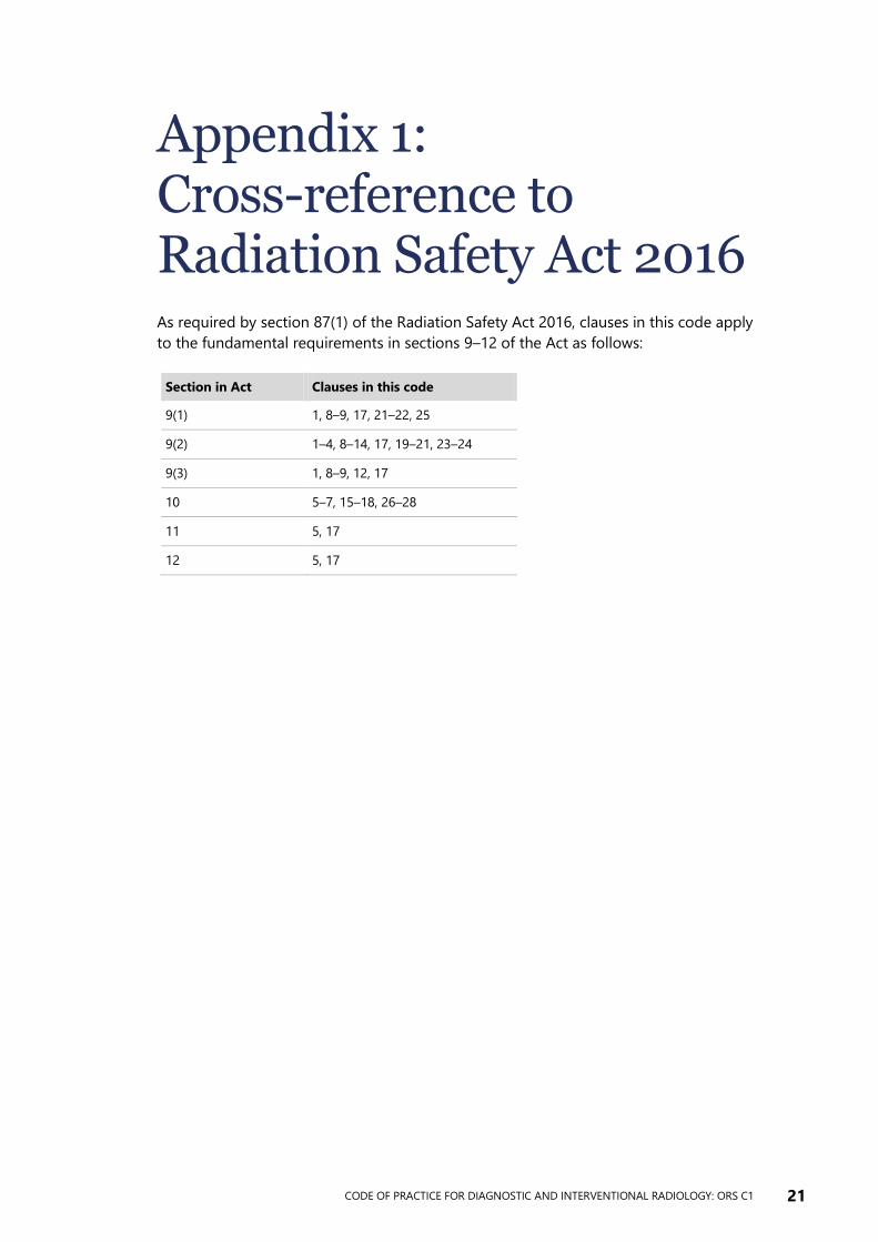
Appendix 1: Cross-reference to Radiation Safety Act 2016 As required by section 87(1) of the Radiation Safety Act 2016, clauses in this code apply
to the fundamental requirements in sections 9–12 of the Act as follows:
Section in Act Clauses in this code
9(1) 1, 8–9, 17, 21–22, 25
9(2) 1–4, 8–14, 17, 19–21, 23–24
9(3) 1, 8–9, 12, 17
10 5–7, 15–18, 26–28
11 5, 17
12 5, 17
22 CODE OF PRACTICE FOR DIAGNOSTIC AND INTERVENTIONAL RADIOLOGY: ORS C1
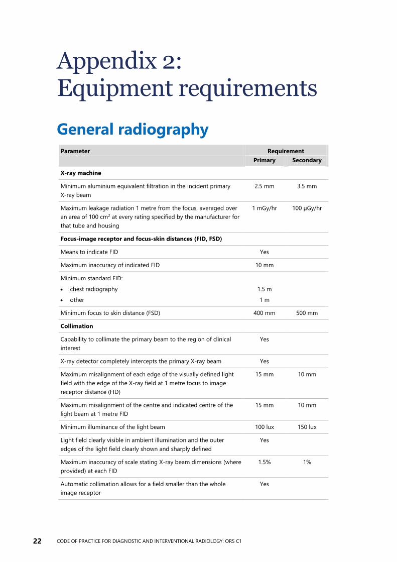
Appendix 2: Equipment requirements
General radiography Parameter Requirement
Primary Secondary
X-ray machine
Minimum aluminium equivalent filtration in the incident primary
X-ray beam
2.5 mm 3.5 mm
Maximum leakage radiation 1 metre from the focus, averaged over
an area of 100 cm2 at every rating specified by the manufacturer for
that tube and housing
1 mGy/hr 100 µGy/hr
Focus-image receptor and focus-skin distances (FID, FSD)
Means to indicate FID Yes
Maximum inaccuracy of indicated FID 10 mm
Minimum standard FID:
chest radiography
other
1.5 m
1 m
Minimum focus to skin distance (FSD) 400 mm 500 mm
Collimation
Capability to collimate the primary beam to the region of clinical
interest
Yes
X-ray detector completely intercepts the primary X-ray beam Yes
Maximum misalignment of each edge of the visually defined light
field with the edge of the X-ray field at 1 metre focus to image
receptor distance (FID)
15 mm 10 mm
Maximum misalignment of the centre and indicated centre of the
light beam at 1 metre FID
15 mm 10 mm
Minimum illuminance of the light beam 100 lux 150 lux
Light field clearly visible in ambient illumination and the outer
edges of the light field clearly shown and sharply defined
Yes
Maximum inaccuracy of scale stating X-ray beam dimensions (where
provided) at each FID
1.5% 1%
Automatic collimation allows for a field smaller than the whole
image receptor
Yes
CODE OF PRACTICE FOR DIAGNOSTIC AND INTERVENTIONAL RADIOLOGY: ORS C1 23
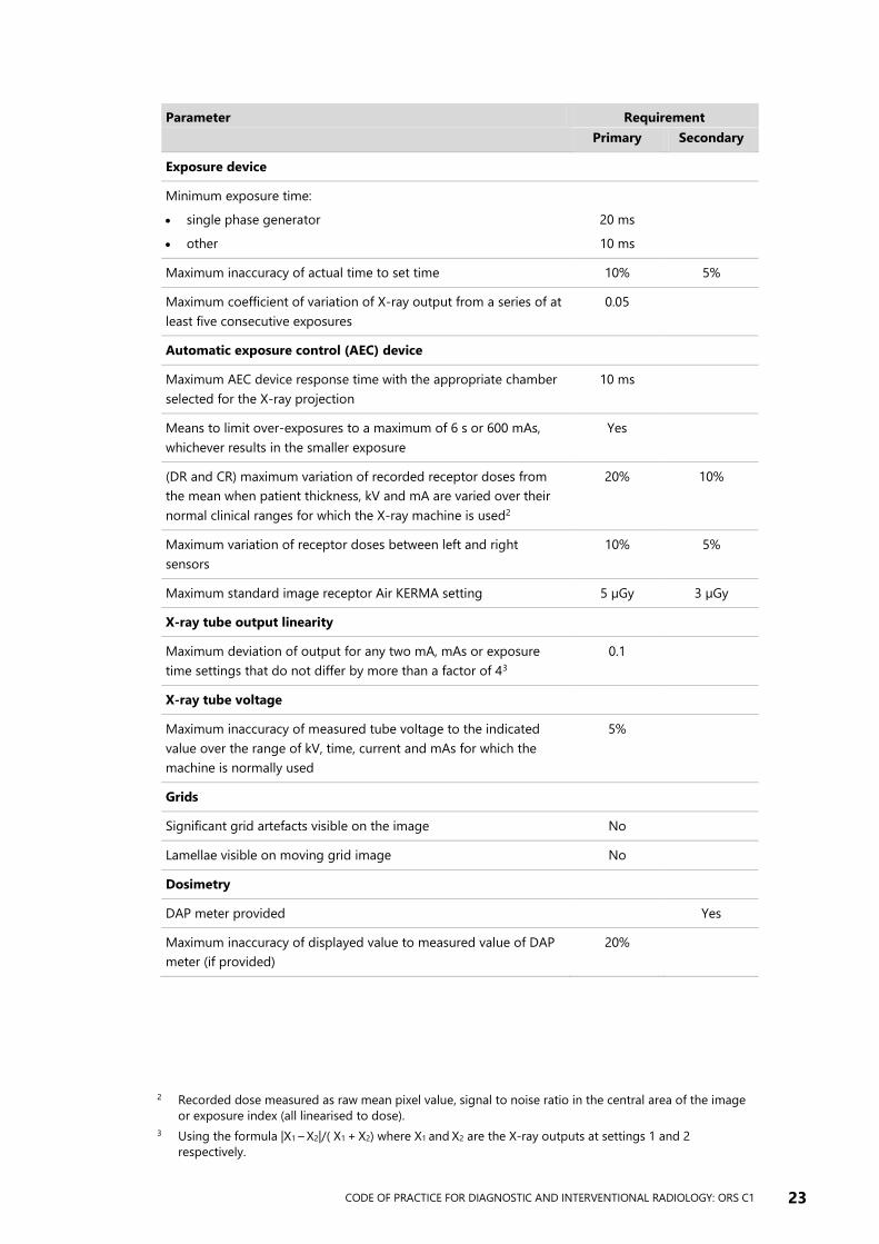
Parameter Requirement
Primary Secondary
Exposure device
Minimum exposure time:
single phase generator
other
20 ms
10 ms
Maximum inaccuracy of actual time to set time 10% 5%
Maximum coefficient of variation of X-ray output from a series of at
least five consecutive exposures
0.05
Automatic exposure control (AEC) device
Maximum AEC device response time with the appropriate chamber
selected for the X-ray projection
10 ms
Means to limit over-exposures to a maximum of 6 s or 600 mAs,
whichever results in the smaller exposure
Yes
(DR and CR) maximum variation of recorded receptor doses from
the mean when patient thickness, kV and mA are varied over their
normal clinical ranges for which the X-ray machine is used2
20% 10%
Maximum variation of receptor doses between left and right
sensors
10% 5%
Maximum standard image receptor Air KERMA setting 5 µGy 3 µGy
X-ray tube output linearity
Maximum deviation of output for any two mA, mAs or exposure
time settings that do not differ by more than a factor of 43
0.1
X-ray tube voltage
Maximum inaccuracy of measured tube voltage to the indicated
value over the range of kV, time, current and mAs for which the
machine is normally used
5%
Grids
Significant grid artefacts visible on the image No
Lamellae visible on moving grid image No
Dosimetry
DAP meter provided Yes
Maximum inaccuracy of displayed value to measured value of DAP
meter (if provided)
20%
2 Recorded dose measured as raw mean pixel value, signal to noise ratio in the central area of the image
or exposure index (all linearised to dose).
3 Using the formula |X1 – X2|/( X1 + X2) where X1 and X2 are the X-ray outputs at settings 1 and 2
respectively.
24 CODE OF PRACTICE FOR DIAGNOSTIC AND INTERVENTIONAL RADIOLOGY: ORS C1
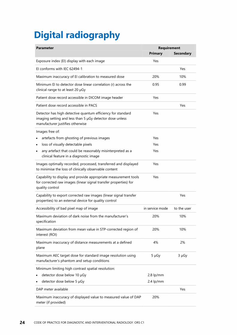
Digital radiography Parameter Requirement
Primary Secondary
Exposure index (EI) display with each image Yes
EI conforms with IEC 62494-1 Yes
Maximum inaccuracy of EI calibration to measured dose 20% 10%
Minimum EI to detector dose linear correlation (r) across the
clinical range to at least 20 µGy
0.95 0.99
Patient dose record accessible in DICOM image header Yes
Patient dose record accessible in PACS Yes
Detector has high detective quantum efficiency for standard
imaging setting and less than 5 µGy detector dose unless
manufacturer justifies otherwise
Yes
Images free of:
artefacts from ghosting of previous images
loss of visually detectable pixels
any artefact that could be reasonably misinterpreted as a
clinical feature in a diagnostic image
Yes
Yes
Yes
Images optimally recorded, processed, transferred and displayed
to minimise the loss of clinically observable content
Yes
Capability to display and provide appropriate measurement tools
for corrected raw images (linear signal transfer properties) for
quality control
Yes
Capability to export corrected raw images (linear signal transfer
properties) to an external device for quality control
Yes
Accessibility of bad pixel map of image in service mode to the user
Maximum deviation of dark noise from the manufacturer’s
specification
20% 10%
Maximum deviation from mean value in STP-corrected region of
interest (ROI)
20% 10%
Maximum inaccuracy of distance measurements at a defined
plane
4% 2%
Maximum AEC target dose for standard image resolution using
manufacturer’s phantom and setup conditions
5 µGy 3 µGy
Minimum limiting high contrast spatial resolution:
detector dose below 10 µGy
detector dose below 5 µGy
2.8 lp/mm
2.4 lp/mm
DAP meter available Yes
Maximum inaccuracy of displayed value to measured value of DAP
meter (if provided)
20%
CODE OF PRACTICE FOR DIAGNOSTIC AND INTERVENTIONAL RADIOLOGY: ORS C1 25
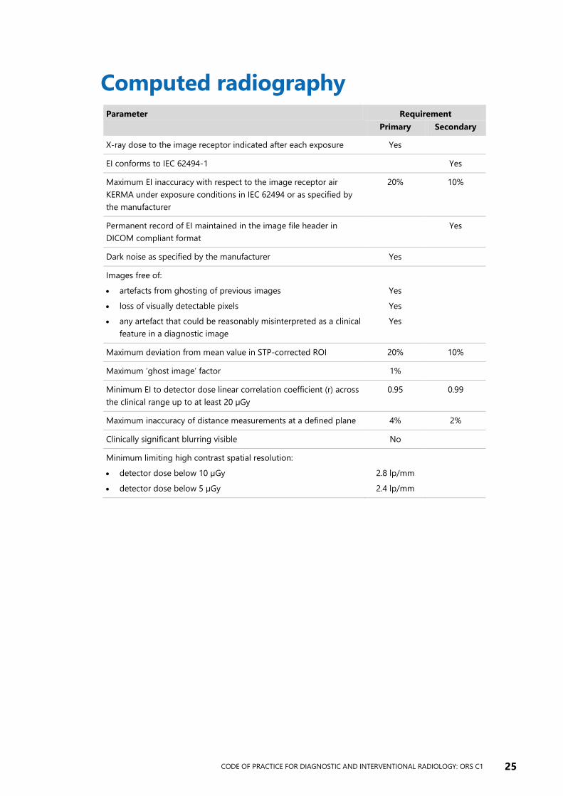
Computed radiography Parameter Requirement
Primary Secondary
X-ray dose to the image receptor indicated after each exposure Yes
EI conforms to IEC 62494-1 Yes
Maximum EI inaccuracy with respect to the image receptor air
KERMA under exposure conditions in IEC 62494 or as specified by
the manufacturer
20% 10%
Permanent record of EI maintained in the image file header in
DICOM compliant format
Yes
Dark noise as specified by the manufacturer Yes
Images free of:
artefacts from ghosting of previous images
loss of visually detectable pixels
any artefact that could be reasonably misinterpreted as a clinical
feature in a diagnostic image
Yes
Yes
Yes
Maximum deviation from mean value in STP-corrected ROI 20% 10%
Maximum ‘ghost image’ factor 1%
Minimum EI to detector dose linear correlation coefficient (r) across
the clinical range up to at least 20 µGy
0.95 0.99
Maximum inaccuracy of distance measurements at a defined plane 4% 2%
Clinically significant blurring visible No
Minimum limiting high contrast spatial resolution:
detector dose below 10 µGy
detector dose below 5 µGy
2.8 lp/mm
2.4 lp/mm
26 CODE OF PRACTICE FOR DIAGNOSTIC AND INTERVENTIONAL RADIOLOGY: ORS C1
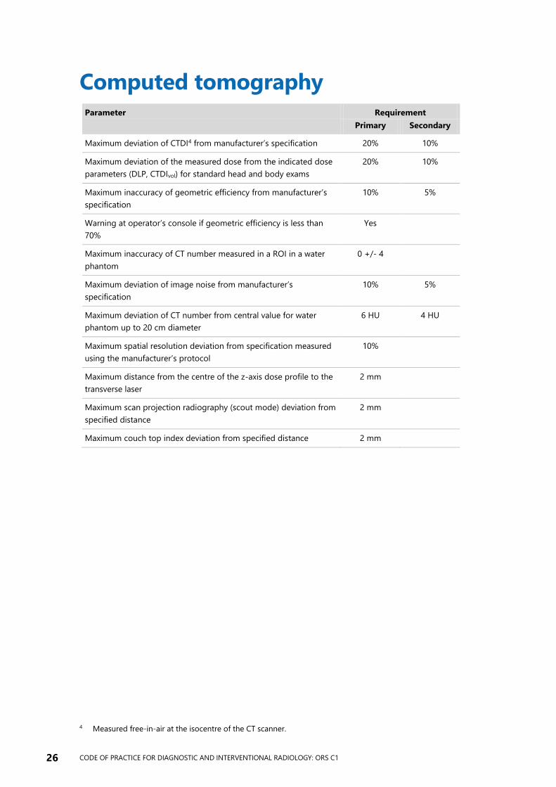
Computed tomography Parameter Requirement
Primary Secondary
Maximum deviation of CTDI4 from manufacturer’s specification 20% 10%
Maximum deviation of the measured dose from the indicated dose
parameters (DLP, CTDIvol) for standard head and body exams
20% 10%
Maximum inaccuracy of geometric efficiency from manufacturer’s
specification
10% 5%
Warning at operator’s console if geometric efficiency is less than
70%
Yes
Maximum inaccuracy of CT number measured in a ROI in a water
phantom
0 +/- 4
Maximum deviation of image noise from manufacturer’s
specification
10% 5%
Maximum deviation of CT number from central value for water
phantom up to 20 cm diameter
6 HU 4 HU
Maximum spatial resolution deviation from specification measured
using the manufacturer’s protocol
10%
Maximum distance from the centre of the z-axis dose profile to the
transverse laser
2 mm
Maximum scan projection radiography (scout mode) deviation from
specified distance
2 mm
Maximum couch top index deviation from specified distance 2 mm
4 Measured free-in-air at the isocentre of the CT scanner.
CODE OF PRACTICE FOR DIAGNOSTIC AND INTERVENTIONAL RADIOLOGY: ORS C1 27
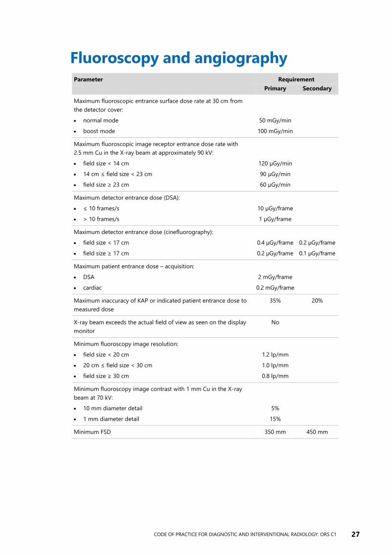
Fluoroscopy and angiography Parameter Requirement
Primary Secondary
Maximum fluoroscopic entrance surface dose rate at 30 cm from
the detector cover:
normal mode
boost mode
50 mGy/min
100 mGy/min
Maximum fluoroscopic image receptor entrance dose rate with
2.5 mm Cu in the X-ray beam at approximately 90 kV:
field size < 14 cm
14 cm ≤ field size < 23 cm
field size ≥ 23 cm
120 µGy/min
90 µGy/min
60 µGy/min
Maximum detector entrance dose (DSA):
≤ 10 frames/s
> 10 frames/s
10 µGy/frame
1 µGy/frame
Maximum detector entrance dose (cinefluorography):
field size < 17 cm
field size ≥ 17 cm
0.4 µGy/frame
0.2 µGy/frame
0.2 µGy/frame
0.1 µGy/frame
Maximum patient entrance dose – acquisition:
DSA
cardiac
2 mGy/frame
0.2 mGy/frame
Maximum inaccuracy of KAP or indicated patient entrance dose to
measured dose
35% 20%
X-ray beam exceeds the actual field of view as seen on the display
monitor
No
Minimum fluoroscopy image resolution:
field size < 20 cm
20 cm ≤ field size < 30 cm
field size ≥ 30 cm
1.2 lp/mm
1.0 lp/mm
0.8 lp/mm
Minimum fluoroscopy image contrast with 1 mm Cu in the X-ray
beam at 70 kV:
10 mm diameter detail
1 mm diameter detail
5%
15%
Minimum FSD 350 mm 450 mm
28 CODE OF PRACTICE FOR DIAGNOSTIC AND INTERVENTIONAL RADIOLOGY: ORS C1
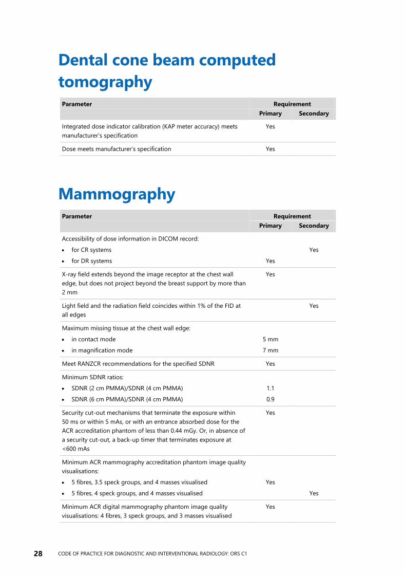
Dental cone beam computed
tomography Parameter Requirement
Primary Secondary
Integrated dose indicator calibration (KAP meter accuracy) meets
manufacturer’s specification
Yes
Dose meets manufacturer’s specification Yes
Mammography Parameter Requirement
Primary Secondary
Accessibility of dose information in DICOM record:
for CR systems
for DR systems
Yes
Yes
X-ray field extends beyond the image receptor at the chest wall
edge, but does not project beyond the breast support by more than
2 mm
Yes
Light field and the radiation field coincides within 1% of the FID at
all edges
Yes
Maximum missing tissue at the chest wall edge:
in contact mode
in magnification mode
5 mm
7 mm
Meet RANZCR recommendations for the specified SDNR Yes
Minimum SDNR ratios:
SDNR (2 cm PMMA)/SDNR (4 cm PMMA)
SDNR (6 cm PMMA)/SDNR (4 cm PMMA)
1.1
0.9
Security cut-out mechanisms that terminate the exposure within
50 ms or within 5 mAs, or with an entrance absorbed dose for the
ACR accreditation phantom of less than 0.44 mGy. Or, in absence of
a security cut-out, a back-up timer that terminates exposure at
<600 mAs
Yes
Minimum ACR mammography accreditation phantom image quality
visualisations:
5 fibres, 3.5 speck groups, and 4 masses visualised
5 fibres, 4 speck groups, and 4 masses visualised
Yes
Yes
Minimum ACR digital mammography phantom image quality
visualisations: 4 fibres, 3 speck groups, and 3 masses visualised
Yes
CODE OF PRACTICE FOR DIAGNOSTIC AND INTERVENTIONAL RADIOLOGY: ORS C1 29
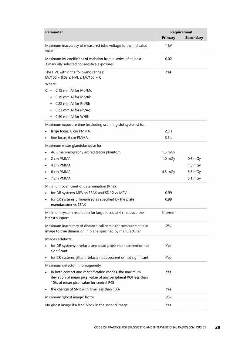
Parameter Requirement
Primary Secondary
Maximum inaccuracy of measured tube voltage to the indicated
value
1 kV
Maximum kV coefficient of variation from a series of at least
3 manually selected consecutive exposures
0.02
The HVL within the following ranges:
kV/100 + 0.03 ≤ HVL ≤ kV/100 + C
Where:
C = 0.12 mm Al for Mo/Mo
= 0.19 mm Al for Mo/Rh
= 0.22 mm Al for Rh/Rh
= 0.23 mm Al for Rh/Ag
= 0.30 mm Al for W/Rh
Yes
Maximum exposure time (excluding scanning slot systems) for:
large focus, 6 cm PMMA
fine focus, 6 cm PMMA
2.0 s
3.5 s
Maximum mean glandular dose for:
ACR mammography accreditation phantom
2 cm PMMA
4 cm PMMA
6 cm PMMA
7 cm PMMA
1.5 mGy
1.0 mGy
4.5 mGy
0.6 mGy
1.5 mGy
3.6 mGy
5.1 mGy
Minimum coefficient of determination (R^2):
for DR systems MPV vs ESAK and SD^2 vs MPV
for CR systems EI linearised as specified by the plate
manufacturer vs ESAK
0.99
0.99
Minimum system resolution for large focus at 4 cm above the
breast support
5 lp/mm
Maximum inaccuracy of distance callipers ruler measurements in
image to true dimension in plane specified by manufacturer
2%
Images artefacts:
for DR systems: artefacts and dead pixels not apparent or not
significant
for CR systems: jitter artefacts not apparent or not significant
Yes
Yes
Maximum detector inhomogeneity:
in both contact and magnification modes, the maximum
deviation of mean pixel value of any peripheral ROI less than
10% of mean pixel value for central ROI
the change of SNR with time less than 10%
Yes
Yes
Maximum ‘ghost image’ factor 2%
No ghost image if a lead block in the second image Yes
30 CODE OF PRACTICE FOR DIAGNOSTIC AND INTERVENTIONAL RADIOLOGY: ORS C1
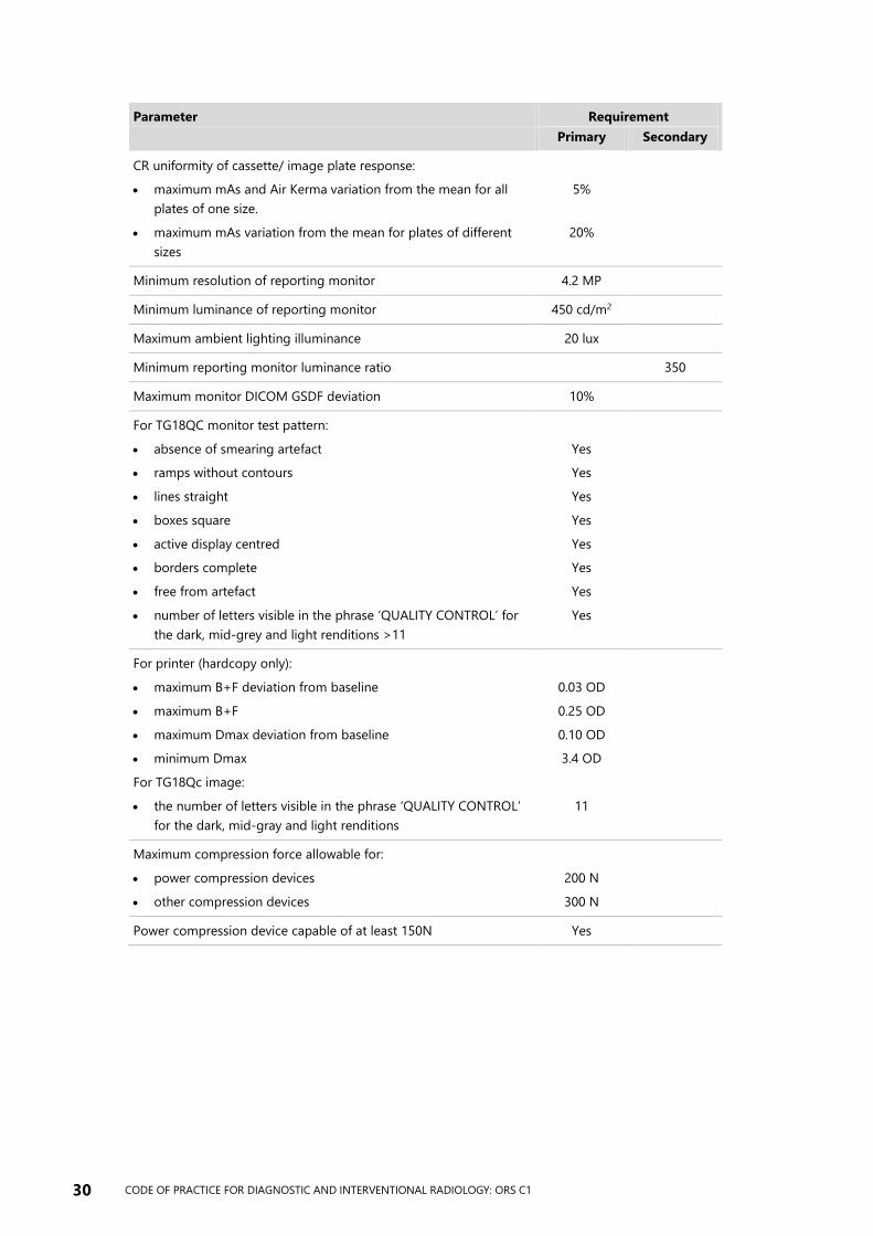
Parameter Requirement
Primary Secondary
CR uniformity of cassette/ image plate response:
maximum mAs and Air Kerma variation from the mean for all
plates of one size.
maximum mAs variation from the mean for plates of different
sizes
5%
20%
Minimum resolution of reporting monitor 4.2 MP
Minimum luminance of reporting monitor 450 cd/m2
Maximum ambient lighting illuminance 20 lux
Minimum reporting monitor luminance ratio 350
Maximum monitor DICOM GSDF deviation 10%
For TG18QC monitor test pattern:
absence of smearing artefact
ramps without contours
lines straight
boxes square
active display centred
borders complete
free from artefact
number of letters visible in the phrase ‘QUALITY CONTROL’ for
the dark, mid-grey and light renditions >11
Yes
Yes
Yes
Yes
Yes
Yes
Yes
Yes
For printer (hardcopy only):
maximum B+F deviation from baseline
maximum B+F
maximum Dmax deviation from baseline
minimum Dmax
For TG18Qc image:
the number of letters visible in the phrase ‘QUALITY CONTROL’
for the dark, mid-gray and light renditions
0.03 OD
0.25 OD
0.10 OD
3.4 OD
11
Maximum compression force allowable for:
power compression devices
other compression devices
200 N
300 N
Power compression device capable of at least 150N Yes
CODE OF PRACTICE FOR DIAGNOSTIC AND INTERVENTIONAL RADIOLOGY: ORS C1 31
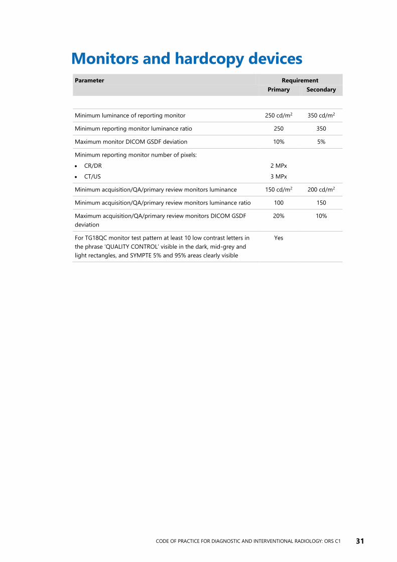
Monitors and hardcopy devices Parameter Requirement
Primary Secondary
Minimum luminance of reporting monitor 250 cd/m2 350 cd/m2
Minimum reporting monitor luminance ratio 250 350
Maximum monitor DICOM GSDF deviation 10% 5%
Minimum reporting monitor number of pixels:
CR/DR
CT/US
2 MPx
3 MPx
Minimum acquisition/QA/primary review monitors luminance 150 cd/m2 200 cd/m2
Minimum acquisition/QA/primary review monitors luminance ratio 100 150
Maximum acquisition/QA/primary review monitors DICOM GSDF
deviation
20% 10%
For TG18QC monitor test pattern at least 10 low contrast letters in
the phrase ‘QUALITY CONTROL’ visible in the dark, mid-grey and
light rectangles, and SYMPTE 5% and 95% areas clearly visible
Yes
32 CODE OF PRACTICE FOR DIAGNOSTIC AND INTERVENTIONAL RADIOLOGY: ORS C1
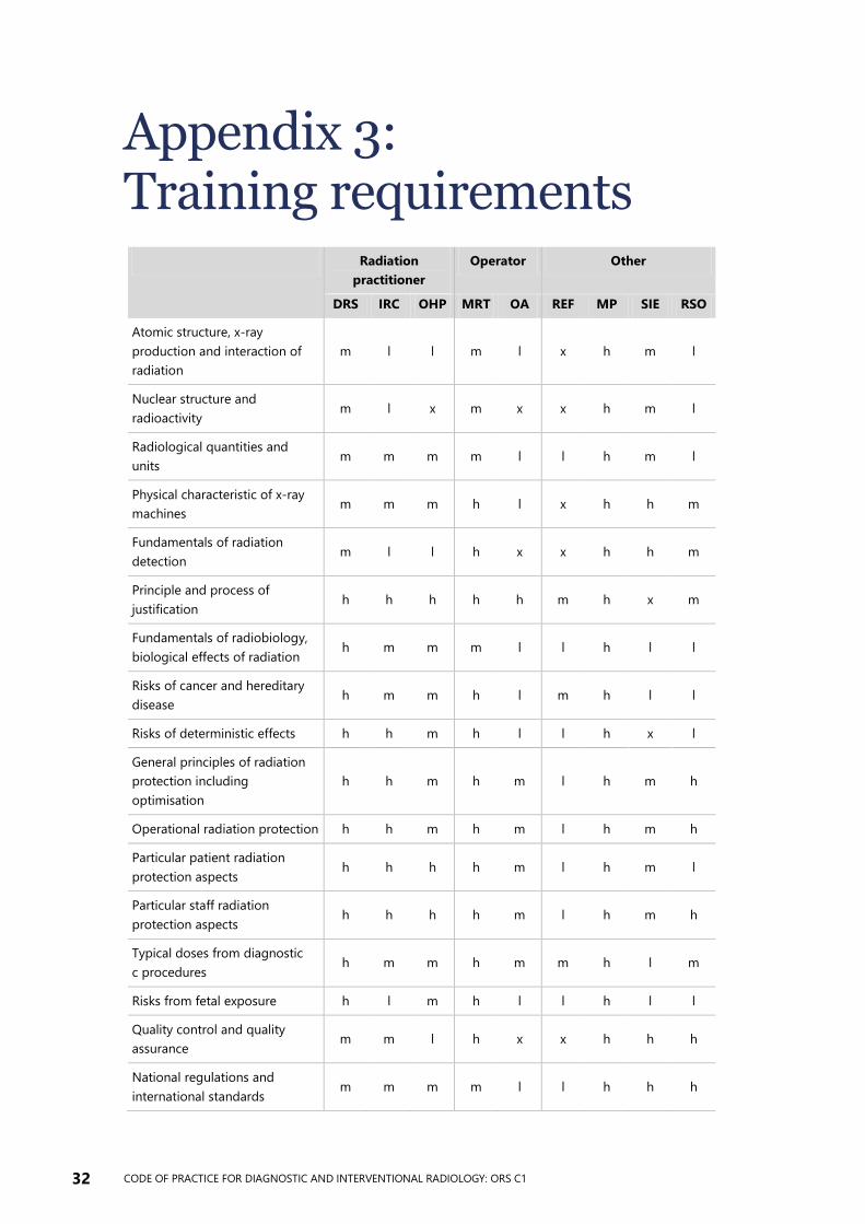
Appendix 3: Training requirements
Radiation
practitioner
Operator Other
DRS IRC OHP MRT OA REF MP SIE RSO
Atomic structure, x-ray
production and interaction of
radiation
m l l m l x h m l
Nuclear structure and
radioactivity m l x m x x h m l
Radiological quantities and
units m m m m l l h m l
Physical characteristic of x-ray
machines m m m h l x h h m
Fundamentals of radiation
detection m l l h x x h h m
Principle and process of
justification h h h h h m h x m
Fundamentals of radiobiology,
biological effects of radiation h m m m l l h l l
Risks of cancer and hereditary
disease h m m h l m h l l
Risks of deterministic effects h h m h l l h x l
General principles of radiation
protection including
optimisation
h h m h m l h m h
Operational radiation protection h h m h m l h m h
Particular patient radiation
protection aspects h h h h m l h m l
Particular staff radiation
protection aspects h h h h m l h m h
Typical doses from diagnostic
c procedures h m m h m m h l m
Risks from fetal exposure h l m h l l h l l
Quality control and quality
assurance m m l h x x h h h
National regulations and
international standards m m m m l l h h h
CODE OF PRACTICE FOR DIAGNOSTIC AND INTERVENTIONAL RADIOLOGY: ORS C1 33
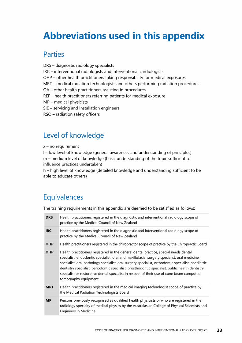
Abbreviations used in this appendix
Parties
DRS – diagnostic radiology specialists
IRC – interventional radiologists and interventional cardiologists
OHP – other health practitioners taking responsibility for medical exposures
MRT – medical radiation technologists and others performing radiation procedures
OA – other health practitioners assisting in procedures
REF – health practitioners referring patients for medical exposure
MP – medical physicists
SIE – servicing and installation engineers
RSO – radiation safety officers
Level of knowledge
x – no requirement
l – low level of knowledge (general awareness and understanding of principles)
m – medium level of knowledge (basic understanding of the topic sufficient to
influence practices undertaken)
h – high level of knowledge (detailed knowledge and understanding sufficient to be
able to educate others)
Equivalences
The training requirements in this appendix are deemed to be satisfied as follows:
DRS Health practitioners registered in the diagnostic and interventional radiology scope of
practice by the Medical Council of New Zealand
IRC Health practitioners registered in the diagnostic and interventional radiology scope of
practice by the Medical Council of New Zealand
OHP Health practitioners registered in the chiropractor scope of practice by the Chiropractic Board
OHP Health practitioners registered in the general dental practice, special needs dental
specialist, endodontic specialist, oral and maxillofacial surgery specialist, oral medicine
specialist, oral pathology specialist, oral surgery specialist, orthodontic specialist, paediatric
dentistry specialist, periodontic specialist, prosthodontic specialist, public health dentistry
specialist or restorative dental specialist in respect of their use of cone beam computed
tomography equipment
MRT Health practitioners registered in the medical imaging technologist scope of practice by
the Medical Radiation Technologists Board
MP Persons previously recognised as qualified health physicists or who are registered in the
radiology specialty of medical physics by the Australasian College of Physical Scientists and
Engineers in Medicine