Embed Size (px)
Citation preview
International Journal for Parasitology xxx (2015) xxx–xxx
Contents lists available at ScienceDirect
International Journal for Parasitology
journal homepage: www.elsevier .com/locate / i jpara
Continuous culture of Cryptosporidium parvum using hollow fibertechnology
http://dx.doi.org/10.1016/j.ijpara.2015.07.0060020-7519/� 2015 Australian Society for Parasitology Inc. Published by Elsevier Ltd. All rights reserved.
⇑ Corresponding author at: Haskins Laboratories, and Department of Chemistryand Physical Sciences, Pace University, 41 Park Row, New York, NY 10038, USA. Tel.:+1 212 346 1853; fax: +1 212 346 1586.
E-mail address: [email protected] (N. Yarlett).
Please cite this article in press as: Morada, M., et al. Continuous culture of Cryptosporidium parvum using hollow fiber technology. Int. J. Parasitol.http://dx.doi.org/10.1016/j.ijpara.2015.07.006
Mary Morada a, Sangun Lee b, Leslie Gunther-Cummins c, Louis M. Weiss d,e, Giovanni Widmer b,Saul Tzipori b, Nigel Yarlett a,⇑aHaskins Laboratories, and Department of Chemistry and Physical Sciences, Pace University, New York, USAbCummings School of Veterinary Medicine, Tufts University, N. Grafton, MA, USAcAnalytical Imaging Facility, Albert Einstein College of Medicine, Bronx, NY, USAdDepartment of Pathology, Albert Einstein College of Medicine, Bronx, NY, USAeDepartment of Medicine, Albert Einstein College of Medicine, Bronx, NY, USA
a r t i c l e i n f o
Article history:Received 21 January 2015Received in revised form 23 July 2015Accepted 24 July 2015Available online xxxx
Keywords:CryptosporidiumIn vitro cultureHollow-fiberAnaerobic
a b s t r a c t
Diarrheal disease is a leading cause of pediatric death in economically low resource countries.Cryptosporidium spp. are the second largest member of this group and the only member for which notreatment exists. One of the handicaps to developing chemotherapy is the lack of a reproducible long-term culture method permitting in vitro drug screening beyond 48 h. We have adapted the well-established hollow fiber technology to provide an environment that mimics the gut by delivering nutri-ents and oxygen from the basal layer upwards while allowing separate redox and nutrient control of thelumen for parasite development. Using this technique, oocyst production was maintained for >6 months,producing approximately 1 � 108 oocysts ml�1 day�1, compared with 48 h with a yield of 1 � 106 -oocysts ml�1 in two-dimensional cultures. Oocysts, after 4 and 20 weeks in culture, produced a chronicinfection in a TCR-a-deficient mouse model. In vivo infectivity of oocysts was confirmed using oocystsfrom a 6 week culture in a dexamethasone immunosuppressed mouse model.
� 2015 Australian Society for Parasitology Inc. Published by Elsevier Ltd. All rights reserved.
1. Introduction
Cryptosporidium spp. are the second (after rotavirus) leadingcause of diarrheal disease and death in infants in economicallylow resource countries (Liu et al., 2012), and remains the onlymember of the major diarrheal diseases for which no consistentlyeffective therapy is available. In parts of Asia and Africa as many as31.5% of all children under 2 years of age are infected with the par-asite, posing a significant health risk (Kotloff et al., 2013; Checkleyet al., 2014). There are five species responsible for infections inhumans, Cryptosporidium hominis, Cryptosporidium parvum, Cryp-tosporidium meleagridis, Cryptosporidium canis and Cryptosporidiumfelis, of which C. hominis or C. parvum are the most common (Xiao,2010). Infection of the epithelial cells of the gastrointestinal tractby this parasite results in an acute watery diarrhea, which canbecome life threatening in children, the elderly and immune-compromised individuals (Mathur et al., 2013). Malnourished chil-
dren are at a greater risk of death from the infection and those thatsurvive are at greater risk of stunted growth and wasting (Banwatet al., 2003; Hamedi et al., 2005; Checkley et al., 2014). The under-lying cause of disease transmission is poor hygiene and contami-nated water sources (Molloy et al., 2011; Mølbak et al., 2013). Insome countries it is endemic due to the poor sanitation and lackof appropriate health practices. Despite the significant morbidityand mortality associated with cryptosporidiosis, drug discoverylags behind that of many other infectious diseases (Striepen,2013). Nitazoxanide, the only FDA-approved drug for treatmentof cryptosporidiosis, is not effective in immune-compromised indi-viduals and is not routinely used in standard care in countries suchas India (Abubakar et al., 2007).
The parasite has a multistage life cycle producing thick walledoocysts that are shed from infected individuals and act as the infec-tive stage; other stages include motile sporozoites, merozoites,macro- and microgametes, as well as thin-walled oocysts thatare responsible for autoinfection. In common with other membersof the Apicomplexa, Cryptosporidium spp. require a host cell tocomplete the life cycle. The parasite is capable of parasitising anumber of in vitro cultured cell lines (Upton et al., 1994) producingthin walled oocysts that are not infective to the primary host.
(2015),
2 M. Morada et al. / International Journal for Parasitology xxx (2015) xxx–xxx
Significant advances have been reported, (reviewed by Arrowood(2008)). More recent is the use of an HCT-8 organoid model toreplicate the intestine (Alcantara Warren et al., 2008), and theuse of primary cultured intestinal epithelial cells (Castellanos-Gonzalez et al., 2013) which have extended the in vitro infectionto >5 days. Recently Varughese et al. (2014) described a modelusing small intestinal epithelial cells, FHs 74 Int, demonstratingimproved infection kinetics that has great potential for host-parasite studies. It was the goal of this study to extend thein vitro culture of C. parvum to >1 month, permitting long-termdrug studies to be performed. We considered the following issuesas critical to the development of a long-term culture system. Thepresence of two controlled environments (biphasic) would providehost cells with oxygen and nutrients from the basal layer, whileallowing a low oxygen nutrient rich environment to be developedon the apical surface where the parasite would be present, thusmimicking the situation in situ. The development of such a systemprovides the added advantage of allowing the development of aparasite-specific growth medium, which is not possible in currenttwo-dimensional (2D) cultures where emphasis is directed tomaintaining host cell growth to support the parasite burden. Theparasite completes its life cycle within the host intestine, a lowoxygen environment. In addition, a microaerophilic parasite meta-bolism is inferred from genome sequence information(Abrahamsen et al., 2004), and is supported by several studiesdemonstrating the loss of typical mitochondria (Hieinz andLithgow, 2013), and dependence upon a low oxygen metabolism,reviewed by Zhu (2008). These observations prompted us to mod-ify the standard medium developed by Upton et al. (1994) toinclude reducing agents and a mixture of fatty acids, which havebeen shown to be important in the growth of various anaerobicprotozoa (Yichoy et al., 2011).
2. Materials and methods
2.1. Culture of HCT-8 host cells
Human ileocecal colorectal adenocarcinoma cells (HCT-8 (HRT-18), ATCC CCL-244) were used to provide a host cell layer on theextra-capillary surface of the fibers. MEM plus supplements andserum (MEMSS) (Upton et al., 1994) circulated through the hollowfibers and glucose levels were recorded using an Accu-Chek Activemonitor (Hoffmann-La Roche Ltd., Indianapolis, IN, USA). The pHwas monitored by sampling 5 mL of the recirculating mediumusing a Corning 440 pH meter (Corning, NY, USA).
2.2. Real-time quantitatice reverse transcription-PCR (qRT-PCR)analysis of parasite 18S rRNA
Total RNA was isolated from 5 ml of C. parvum and host cellscollected from the cartridge. The sample was centrifuged at6449g for 5 min in a swing-out rotor (Beckman-Coulter, Indi-anapolis, IN, USA), then the pellet was resuspended in 200 ll ofPBS and washed three times; 50 ll was removed and the remain-der centrifuged, resuspended in lysis buffer (iScript RT-qPCR sam-ple preparation reagent, Bio-Rad Laboratories, Hercules, CA, USA)and subjected to six cycles of freezing-thawing using liquid nitro-gen and a 65 �C heating block. Total RNA was isolated using RNeasy(Qiagen Inc., Valencia, CA, USA) and the total RNA was determinedusing a Qubit 3.0 fluorometer (Life Technologies, Thermo-FisherScientific Inc., Waltham, MA, USA). RNA was adjusted to 5 ng/lland the parasite, C. parvum (Cp)18S RNA, and human, (h)18S rRNA,detected using the primers described by Cai et al. (2005), Cp18S-995F: 50-TAGAGATTGGAGGTTCCT-30 and Cp18S-1206R: 50-CTCCACCAACTAAGAACGCC-30; the primers for human 18S-RNA were
Please cite this article in press as: Morada, M., et al. Continuous culture of Cryphttp://dx.doi.org/10.1016/j.ijpara.2015.07.006
Hs18S-F1373: 50-CCGATAACGAACGAGACTCTGG-30, and Hs18S-R1561: 50-TAGGGTAGGCACACGCTGAGCC-30. The expected sizesof the parasite and human amplicons obtained were 212 bp and189 bp, respectively. Cp18S rRNA and h18S rRNA was quantitatedby qRT-PCR using an iScript One-Step qRT-PCR kit with SYBR green(Bio-Rad Laboratories). Total RNA (5 ng), reagents and primerswere incubated at 48 �C for 30 min, followed by 95 �C for 10 min,and then subjected to 40 thermal cycles of 95 �C for 15 s and60 �C for 1 min. A melting curve was performed by heating to95 �C for 15 s, followed by 60 �C for 15 s and 95 �C for 15 s, usingan QuantStudio 6 Flex Real-Time PCR System (Life Technologies,Thermo-Fisher Scientific Inc., Waltham, MA, USA). The qRT-PCRanalysis was also performed for the Iowa isolate (Bunch GrassFarms, Deary, ID, USA), diluted to give 105, 106 and 107 oocysts.The DCT for parasite 18S rRNA was determined by subtraction ofthe CT for h18S rRNA from the CT for parasite Cp18S rRNA.
2.3. Immunocompromised and immunosuppressed mouse models
Oocysts collected from the culture system were tested for theability to infect two well established mouse models. Oocysts werecollected at specified times from the culture system, stored in 2.5%potassium dichromate at 4 �C for less than 1 month, washed withdistilled water three times to remove the potassium dichromate,and enumerated using a hemacytometer prior to use.
The first model employed nine 6 week old female TCR-a-deficient mice on a C57BL/6 background (Jackson Laboratories,Bar Harbor, ME, USA); mice were segregated into groups of three;three were infected per os with 104 oocysts in 200 lL of PBS col-lected after 4 and 20 weeks in culture; three were infected with104 oocysts of the Iowa isolate used to establish the culture; threewere administered 200 lL of PBS as negative controls. All micewere housed in metabolic chambers (VWR, Atlanta, GA, USA) andevery 24 h feces were removed, weighed and placed into potas-sium dichromate for storage. After 7 days, mice were euthanisedby CO2 asphyxiation as recommended by the American VeterinaryMedical Association (AVMA) (Pace University, NY, USA) andintestinal sections from the distal ileum-cecum and proximal colonremoved and fixed in 10% neutral buffered formalin (3.7–4.0%formaldehyde in 33.3 mM monosodium phosphate and 45.8 mMdisodium phosphate buffer).
The second model employed 15 female CD-1� IGS mice (CharlesRiver Laboratory, Wilmington, MA, USA) aged 3–5 weeks, weighing17–22 g each, which were immunosuppressed with dexametha-sone 21-phosphate (D1159, Sigma, USA) administered ad libidumin drinking water (16 lg/ml) from 4 days prior to inoculation tothe end of the study. Mice were separated into the following threegroups of five per group, and infected per os with 200 ll of sterilewater containing either 106 oocysts from 6 weeks in culture (group1), the parent Iowa isolate used to initiate the culture (group 2) orwere an uninfected control group (group 3). Mice were monitoredtwice daily following the infection and weighed three times perweek; fecal samples were collected daily for 17 days and fecalsmears stained using the modified Kinyoun acid fast method (Maand Soave, 1983). After 17 days, mice were euthanised by CO2
asphyxiation as recommended in the AVMA (Tufts University,MA, USA) and gastrointestinal sections removed for histopatholog-ical examination. The ileum, cecum and proximal colon were fixedin 10% natural buffered formalin and processed for H&E stains.
Animal studies were performed in strict accordance with therecommendations in the Guide for the Care and Use of LaboratoryAnimals of the National Institutes of Health, USA under the AnimalWelfare Assurance numbers A3112-01 (Pace University, HaskinsLaboratories, NYC, USA) and A4059-01 (Tufts University, Cum-mings School of Veterinary Medicine, USA). Animal experimentswere performed in accordance with the procedures approved by
tosporidium parvum using hollow fiber technology. Int. J. Parasitol. (2015),
M. Morada et al. / International Journal for Parasitology xxx (2015) xxx–xxx 3
the Institutional Animal Care and Use Committees of Pace Univer-sity, NYC and Tufts University, Cummings School Veterinary Med-icine, MA, USA.
2.4. Staining with fluorescently labeled antibodies
Samples (5–10 mL) were removed from the extra capillaryspace of the cartridge by displacement using two 10 mL sterile syr-inges. Samples were centrifuged at 16,162g for 1 min (Sorvall Bio-fuge Fresco, Thermo Fisher Scientific) and the pellet resuspendedin Dulbecco’s PBS. Oocysts were stained using 50 lL of FITC-labeled mouse monoclonal antibody to C. parvum oocyst surfaceproteins (Crypt-a-Glo�, Waterborne Inc., New Orleans, LA, USA)and incubated in the dark at room temperature for 30 min, afterwhich time they were washed twice with PBS and examined usinga fluorescence microscope (Nikon Optiphot, Melville, NY, USA)with an excitation k 410–485 nm, and an emission k 515 nm. Sam-ples were counter-stained with a fluorescent-labeled polyclonalantibody specific for sporozoites and other motile intracellularstages (Sporo-Glo�, Waterborne Inc.), and examined using an exci-tation k 535–550 nm, and an emission k 580 nm.
2.5. Preparation of oocysts and excystation
Cryptosporidium parvum oocysts were purified from the otherstages (empty oocyst shells, host cells, etc) using isopycnic percollgradient centrifugation (Arrowood and Sterling, 1987) consistingof 3 mL of 1.04 g mL�1 percoll overlaid onto 5 mL of 1.08 g mL�1
percoll. One millilitre samples from the cartridge were layeredonto these gradients and centrifuged at 400g for 30 min. Theoocysts formed a light colored band approximately 1/3 of the dis-tance from the bottom of the tube and were then aspirated andwashed with PBS by centrifugation at 1000g for 10 min. Sporo-zoites were obtained by excysting oocysts in MEM containing0.25% trypsin, 0.75% taurodeoxycholate at 37 �C in a shaking waterbath.
2.6. Polyamine analysis
Polyamines were separated and identified by reverse phaseHPLC using a Perkin Elmer LC410 system coupled to a C18 10 lmcolumn (4.5 � 250 mm) at a flow rate of 1 mL min�1. The methodemployed a 40 min discontinuous gradient starting with 90% (v/v) buffer A (0.1 M NaH2PO4, 8 mM octanesulfonic acid, and0.05 mM EDTA) and 10% (v/v) buffer B (acetonitrile). The initialconditions were held for 5 min before changing to 20% buffer Bfor 15 min, 40% buffer B for a further 10 min, and then returningto 10% buffer B for 10 min (Morada et al., 2013). Samples and stan-dards were mixed 1:1 with 1.5 mM o-phthalaldehyde (dissolved in0.5 M boric acid, 0.43 M KOH and 0.014 M b-mercaptoethanol)using a post column pump and detected using a Perkin Elmer flu-orescence detector (excitation wavelength 320 nm, emissionwavelength 455 nm). Areas under the peaks were determinedusing b-RAM computer software (IN/US Systems). Results wereobtained using three separate oocyst preparations obtained fromthe cartridge.
2.7. Enzyme assays
Spermidine/spermine N1-acetyltransferase (SSAT) was deter-mined using 100 lM bicine buffer (pH 8.0) containing 17 lM[1-14C]acetyl-CoA (60 lCi/mmol) and supplemented with 50 lMunlabeled acetyl-CoA, 500 lM spermine and 25 lg of protein.The reaction was stopped after 30 min with ice-cold 50 mMhydroxylamine, placed in a boiling water bath for 3 min, cooledand centrifuged at 9000g for 1 min to remove precipitated protein.
Please cite this article in press as: Morada, M., et al. Continuous culture of Cryphttp://dx.doi.org/10.1016/j.ijpara.2015.07.006
The supernatant (50 lL) was spotted onto filter discs, dried andwashed with 6 � 200 mL changes of distilled water to removeunreacted [1-14C]-acetyl-CoA, with a final wash with 200 mL ofmethanol. The dried discs were placed in 10 mL of Omni Fluorand the radioactivity present as [14C]-acetylspermine was countedusing a Beckman Tri-Carb 1600CA liquid scintillation counter (Per-kin Elmer Life Sciences, Waltham, MA, USA). Blanks containing[1-14C] acetyl-CoA and spermine without protein were also ana-lyzed and subtracted from the experimental results (Moradaet al., 2013).
Spermidine acetyl transferase (SAT) was assayed using the samemethod for SSAT but using 500 lM spermidine in place of sper-mine (Keithly et al., 1997).
S-adenosyl-L-methionine decarboxylase (putrescine stimu-lated) was determined by measuring the 14CO2 produced from0.5 lCi [14C-carboxyl]-S-adenosylmethionine (55 mCi/mmol) incu-bations containing 10 mM Tris–HCl pH 6.5 and 1 mM putrescine(Keithly et al., 1997). The 14CO2 released in 60 min was trappedon benzethonium hydroxide soaked filter paper and counted byscintillation using a Beckman Tri-Carb 1600CA liquid scintillationcounter (Perkin Elmer Life Sciences).
2.8. Electron microscopy
For Scanning Electron Microscopy (SEM), samples were fixed in2.5% glutaraldehyde, 0.1 M sodium cacodylate pH 7.4, dehydratedthrough a graded series of ethanol, critical point dried using liquidcarbon dioxide in a Tousimis Samdri 790 Critical Point Drier (Rock-ville, MD, USA) and sputter coated with gold–palladium in a Den-ton Vacuum Desk-2 Sputter Coater (Cherry Hill, NJ, USA). Thesamples were examined in a JEOL JSM6400 Scanning ElectronMicroscope (Peabody, MA, USA), using an accelerating voltage of10 kV. Images were recorded with AnalySIS, Soft Imaging Systems(Munster, Germany).
2.9. Statistics
The statistical comparison of the differences among experimen-tal groups was made by Mann–Whitney U test using GraphPadPrism 5 software (GraphPad Software, San Diego, CA, USA). Resultswere considered to be statistically significant at P < 0.05.
3. Results
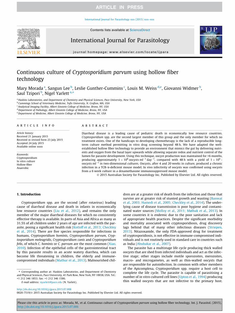
We describe a method for the long-term in vitro culture of C.parvum in a simulated gut-like environment using hollow fibertechnology. Human ileocecal adenocarcinoma (HCT-8; ATCC CCL244) cells were selected as the cell line to support growth of C. par-vum because previous studies found this cell line supported agreater infection than alternative ones (Upton et al., 1994;Meloni and Thompson, 1996), a feature that does not diminishwith age of the cells (Sifuentes and DiGiovanni, 2007). HCT-8 cellshave also been shown to differentiate into organoids that expressnormal intestinal tissue markers, and morphologically producemicrovilli and desmosomes characteristic of normal intestinal tis-sue (Carvalho et al., 2005). We considered these features ideal fora long-term in vitro culture system. A 20 ml hollow fiber cartridge(CS2011; FiberCell� Systems, Inc., Frederick, MD, USA), conditionedaccording to the manufacturer’s instructions, was inoculated with108 HCT-8 cells via the side port, and a 125 mL reservoir containingMEMSS (Upton et al., 1994) circulated through the hollow fibers ata flow rate of 51 mL min�1 (setting five; Fig. 1). The fiber surfacearea (2100 cm2) provides efficient nutrient, metabolite and gasexchange between the HCT-8 cell layer and the re-circulating med-ium. The cartridge, pump and growth medium were kept inside a
tosporidium parvum using hollow fiber technology. Int. J. Parasitol. (2015),
Fig. 1. Diagram of the hollow fiber system. The cartridge is composed of 200 lm diameter polysulfone hollow fibers with a 20 kDa molecular weight cut-off. The growthmedium (MEM plus supplements and serum, MEMSS) was pumped through the fibers to supply nutrients and oxygen to, and remove waste products from, the host cells(HCT-8) which colonize the outside of the hollow fibers. The extra capillary space was inoculated with Cryptosporidium parvum and the MEMSS medium in this environmentwas modified to include additives that promote parasite growth - lipids, redox buffers and vitamins (as described in Section 3). Additions can be made and parasites removedfrom the extra capillary space using the side port shown on the top of the cartridge.
Fig. 2. Daily glucose and cell counts from the hollow fiber culture system. (A) The cartridge was inoculated with 108 HCT-8 cells and allowed to achieve confluence for11 days. On day 12 the cartridge was inoculated with 106 Cryptosporidium parvum oocysts and the glucose levels monitored daily. Twenty-four hours p.i. there was an initialincrease in medium glucose levels associated with infection of host cells by C. parvum. The increase in glucose levels may be due to disruption of the host cell monolayer,resulting in lysis and release of stored glycogen. (B) The pattern of medium glucose levels (bars) cycled for approximately 20 days and exhibited a dampened oscillation,indicative of loss of synchrony of the parasitemia of host cells by C. parvum. The line at 350 mg dL�1 is the glucose concentration of the MEM plus supplements and serum(MEMSS) growth media circulated through the microfibers. (C) Seven days p.i., half of the extra capillary volume (7.5 mL) was removed for enumeration of oocysts andsporozoites. This was repeated at 3–4 day intervals; asterisks indicate removal of 50% of the extra capillary volume and replacement with fresh medium. Oocysts were stainedwith a fluorescent-labeled mouse anti-C. parvum oocyst wall monoclonal antibody and counted using a hemacytometer by fluorescence microscopy with an excitation k410 nm–485 nm and an emission k of 515 nm. (D) Sporozoites were counted using a hemacytometer after staining with a polyclonal antibody to intracellular and motilestages (Sporo-Glo�, Waterborne Inc., New Orleans, LA, USA) using an excitation k 535 nm–550 nm and an emission wavelength of 580 nm.
4 M. Morada et al. / International Journal for Parasitology xxx (2015) xxx–xxx
Please cite this article in press as: Morada, M., et al. Continuous culture of Cryptosporidium parvum using hollow fiber technology. Int. J. Parasitol. (2015),http://dx.doi.org/10.1016/j.ijpara.2015.07.006
M. Morada et al. / International Journal for Parasitology xxx (2015) xxx–xxx 5
5% CO2 incubator maintained at 37 �C. At 24 h intervals the glucose(Fig. 2A) and pH levels (data not shown) were recorded. When theglucose fell below 50% of the fresh MEMSS, the reservoir volumewas doubled to 250 ml, and this process was continued until areservoir volume of 1 L was achieved (8–11 days); when theHCT-8 cells were at full confluent density the pump rate wasincreased to 105 ml min�1 (setting 10).
3.1. Redox and nutrient requirements
The provision of oxygen and nutrients for the host cells from thebasal layer upwards provided us with the opportunity to develop aC. parvum culture medium that more closely approximates theintestinal lumen on the apical epithelial surface. Cryptosporidiumparvum parasitises the lumen of the gut that has sub-micromolaroxygen tensions and the biochemistry of the parasite supportsthe presence of an anaerobic metabolism; the C. parvum genomereveals a lack of a Krebs cycle and oxidative phosphorylation anda striking increase in the number of amino acid and fatty acidtransporters (Abrahamsen et al., 2004; Zhu, 2008). Addressingthese nutritional and environmental requirements is necessary todeveloping a long-term culture system. To this end we developeda redox buffer with the aim of creating a low redox environmentthat would mimic that present in the gut (Circu and Aw, 2011).The redox buffer contained 20 mg mL�1 each of glutathione, tau-rine, betaine and cysteine prepared in nitrogen gassed distilledwater. This mix resulted in a higher oocyst production than theaddition of a mix containing 20 mg mL�1 each of DTT/cysteine or20 mg mL�1 each of mercaptoethanol/cysteine. Many parasiteshave specific lipid requirements that may not be provided by theaddition of serum alone, and there is some evidence in the litera-ture that C. parvum also has a selective lipid requirement(Mazumdar and Striepen, 2007; Bushkin et al., 2013) and to thisend we focused on developing a lipid mix composed primarily ofomega-3 lipids. The composition and final amount was determinedby titration of individual components of the mix that produced themaximum number of oocysts. The lipid mix was composed of 1.5%deoxycholate, 6.7 mg mL�1 oleic acid, 10 mg mL�1 phosphocholine,1.6 mg mL�1 a-linolenic acid, 6.8 mg mL�1 eicosapentaenoic acid,2 mg mL�1 docosahexaenoic acid, 18 mg mL�1 cholesterol (dis-solved in ethanol and mixed 1:1 with Tween 80). The extra capil-lary space of the cartridge (Fig. 1) was flushed with 50 mL ofMEMSS (Upton et al., 1994) containing 1 lg mL�1 resazurin (7-hydroxy-3H-phenoxazin-3-one 10 oxide) as a redox indicator, inplace of phenol red, and containing the redox buffer and lipidmix as additives.
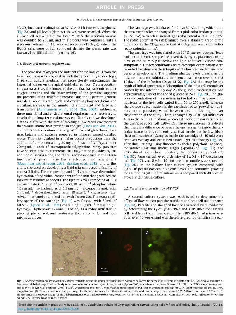
Fig. 3. Specificity of fluorescent antibody stages from the Cryptosporidium parvum culturefluorescein-labeled polyclonal antibody to intracellular and motile stages of the parasiteantibody to oocyst wall proteins (Crypt-a-Glo�, Waterborne Inc.) for 30 min, washed thmagnification. (B) Fluorescence microscopic image for fluorescein-labeled antibody toFluorescence microscopic image for FITC-labeled monoclonal antibody to oocysts, excitatido not label intracellular or motile stages.
Please cite this article in press as: Morada, M., et al. Continuous culture of Cryphttp://dx.doi.org/10.1016/j.ijpara.2015.07.006
The cartridge was incubated for 2 h at 37 �C, during which timethe resazurin indicator changed from a pink color (redox potentialP�51 mV) to colorless, indicating a redox potential of6�110 mV.The redox potential was determined from a standard graph of thedifference in the OD570 nm to that at OD600 nm versus the bufferredox potential in mV.
The cartridge was inoculated with 106 C. parvum oocysts (Iowaisolate), and 3 mL samples removed daily by displacement using3 mL of the MEMSS plus redox and lipid additives. Glucose con-sumption, pH, redox conditions and microscopic examination wererecorded to determine the integrity of the host cell feeder layer andparasite development. The medium glucose levels present in thehost cell medium exhibited a dampened oscillation over the first10 days of the infection (Days 12–22, Fig. 2A) that may be theresult of initial synchrony of disruption of the host cell monolayercaused by the infection. By day 23 the glucose consumption wasapproximately 50% of the added glucose in 24 h (Fig. 2B). The glu-cose concentration of the medium in the hollow fibers, providingnutrients to the host cells varied from 50 to 250 mg/dL, whereasthe glucose concentration in the cartridge space (providing nutri-ents to the parasites) varied between 270 and 334 mg/dL overthe duration of the study. The pH changed by �0.81 pH units over48 h in the host cell medium, whereas it showed minor variation inthe cartridge space (pH 6.99–7.09). These measurements indicatethat there is a difference between the environments inside the car-tridge (parasite environment) and that inside the hollow fibers(host cell nutrients). Samples inside the cartridge (5–10 mL) wereremoved weekly and examined under light microscopy (Fig. 3A)after duel staining using fluorescein-labeled polyclonal antibodyfor intracellular and motile stages (Sporo-Glo�; Fig. 3B), andFITC-labeled monoclonal antibody for oocysts (Crypt-a-Glo�;Fig. 3C). Parasites achieved a density of 1 ± 0.1 � 108 oocysts permL (Fig. 2C), and 8 ± 2 � 107 intracellular motile stages per mL(Fig. 2D), in the hollow fiber culture system compared with0.8 � 106 per mL oocysts in 25 cm2 flasks, and continued growingfor >6 months (at time of submission) compared with 48 h whengrown in 2D tissue culture.
3.2. Parasite enumeration by qRT-PCR
A second culture system was established to determine theeffects of flow rate on parasite numbers and host cell maintenance(Fig. 4A). Parasite and sloughed host cell numbers were evaluatedby determining the CT of Cp18S rRNA and h18S rRNA for samplescollected from the culture system. The h18S rRNA had minor vari-ation over 15 weeks, and was therefore used to normalise the par-
. Samples collected from the culture were incubated at 26 �C with equal volumes of(Sporo-Glo�, Waterborne Inc., New Orleans, LA, USA) and FITC-labeled monoclonalree-times in PBS and examined microscopically. (A) Light microscopic image, �400intracellular and motile stages, excitation k 535–550 nm, emission k 580 nm. (C)on k 418–485 nm, emission k 575 nm. Magnification 400-fold, antibodies for oocysts
tosporidium parvum using hollow fiber technology. Int. J. Parasitol. (2015),
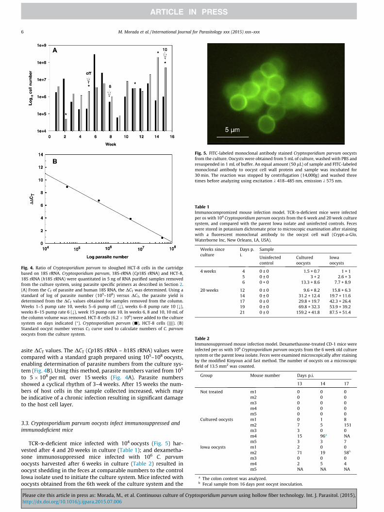
Fig. 4. Ratio of Cryptosporidium parvum to sloughed HCT-8 cells in the cartridgebased on 18S rRNA. Cryptosporidium parvum, 18S-rRNA (Cp18S rRNA) and HCT-8,18S rRNA (h18S rRNA) were quantitated in 5 ng of RNA purified samples removedfrom the culture system, using parasite specific primers as described in Section 2.(A) From the CT of parasite and human 18S RNA, the DCT was determined. Using astandard of log of parasite number (105–108) versus DCT, the parasite yield isdetermined from the DCT values obtained for samples removed from the column.Weeks 1–5 pump rate 10, weeks 5–6 pump off (;), weeks 6–8 pump rate 10 (;),weeks 8–15 pump rate 6 (;), week 15 pump rate 10. In weeks 6, 8 and 10, 10 mL ofthe column volume was removed. HCT-8 cells (6.2 � 106) were added to the culturesystem on days indicated (⁄). Cryptosporidium parvum (j), HCT-8 cells ( ). (B)Standard oocyst number versus CT curve used to calculate numbers of C. parvumoocysts from the culture system.
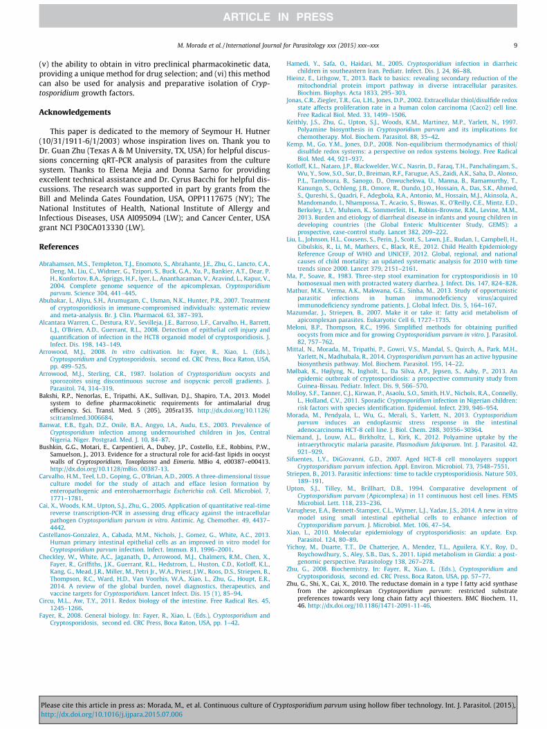
Fig. 5. FITC-labeled monoclonal antibody stained Cryptosporidium parvum oocystsfrom the culture. Oocysts were obtained from 5 mL of culture, washed with PBS andresuspended in 1 mL of buffer. An equal amount (50 lL) of sample and FITC-labeledmonoclonal antibody to oocyst cell wall protein and sample was incubated for30 min. The reaction was stopped by centrifugation (14,000g) and washed threetimes before analyzing using excitation k 418–485 nm, emission k 575 nm.
Table 2Immunosuppressed mouse infection model. Dexamethasone-treated CD-1 mice wereinfected per os with 106 Cryptosporidium parvum oocysts from the 6 week old culturesystem or the parent Iowa isolate. Feces were examined microscopically after stainingby the modified Kinyoun acid fast method. The number of oocysts on a microscopicfield of 13.5 mm2 was counted.
Group Mouse number Days p.i.
13 14 17
Not treated m1 0 0 0m2 0 0 0m3 0 0 0
Table 1Immunocompromized mouse infection model. TCR-a-deficient mice were infectedper os with 104 Cryptosporidium parvum oocysts from the 6 week and 20 week culturesystem, and compared with the parent Iowa isolate and uninfected controls. Feceswere stored in potassium dichromate prior to microscopic examination after stainingwith a fluorescent monoclonal antibody to the oocyst cell wall (Crypt-a-Glo,Waterborne Inc, New Orleans, LA, USA).
Weeks sinceculture
Days p.i.
Sample
Uninfectedcontrol
Culturedoocysts
Iowaoocysts
4 weeks 4 0 ± 0 1.5 + 0.7 1 + 15 0 ± 0 3 + 2 2.6 + 36 0 + 0 13.3 + 8.6 7.7 + 8.9
20 weeks 12 0 ± 0 9.6 + 8.2 15.8 + 6.314 0 ± 0 31.2 + 12.4 19.7 + 11.617 0 ± 0 29.8 + 19.7 42.3 + 26.419 0 ± 0 69.8 + 32.3 53.9 + 39.221 0 ± 0 159.2 + 41.8 87.5 + 51.4
6 M. Morada et al. / International Journal for Parasitology xxx (2015) xxx–xxx
asite DCT values. The DCT (Cp18S rRNA – h18S rRNA) values werecompared with a standard graph prepared using 105–108 oocysts,enabling determination of parasite numbers from the culture sys-tem (Fig. 4B). Using this method, parasite numbers varied from 105
to 5 � 108 per mL over 15 weeks (Fig. 4A). Parasite numbersshowed a cyclical rhythm of 3–4 weeks. After 15 weeks the num-bers of host cells in the sample collected increased, which maybe indicative of a chronic infection resulting in significant damageto the host cell layer.
m4 0 0 0m5 0 0 0
Cultured oocysts m1 0 1 8m2 7 5 151m3 3 0 0m4 15 96a NAm5 3 3 7
Iowa oocysts m1 2 0 0m2 71 19 58b
m3 0 0 0m4 2 5 4m5 NA NA NA
a The colon content was analyzed.b Fecal sample from 16 days post oocyst inoculation.
3.3. Cryptosporidium parvum oocysts infect immunosuppressed andimmunodeficient mice
TCR-a-deficient mice infected with 104 oocysts (Fig. 5) har-vested after 4 and 20 weeks in culture (Table 1); and dexametha-sone immunosuppressed mice infected with 106 C. parvumoocysts harvested after 6 weeks in culture (Table 2) resulted inoocyst shedding in the feces at comparable numbers to the controlIowa isolate used to initiate the culture system. Mice infected withoocysts obtained from the 6th week of the culture system and the
Please cite this article in press as: Morada, M., et al. Continuous culture of Cryptosporidium parvum using hollow fiber technology. Int. J. Parasitol. (2015),http://dx.doi.org/10.1016/j.ijpara.2015.07.006
M. Morada et al. / International Journal for Parasitology xxx (2015) xxx–xxx 7
parent Iowa isolate demonstrated an average 17% weight loss com-pared with uninfected immunosuppressed control mice after day10 (Fig. 6A). Post 10 days, oocyst shedding was detected in fecalsamples from all of the mice infected with the culture system(Table 2). Oocyst shedding intensities were similar between themice from both the culture-derived oocyst infection and theparent-derived oocyst infection. On day 14 the colon content wasanalyzed for mouse four from the culture-derived oocyst group(Table 2). Histological sections of the intestine (ileum) from mice17 days p.i. demonstrated the presence of C. parvum life cyclestages in the terminal ilea and proximal colon (Fig. 6B).
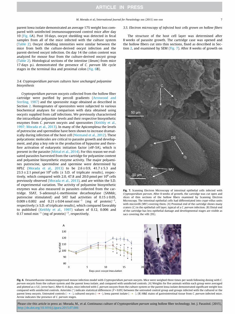
Fig. 7. Scanning Electron Microscopy of intestinal epithelial cells infected withCryptosporidium parvum. After 8 weeks of growth, the cartridge was cut open andslices of thin sections of the hollow fibers examined by Scanning ElectronMicroscopy. The intestinal epithelial cells had differentiated into crypt-villus unitswith microvilli (MV) covering them. (A) Proximal end of the cartridge shows manycraters (C) in the epithelial cell layer, typical of a C. parvum infection. (B) Distal endof the cartridge has less epithelial damage and developmental stages are visible assacs covering the villi (DS).
3.4. Cryptosporidium parvum cultures have unchanged polyaminebiosynthesis
Cryptosporidium parvum oocysts collected from the hollow fibercartridge were purified by percoll gradients (Arrowood andSterling, 1987) and the sporozoite stage obtained as described inSection 2. Homogenates of sporozoites were subjected to variousbiochemical analyses for comparison with data obtained usingoocysts supplied from calf infections. We previously characterisedthe intracellular polyamine levels and their respective biosyntheticenzymes from C. parvum oocysts and sporozoites (Keithly et al.,1997; Morada et al., 2013). In many of the Apicomplexa the levelsof putrescine and spermidine have been shown to increase dramat-ically during infection of the host cell (Niemand et al., 2012). Thesepolycationic molecules are critical to parasite growth and develop-ment, and play a key role in the production of hypusine and there-fore activation of eukaryotic initiation factor (eIF-5A), which ispresent in the parasite (Mittal et al., 2014). For this reason we eval-uated parasites harvested from the cartridge for polyamine contentand polyamine biosynthetic enzyme activity. The major polyami-nes putrescine, spermidine and spermine were determined byHPLC (Morada et al., 2013) to be 2.6 ± 0.9, 41.7 ± 5.3 and23.5 ± 2.1 pmol per 106 cells (± S.D. of triplicate results), respec-tively, which compared with 2.0, 47.8 and 20.0 pmol per 106 cellspreviously observed (Morada et al., 2013), and are within the S.D.of experimental variation. The activity of polyamine biosyntheticenzymes was also measured in parasites collected from the car-tridge. SSAT, S-adenosyl-L-methionine decarboxylase (SAMdc,putrescine stimulated) and SAT had activities of 0.15 ± 0.05,0.009 ± 0.002 and 0.21 ± 0.04 nmol min�1 (mg of protein)�1,respectively (± S.D. of triplicate results), which compared favorablyto published (Keithly et al., 1997) values of 0.12, 0.006 and0.17 nmol min�1 (mg of protein)�1, respectively.
Fig. 6. Dexamethasone-immunosuppressed mouse infection model with Cryptosporidiumparvum oocysts from the culture system and the parent Iowa isolate, and compared withand plotted as ± S.E. (error bars). After 6–8 days, mice infected with C. parvum oocysts fromcompared with uninfected controls. Asterisks (⁄) indicate statistical differences (P < 0.05)parent Iowa oocysts. Untreated controls ( ), cultured oocysts ( ), Iowa parent isoArrow indicates the presence of C. parvum stages.
Please cite this article in press as: Morada, M., et al. Continuous culture of Cryphttp://dx.doi.org/10.1016/j.ijpara.2015.07.006
3.5. Electron microscopy of infected host cells grown on hollow fibers
The structure of the host cell layer was determined after8 weeks of parasite growth. The cartridge case was opened andthe hollow fibers cut into thin sections, fixed as described in Sec-tion 2, and examined by SEM (Fig. 7). After 8 weeks of growth on
parvum oocysts. Mice were weighed three times per week following dosing with C.uninfected controls. (A) Weights for five animals within each group were averagedthe culture system or the parent Iowa isolate demonstrated significant weight lossbetween the untreated control group and groups infected with the cultured or thelate ( ). (B) H&E stains of gastrointestinal tissue from C. parvum-infected mice.
tosporidium parvum using hollow fiber technology. Int. J. Parasitol. (2015),

8 M. Morada et al. / International Journal for Parasitology xxx (2015) xxx–xxx
hollow fibers, the intestinal epithelial cells had differentiated intocrypt-villus units (Fig. 7). Developmental parasite stages were evi-dent as sacs covering the epithelial cells, and at this early stagepopulated mainly the proximal end of the cartridge (Fig. 7A) wherethey were introduced. The epithelial layer has many craters andscars (Fig. 7A) characteristic of those previously observed in Cryp-tosporidium sp.-infected intestinal sections (Fayer, 2008). At the8 week stage the distal end of the cartridge has a lower parasiteload (Fig. 7B), which was mainly due to oocysts produced fromthe initial infection as the parasites move down the cartridge,mimicking the situation found in the gut of infected animals(Fayer, 2008). Oocysts collected from the cartridge were identifiedusing a specific FITC labeled mouse anti C. parvum oocyst mono-clonal antibody, and excysted (as described in Section 2) producingmotile sporozoites. Oocysts (106) collected from the cartridge wereused to infect a HCT-8 monolayer in 2D cultures using 75 cm2
flasks containing 10 mL of MEM plus 10% horse serum. The med-ium was removed 3 h p.i. and replaced with fresh MEM plus 10%horse serum. After 24 h at 37 �C in a 5% CO2 incubator, the flasksproduced 2.6 � 106 oocysts compared with 3.2 � 106 oocysts ml�1
in control flasks infected with 106 oocysts obtained from neonatalcalves.
4. Discussion
Building on recent advances in the genomic, biochemical andin vitro culture methods of Cryptosporidium research, we havedeveloped a method for the long-term in vitro culture of C. parvumusing hollow fiber technology. In developing the method, we con-centrated on a design that would permit control of two separateenvironments (biphasic), permitting an aerobic nutrient supplyto the host cells, while also allowing a separate anaerobic nutrientrich medium to be established for the parasite environment. Theability to generate the intestinal redox conditions is a critical factorin the success of the method. It has been determined that underphysiological conditions the Eh for the oxidised/reduced (GSH/GSSG) glutathione couple is between �260 and �200 mV (Kempet al., 2008). Intestinal epithelial cells have a highly reducedcytosolic glutathione pool (Eh for GSH/GSSG of �260 mV) whereasthe inter-membranous space is slightly more oxidised (Eh of�255 mV) and the endoplasmic reticulum matrix is highly oxi-dised (Eh �170 to �185 mV), ensuring correct folding of nascentproteins (Circu and Aw, 2011). The luminal glutathione pool is effi-ciently taken up by the intestinal epithelial cells and is importantfor normal growth and development of these cells. It has beenshown that changes in the redox potential correlate with intestinalcell phenotypes. A reducing redox environment (Eh �260 mV to�240 mV) favors proliferation, whereas an oxidised one (Eh�220 mV to �200 mV) favors differentiation and under highly oxi-dised conditions growth arrest occurs (Eh �170 mV), which is fol-lowed by necrosis or apoptosis at an Eh �150 mV (Jonas et al.,2002). The luminal cysteine pool is also a key player in redox con-trol as a significant proportion of the extracellular glutathione poolis hydrolysed to cysteine (Kemp et al., 2008). Redox control ofextracellular surface proteins by luminal cysteine is believed toregulate signaling processes at the apical plasma membrane(Jonas et al., 2002). Glutamine was included as an additive becauseit has been shown that this amino acid contributed to a morereduced extracellular cysteine pool, resulting in enhanced CaCo-2cell growth by activation of redox signaling at the plasma mem-brane (Jonas et al., 2002). In addition glutathione is essential forthe intestinal elimination of luminal peroxidised lipids (Circu andAw, 2011), which if not removed would deter parasite colonisationof the epithelial cells. In this regard C. parvum sporozoites infecthost cells in the crypts of the villi and develop into meronts as
Please cite this article in press as: Morada, M., et al. Continuous culture of Cryphttp://dx.doi.org/10.1016/j.ijpara.2015.07.006
the epithelial cell migrates to the apical tip where the merozoitesescape from the parasitophorous vesicle.
Biochemical and genomic data agree that C. parvum lacks a typ-ical mitochondrion, oxidative phosphorylation pathway and aKrebs cycle, relying upon glycolysis and fatty acid oxidation forenergy production (Mazumdar and Striepen, 2007; Zhu, 2008).The low redox environment of the media employed in this studywould therefore favor the growth and development of a parasitewith a fermentative metabolism.
In support of this hypothesis, the yield of parasites obtainedfrom the culture system was significantly improved by includinga lipid supplement which included the omega-3 fatty acids, a-linolenic acid, eicosapentaenoic acid and docosahexaenoic acid.Recently it has been shown that C. parvum oocyst walls are acid-fast and contain a complex set of triglycerides rich in polyhydroxyand long fatty acyl chains that may be synthesized by a polyketidesynthase (Zhu et al., 2010; Bushkin et al., 2013). This wax-like rigidbilayer is impermeable to disinfectants and environmental stress,and is essential for continued propagation of the parasite. Cryp-tosporidium parvum lacks fatty acid synthase II biosyntheticmachinery, suggesting they are dependent upon fatty acid salvagefrom the host (Zhu et al., 2010). However the parasite does possessa fatty acid synthase I complex that resembles a polyketide syn-thase that is proposed to act as a fatty acyl elongase and is respon-sible for the production of the long acyl chain fatty acids requiredfor the formation of the oocyst cell wall (Mazumdar and Striepen,2007). Hence it is likely that the medium chain fatty acids added tothe extra capillary space favor formation of the thick cell walloocyst stage and propagate the infection. Thick walled oocystsare shed in the feces of infected individuals and serve to transmitthe infection; once ingested these thick walled oocysts passthrough the stomach and produce motile sporozoites in the smallintestine that infect the host epithelial cells, resulting in an asexualcycle that produces Type I meronts which reinfect epithelial cells;the problem with current culture conditions is that the merozoitesproduced at this stage fail to enter the sexual cycle, producing TypeII meronts that ultimately produce zygotes which transform intothin walled oocysts, resulting in auto-infection or thick walledoocysts that are excreted and are responsible for disease transmis-sion. That the oocysts from the culture system undergo a completelife cycle is demonstrated using the well described and validatedmouse model that employs either the dexamethasone immuno-suppressed mice, or the genetically modified TCR-a-immunocompromised mouse model.
Hollow fiber technology has successfully been used to producea constant supply of large numbers of Plasmodium falciparum forpharmacokinetic analysis of potential chemotherapeutic com-pounds (Bakshi et al., 2013) which mimicked the dynamic fluctua-tions of the drug in vivo, providing clinically relevant data thatcould be used to select antimalarial lead compounds based upontotal exposure, peak concentration or time above the minimuminhibitory concentration, providing preclinical pharmacokineticdata in the absence of animal models. We are currently adaptingour method to perform a similar pharmacokinetic function thatwill allow us to obtain critical information needed for the develop-ment of chemotherapeutic agents to treat disease caused by thisparasite.
Hollow fiber technology provides several unique features: (i) alarge surface area for metabolite and gas exchange, which areneeded for efficient growth of host cells; (ii) the creation of abiphasic medium providing an oxygen rich nutrient supply to thebasal layer of the host cells, while permitting the provision of ananaerobic nutrient rich supply to the apical side mimicking thegut; (iii) the ability to obtain high numbers of in vitro cultured C.parvum oocysts for biochemical and molecular studies; (iv) studyof the host-parasite relationship in a long-term in vitro infection;
tosporidium parvum using hollow fiber technology. Int. J. Parasitol. (2015),
M. Morada et al. / International Journal for Parasitology xxx (2015) xxx–xxx 9
(v) the ability to obtain in vitro preclinical pharmacokinetic data,providing a unique method for drug selection; and (vi) this methodcan also be used for analysis and preparative isolation of Cryp-tosporidium growth factors.
Acknowledgements
This paper is dedicated to the memory of Seymour H. Hutner(10/31/1911-6/1/2003) whose inspiration lives on. Thank you toDr. Guan Zhu (Texas A & M University, TX, USA) for helpful discus-sions concerning qRT-PCR analysis of parasites from the culturesystem. Thanks to Elena Mejia and Donna Sarno for providingexcellent technical assistance and Dr. Cyrus Bacchi for helpful dis-cussions. The research was supported in part by grants from theBill and Melinda Gates Foundation, USA, OPP1117675 (NY); TheNational Institutes of Health, National Institute of Allergy andInfectious Diseases, USA AI095094 (LW); and Cancer Center, USAgrant NCI P30CA013330 (LW).
References
Abrahamsen, M.S., Templeton, T.J., Enomoto, S., Abrahante, J.E., Zhu, G., Lancto, C.A.,Deng, M., Liu, C., Widmer, G., Tzipori, S., Buck, G.A., Xu, P., Bankier, A.T., Dear, P.H., Konfortov, B.A., Spriggs, H.F., Iyer, L., Anantharaman, V., Aravind, L., Kapur, V.,2004. Complete genome sequence of the apicomplexan, Cryptosporidiumparvum. Science 304, 441–445.
Abubakar, I., Aliyu, S.H., Arumugam, C., Usman, N.K., Hunter, P.R., 2007. Treatmentof cryptosporidiosis in immune-compromised individuals: systematic reviewand meta-analysis. Br. J. Clin. Pharmacol. 63, 387–393.
Alcantara Warren, C., Destura, R.V., Sevilleja, J.E., Barroso, L.F., Carvalho, H., Barrett,L.J., O’Brien, A.D., Guerrant, R.L., 2008. Detection of epithelial cell injury andquantification of infection in the HCT8 organoid model of cryptosporidiosis. J.Infect. Dis. 198, 143–149.
Arrowood, M.J., 2008. In vitro cultivation. In: Fayer, R., Xiao, L. (Eds.),Cryptosporidium and Cryptosporidosis, second ed. CRC Press, Boca Raton, USA,pp. 499–525.
Arrowood, M.J., Sterling, C.R., 1987. Isolation of Cryptosporidium oocysts andsporozoites using discontinuous sucrose and isopycnic percoll gradients. J.Parasitol. 74, 314–319.
Bakshi, R.P., Nenorlas, E., Tripathi, A.K., Sullivan, D.J., Shapiro, T.A., 2013. Modelsystem to define pharmacokinetic requirements for antimalarial drugefficiency. Sci. Transl. Med. 5 (205), 205ra135. http://dx.doi.org/10.1126/scitranslmed.3006684.
Banwat, E.B., Egah, D.Z., Onile, B.A., Angyo, I.A., Audu, E.S., 2003. Prevalence ofCryptosporidium infection among undernourished children in Jos, CentralNigeria. Niger. Postgrad. Med. J. 10, 84–87.
Bushkin, G.G., Motari, E., Carpentieri, A., Dubey, J.P., Costello, E.E., Robbins, P.W.,Samuelson, J., 2013. Evidence for a structural role for acid-fast lipids in oocystwalls of Cryptosporidium, Toxoplasma and Eimeria. MBio 4, e00387–e00413.http://dx.doi.org/10.1128/mBio. 00387-13.
Carvalho, H.M., Teel, L.D., Goping, G., O’Brian, A.D., 2005. A three-dimensional tissueculture model for the study of attach and efface lesion formation byenteropathogenic and enterohaemorrhagic Escherichia coli. Cell. Microbiol. 7,1771–1781.
Cai, X., Woods, K.M., Upton, S.J., Zhu, G., 2005. Application of quantitative real-timereverse transcription-PCR in assessing drug efficacy against the intracellularpathogen Cryptosporidium parvum in vitro. Antimic. Ag. Chemother. 49, 4437–4442.
Castellanos-Gonzalez, A., Cabada, M.M., Nichols, J., Gomez, G., White, A.C., 2013.Human primary intestinal epithelial cells as an improved in vitro model forCryptosporidium parvum infection. Infect. Immun. 81, 1996–2001.
Checkley, W., White, A.C., Jaganath, D., Arrowood, M.J., Chalmers, R.M., Chen, X.,Fayer, R., Griffiths, J.K., Guerrant, R.L., Hedstrom, L., Huston, C.D., Kotloff, K.L.,Kang, G., Mead, J.R., Miller, M., Petri Jr., W.A., Priest, J.W., Roos, D.S., Striepen, B.,Thompson, R.C., Ward, H.D., Van Voorhis, W.A., Xiao, L., Zhu, G., Houpt, E.R.,2014. A review of the global burden, novel diagnostics, therapeutics, andvaccine targets for Cryptosporidium. Lancet Infect. Dis. 15 (1), 85–94.
Circu, M.L., Aw, T.Y., 2011. Redox biology of the intestine. Free Radical Res. 45,1245–1266.
Fayer, R., 2008. General biology. In: Fayer, R., Xiao, L. (Eds.), Cryptosporidium andCryptosporidosis, second ed. CRC Press, Boca Raton, USA, pp. 1–42.
Please cite this article in press as: Morada, M., et al. Continuous culture of Cryphttp://dx.doi.org/10.1016/j.ijpara.2015.07.006
Hamedi, Y., Safa, O., Haidari, M., 2005. Cryptosporidium infection in diarrheicchildren in southeastern Iran. Pediatr. Infect. Dis. J. 24, 86–88.
Hieinz, E., Lithgow, T., 2013. Back to basics: revealing secondary reduction of themitochondrial protein import pathway in diverse intracellular parasites.Biochim. Biophys. Acta 1833, 295–303.
Jonas, C.R., Ziegler, T.R., Gu, L.H., Jones, D.P., 2002. Extracellular thiol/disulfide redoxstate affects proliferation rate in a human colon carcinoma (Caco2) cell line.Free Radical Biol. Med. 33, 1499–1506.
Keithly, J.S., Zhu, G., Upton, S.J., Woods, K.M., Martinez, M.P., Yarlett, N., 1997.Polyamine biosynthesis in Cryptosporidium parvum and its implications forchemotherapy. Mol. Biochem. Parasitol. 88, 35–42.
Kemp, M., Go, Y.M., Jones, D.P., 2008. Non-equilibrium thermodynamics of thiol/disulfide redox systems: a perspective on redox systems biology. Free RadicalBiol. Med. 44, 921–937.
Kotloff, K.L., Nataro, J.P., Blackwelder, W.C., Nasrin, D., Faraq, T.H., Panchalingam, S.,Wu, Y., Sow, S.O., Sur, D., Breiman, R.F., Farugue, A.S., Zaidi, A.K., Saha, D., Alonso,P.L., Tamboura, B., Sanogo, D., Onwuchekwa, U., Manna, B., Ramamurthy, T.,Kanungo, S., Ochleng, J.B., Omore, R., Oundo, J.O., Hossain, A., Das, S.K., Ahmed,S., Qureshi, S., Quadri, F., Adegbola, R.A., Antonio, M., Hossain, M.J., Akinsola, A.,Mandomando, I., Nhampossa, T., Acacio, S., Biswas, K., O’Reilly, C.E., Mintz, E.D.,Berkeley, L.Y., Muhsen, K., Sommerfeit, H., Robins-Browne, R.M., Levine, M.M.,2013. Burden and etiology of diarrheal disease in infants and young children indeveloping countries (the Global Enteric Multicenter Study, GEMS): aprospective, case-control study. Lancet 382, 209–222.
Liu, L., Johnson, H.L., Cousens, S., Perin, J., Scott, S., Lawn, J.E., Rudan, I., Campbell, H.,Cibulskis, R., Li, M., Mathers, C., Black, R.E., 2012. Child Health EpidemiologyReference Group of WHO and UNICEF, 2012. Global, regional, and nationalcauses of child mortality: an updated systematic analysis for 2010 with timetrends since 2000. Lancet 379, 2151–2161.
Ma, P., Soave, R., 1983. Three-step stool examination for cryptosporidiosis in 10homosexual men with protracted watery diarrhea. J. Infect. Dis. 147, 824–828.
Mathur, M.K., Verma, A.K., Makwana, G.E., Sinha, M., 2013. Study of opportunisticparasitic infections in human immunodeficiency virus/acquiredimmunodeficiency syndrome patients. J. Global Infect. Dis. 5, 164–167.
Mazumdar, J., Striepen, B., 2007. Make it or take it: fatty acid metabolism ofapicomplexan parasites. Eukaryotic Cell 6, 1727–1735.
Meloni, B.P., Thompson, R.C., 1996. Simplified methods for obtaining purifiedoocysts from mice and for growing Cryptosporidium parvum in vitro. J. Parasitol.82, 757–762.
Mittal, N., Morada, M., Tripathi, P., Gowri, V.S., Mandal, S., Quirch, A., Park, M.H.,Yarlett, N., Madhubala, R., 2014. Cryptosporidium parvum has an active hypusinebiosynthesis pathway. Mol. Biochem. Parasitol. 195, 14–22.
Mølbak, K., Højlyng, N., Ingholt, L., Da Silva, A.P., Jepsen, S., Aaby, P., 2013. Anepidemic outbreak of cryptosporidiosis: a prospective community study fromGuinea-Bissau. Pediatr. Infect. Dis. 9, 566–570.
Molloy, S.F., Tanner, C.J., Kirwan, P., Asaolu, S.O., Smith, H.V., Nichols, R.A., Connelly,L., Holland, C.V., 2011. Sporadic Cryptosporidium infection in Nigerian children:risk factors with species identification. Epidemiol. Infect. 239, 946–954.
Morada, M., Pendyala, L., Wu, G., Merali, S., Yarlett, N., 2013. Cryptosporidiumparvum induces an endoplasmic stress response in the intestinaladenocarcinoma HCT-8 cell line. J. Biol. Chem. 288, 30356–30364.
Niemand, J., Louw, A.L., Birkholtz, L., Kirk, K., 2012. Polyamine uptake by theintraerythrocytic malaria parasite, Plasmodium falciparum. Int. J. Parasitol. 42,921–929.
Sifuentes, L.Y., DiGiovanni, G.D., 2007. Aged HCT-8 cell monolayers supportCryptosporidium parvum infection. Appl. Environ. Microbiol. 73, 7548–7551.
Striepen, B., 2013. Parasitic infections: time to tackle cryptosporidiosis. Nature 503,189–191.
Upton, S.J., Tilley, M., Brillhart, D.B., 1994. Comparative development ofCryptosporidium parvum (Apicomplexa) in 11 continuous host cell lines. FEMSMicrobiol. Lett. 118, 233–236.
Varughese, E.A., Bennett-Stamper, C.L., Wymer, L.J., Yadav, J.S., 2014. A new in vitromodel using small intestinal epithelial cells to enhance infection ofCryptosporidium parvum. J. Microbiol. Met. 106, 47–54.
Xiao, L., 2010. Molecular epidemiology of cryptosporidiosis: an update. Exp.Parasitol. 124, 80–89.
Yichoy, M., Duarte, T.T., De Chatterjee, A., Mendez, T.L., Aguilera, K.Y., Roy, D.,Roychowdhury, S., Aley, S.B., Das, S., 2011. Lipid metabolism in Giardia: a post-genomic perspective. Parasitology 138, 267–278.
Zhu, G., 2008. Biochemistry. In: Fayer, R., Xiao, L. (Eds.), Cryptosporidium andCryptosporidosis, second ed. CRC Press, Boca Raton, USA, pp. 57–77.
Zhu, G., Shi, X., Cai, X., 2010. The reductase domain in a type I fatty acid synthasefrom the apicomplexan Cryptosporidium parvum: restricted substratepreferences towards very long chain fatty acyl thioesters. BMC Biochem. 11,46. http://dx.doi.org/10.1186/1471-2091-11-46.
tosporidium parvum using hollow fiber technology. Int. J. Parasitol. (2015),