Embed Size (px)
Citation preview
Veterinary Parasitology 113 (2003) 35–57
Pathogenicity ofCryptosporidiumparvum—evaluation of an animal infection model
H.L. Enemarka,∗, V. Bille-Hansena, P. Linda, P.M.H. Heegaarda,H. Vigrea, P. Ahrensa, S.M. Thamsborgb
a Danish Veterinary Institute, Bülowsvej 27, DK-1790 Copenhagen V, Denmarkb The Royal and Agricultural University, Bülowsvej 17, 1870 Frederiksberg C, Denmark
Received 6 June 2002; received in revised form 18 December 2002; accepted 18 December 2002
Abstract
With the intention of developing a standardised method for assessment of pathogenicity ofCryp-tosporidium parvum, the CPB-0 isolate was studied by propagation in 1-day-old calves followedby inoculation into specific pathogen free (SPF) piglets. The experiment was repeated. Diarrhoeaand shedding of oocysts were seen in all animals infected with the CPB-0 isolate. Clinical signsincluded depression, inappetence, vomiting (exclusively in the piglets), and death. Histologicalexamination at 17 and 19 days post-infection revealed parasitic stages and microscopic changesprimarily restricted to colon and rectum.
The unintended presence of rotavirus in some of the experimental animals revealed an additive orsynergistic effect between rotavirus andC. parvum as indicated by prolonged diarrhoea, increasedoocyst shedding, decreased weight gain and elevated levels of serum haptoglobin and serum amyloidA (SAA) in piglets infected simultaneously with both pathogens. The difference in daily weight gainbetween infected and control animals was significant only for piglets co-infected with rotavirus. Theacute phase response of haptoglobin and SAA was characterised by a large individual variation. Inpiglets, co-infected with rotavirus, the levels of serum haptoglobin were 3.5 and 4.6 times higher inthe infected versus the controls 6 and 9 dpi, respectively (mean values: 2411�g/ml±S.D. 2023 and1840�g/ml ± S.D. 1697). In the controls infected with rotavirus, peak haptoglobin concentrationwas seen 3 dpi (mean: 1022�g/ml ± S.D. 425). Elevated levels of SAA were seen in 1 of 6 pigletsinfected withC. parvum, and in 5 of 6 piglets co-infected with rotavirus. Tumour necrosis factoralpha (TNF�) was undetectable in all serum samples from piglets.
The obvious advantages of the SPF pig model are the naturally acquired intestinal microflora, thedevelopment of distinct clinical signs similar to cryptosporidiosis in humans and calves, the sizeof the animals, and the accessibility of individuals born within a short time span. This makes themodel ideal for dose–response studies, evaluation of therapeutic agents as well as for assessmentof differences in the clinical response to isolates of diverse genetic background. In conclusion, it was
∗ Corresponding author. Tel.:+45-35300211; fax:+45-35300120.E-mail address: [email protected] (H.L. Enemark).
0304-4017/03/$ – see front matter © 2003 Elsevier Science B.V. All rights reserved.doi:10.1016/S0304-4017(03)00034-7
36 H.L. Enemark et al. / Veterinary Parasitology 113 (2003) 35–57
shown that the CPB-0 isolate was pathogenic to calves and piglets at a dose of 2.5 × 10⁵ oocysts,and that the clinical signs could be replicated during separate experiments. Moreover, diarrhoea,oocyst shedding, body weight changes, histological alterations, and the acute phase response ofhaptoglobin and SAA were identified as useful parameters for discrimination of isolate-specificdifferences of pathogenicity.© 2003 Elsevier Science B.V. All rights reserved.
Keywords: Cryptosporidium parvum; Pathogenicity; Oocyst shedding; Acute phase response; Cattle–protozoa;Pig–protozoa
1. Introduction
Our knowledge about the protozoan parasiteCryptosporidium parvum has increasedconsiderably since the 1970s when it was first discovered as a cause of diarrhoea in humansand animals (Fayer et al., 1997), and today the organism is recognised as one of the mostcommon opportunistic parasites (Guerrant, 1997). There are at least 11Cryptosporidiumspecies, and new species as well as genotypes are regularly described (Fayer et al., 2000,2001). While current studies concerning the genetic structure ofCryptosporidium have beennumerous, the number of biological studies has been relatively restricted. Consequently,there is an urgent need for biological studies so that the genetic variation may be correlatedwith clinically important characters of significance for the diagnosis, treatment and controlof cryptosporidiosis.
The severity of infection in both man and animals has been found to vary depending on thespecificC. parvum isolate (Pozio et al., 1992; Tzipori et al., 1994; Okhuysen et al., 1999).Moreover, differences between isolates ofC. parvum have been shown at the molecularlevel by several techniques among those, analysis of microsatellites. Thus, a recent studydemonstrated the existence of four different alleles in DanishCryptosporidium isolates ofhuman and bovine origin, in addition to the genetically distinct porcine genotype, whichwas found in Danish pigs (Enemark et al., 2002). As a consequence, the present experimentswere performed, the purposes being (1) to develop an animal model for pathogenicity stud-ies of genetically diverse isolates, (2) to evaluate and identify parameters of pathogenicityincluding well-known parameters such as clinical signs, oocyst shedding and histopatholog-ical changes in the intestine, (3) to study the acute phase response of tumour necrosis factoralpha (TNF�), haptoglobin and serum amyloid A (SAA), and finally (4) to characterise the‘Copenhagen calf laboratory isolate’ with special reference to virulence.
To establish a method in which the relatively small numbers of oocysts, purified fromlaboratory samples, could be used, an animal infection model system was developed, i.e.Cryptosporidium isolates were propagated initially in a calf, and the pathogenicity subse-quently evaluated in a group of piglets. This system enables an indication of possible hostspp. related differences in pathogenicity, although the effect of cryptosporidiosis in calvescannot be statistically evaluated on this background.
As model, neonatal piglets were chosen for the following reasons: (1) they are small com-pared to calves, thus they can be kept in containers which prevent the spread of cryptosporidiato the environment, (2) contrary to most mice models, they develop distinct clinical signs
H.L. Enemark et al. / Veterinary Parasitology 113 (2003) 35–57 37
similar to cryptosporidiosis in calves and humans, (3) using specific pathogen free (SPF)piglets, the study of cryptosporidiosis in a natural intestinal micro-flora can be undertakenwithout the risk of several highly pathogenic infections. Furthermore, (4) the widely dis-seminated SPF system, ensure that large numbers of healthy piglets are easily obtainableunder Danish conditions, and finally (5) heat synchronisation in sow-herds make piglets bornwithin a short time span obtainable in adequate numbers for statistical comparison of groups.
The clinical response to cryptosporidiosis has been shown to differ in gnotobiotic andconventionally reared piglets (Vitovec and Koudela, 1992). In addition, colostrum deprivedgnotobiotic piglets are more susceptible to infections, and do not possess a normal bacterialflora (Dom and Haesebrouck, 1992). On the basis of this, animals from commercial cattleand sow herds were chosen in preference to gnotobiots, to give a more realistic course ofdisease. Moreover, the neonates received colostrum during the first 24 h of their lives.
Previous studies have described cryptosporidiosis in pigs caused by inoculation of bovinederivedCryptosporidium oocysts. However, many of these studies used gnotobiotic pigs,did not include examination for other pathogens or used oocysts that were not characterisedby molecular techniques. These circumstances were taken into account in the present studyof C. parvum using the neonate calf/SPF pig model system.
2. Materials and methods
2.1. Inoculum
C. parvum, ‘the Copenhagen calf laboratory isolate’ (CPB-0) was isolated from a Danishdairy herd in 1990, and subsequently propagated in calves approximately every third month.Pathogenicity of the isolate has been demonstrated by several fatalities in calves caused by aninoculation dose of 5×10⁶ (data not shown), thus the dose used for propagation has recentlybeen changed to 2.5× 10⁵. The isolate has been characterised as genotype II by analysis ofthe oocyst wall protein (COWP) gene, the ‘bovine genotype’ by partial sequencing of the18S rDNA, and assigned to subgenotype C2 by sequencing of a locus containing a GAGmicrosatellite (Cacciò et al., 2000; Enemark et al., 2002).
Oocysts were concentrated with diethyl ether, as described byPeeters and Villacorta(1995), and stored in 2.5% aqueous potassium dichromate solution at 4◦C between inoc-ulations. Prior to inoculation the oocysts were treated with 99.9 vol.% ethanol for 10 min,washed three times by centrifugation in sterile phosphate-buffered saline (PBS, pH 7.2) andcounted using a haemocytometer. A minimum of five counts was done to get an accurateinoculum size. The inoculation dose used in both calves and piglets was 2.5× 10⁵ oocystsin one ml of PBS, given orally with a syringe.
2.2. DNA extraction and genetic analysis
Molecular characterisation was carried out before and after each passage of the isolate toensure that the isolate had not been contaminated by wild type strains ofC. parvum. DNAextraction, geno- and sub-genotype assignments were performed as described previously(Enemark et al., 2002).
38 H.L. Enemark et al. / Veterinary Parasitology 113 (2003) 35–57
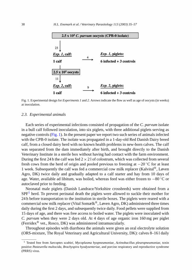
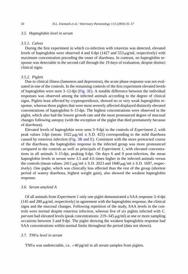
Fig. 1. Experimental design forExperiments 1 and2. Arrows indicate the flow as well as age of oocysts (in weeks)at inoculation.
2.3. Experimental animals
Each series of experimental infections consisted of propagation of theC. parvum isolatein a bull calf followed inoculation, into six piglets, with three additional piglets serving asnegative controls (Fig. 1). In the present paper we report two such series of animals infectedwith the CPB-0 isolate. The isolate was propagated in a 1-day-old Red Danish Dairy breedcalf, from a closed dairy herd with no known health problems in new-born calves. The calfwas separated from the dam immediately after birth, and brought directly to the DanishVeterinary Institute in a sterile box without having had contact with the farm environment.During the first 24 h the calf was fed 2×2 l of colostrum, which was collected from severalfresh cows from the herd of origin and pooled previous to freezing at−20◦C for at least1 week. Subsequently the calf was fed a commercial cow milk replacer (Kalvital®, LøvenAgro, DK) twice daily and gradually adapted to a calf starter and hay from 10 days ofage. Water, available ad libitum, was boiled, whereas feed was either frozen to−80◦C orautoclaved prior to feeding.
Neonatal male piglets (Danish Landrace/Yorkshire crossbreds) were obtained from aSPF1 herd. To prevent perinatal death the piglets were allowed to suckle their mother for24 h before transportation to the institution in sterile boxes. The piglets were reared with acommercial sow milk replacer (Vital Somælk®, Løven Agro, DK) administered three timesdaily during the first 2 days, and subsequently twice daily. Food pellets were supplied from15 days of age, and there was free access to boiled water. The piglets were inoculated withC. parvum when they were 2 days old. At 4 days of age organic iron 160 mg per piglet(Ferridex® vet., Rosco, DK) was administered intramuscularly.
Throughout episodes with diarrhoea the animals were given an oral electrolyte solution(ORS-mixture, The Royal Veterinary and Agricultural University, DK): calves 8–16 l daily
1 Tested free fromSarcoptes scabiei, Mycoplasma hyopneumoniae, Actinobacillus pleuropneumoniae, toxinpositivePasteurella multocida, Brachyspira hyodysenteriae, and porcine respiratory and reproductive syndrome(PRRS) virus.
H.L. Enemark et al. / Veterinary Parasitology 113 (2003) 35–57 39
via bottle, piglets ad libitum or 50–100 ml via tube 3–6 times daily, if the animals wereseverely dehydrated. During the first experiment, there was a delay in the administrationof electrolytes, and therefore, the piglets did not receive electrolytes until 2 days after theinitial signs. Experimental animals were killed by intravenously injection of a lethal doseof pentobarbital, 20 mg per ml; calves 19 days post-infection (dpi), piglets 17 dpi.
2.4. Experimental facilities and sanitary measures
The study was carried out under very strict precautions to minimise the possibility ofcontamination of the isolate, including totally separate facilities for infected and controlgroups. The piglets were kept in smooth plastic containers (137 cm×97 cm×92 cm), threeanimals together in each. Access to the stables was restricted to a few people who had nocontact with other farm animals. Washing of hands, wearing sterile gloves, and change ofclothing was obligatory prior to admittance. Between experiments the stables were cleanedmechanically with a high-pressure hose, fumigated with a potassium hydroxide, phosphoracid solution (trinol, Trinol A/S, DK), and disinfected with ammonium chloride (OO-cide®,Antec Int. Ltd., UK). Equipment was disinfected with 5% hydrogen peroxide, and frozen at−80◦C. As bedding, sterile sawdust pellets (Tørstrø, Dansk Træmel I/S, DK) and autoclavedstraw was used.
2.5. Clinical examination
The experimental animals were examined at least twice daily for signs of clinical illnessand the milk intake was recorded. In addition, daily faecal samples were collected directlyfrom each animal and rectal body temperature was measured every day. Blood sampleswere taken from the jugular vein at irregular intervals in the calves, whereas blood samplesfrom the piglets were taken every third day during the entire period with a single exception9 dpi, due to severe clinical disease in one piglet. All piglets were weighed 3–4 times duringthe course of the experiment.
2.6. Faecal consistency and oocyst excretion
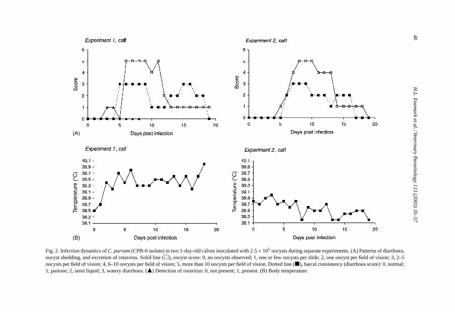
To classify the severity of diarrhoea, samples were scored on a scale from 0 to 3, asoutlined in the text toFig. 2. Moreover, it was noted whether the faecal samples containedmucus or not. Shedding ofC. parvum oocysts was analysed by a modified Ziehl-Neelsentechnique (Henriksen and Pohlenz, 1981), and graded between 0 and 5 corresponding tothe number of oocysts per slide (text toFig. 2).
2.7. Examination for other enteropathogens
Daily faecal samples from calves taken 0–7 dpi were analysed for co-infection with rota-and coronavirus by an enzyme linked immunosorbent assay (ELISA) (Grauballe et al.,1981). Furthermore, samples taken 0 dpi were examined by routine culture forEscherichiacoli (E. coli), and a proportion of colonies screened for pilus antigens by slide agglutination.During episodes with diarrhoea additional samples were examined forSalmonella spp. by
40H
.L.E
nemark
etal./VeterinaryParasitology
113(2003)
35–57
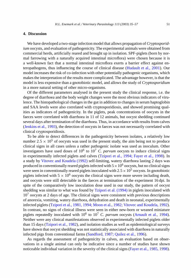
Fig. 2. Infection dynamics ofC. parvum (CPB-0 isolate) in two 1-day-old calves inoculated with 2.5×10⁵ oocysts during separate experiments. (A) Patterns of diarrhoea,oocyst shedding, and excretion of rotavirus. Solid line (�), oocyst score: 0, no oocysts observed; 1, one or few oocysts per slide; 2, one oocyst per field of vision; 3, 2–5oocysts per field of vision; 4, 6–10 oocysts per field of vision; 5, more than 10 oocysts per field of vision. Dotted line (�), faecal consistency (diarrhoea score): 0, normal;1, pastose; 2, semi liquid; 3, watery diarrhoea. (�) Detection of rotavirus: 0, not present; 1, present. (B) Body temperature.
H.L. Enemark et al. / Veterinary Parasitology 113 (2003) 35–57 41
routine culture as well as for rotavirus, coronavirus, andE. coli. The same procedure, notincluding analysis for coronavirus, was followed in the piglets. Additionally, the presence ofIsospora suis was examined 11 dpi by a modified McMaster technique (Henriksen, 1995).The occurrence of rotavirus in calves as well as in piglets was scored: 1, present; 0, notpresent.
2.8. Pathological examination
During post-mortem examination the gastrointestinal tract was given special attentionin all test and control animals. Tissue samples, taken for each 50 cm throughout the in-testines in calves, and from duodenum, jejunum (proximal, mid and distal), ileum, caecum,colon, and rectum in piglets, were fixed in 10% neutral buffered formalin within 15 minafter euthanasia. Paraffin embedded tissue blocks were cut into 3�m sections, stainedby hematoxylin and eosin (H&E), and examined by light microscopy according to stan-dard operation procedures. The degree of cryptosporidial infection as well as the range ofhistopathological alterations in the mucosa were given a score between 0 and 3 (Tziporiet al., 1981). For specific verification of cryptosporidia, especially during low-grade infec-tion, immunohistochemistry was performed either as immunofluorescense (IF) detectionwith a FITC conjugated monoclonal antibody directed against the oocyst wall (M85, Mi-crogen Bioproducts, UK) or as a two-step visualisation system (DAKO EnVisionTM, DAKOCorporation, USA), using the same antibody without FITC conjugation.
Animals dying during the experimental period were submitted to necropsy including abacteriological examination of liver, spleen and intestines.
2.9. Haptoglobin level in serum
Bovine serum haptoglobin was determined by a sandwich ELISA (Godson et al., 1996) asdescribed previously (Heegaard et al., 2000) using a pool of bovine serum as standard. Thebovine serum standard was calibrated against affinity-purified bovine haptoglobin (Godsonet al., 1996). The lower detection limit of the assay as defined by the linear range of thestandard curve was 1.15�g/ml. Serum samples were tested in serial dilutions of 1:100,1:300, 1:900, resulting in a lower limit of detection of 115�g/ml.
Porcine haptoglobin was analysed by an in-house sandwich ELISA, using an in-housemonoclonal antibody directed against porcine haptoglobin (3.8/D7) as catching antibody,and biotinylated rabbit anti human haptoglobin (DAKO A0030) as detection antibody,followed by peroxidase-conjugated streptavidin (DAKO P397). As standard was used apool of pig serum calibrated against a porcine haptoglobin standard from Saikin KagakuCo. Ltd. (Japan). All samples were applied in duplicate and ODs were read at 490 nmsubtracting 650 nm.
2.10. Serum amyloid A
The concentration of SAA in the porcine serum samples was assessed by a sandwichELISA from Tridelta (Tridelta Development Ltd., Bray Co. Wicklow, Ireland) in accordancewith the manufacturers instructions. The lower detection limit of the assay as defined by
42 H.L. Enemark et al. / Veterinary Parasitology 113 (2003) 35–57
the linear range of the standard curve was 9.4 ng/ml. Serum samples were initially diluted1:500, resulting in a lower limit of detection of 4.69�g/ml. When concentrations exceededthe range of the assay, samples were diluted further and re-tested.
2.11. Tumour necrosis factor alpha level in serum
Duplicate serum samples from piglets were analysed by a commercial ELISA kit (PigELISA TNF�, Endogen, Inc., USA) according to manufactures instructions. A standardcurve was generated from the OD values of increasing concentrations of recombinantporcine TNF�, and the TNF� level in the serum samples was calculated form the stan-dard curve. The assays produced OD values in the range from 0.09 to 2.44 for standardscontaining 40–1500 pg/ml. Negative control preparations gave results below 0.03.
2.12. Statistical analysis
The units of analyses were the individual measures (diarrhoea and excretion of oocysts)or the difference between two measures (daily weight gain). To compare differences in theoccurrence of diarrhoea between inoculated and control piglets, logistic regression of theproportion of days with diarrhoea in the experimental period was used. Logistic regressionwas performed by the GENMOD procedure in SAS© System for Elementary StatisticalAnalysis (SAS Institute, Inc., USA).
Thet-tests, performed by the procedure TTEST in SAS, were used to compare differencesin average daily weight gain during the experimental period between inoculated and controlpiglets.
Potential differences in the excretion of oocysts by inoculated piglets were tried estimated.It was assumed that the observed measurement of quantity of excreted oocysts, 0–5 on anordinal scale, was the result of passing thresholds on an underlying latent continuous scale.By assuming a latent scale, it was possible to fit an ordinal probit model to data, where thepotential difference of excretion of oocysts between the experiments was estimated. Theestimate was adjusted for the effect of time relapse after inoculation by adding the lineareffect as well as the squared linear effect of time to the model. Because the measures wereobtained repeatedly from each individual animal, a pig-level random effect was added tothe model to adjust for the dependency of measures obtained from the same individual. Themodel was estimated using the NLMIXED procedure in SAS. To evaluate the adequacy ofthe model assumptions and estimated values, the fit of the model was evaluated by inspectionof plots in which predicted values of the model were superimposed on the observed values.
3. Results
3.1. Molecular characterisation of Cryptosporidium
Following passage through inoculated calves and piglets inExperiments 1 and2, respec-tively, no genetic changes were observed. In other words, the isolate was characterised asgenotype II by analysis of the COWP gene, the ‘bovine genotype’ by partial sequencing
H.L. Enemark et al. / Veterinary Parasitology 113 (2003) 35–57 43
of the 18S rDNA, and assigned to subgenotype C2 by sequencing of a locus containing aGAG microsatellite. No otherCryptosporidium spp. or genotypes were detected.
3.2. Clinical signs and oocyst excretion
3.2.1. CalvesExperiment 1: Voluminous, watery diarrhoea was seen from 5 dpi, and continued with
varying intensity until 18 dpi (Fig. 2A). During the third week of infection the faeces hadhigh mucus content. Concurrent with incipient diarrhoea, the calf became mildly depressedand listless. Mild fever (temperatures between 39.6 and 40.0◦C) was present 4, 6 dpi, andtowards the end of the experimental period (Fig. 2B). Regardless of this, the appetite wasgood throughout the entire period, and the calf remained relatively unaffected except frommild lethargy 9, 13 and 18 dpi. The prepatent period was 6 days with peak numbers ofoocysts occurring from 6 to 12 dpi.
Experiment 2: Essentially, the clinical picture was repeated in the second experiment.Watery diarrhoea started 6 dpi simultaneously with the oocyst shedding, and continueduntil 16 dpi (Fig. 2A). Apathy, muscle tremor, and cold extremities were seen 8–9 dpi, butthe appetite was retained throughout the period, and fever was not present (Fig. 2B).
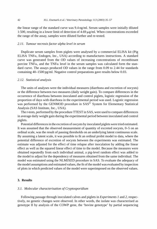
3.2.2. PigletsExperiment 1: The first clinical response, 2–3 days after inoculation, was vomiting in
four of six experimentally infected piglets (lasting 1–3 days), succeeded by inappetence(1–8 days), depression and watery, yellow diarrhoea with clumps of mucus (1–6 days)(Table 1, Fig. 3A). Intermittent, dry coughing was observed 3–6 dpi in several piglets in-cluding the controls. Following the onset of vomiting and diarrhoea, subnormal temperatures(<38.8◦C) were observed in five of six piglets (Fig. 3C). A large individual variation inthe clinical signs was seen between the piglets. Thus, one piglet had semi-liquid faecesfor a single day succeeded by the excretion of high numbers of oocysts through severaldays without any clinical signs, whereas another piglet had to be euthanised 7 dpi dueto dehydration and circulatory failure. The remaining piglets recovered after a period of
Table 1Infection dynamics ofC. parvum (CPB-0 isolate) in piglets inoculated with 2.5 × 10⁵ oocysts during repeatedexperiments
Experiment 1 (n = 6)a Experiment 2 (n = 6)
Incubation period 2.8 (range 2–4) 2.3 (range 1–3)Maximum diarrhoea score 2.8 (range 2–3), 4 dpi 2.7 (range 2–3), 4 dpiDuration of diarrhoea (score= 2) 3.5 days (range 1–6) 6.0 days (range 3–7)Prepatent period 3.5 days (range 2–5) 3.0 days (range 2–4)Maximum oocyst score 3.8, 4 dpi 4.7, 6 dpiDuration of oocyst excretionb 12.3 days (5–16) 14.3 days (13–16)Daily weight gain (infected) 279 g (±S.D. 77) 247 g (±S.D. 25)Daily weight gain (controls,n = 3) 318 g (±S.D. 55) 300 g (±S.D. 16)
Experiment 1: mono-infection withC. parvum. Experiment 2: co-infection with rotavirus.a One piglet was euthanised 7 days post-infection because of poor health associated with diarrhoea.b Oocyst excretion was not completed at termination of the observational period.
44H
.L.E
nemark
etal./VeterinaryParasitology
113(2003)
35–57
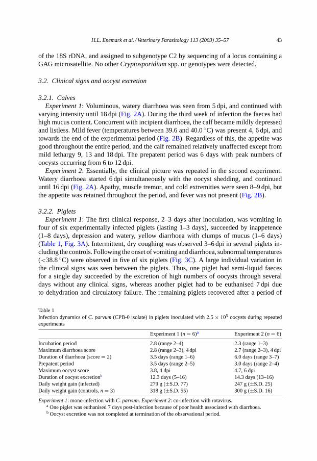
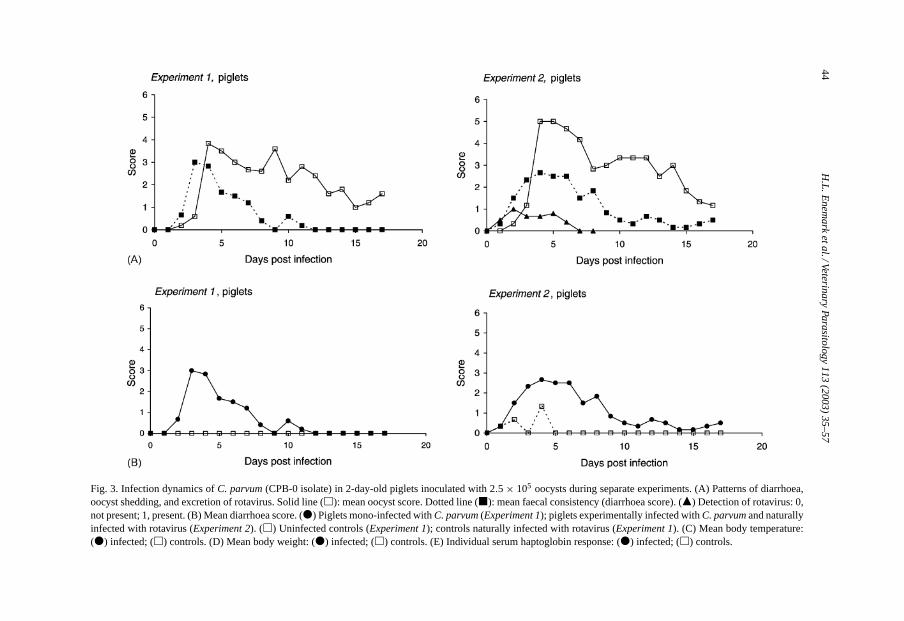
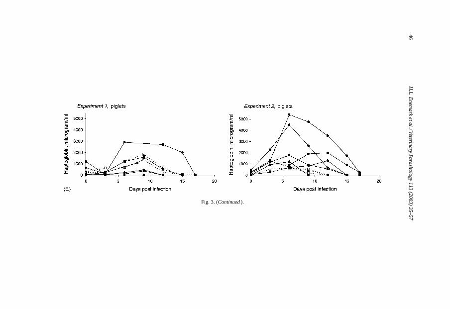
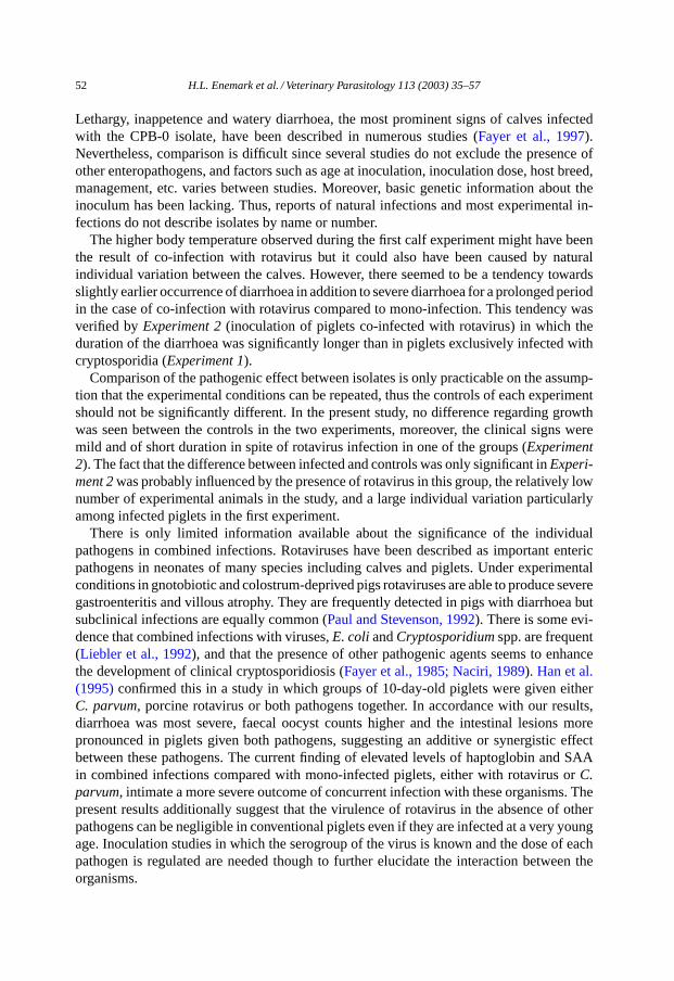
Fig. 3. Infection dynamics ofC. parvum (CPB-0 isolate) in 2-day-old piglets inoculated with 2.5 × 10⁵ oocysts during separate experiments. (A) Patterns of diarrhoea,oocyst shedding, and excretion of rotavirus. Solid line (�): mean oocyst score. Dotted line (�): mean faecal consistency (diarrhoea score). (�) Detection of rotavirus: 0,not present; 1, present. (B) Mean diarrhoea score. (�) Piglets mono-infected withC. parvum (Experiment 1); piglets experimentally infected withC. parvum and naturallyinfected with rotavirus (Experiment 2). (�) Uninfected controls (Experiment 1); controls naturally infected with rotavirus (Experiment 1). (C) Mean body temperature:(�) infected; (�) controls. (D) Mean body weight: (�) infected; (�) controls. (E) Individual serum haptoglobin response: (�) infected; (�) controls.
H.L
.Enem
arketal./Veterinary
Parasitology113
(2003)35–57
45
Fig. 3. (Continued )
46H
.L.E
nemark
etal./VeterinaryParasitology
113(2003)
35–57
Fig. 3. (Continued ).
H.L. Enemark et al. / Veterinary Parasitology 113 (2003) 35–57 47
diarrhoea, despite temperatures as low as 35.4◦C. Subnormal temperatures were also ob-served in controls as well as infected piglets 1 dpi, but this was normalised following adjust-ment of the heat lamps.Cryptosporidium oocysts in the faeces commenced from between1 day before till 2 days after the onset of clinical illness and were shed for 12–16 days, i.e.all infected piglets, except the one that was euthanised, continued to shed oocysts after thediarrhoea had ended. Towards the end of the experiment oocysts were excreted intermit-tently by three piglets. Three littermates (controls) remained free from enteric signs as wellas cryptosporidia throughout the study, however mild depression (0–7 dpi), and lamenesson one leg (0–8 dpi) following injury during transport were seen in one piglet in this group(Table 1, Fig. 3B).
Experiment 2: At 1–3 dpi, three piglets vomited (1–2 days), and all of the six piglets devel-oped anorexia (lasting 3–6 days) as well as diarrhoea (3–7 days). Lethargy and depressionwere present during periods with watery diarrhoea. Except for one piglet the onset of diar-rhoea preceeded detection of oocysts in the faeces. As in the first experiment oocysts wereshed intermittently by three animals towards the end of the period, and all but one excretedoocysts when they were killed 17 dpi (Table 1, Fig. 3A). Cryptosporidium oocysts were notdetected in the control piglets. Nevertheless, these animals went through a short period, lessthan 24 h, of diarrhoea with scores between 1 and 3 (Fig. 3B) and a corresponding drop ofbody temperature (Fig. 3C).
The proportion of days with diarrhoea was significantly higher in the infected groupscompared to the controls (P < 0.0001) in both experiments. Compared toExperiment 1 theproportion of days with diarrhoea as well as the oocyst shedding was significantly higherin Experiment 2 (P = 0.0007 andP = 0.0341, respectively). The plots of predicted valuesof oocyst excretion correlated with the observed values indicating that the estimated probitmodel described the observed data relatively well.
The mean body weights of infected and controls, measured at different intervals through-out the study, are illustrated inFig. 3D, whereas the mean daily weight gain of the differentgroups are shown inTable 1. The weight gain during the experimental period did not differsignificantly between the control groups (P = 0.5682). In contrast, the mean daily weightgain was larger in the controls compared to the infected animals (difference between infectedand controls: 0.039 kg± 0.068 S.D. (Experiment 1), 0.053 kg± 0.023 S.D. (Experiment2)), but the difference was only significant inExperiment 2 (P = 0.4665,Experiment 1;P = 0.0137,Experiment 2).
3.3. Examination for other enteropathogens
3.3.1. CalvesExperiment 1: A mixed culture of non-haemolyticE. coli was found during episodes with
diarrhoea in addition to rotavirus, which were shed 3–4 dpi (Fig. 2A).Experiment 2: Non-haemolyticE. coli were detected in the second calf, but no entero-
toxigenicE. coli (ETEC-F5+), rota-, coronavirus orSalmonella were found.
3.3.2. PigletsExperiment 1: Bacteriological examination of faecal samples revealed a mixed culture
of non-haemolyticE. coli in a few samples from four of six piglets, but there was no
48 H.L. Enemark et al. / Veterinary Parasitology 113 (2003) 35–57
histological evidence of coliform adherence to the mucosa in any of these piglets at necropsy.The majority of samples, including controls, contained no pathogenic bacteria, and neitherrotavirus norI. suis were found. By post-mortem examination of one piglet, which waseuthanised prematurely, no bacteria were detected in the parenchymatous organs. A possibleinfectious cause to the respiratory signs was not further investigated.
Experiment 2: Non-haemolyticE. coli O8 in mixed culture was detected in one pigon one occasion 3 dpi. Virological examination of daily faecal samples from 0 to 8 dpi,demonstrated the presence of rotavirus in all but one animal, including the three controls.Excretion of rotavirus was detected from 2 to 3 dpi in the infected group (lasting 3–5 days),and from 3 to 4 dpi in the controls, that were still excreting virus 8 dpi, several days afterany signs of clinical illness. Pastose stools lasting 2 days were seen in one of the controls;in another, faeces was semi-liquid for 1 day, whereas the third control piglet had waterydiarrhoea at one sampling occasion without inappetence or any other clinical signs (Fig. 3B).No other pathogens were detected in samples fromExperiment 2.
3.4. Pathology
3.4.1. CalvesExperiment 1: At autopsy, the only finding was moderately enlarged mesenteric lymph
nodes. Histological examination of the intestine revealed no parasitic stages. However,stunting and fusion of villi, replacement of enterocytes by immature cells, and increasedcellularity of lamina propria were observed focally in the distal jejunum, ileum and caecum.
Experiment 2: The mesenteric lymph nodes were moderately enlarged. Solitary cryp-tosporidia were observed in the distal colon and rectum. Stunting and fusion of villi, re-placement of enterocytes by immature cells, increased cellularity and marked eosinophiliaof lamina propria were seen in the terminal jejunum, ileum and colon.
3.4.2. PigletsExperiment 1: Catarrhal enteritis and severe dehydration was seen in one piglet, which
was euthanised 7 dpi because of poor health associated with diarrhoea. The content of thestomach and the small intestines was sparse and watery. In the remaining piglets, no grosslesions were detected neither among infected nor among controls following autopsy 17 dpi.
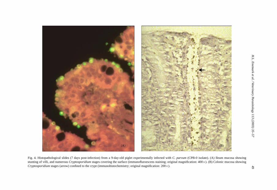
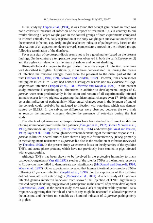
Parasitic stages and microscopic lesions were absent in the controls as well as in two in-fected piglets. With the exception of duodenum,Cryptosporidium stages were seen through-out the entire intestine of the one piglet that died prematurely, in addition to catarrhalenteritis and severe mucosal damage (Fig. 4). In the remaining piglets, infection was re-stricted to colon and rectum. Corresponding to the severity of the clinical signs, stuntingand fusion of villi, and replacement of enterocytes by immature cells were present in twopiglets.
Experiment 2: Following necropsy, no macroscopic lesions were observed. Histologicalexamination revealed low numbers of cryptosporidia in addition to lesions, characterisedby replacement of enterocytes by immature cells in the colon and rectum of all experimen-tally infected piglets. Furthermore, stunting and fusion of villi as well as replacement ofenterocytes by immature cells were observed in the jejunum of one control piglet despitenormal consistency of faeces and the absence of macropic changes.
H.L
.Enem
arketal./Veterinary
Parasitology113
(2003)35–57
49
Fig. 4. Histopathological slides (7 days post-infection) from a 9-day-old piglet experimentally infected withC. parvum (CPB-0 isolate). (A) Ileum mucosa showingstunting of villi, and numerousCryptosporidium stages covering the surface (immunofluroescens staining; original magnification: 400×). (B) Colonic mucosa showingCryptosporidium stages (arrow) confined to the crypt (immunohistochemistry; original magnification: 200×).
50 H.L. Enemark et al. / Veterinary Parasitology 113 (2003) 35–57
3.5. Haptoglobin level in serum
3.5.1. CalvesDuring the first experiment in which co-infection with rotavirus was detected, elevated
levels of haptoglobin were observed 4 and 6 dpi (1427 and 553�g/ml, respectively) withmaximum concentration preceding the onset of diarrhoea. In contrast, no haptoglobin re-sponse was detectable in the second calf through the 19 days of evaluation, despite distinctclinical signs.
3.5.2. PigletsDue to clinical illness (lameness and depression), the acute phase response was not eval-
uated in one of the controls. In the remaining controls of the first experiment elevated levelsof haptoglobin were seen 3–12 dpi (Fig. 3E). A notable difference between the individualresponses was observed among the infected animals according to the degree of clinicalsigns. Piglets least affected by cryptosporidiosis, showed no or very weak haptoglobin re-sponse, whereas those piglets that were most severely affected displayed distinctly elevatedconcentrations of haptoglobin 6–15 dpi. The highest concentrations were observed in thepiglet, which also had the lowest growth rate and the most pronounced degree of mucosalchanges following autopsy (with the exception of the piglet that died prematurely becauseof diarrhoea).
Elevated levels of haptoglobin were seen 3–9 dpi in the controls ofExperiment 2, withpeak values 3 dpi (mean: 1022�g/ml ± S.D. 425) corresponding to the mild diarrhoeacaused by rotavirus infection (Fig. 3B and E). Consistent with the more protracted courseof the diarrhoea, the haptoglobin response in the infected group was more pronouncedcompared to the controls as well as principals ofExperiment 1, with elevated concentra-tions in all animals 3–15 dpi, peaking 6 dpi. On days 6 and 9 post-infection, the meanhaptoglobin levels in serum were 3.5 and 4.6 times higher in the infected animals versusthe controls (mean values: 2411�g/ml ± S.D. 2023 and 1840�g/ml ± S.D. 1697, respec-tively). One piglet, which was clinically less affected than the rest of the group (shortestperiod of watery diarrhoea, highest weight gain), also showed the weakest haptoglobinresponse.
3.6. Serum amyloid A
Of all animals fromExperiment 1 only one piglet demonstrated a SAA response 3–6 dpi(145 and 280�g/ml, respectively) in agreement with the haptoglobin response, the clinicalsigns and the mucosal changes. Following repetition of the study, SAA levels in the con-trols were normal despite rotavirus infection, whereas five of six piglets infected withC.parvum had elevated levels (peak concentrations: 219–345�g/ml) at one or more samplingoccasions between 3 and 9 dpi. The piglet showing the weakest haptoglobin response hadSAA concentrations within normal limits throughout the period (data not shown).
3.7. TNFα level in serum
TNF� was undetectable, i.e.<40 pg/ml in all serum samples from piglets.
H.L. Enemark et al. / Veterinary Parasitology 113 (2003) 35–57 51
4. Discussion
We have developed a two-stage infection model that allows propagation ofCryptosporid-ium oocysts, and evaluation of pathogenicity. The experimental animals were obtained fromcommercial herds, artificially reared and brought up in isolation. SPF-piglets (born by nor-mal farrowing with a naturally acquired intestinal microflora) were chosen because it isa well-known fact that a normal intestinal microflora exerts a barrier effect against en-teropathogens, thus influencing the course of clinical disease (Hudault et al., 2001). Ourmodel increases the risk of co-infection with other potentially pathogenic organisms, whichmakes the interpretation of the results more complicated. The advantage however, is that themodel is less expensive than a gnotobiotic model, and allows the study ofCryptosporidiumin a more natural setting of other micro-organisms.
Of the different parameters analysed in the present study the clinical response, i.e. thedegree of diarrhoea and the body weight changes were the most obvious indicators of viru-lence. The histopathological changes in the gut in addition to changes in serum haptoglobinand SAA levels were also correlated with cryptosporidiosis, and showed promising qual-ities as indicators of pathogenicity. In the piglets, peak concentrations of oocysts in thefaeces were correlated with diarrhoea in 11 of 12 animals, but oocyst shedding continuedseveral days after termination of the diarrhoea. Thus, in accordance with results from calves(Jenkins et al., 1993), the detection of oocysts in faeces was not necessarily correlated withclinical cryptosporidiosis.
To be able to detect differences in the pathogenicity between isolates, a relatively lownumber 2.5 × 10⁵ of oocysts was used in the present study, the aim being not to produceclinical signs in all cases unless a rather pathogenic isolate was used as inoculum. Otherinvestigators have used doses of 106 to 107 C. parvum oocysts to induce clinical signsin experimentally infected piglets and calves (Tzipori et al., 1994; Fayer et al., 1998). Ina study byVitovec and Koudela (1992)self-limiting, watery diarrhoea lasting 2 days wasproduced in conventionally reared piglets infected with 5×10⁶ oocysts, but no clinical signswere seen in conventionally reared piglets inoculated with 2.5×10⁵ oocysts. In gnotobioticpiglets infected with 5× 10⁶ oocysts the clinical signs were more severe including death,and oocysts were still detectable in the faeces at termination of the experiment 16 dpi. Inspite of the comparatively low inoculation dose used in our study, the pattern of oocystshedding was similar to what was found byTzipori et al. (1994)in piglets inoculated with107 oocysts at 2 days of age. The clinical signs were consistent with previous descriptionsof anorexia, vomiting, watery diarrhoea, dehydration and death in neonatal, experimentallyinfected piglets (Tzipori et al., 1981, 1994; Moon et al., 1982; Vitovec and Koudela, 1992).In contrast, no signs of clinical illness were seen in either new-born or weaned miniaturepiglets repeatedly inoculated with 106 to 107 C. parvum oocysts (Arnault et al., 1994).Neither were any clinical manifestations observed in experimentally infected piglets olderthan 15 days (Tzipori et al., 1982), and isolation studies as well as epidemiological surveyshave shown that oocyst shedding was not statistically associated with diarrhoea in naturallyinfected pigs from conventional farms (Sandford, 1987; Quilez et al., 1996).
As regards the assessment of pathogenicity in calves, an evaluation based on obser-vations in a single animal can only be indicative since a number of studies have shownnoticeable individual variation in the severity of the clinical signs (Fayer et al., 1985, 1998).
52 H.L. Enemark et al. / Veterinary Parasitology 113 (2003) 35–57
Lethargy, inappetence and watery diarrhoea, the most prominent signs of calves infectedwith the CPB-0 isolate, have been described in numerous studies (Fayer et al., 1997).Nevertheless, comparison is difficult since several studies do not exclude the presence ofother enteropathogens, and factors such as age at inoculation, inoculation dose, host breed,management, etc. varies between studies. Moreover, basic genetic information about theinoculum has been lacking. Thus, reports of natural infections and most experimental in-fections do not describe isolates by name or number.
The higher body temperature observed during the first calf experiment might have beenthe result of co-infection with rotavirus but it could also have been caused by naturalindividual variation between the calves. However, there seemed to be a tendency towardsslightly earlier occurrence of diarrhoea in addition to severe diarrhoea for a prolonged periodin the case of co-infection with rotavirus compared to mono-infection. This tendency wasverified byExperiment 2 (inoculation of piglets co-infected with rotavirus) in which theduration of the diarrhoea was significantly longer than in piglets exclusively infected withcryptosporidia (Experiment 1).
Comparison of the pathogenic effect between isolates is only practicable on the assump-tion that the experimental conditions can be repeated, thus the controls of each experimentshould not be significantly different. In the present study, no difference regarding growthwas seen between the controls in the two experiments, moreover, the clinical signs weremild and of short duration in spite of rotavirus infection in one of the groups (Experiment2). The fact that the difference between infected and controls was only significant inExperi-ment 2 was probably influenced by the presence of rotavirus in this group, the relatively lownumber of experimental animals in the study, and a large individual variation particularlyamong infected piglets in the first experiment.
There is only limited information available about the significance of the individualpathogens in combined infections. Rotaviruses have been described as important entericpathogens in neonates of many species including calves and piglets. Under experimentalconditions in gnotobiotic and colostrum-deprived pigs rotaviruses are able to produce severegastroenteritis and villous atrophy. They are frequently detected in pigs with diarrhoea butsubclinical infections are equally common (Paul and Stevenson, 1992). There is some evi-dence that combined infections with viruses,E. coli andCryptosporidium spp. are frequent(Liebler et al., 1992), and that the presence of other pathogenic agents seems to enhancethe development of clinical cryptosporidiosis (Fayer et al., 1985; Naciri, 1989). Han et al.(1995)confirmed this in a study in which groups of 10-day-old piglets were given eitherC. parvum, porcine rotavirus or both pathogens together. In accordance with our results,diarrhoea was most severe, faecal oocyst counts higher and the intestinal lesions morepronounced in piglets given both pathogens, suggesting an additive or synergistic effectbetween these pathogens. The current finding of elevated levels of haptoglobin and SAAin combined infections compared with mono-infected piglets, either with rotavirus orC.parvum, intimate a more severe outcome of concurrent infection with these organisms. Thepresent results additionally suggest that the virulence of rotavirus in the absence of otherpathogens can be negligible in conventional piglets even if they are infected at a very youngage. Inoculation studies in which the serogroup of the virus is known and the dose of eachpathogen is regulated are needed though to further elucidate the interaction between theorganisms.
H.L. Enemark et al. / Veterinary Parasitology 113 (2003) 35–57 53
In the study byTzipori et al. (1994), it was found that weight gain or loss in mice wasnot a consistent measure of infection or the impact of treatment. This is contrary to ourresults showing a larger weight gain in the control groups of both experiments comparedto infected animals. Yet, daily registration of the body weight gain and evaluation earlier inthe course of infection, e.g. 10 dpi might be a better indicator of pathogenicity based on theobservation of an apparent tendency towards compensatory growth in the infected groupsfollowing termination of the diarrhoea.
Fever as a sign of cryptosporidiosis seems not to be a good marker based on the presentfindings. On the contrary a temperature drop was observed in both the calf (Experiment 2)and the piglets correlated with maximum diarrhoea and oocyst shedding.
Histopathological changes in the gut during the acute stages of infection have beenwell described in piglets. Additionally, it has been demonstrated that during the courseof infection the mucosal changes move from the proximal to the distal part of the GItract (Tzipori et al., 1982, 1994; Vitovec and Koudela, 1992). However, it has been shownthat piglets killed 11 to 17 dpi had neither histological lesions nor any evidence ofCryp-tosporidium infection (Tzipori et al., 1981; Vitovec and Koudela, 1992). In the presentstudy, moderate histopathological alterations in addition to developmental stages ofC.parvum were seen predominantly in the colon and rectum of all experimentally infectedanimals except for two piglets, suggesting that histological changes even 17–19 dpi mightbe useful indicators of pathogenicity. Histological changes seen in the jejunum of one ofthe controls could probably be attributed to infection with rotavirus, which was demon-strated by ELISA. In the calves, no differences were observed between the two trialsas regards the mucosal changes, despite the presence of rotavirus during the firststudy.
The effects of cytokines on cryptosporidiosis have been studied in different models in-cluding immunocompromised human patients (Flanigan et al., 1992; Gomez Morales et al.,1996), mice models (Ungar et al., 1991; Urban et al., 1996), and calves (de Graaf and Peeters,1997; Fayer et al., 1998). Although our current understanding of the immune response toC.parvum is limited, several studies have shown a key role for interferon-� (IFN�) primarilyin mediating innate resistance toC. parvum but also in the resolution of infection (reviewedby Theodos, 1998). In the present study we chose to focus on the dynamics of the cytokineTNF� and acute phase proteins, which have not previously been studied in pigs infectedwith cryptosporidia.
Although TNF� has been shown to be involved in the protective immunity to manypathogenic organisms (Vassalli, 1992), studies of the role for TNF� in the immune responseto C. parvum have failed to demonstrate any significance (McDonald and Bancroft, 1994;Wyatt et al., 1997). Other experiments revealed that human intestinal cells produce TNF�following C. parvum infection (Seydel et al., 1998), but the expression of this cytokinedid not correlate with enteric signs (Robinson et al., 2001). A recent study ofC. parvuminfected gamma interferon knockout mice showed that injection of TNF� significantlyreduced oocyst shedding, suggestive of participation in the control of parasite development(Lacroix et al., 2001). In the present study, there was a lack of any detectable systemic TNF�response, suggesting that the role of TNF�, if any, might be restricted to a local response inthe intestine, and therefore not suitable as a humoral indicator ofC. parvum pathogenicityin piglets.
54 H.L. Enemark et al. / Veterinary Parasitology 113 (2003) 35–57
In contrast, the SPF piglet model seems to be relevant for the study of haptoglobin andSAA responses, as illustrated by the distinct reactions in those piglets most severely affectedclinically. Haptoglobin and SAA are major acute phase proteins in most species studied(Mackiewicz et al., 1993), and well-known general indicators of inflammation, trauma andother pathological conditions (Kushner and Rzewnicki, 1994; Baumann and Gauldie, 1994).Serum haptoglobin has been shown to be highly correlated with the inoculation dose as wellas with strain virulence in, e.g. mice and pigs inoculated withToxoplasma gondii (Jensenet al., 1998; Jungersen et al., 1999), and therefore, potentially attractive as a measure ofpathogenicity in hosts infected with other protozoa such asC. parvum. In the present study,increased serum levels of haptoglobin and SAA were observed during the first as wellas the second experiment, i.e. irrespective of concurrent rotavirus infection. However, theresponses were more dramatic in piglets infected with both pathogens as an indication ofmore severe enteric damage. In the infected piglets ofExperiment 2, haptoglobin reachedmaximum concentrations up to above 5000�g/ml while SAA increased up to 280�g/ml. NoSAA response was observed in the controls ofExperiment 2, but a haptoglobin response withpeak values 3 dpi (mean 1022�g/ml) was found to be correlated to the mild diarrhoea causedby rotavirus. InExperiment 1, serum concentrations of haptoglobin peaked around 9 dpi ininfected plus controls. In the light of this observation, it is concluded that the respiratory signsseen possibly as a result of cooling influenced the acute phase response. Therefore, the acutephase response toC. parvum cannot be evaluated based on this experiment. Nevertheless,there was a correlation to the clinical signs, as manifested by the clear response in one piglet,which also showed elevated levels of SAA, indicating that a measurable SAA response couldonly be induced by severe enteric signs. Thus, this acute phase protein might be a valuabletool to point out the most severely affected individuals.
In conclusion, the infection dynamics ofC. parvum (CPB-0 isolate) was described in onecalf (Experiment 2) and a group of piglets (Experiment 1) in which no other enteropathogenswere detected. Further, it was shown that this particular isolate was pathogenic to calvesas well as piglets even at a relatively low dose, and that the clinical signs could be repli-cated during separate experiments. Moreover, diarrhoea, oocyst shedding, body weightchanges, histological alterations, and the acute phase response of haptoglobin and SAAwere identified as useful parameters of pathogenicity. These results demonstrate that clin-ical cryptosporidiosis can be induced in SPF piglets with a naturally acquired intestinalmicroflora, and thus should be included as a causative agent of diarrhoea in young pigs.
Diarrhoea and shedding of oocysts were present in animals infected with the CPB-0 isolateindependent of simultaneous infection with rotavirus. In addition, the unintended presenceof rotavirus in some of the experimental animals revealed an additive or synergistic effectbetween rotavirus andC. parvum as demonstrated by the prolonged diarrhoea, increasedoocyst shedding, decreased weight gain and elevated levels of serum haptoglobin and SAAin piglets infected simultaneously with both pathogens.
The obvious advantages of the SPF pig model are the naturally acquired intestinal mi-croflora, the development of distinct clinical signs similar to cryptosporidiosis in humansand calves, the size of the animals, and the accessibility of individuals born within a shorttime span. This makes the model ideal for dose–response studies, evaluation of therapeuticagents as well as for assessment of differences in the clinical response to isolates of diversegenetic background.
H.L. Enemark et al. / Veterinary Parasitology 113 (2003) 35–57 55
Acknowledgements
The authors would like to thank John Pedersen and his staff for vigorous disinfectionof the stables, and Cynthia D. Juel, Annie Ravn, Ulla L. Andreasen, Heidi G. Pedersenand Tine Petersen for skilled technical assistance. This work was carried out as part of theDanish research programme: Food quality with a focus on food safety (FØSI00-SVS-8).
References
Arnault, I., Répérant, J.M., Naciri, M., 1994. Humoral antibody response and oocyst shedding after experimentalinfection of histocompatible new-born and weaned piglets withCryptosporidium parvum. Vet. Res. 25, 371–383.
Baumann, H., Gauldie, J., 1994. The acute phase response. Immunol. Today 15, 74–80.Cacciò, S., Homan, W., Camilli, R., Traldi, G., Kortbeek, T., Pozio, E., 2000. A microsatellite marker reveals
population heterogeneity within human and animal genotypes ofCryptosporidium parvum. Parasitology 120,237–244.
de Graaf, D.C., Peeters, J., 1997. Specific interferon-gamma IgA and IgM responses after experimental infectionof neonatal calves withCryptosporidium parvum. Int. J. Parasitol. 27, 131–134.
Dom, P., Haesebrouck, F., 1992. Comparative virulence of NAD-dependent and NAD-independentActinobacilluspleuropneumoniae strains. J. Vet. Med. 39, 303–306.
Enemark, H.L., Ahrens, P., Juel, C.D., Petersen, E., Petersen, R.F., Andersen, J.S., Lind, P., Thamsborg, S.M.,2002. Molecular characterization of DanishCryptosporidium parvum isolates. Parasitology 125, 331–341.
Fayer, R., Ernst, J.V., Miller, R.G., Leek, R.G., 1985. Factors contributing to clinical illness in calves experimentallyinfected with a bovine isolate ofCryptosporidium. Proc. Helminthol. Soc. Wash. 52, 64–70.
Fayer, R., Speer, C.A., Dubey, J.P., 1997. The general biology ofCryptosporidium. In: Fayer, R. (Ed.),Cryptosporidium and Cryptosporidiosis. CRC Press, Boca Raton, pp. 1–41.
Fayer, R., Gasbarre, L., Pasquali, P., Canals, A., Almeria, S., Zarlenga, D., 1998.Cryptosporidium parvum infectionin bovine neonates: dynamic clinical, parasitic and immunologic patterns. Int. J. Parasitol. 28, 49–56.
Fayer, R., Morgan, U., Upton, S.J., 2000. Epidemiology ofCryptosporidium: transmission, detection andidentification. Int. J. Parasitol. 30, 1305–1322.
Fayer, R., Trout, J.M., Xiao, L., Morgan, U.M., Lal, A.A., Dubey, J.P., 2001.Cryptosporidium canis n. sp. fromdomestic dogs. J. Parasitol. 87, 1415–1422.
Flanigan, T., Whalen, C., Turner, J., Soave, R., Toerner, J., Havlir, D., Kotler, D., 1992.Cryptosporidium infectionand CD4 counts. Ann. Intern. Med. 116, 840–842.
Godson, D.L., Campos, M., Attah-Poku, S.K., Redmond, M.J., Cordeiro, D.M., Sethi, M.S., Harland, R.J., Babiuk,L.A., 1996. Serum haptoglobin as an indicator of the acute phase response in bovine respiratory disease. Vet.Immunol. Immunopathol. 51, 277–292.
Gomez Morales, M.A., Ausiello, C.M., Guarino, A., Urbani, F., Spanuolo, M.I., Pignata, C., Pozio, E., 1996.Severe protracted intestinal cryptosporidiosis associated with interferon� deficiency: pediatric case report. Int.J. Parasitol. 22, 848–850.
Grauballe, P.C., Vestergaard, B.F., Meyling, A., Genner, J., 1981. Optimized enzyme-linked immunosorbentassay for detection of human and bovine rotavirus in stools: comparison with electron-microscopy,immunoelectro-osmophoresis, and fluorescent antibody techniques. J. Med. Virol. 7, 29–40.
Guerrant, R.L., 1997. Cryptosporidiosis: an emerging, highly infectious threat. Emerg. Infect. Dis. 3, 51–57.Han, D.U., Kang, M.I., Park, N.Y., Wee, S.H., 1995. Pathogenesis of enteritis induced byCryptosporidium parvum
alone and combined with porcine rotavirus in piglets. Korean J. Vet. Res. 35, 149–158.Heegaard, P.M.H., Godson, D.L., Toussaint, M.J., Toornehooj, K., Larsen, L.E., Viuff, B., Rønsholt, L., 2000. The
acute phase response of haptoglobin and serum amyloid A (SAA) in cattle undergoing experimental infectionwith bovine respiratory syncytial virus. Vet. Immunol. Immunopathol. 77, 151–159.
Henriksen, S.A., 1995.Eimeria and Isospora. In: Eckert, J., Braun, R., Shirley, M.W., Coudert, P. (Eds.),Biotechnology—Guidelines on Techniques in Coccidiosis Research. Office for Official Publications of theEuropean Communities, Luxembourg, pp. 74–78.
56 H.L. Enemark et al. / Veterinary Parasitology 113 (2003) 35–57
Henriksen, S.A., Pohlenz, J.F.L., 1981. Staining of cryptosporidia by a modified Ziehl-Neelsen technique. ActaVet. Scand. 22, 594–596.
Hudault, S., Guignot, J., Servin, A.L., 2001.Escherichia coli strains colonising the gastrointestinal tract protectgermfree mice againstSalmonella typhimurium infection. Gut 49, 47–55.
Jenkins, M.C., Fayer, R., Tilley, M., Upton, S.J., 1993. Cloning and expression of a cDNA encoding epitopes sharedby 15- and 60-kilodalton proteins ofCryptosporidium parvum sporozoites. Infect. Immun. 61, 2377–2382.
Jensen, L., Heegaard, P.M.H., Lind, P., 1998. A study of virulence parameters forToxoplasma gondii infectionsin mice. Parasitol. Res. 84, 382–387.
Jungersen, G., Jensen, L., Riber, U., Heegaard, P.M.H., Petersen, E., Poulsen, J.S.D., Bille-Hansen, V., Lind, P.,1999. Pathogenicity of selectedToxoplasma gondii isolates in young pigs. Int. J. Parasitol. 29, 1307–1319.
Kushner, I., Rzewnicki, D.L., 1994. The acute phase response: general aspects. Ballière’s Clin. Rheumatol. 8,513–530.
Lacroix, S., Manacassola, R., Naciri, M., Laurent, F., 2001.Cryptosporidium parvum-specific mucosal immuneresponse in C57BL/6 neonatal and gamma interferon-deficient mice: role of tumour necrosis factor alpha inprotection. Infect. Immun. 69, 1635–1642.
Liebler, E.M., Pohlenz, J.F., Whipp, S.C., 1992. Digestive system. In: Leman, A.D., Straw, B.E., Mengeling, W.L.,D’Allaire, S., Taylor, D.J. (Eds.), Diseases of Swine. Iowa State University Press, Iowa, pp. 12–20.
Mackiewicz, A., Kushner, I., Baumann, H. (Eds.), 1993. Acute phase proteins molecular biology, biochemistryand clinical applications. CRC Press, Boca Raton, FL.
McDonald, V., Bancroft, G., 1994. Mechanisms of innate and acquired resistance toCryptosporidium parvuminfection in SCID mice. Parasite Immunol. 16, 315–320.
Moon, H.W., Schwartz, A., Welch, M.J., McCann, P.P., Runnels, P.L., 1982. Experimental fecal transmission ofhuman cryptosporidia to pigs, and attempted treatment with an Ornithine Decarboxylase inhibitor. Vet. Pathol.19, 700–707.
Naciri, M., 1989. Animal and human cryptosporidiosis: opportunist infections? Pathogenicity of the genusCryptosporidium. In: Yvore, P. (Ed.), Coccidia and Intestinal Coccidiomorphs. Proceedings of the 5thInternational Coccidiosis Conference. INRA Publ., pp. 51–63.
Okhuysen, P.C., Chappell, C.L., Crabb, J.H., Sterling, C.R., DuPont, H.L., 1999. Virulence of three distinctCryptosporidium parvum isolates for healthy adults. J. Infect. Dis. 180, 1275–1281.
Paul, P.S., Stevenson, G.W., 1992. Rotavirus and reovirus. In: Leman, A.D., Straw, B.E., Mengeling, W.L.,D’Allaire, S. (Eds.), Diseases of Swine. Iowa State University Press, Iowa, pp. 331–348.
Peeters, J.E., Villacorta, I., 1995.Cryptosporidium. In: Eckert, J., Braun, R., Shirley, M.W., Coudert, P. (Eds.),Biotechnology—Guidelines on Techniques in Coccidiosis Research. Office for Official Publications of theEuropean Communities, Luxembourg, pp. 202–240.
Pozio, E., Morales, M.A.G., Barbieri, M., La Rosa, G., 1992.Cryptosporidium: different behaviour in calves ofisolates of human origin. Trans. R. Soc. Trop. Med. Hyg. 86, 636–638.
Quilez, J., Area-Mazás, E., Sanchez-Acedo, C., del Cacho, E., Clavel, A., 1996. Comparison of oocyst sheddingand the serum immune response toCryptosporidium parvum in cattle and pigs. Parasitol. Res. 82, 529–534.
Robinson, P., Okhuysen, P.C., Chappell, C.L., Lewis, D.E., Shahab, I., Janecki, A., White Jr., A.C., 2001.Expression of tumour necrosis factor alpha and interleukin 1beta in jejuna of volunteers after experimentalchallenge withCryptosporidium parvum correlates with exposure but not with symptoms. Infect. Immun. 69,1172–1174.
Sandford, S.E., 1987. Enteric cryptosporidial infection in pigs: 184 cases (1981–1985). JAVMA 190, 695–698.Seydel, K.B., Zhang, T., Champion, G.A., Fichtenbaum, C., Swanson, P.E., Tzipori, S., Griffiths, J.K., Stanley,
S.L., Zhang, T.H., 1998.Cryptosporidium parvum infection of human intestinal xenografts in SCID miceinduces production of human tumour necrosis fractor alpha and interleukin-8. Infect. Immun. 66, 2379–2382.
Theodos, C.M., 1998. Innate and cell-mediated immune responses toCryptosporidium parvum. Adv. Parasitol.40, 88–119.
Tzipori, S., McCartney, E., Lawson, G.H.K., Rowland, A.C., Campbell, I., 1981. Experimental infection of pigletswith Cryptosporidium. Res. Vet. Sci. 31, 358–368.
Tzipori, S., Rand, W., Griffiths, J., Widmer, G., Crabb, J., 1994. Evaluation of an animal model system forcryptosporidiosis: therapeutic efficacy of paromomycin and hyperimmune bovine colostrum-immunoglobulin.Clin. Diagn. Lab. Immunol. 1, 450–463.
H.L. Enemark et al. / Veterinary Parasitology 113 (2003) 35–57 57
Tzipori, S., Smith, M., Makin, T., Halpin, C., 1982. Enterocolitis in piglets caused byCryptosporidium sp. purifiedfrom calf faeces. Vet. Parasitol. 11, 121–126.
Ungar, B.L.P., Kao, T., Burris, J.A., Finkelman, F.D., 1991.Cryptosporidium infection in an adult mouse model:independent roles for IFN-� and CD4+ T lymphocytes in protective immunity. J. Immunol. 147, 1014–1022.
Urban, J.F., Fayer, R., Chen, S., Gause, W.C., Gately, M.K., Finkelman, F.D., 1996. IL-12 protectsimmunocompetent and immunodeficient neonatal mice against infection withCryptosporidium parvum. J.Immunol. 156, 263–268.
Vassalli, P., 1992. The pathophysiology of tumour necrosis factors. Annu. Rev. Immunol. 10, 411–452.Vitovec, J., Koudela, B., 1992. Pathogenesis of intestinal cryptosporidiosis in conventional and gnotobiotic piglets.
Vet. Parasitol. 43, 25–36.Wyatt, C.R., Brackett, E.J., Perryman, L.E., Rice-Ficht, A.C., Brown, W.C., O’Rourke, K.I., 1997. Activation of
intestinal intraepihelial T lymphocytes in calves infected withCryptosporidium parvum. Infect. Immun. 65,185–190.