Embed Size (px)
Citation preview
77
Development, Anatomy and Physiology of the Eye
The word perspective comes from the Latin per-
“through” and specere “look at”. Last week we discussed
vision from a historical perspective in order to
understand how Johannes Kepler, René Descartes and
Bishop Berkeley discovered the importance of the mind
in effecting vision. Next we discussed image formation
from a geometrical and analytical perspective in order
to understand how Euclid, Ptolemy, Alhazen, Kepler,
Snel and Descartes discovered how images were formed
by reflecting and refracting elements, including metallic
mirrors, glass lenses, and the proteinaceous cornea and crystalline lens of our eye.
Today we will talk about the eye and its connections with the brain from the
perspective of development, anatomy and physiology.
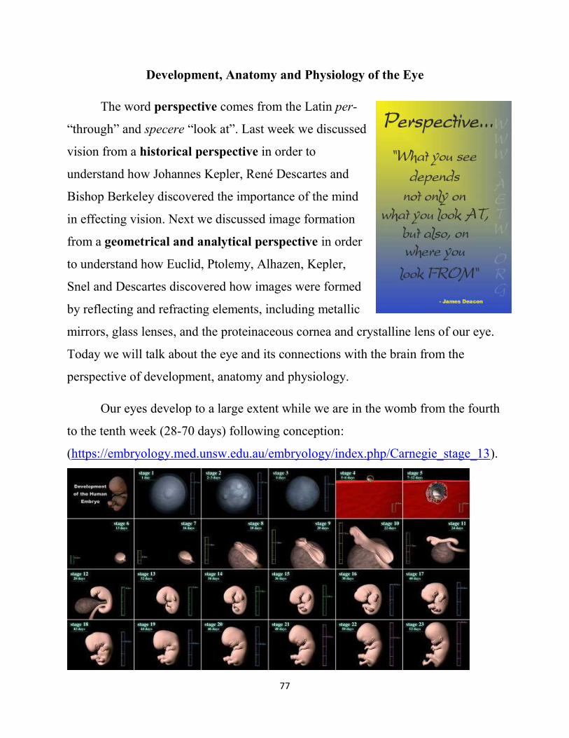
Our eyes develop to a large extent while we are in the womb from the fourth
to the tenth week (28-70 days) following conception:
(https://embryology.med.unsw.edu.au/embryology/index.php/Carnegie_stage_13).
78
Following conception, the fertilized egg divides to
form tissues that will connect the embryo to the mother,
yolk cells that will give rise to the germ cells, and stem
cells. The embryonic stem cells give rise to the three
embryonic tissues. Our eyes have their origin in these
three embryonic tissues: the lens and the cornea as well as
the optic nerve, the retina and the epithelial layers of the
iris and ciliary body are derived from the ectoderm, and the rest is derived from
the mesoderm.
Approximately four
weeks (28 days) after
conception, the forebrain,
which is derived from the
ectoderm and from which
the optic nerve, the retina,
and the epithelia of the
ciliary body develop,
pushes its way into the
surrounding loosely-associated cells known as the mesochyme, which is derived
from the mesoderm, to form the optic vesicles.
79
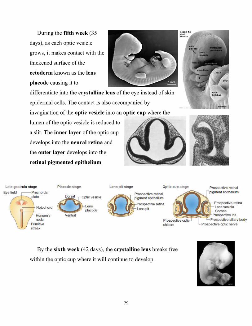
During the fifth week (35
days), as each optic vesicle
grows, it makes contact with the
thickened surface of the
ectoderm known as the lens
placode causing it to
differentiate into the crystalline lens of the eye instead of skin
epidermal cells. The contact is also accompanied by
invagination of the optic vesicle into an optic cup where the
lumen of the optic vesicle is reduced to
a slit. The inner layer of the optic cup
develops into the neural retina and
the outer layer develops into the
retinal pigmented epithelium.
By the sixth week (42 days), the crystalline lens breaks free
within the optic cup where it will continue to develop.
80
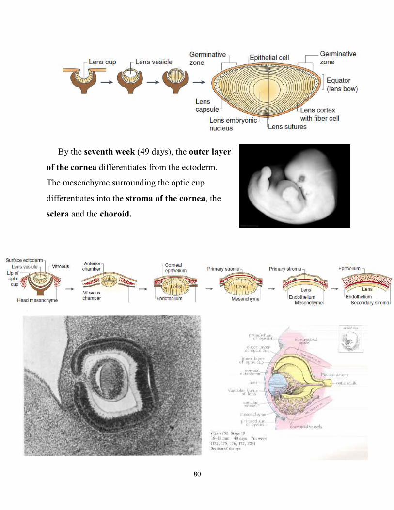
By the seventh week (49 days), the outer layer
of the cornea differentiates from the ectoderm.
The mesenchyme surrounding the optic cup
differentiates into the stroma of the cornea, the
sclera and the choroid.
81
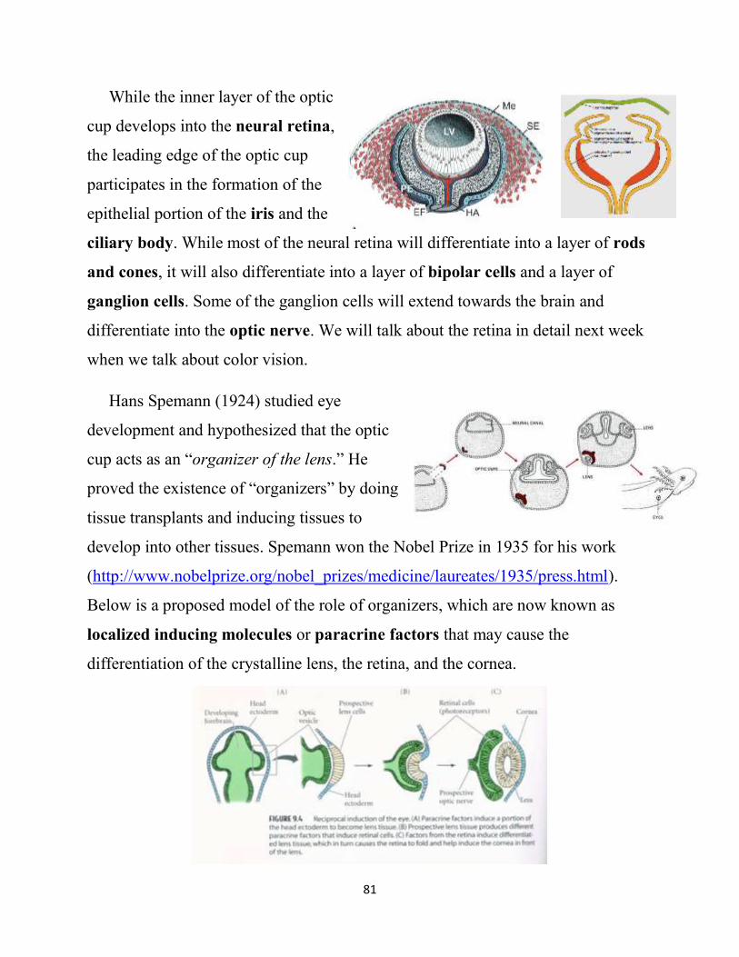
While the inner layer of the optic
cup develops into the neural retina,
the leading edge of the optic cup
participates in the formation of the
epithelial portion of the iris and the
ciliary body. While most of the neural retina will differentiate into a layer of rods
and cones, it will also differentiate into a layer of bipolar cells and a layer of
ganglion cells. Some of the ganglion cells will extend towards the brain and
differentiate into the optic nerve. We will talk about the retina in detail next week
when we talk about color vision.
Hans Spemann (1924) studied eye
development and hypothesized that the optic
cup acts as an “organizer of the lens.” He
proved the existence of “organizers” by doing
tissue transplants and inducing tissues to
develop into other tissues. Spemann won the Nobel Prize in 1935 for his work
(http://www.nobelprize.org/nobel_prizes/medicine/laureates/1935/press.html).
Below is a proposed model of the role of organizers, which are now known as
localized inducing molecules or paracrine factors that may cause the
differentiation of the crystalline lens, the retina, and the cornea.
82
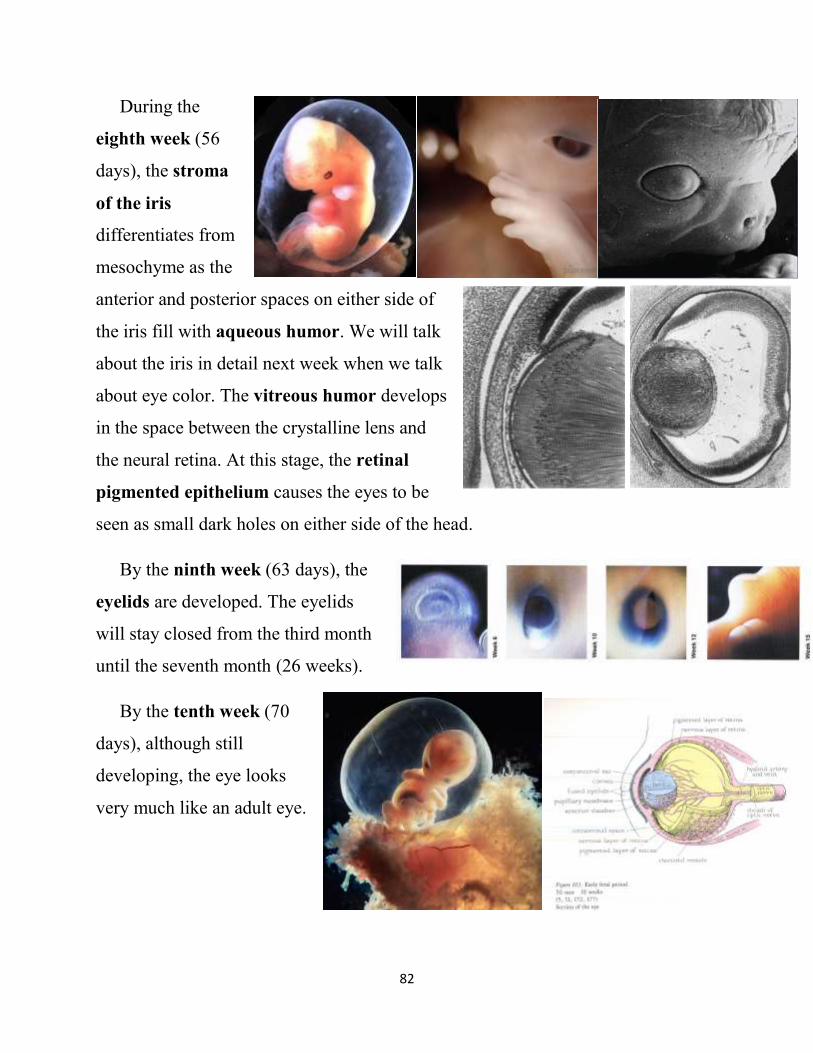
During the
eighth week (56
days), the stroma
of the iris
differentiates from
mesochyme as the
anterior and posterior spaces on either side of
the iris fill with aqueous humor. We will talk
about the iris in detail next week when we talk
about eye color. The vitreous humor develops
in the space between the crystalline lens and
the neural retina. At this stage, the retinal
pigmented epithelium causes the eyes to be
seen as small dark holes on either side of the head.
By the ninth week (63 days), the
eyelids are developed. The eyelids
will stay closed from the third month
until the seventh month (26 weeks).
By the tenth week (70
days), although still
developing, the eye looks
very much like an adult eye.
83
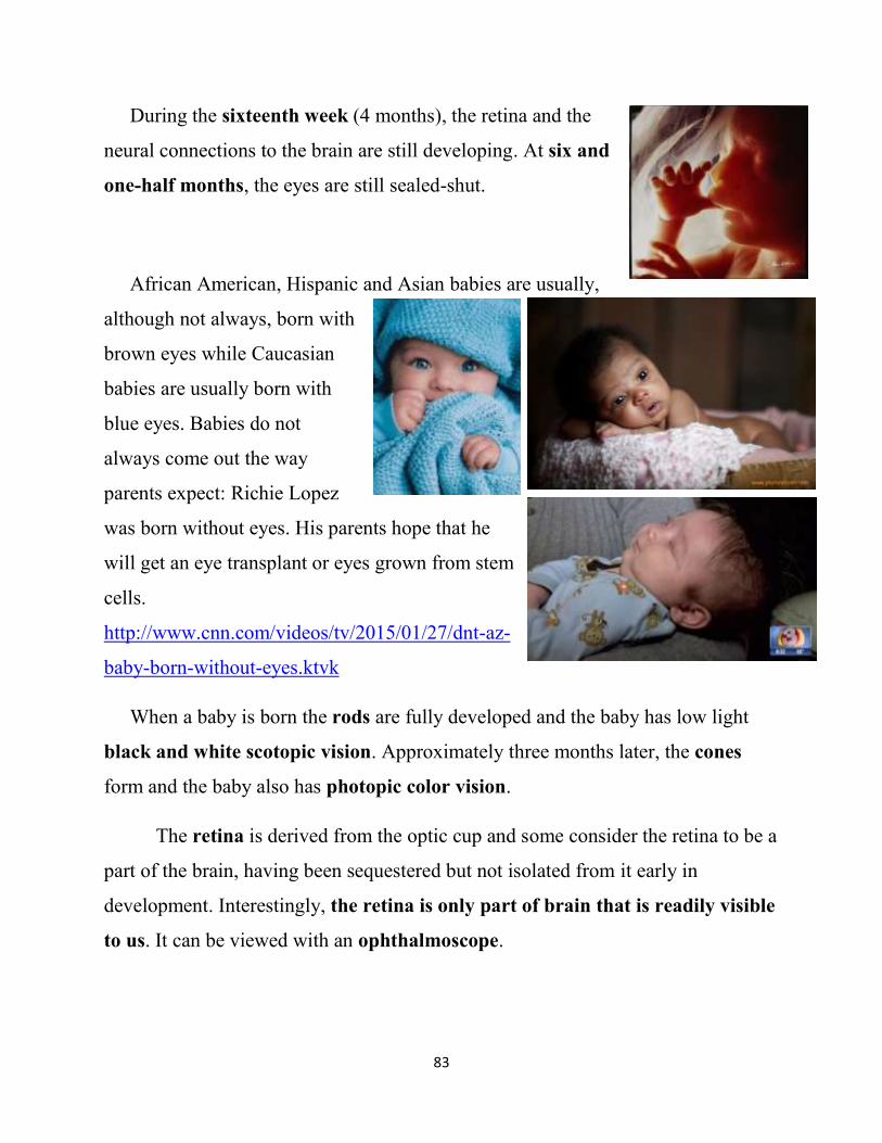
During the sixteenth week (4 months), the retina and the
neural connections to the brain are still developing. At six and
one-half months, the eyes are still sealed-shut.
African American, Hispanic and Asian babies are usually,
although not always, born with
brown eyes while Caucasian
babies are usually born with
blue eyes. Babies do not
always come out the way
parents expect: Richie Lopez
was born without eyes. His parents hope that he
will get an eye transplant or eyes grown from stem
cells.
http://www.cnn.com/videos/tv/2015/01/27/dnt-az-
baby-born-without-eyes.ktvk
When a baby is born the rods are fully developed and the baby has low light
black and white scotopic vision. Approximately three months later, the cones
form and the baby also has photopic color vision.
The retina is derived from the optic cup and some consider the retina to be a
part of the brain, having been sequestered but not isolated from it early in
development. Interestingly, the retina is only part of brain that is readily visible
to us. It can be viewed with an ophthalmoscope.
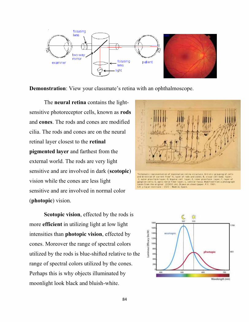
84
Demonstration: View your classmate’s retina with an ophthalmoscope.
The neural retina contains the light-
sensitive photoreceptor cells, known as rods
and cones. The rods and cones are modified
cilia. The rods and cones are on the neural
retinal layer closest to the retinal
pigmented layer and farthest from the
external world. The rods are very light
sensitive and are involved in dark (scotopic)
vision while the cones are less light
sensitive and are involved in normal color
(photopic) vision.
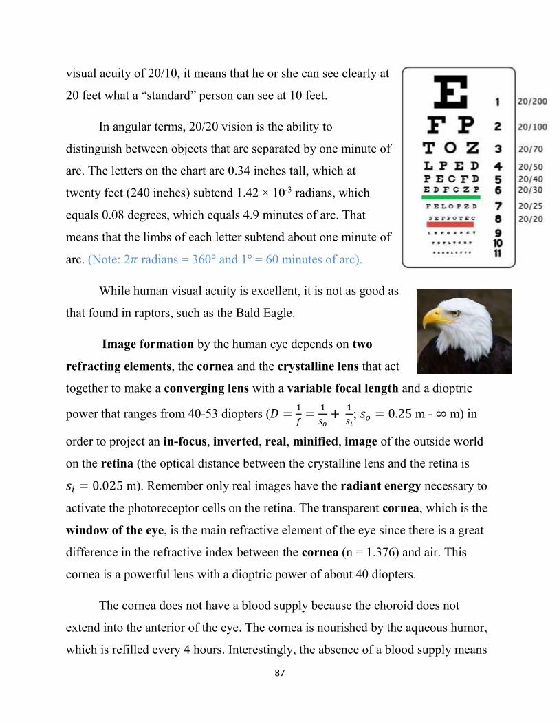
Scotopic vision, effected by the rods is
more efficient in utilizing light at low light
intensities than photopic vision, effected by
cones. Moreover the range of spectral colors
utilized by the rods is blue-shifted relative to the
range of spectral colors utilized by the cones.
Perhaps this is why objects illuminated by
moonlight look black and bluish-white.

85
Remember from the Pulfrich pendulum effect, that when we use our
scotopic vision, we see things “in the past.” Night lights in baseball stadiums
allow players to play with their photopic vision, so they see, and catch or hit the
ball “in the present.”
The rods and cones are connected to
bipolar cells which in turn are connected to
retinal ganglion cells. The retinal ganglion cells
are in the layer of the retina closest to the external
world. The axons of the retinal ganglia pass
through the retina at the optic disc to connect with
the optic nerve. Since two cells cannot be in the
same place at the same time, this precludes the
photoreceptor cells from being in the optic disc,
thus creating a blind spot on the nasal side of the
retina.
The rods, which are used for scotopic vision, are located around the
periphery of the retina. The most peripheral rods are capable of sensing motion but
are not able to produce an image of what is moving. You can tell this by having a
friend wave an object such as a fork or a spoon at the very edge of your visual
field near your ear. You will be able to tell something is moving, and in which
direction, but you will have no idea what is moving!
86
The cones that are involved in photopic color vision are enriched in the
center of the retina known as the macula, which is 2.5-3 mm in diameter. The
macula is a region of the retina that is rich in retinal ganglion cells as well as
cones. The fovea is in the center of the macula. Just as the optic disc excludes the
photoreceptor cells, the fovea is a depressed area of the retina, about 0.3 mm in
diameter, that excludes the bipolar cells and the retinal ganglion cells so that light
travels directly and unhindered through the rest of the neural retina layer to
the cones. Consequently, this region of the retina gives us the greatest visual
acuity.
Light is hindered from reaching the rods and cones outside the fovea by the
bipolar cells and retinal ganglion cells since the rods and cones outside the fovea
face the retinal pigmented layer instead of the outside world. The rods and cones
may face “backwards” so that old discs from the photoreceptor cells are sloughed
off to the back of the retina so that they do not accumulate in the vitreous humor.
It has been suggested that the melanin-containing cells are adjacent to the rods and
cones to help them chemically restore the light-sensitive visual pigment in the
receptors after it has been bleached by light.
Visual acuity is measured with a Snellen eye chart, developed by Hermann
Snellen in 1862. To measure visual acuity, a person stands 20 feet away from the
chart, covers one eye, and reads the letters starting at the top until they get to the
line where they can no longer make out the letters. The last line that they can
clearly read, gives their visual acuity. A “standard” person has a visual acuity of
20/20. Others have better or worse vision. If someone has a visual acuity of
20/200, it means that he or she can see as clearly at 20 feet as a “standard” person
can see at 200 feet. Someone with 20/200 vision is legally blind. If someone has a
87
visual acuity of 20/10, it means that he or she can see clearly at
20 feet what a “standard” person can see at 10 feet.
In angular terms, 20/20 vision is the ability to
distinguish between objects that are separated by one minute of
arc. The letters on the chart are 0.34 inches tall, which at
twenty feet (240 inches) subtend 1.42 × 10⁻³ radians, which
equals 0.08 degrees, which equals 4.9 minutes of arc. That
means that the limbs of each letter subtend about one minute of
arc. (Note: 2𝜋 radians = 360° and 1° = 60 minutes of arc).
While human visual acuity is excellent, it is not as good as
that found in raptors, such as the Bald Eagle.
Image formation by the human eye depends on two
refracting elements, the cornea and the crystalline lens that act
together to make a converging lens with a variable focal length and a dioptric
power that ranges from 40-53 diopters (𝐷 =1
𝑓=
1
𝑠𝑜+
1
𝑠𝑖; 𝑠𝑜 = 0.25 m - ∞ m) in
order to project an in-focus, inverted, real, minified, image of the outside world
on the retina (the optical distance between the crystalline lens and the retina is
𝑠𝑖 = 0.025 m). Remember only real images have the radiant energy necessary to
activate the photoreceptor cells on the retina. The transparent cornea, which is the
window of the eye, is the main refractive element of the eye since there is a great
difference in the refractive index between the cornea (n = 1.376) and air. This
cornea is a powerful lens with a dioptric power of about 40 diopters.
The cornea does not have a blood supply because the choroid does not
extend into the anterior of the eye. The cornea is nourished by the aqueous humor,
which is refilled every 4 hours. Interestingly, the absence of a blood supply means
88
that antibodies made by the immune system of the body do not reach the cornea.
The absence of any rejection response by the immune system has made corneal
transplants successful since as early as 1905.
The crystalline lens (n = 1.386-1.406) is the second refracting element of
the eye. When we are young, the crystalline lens can increase the dioptric power of
the eye by an additional 13 diopters, giving a total dioptric power of about 53
diopters. The reason that the dioptric power of the crystalline lens, with a refractive
index of 1.386-1.406, is less than the dioptric power of the cornea with a smaller
refractive index of 1.376, is that the crystalline lens is surrounded by the aqueous
and vitreous humors that have refractive indices 1.336,and 1.337, respectively.
The small difference in refractive index, like that of Pyrex glass in Wesson oil,
does not allow for much light bending or coarse focusing—only fine focusing.
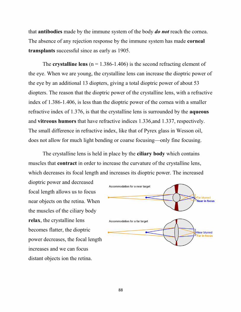
The crystalline lens is held in place by the ciliary body which contains
muscles that contract in order to increase the curvature of the crystalline lens,
which decreases its focal length and increases its dioptric power. The increased
dioptric power and decreased
focal length allows us to focus
near objects on the retina. When
the muscles of the ciliary body
relax, the crystalline lens
becomes flatter, the dioptric
power decreases, the focal length
increases and we can focus
distant objects ion the retina.
89
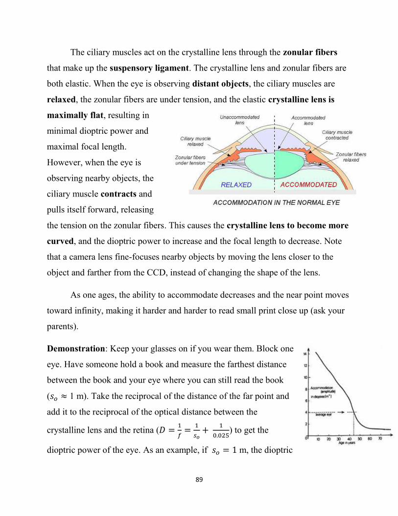
The ciliary muscles act on the crystalline lens through the zonular fibers
that make up the suspensory ligament. The crystalline lens and zonular fibers are
both elastic. When the eye is observing distant objects, the ciliary muscles are
relaxed, the zonular fibers are under tension, and the elastic crystalline lens is
maximally flat, resulting in
minimal dioptric power and
maximal focal length.
However, when the eye is
observing nearby objects, the
ciliary muscle contracts and
pulls itself forward, releasing
the tension on the zonular fibers. This causes the crystalline lens to become more
curved, and the dioptric power to increase and the focal length to decrease. Note
that a camera lens fine-focuses nearby objects by moving the lens closer to the
object and farther from the CCD, instead of changing the shape of the lens.
As one ages, the ability to accommodate decreases and the near point moves
toward infinity, making it harder and harder to read small print close up (ask your
parents).
Demonstration: Keep your glasses on if you wear them. Block one
eye. Have someone hold a book and measure the farthest distance
between the book and your eye where you can still read the book
(𝑠𝑜 ≈ 1 m). Take the reciprocal of the distance of the far point and
add it to the reciprocal of the optical distance between the
crystalline lens and the retina (𝐷 =1
𝑓=
1
𝑠𝑜+
1
0.025) to get the
dioptric power of the eye. As an example, if 𝑠𝑜 = 1 m, the dioptric
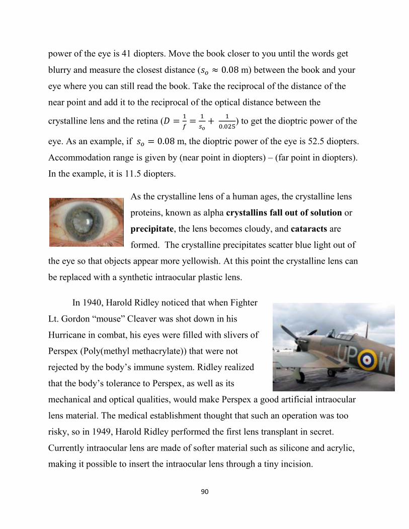
90
power of the eye is 41 diopters. Move the book closer to you until the words get
blurry and measure the closest distance (𝑠𝑜 ≈ 0.08 m) between the book and your
eye where you can still read the book. Take the reciprocal of the distance of the
near point and add it to the reciprocal of the optical distance between the
crystalline lens and the retina (𝐷 =1
𝑓=
1
𝑠𝑜+
1
0.025) to get the dioptric power of the
eye. As an example, if 𝑠𝑜 = 0.08 m, the dioptric power of the eye is 52.5 diopters.
Accommodation range is given by (near point in diopters) – (far point in diopters).
In the example, it is 11.5 diopters.
As the crystalline lens of a human ages, the crystalline lens
proteins, known as alpha crystallins fall out of solution or
precipitate, the lens becomes cloudy, and cataracts are
formed. The crystalline precipitates scatter blue light out of
the eye so that objects appear more yellowish. At this point the crystalline lens can
be replaced with a synthetic intraocular plastic lens.
In 1940, Harold Ridley noticed that when Fighter
Lt. Gordon “mouse” Cleaver was shot down in his
Hurricane in combat, his eyes were filled with slivers of
Perspex (Poly(methyl methacrylate)) that were not
rejected by the body’s immune system. Ridley realized
that the body’s tolerance to Perspex, as well as its
mechanical and optical qualities, would make Perspex a good artificial intraocular
lens material. The medical establishment thought that such an operation was too
risky, so in 1949, Harold Ridley performed the first lens transplant in secret.
Currently intraocular lens are made of softer material such as silicone and acrylic,
making it possible to insert the intraocular lens through a tiny incision.
91
The iris is the colored part of the eye and we will talk about its color next
week. The iris contains circularly-arranged sphincter and radially-arranged
dilator muscles. Contraction of the sphincter muscle, which is a striated muscle,
closes the pupil in response to stimulation by the parasympathetic nervous system,
which is active when the body is in the rest and digest state. Contraction of the
dilator muscle, which is a smooth muscle, opens the pupil in response to
stimulation by the sympathetic nervous system, which is active when the body is in
the fight or flight state.
Oxytocin, which is produced by the human body during sexual arousal, also
causes pupils to dilate. Eckhard Hess discovered the role of pupil size in
communicating attitude. The size of the pupil lets people know our emotional state.
We can mimic emotional states in terms of pupil size with drugs, such as
atropine (belladonna), hysocyamine, and scopolamine that cause dilation of the
pupils known as mydriasis by inhibiting the parasympatheitic nervous system.
Opiates mimic the rest and digest state by inhibiting the sympathetic nervous
system and causing an extreme contraction of the pupils known as miosis.
Typically, the adult human pupil is about 2-3 mm in diameter and children
have larger pupils than adults. The adult human pupil varies from 2 mm in bright
light to 8 mm in dim light.
Demonstration: Perform the flashlight test to see your classmate’s pupils contract
in bright light and dilate in dim light. Both pupils respond the same way, even if
you only illuminate one. This consensual response indicates that there is higher-
level control of pupil size. Also notice that the pupils constrict when your
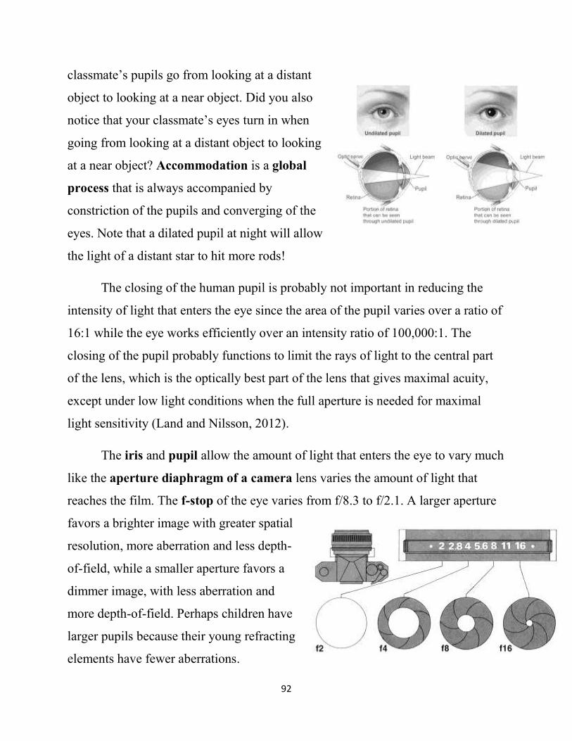
92
classmate’s pupils go from looking at a distant
object to looking at a near object. Did you also
notice that your classmate’s eyes turn in when
going from looking at a distant object to looking
at a near object? Accommodation is a global
process that is always accompanied by
constriction of the pupils and converging of the
eyes. Note that a dilated pupil at night will allow
the light of a distant star to hit more rods!
The closing of the human pupil is probably not important in reducing the
intensity of light that enters the eye since the area of the pupil varies over a ratio of
16:1 while the eye works efficiently over an intensity ratio of 100,000:1. The
closing of the pupil probably functions to limit the rays of light to the central part
of the lens, which is the optically best part of the lens that gives maximal acuity,
except under low light conditions when the full aperture is needed for maximal
light sensitivity (Land and Nilsson, 2012).
The iris and pupil allow the amount of light that enters the eye to vary much
like the aperture diaphragm of a camera lens varies the amount of light that
reaches the film. The f-stop of the eye varies from f/8.3 to f/2.1. A larger aperture
favors a brighter image with greater spatial
resolution, more aberration and less depth-
of-field, while a smaller aperture favors a
dimmer image, with less aberration and
more depth-of-field. Perhaps children have
larger pupils because their young refracting
elements have fewer aberrations.
93
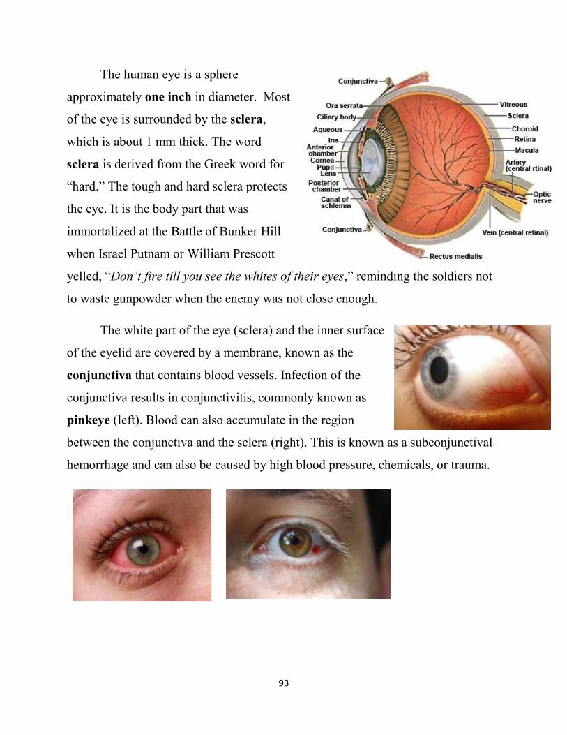
The human eye is a sphere
approximately one inch in diameter. Most
of the eye is surrounded by the sclera,
which is about 1 mm thick. The word
sclera is derived from the Greek word for
“hard.” The tough and hard sclera protects
the eye. It is the body part that was
immortalized at the Battle of Bunker Hill
when Israel Putnam or William Prescott
yelled, “Don’t fire till you see the whites of their eyes,” reminding the soldiers not
to waste gunpowder when the enemy was not close enough.
The white part of the eye (sclera) and the inner surface
of the eyelid are covered by a membrane, known as the
conjunctiva that contains blood vessels. Infection of the
conjunctiva results in conjunctivitis, commonly known as
pinkeye (left). Blood can also accumulate in the region
between the conjunctiva and the sclera (right). This is known as a subconjunctival
hemorrhage and can also be caused by high blood pressure, chemicals, or trauma.
94
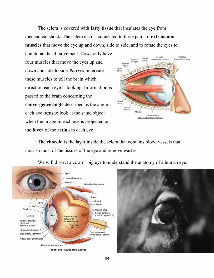
The sclera is covered with fatty tissue that insulates the eye from
mechanical shock. The sclera also is connected to three pairs of extraocular
muscles that move the eye up and down, side to side, and to rotate the eyes to
counteract head movement. Cows only have
four muscles that move the eyes up and
down and side to side. Nerves innervate
these muscles to tell the brain which
direction each eye is looking. Information is
passed to the brain concerning the
convergence angle described as the angle
each eye turns to look at the same object
when the image in each eye is projected on
the fovea of the retina in each eye.
The choroid is the layer inside the sclera that contains blood vessels that
nourish most of the tissues of the eye and remove wastes.
We will dissect a cow or pig eye to understand the anatomy of a human eye:
95
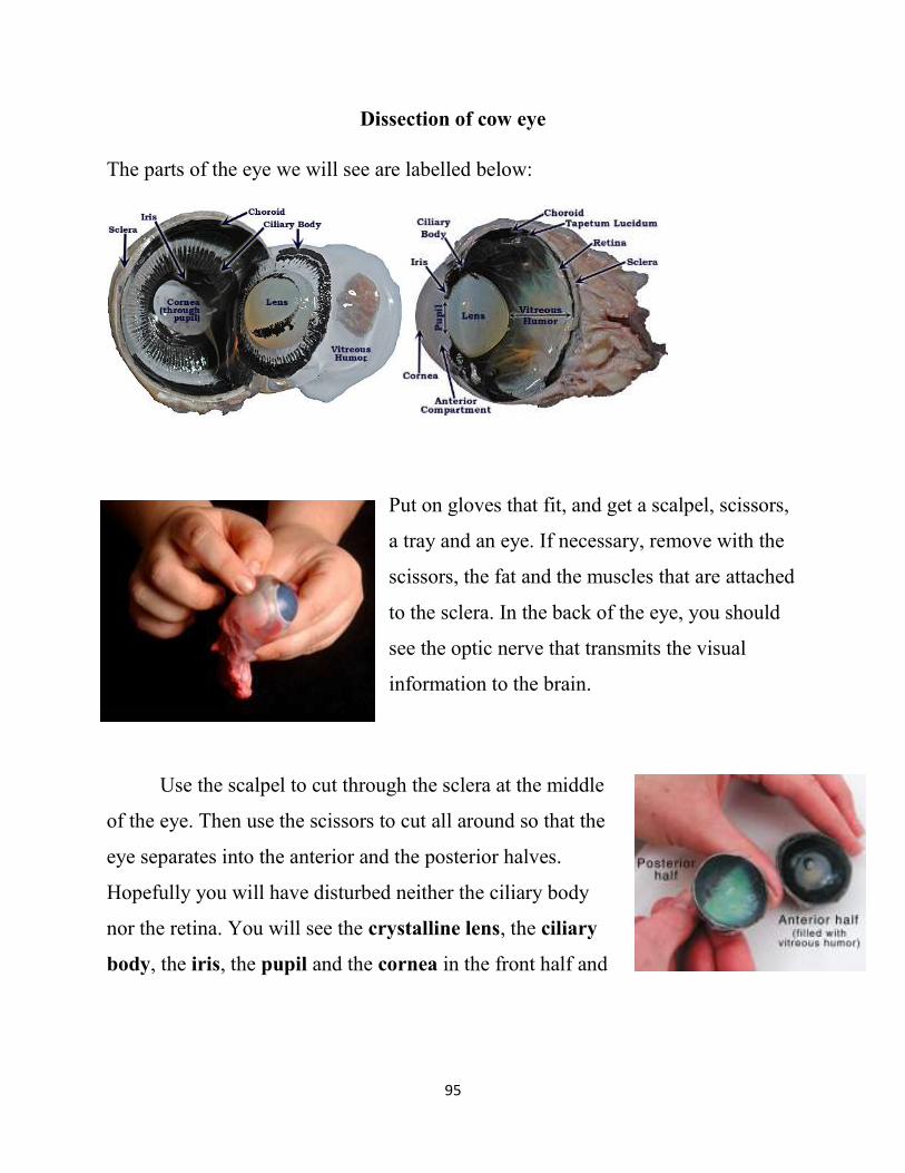
Dissection of cow eye
The parts of the eye we will see are labelled below:
Put on gloves that fit, and get a scalpel, scissors,
a tray and an eye. If necessary, remove with the
scissors, the fat and the muscles that are attached
to the sclera. In the back of the eye, you should
see the optic nerve that transmits the visual
information to the brain.
Use the scalpel to cut through the sclera at the middle
of the eye. Then use the scissors to cut all around so that the
eye separates into the anterior and the posterior halves.
Hopefully you will have disturbed neither the ciliary body
nor the retina. You will see the crystalline lens, the ciliary
body, the iris, the pupil and the cornea in the front half and
96
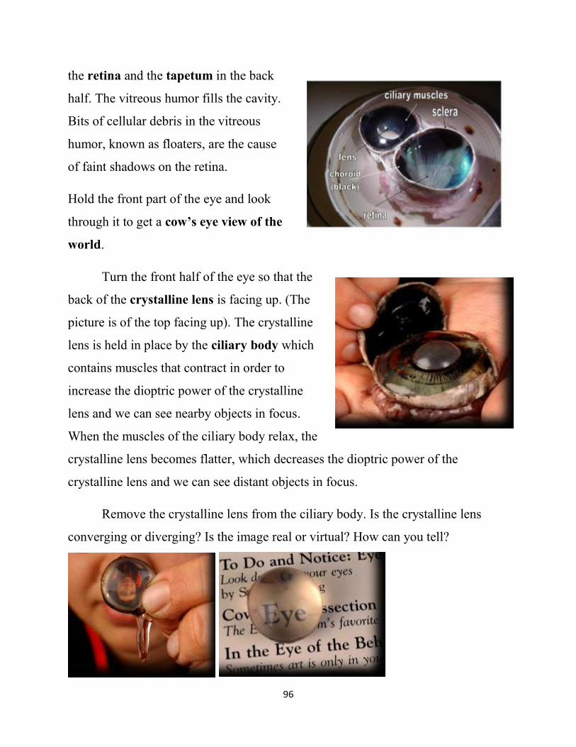
the retina and the tapetum in the back
half. The vitreous humor fills the cavity.
Bits of cellular debris in the vitreous
humor, known as floaters, are the cause
of faint shadows on the retina.
Hold the front part of the eye and look
through it to get a cow’s eye view of the
world.
Turn the front half of the eye so that the
back of the crystalline lens is facing up. (The
picture is of the top facing up). The crystalline
lens is held in place by the ciliary body which
contains muscles that contract in order to
increase the dioptric power of the crystalline
lens and we can see nearby objects in focus.
When the muscles of the ciliary body relax, the
crystalline lens becomes flatter, which decreases the dioptric power of the
crystalline lens and we can see distant objects in focus.
Remove the crystalline lens from the ciliary body. Is the crystalline lens
converging or diverging? Is the image real or virtual? How can you tell?
97
Look through the crystalline lens. The lens is a double convex converging
lens that produces an inverted, real image of an object that is more distant than its
focal length. The crystalline lens produces an erect, virtual image when the object
is closer than the focal point. The elastic properties of the crystalline lens cause it
to round up and take the form of an accommodated lens when it is separated from
the zonal fibers and the ciliary body. Feel how elastic the crystalline lens is.
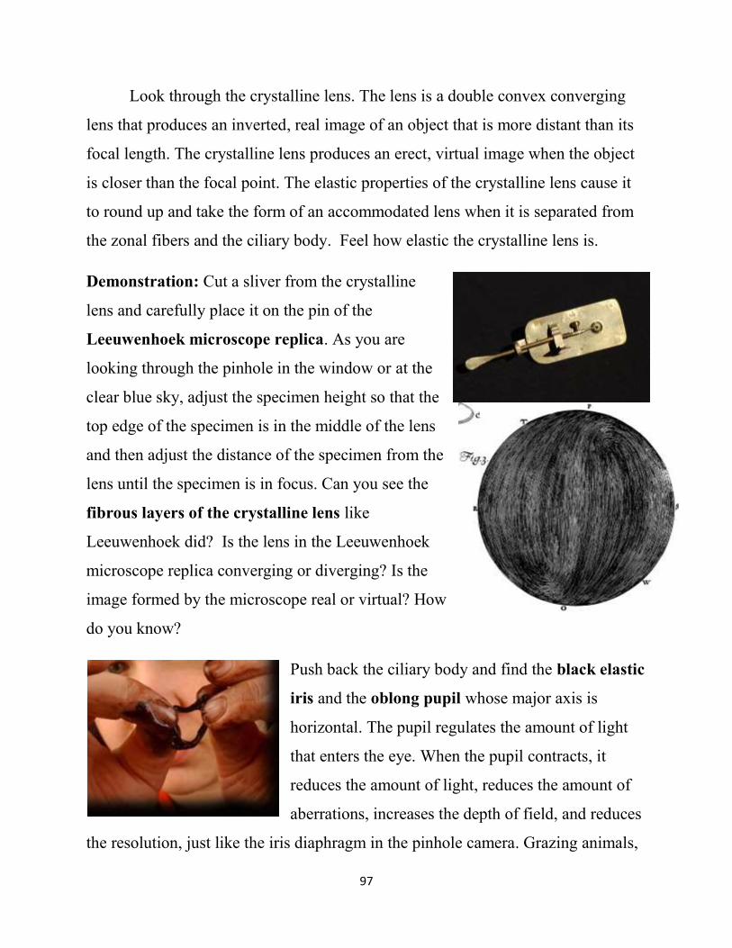
Demonstration: Cut a sliver from the crystalline
lens and carefully place it on the pin of the
Leeuwenhoek microscope replica. As you are
looking through the pinhole in the window or at the
clear blue sky, adjust the specimen height so that the
top edge of the specimen is in the middle of the lens
and then adjust the distance of the specimen from the
lens until the specimen is in focus. Can you see the
fibrous layers of the crystalline lens like
Leeuwenhoek did? Is the lens in the Leeuwenhoek
microscope replica converging or diverging? Is the
image formed by the microscope real or virtual? How
do you know?
Push back the ciliary body and find the black elastic
iris and the oblong pupil whose major axis is
horizontal. The pupil regulates the amount of light
that enters the eye. When the pupil contracts, it
reduces the amount of light, reduces the amount of
aberrations, increases the depth of field, and reduces
the resolution, just like the iris diaphragm in the pinhole camera. Grazing animals,
98
like the cow, tend to have horizontal pupils. Since the pupil of the cow is greater in
the horizontal direction, the cow can resolve horizontal details more sharply than it
can resolve vertical details. Humans, with a round iris, see details equally sharply,
independent of their orientation. Remove the iris and look at the cornea. The
cornea is relatively thin; neither delicate nor tough. Imagine doing Lasik surgery
on it!
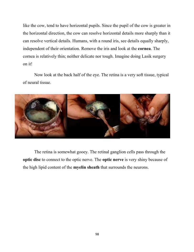
Now look at the back half of the eye. The retina is a very soft tissue, typical
of neural tissue.
The retina is somewhat gooey. The retinal ganglion cells pass through the
optic disc to connect to the optic nerve. The optic nerve is very shiny because of
the high lipid content of the myelin sheath that surrounds the neurons.
99
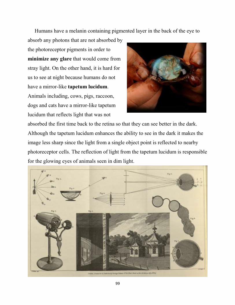
Humans have a melanin containing pigmented layer in the back of the eye to
absorb any photons that are not absorbed by
the photoreceptor pigments in order to
minimize any glare that would come from
stray light. On the other hand, it is hard for
us to see at night because humans do not
have a mirror-like tapetum lucidum.
Animals including, cows, pigs, raccoon,
dogs and cats have a mirror-like tapetum
lucidum that reflects light that was not
absorbed the first time back to the retina so that they can see better in the dark.
Although the tapetum lucidum enhances the ability to see in the dark it makes the
image less sharp since the light from a single object point is reflected to nearby
photoreceptor cells. The reflection of light from the tapetum lucidum is responsible
for the glowing eyes of animals seen in dim light.
100
The eye is a simple and elegant instrument. George Adams (1789, 1792), the
instrument maker to King George III, wrote in his An Essay on Vision, “In the
structure of the eye we find the most evident manifestations of exquisite art and
design, every part elegantly framed, nicely adjusted, and commodiously placed, to
answer in the most perfect manner every possible good purpose, and thus evince
that it is the work of unerring wisdom, prompted to action by infinite love. So
manifold are the blessings we derive from this organ….To it we are indebted for
that delightful sensations that arise from the proportion and variety of forms, the
harmonious mixture of colours, and the graces of beauty. It enables us to seek, to
see, and to chuse our food; to go here and there, as the calls of friendship, or the
occasions of business, require; to traverse the ocean, ransack the bowels of the
earth, visit distant regions, accumulate wealth, and multiply knowledge. Assisted
by it, we become acquainted with the works of the Creator, and can trace his
wisdom, his power, and his goodness, in the texture of plants, the mechanism of
animals, and the glories of the heavens. ”
Percy Shelley wrote in his Hymn of Apollo:
I am the eye with which the Universe
Beholds itself, and knows it is divine;
All harmony of instrument or verse,
All prophecy, all medicine, is mine,
All light of art or nature; - to my song
Victory and praise in its own right belong.