Embed Size (px)
Citation preview

Zurich Open Repository and Archive
University of ZurichMain LibraryWinterthurerstr. 190CH-8057 Zurichwww.zora.uzh.ch
Year: 2011
Duodenal ileus caused by a calf feeding nipple in a cow
Braun, U; Schnetzler, C; Previtali, M; Gerspach, C; Schmid, T
http://www.ncbi.nlm.nih.gov/pubmed/21211012.Postprint available at:http://www.zora.uzh.ch
Posted at the Zurich Open Repository and Archive, University of Zurich.http://www.zora.uzh.ch
Originally published at:Braun, U; Schnetzler, C; Previtali, M; Gerspach, C; Schmid, T (2011). Duodenal ileus caused by a calffeeding nipple in a cow. BMC Veterinary Research, 7(2):2.

Duodenal ileus caused by a calf feeding nipple in a cow
Abstract
Background: The aim of this report was to describe duodenal obstruction caused by a rubberforeign body in a cow. Case Presentation: The clinical, biochemical and ultrasonographicfindings in a five-year-old Swiss Braunvieh cow with duodenal ileus caused by a calf feedingnipple are described. The main clinical signs were anorexia, ruminal tympany, decreasedfaecal output and abomasal reflux syndrome. Ultrasonographic examination revealed reticularhyperactivity and a dilated duodenum. A diagnosis of duodenal ileus was made and the cowunderwent rightflank laparotomy, which revealed a dilation of the cranial part of theduodenum because of obstruction by a pliable foreign body. This was identified viaenterotomy as a calf feeding nipple. The cow was healthy at the time of discharge four daysafter surgery and went on to complete a successful lactation. Conclusions: To our knowledge,this is the first description of duodenal obstruction by a calf feeding nipple. This is aninteresting case, which broadens the spectrum of the causes of duodenal ileus, which isusually caused by obstruction of the duodenum by a phytobezoar.
CASE REPORT Open Access
Duodenal ileus caused by a calf feeding nipple ina cowUeli Braun*, Charlotte Schnetzler, Matteo Previtali, Christian Gerspach, Tanja Schmid
Abstract
Background: The aim of this report was to describe duodenal obstruction caused by a rubber foreign body in acow.
Case Presentation: The clinical, biochemical and ultrasonographic findings in a five-year-old Swiss Braunvieh cowwith duodenal ileus caused by a calf feeding nipple are described. The main clinical signs were anorexia, ruminaltympany, decreased faecal output and abomasal reflux syndrome. Ultrasonographic examination revealed reticularhyperactivity and a dilated duodenum. A diagnosis of duodenal ileus was made and the cow underwent right-flank laparotomy, which revealed a dilation of the cranial part of the duodenum because of obstruction by apliable foreign body. This was identified via enterotomy as a calf feeding nipple. The cow was healthy at the timeof discharge four days after surgery and went on to complete a successful lactation.
Conclusions: To our knowledge, this is the first description of duodenal obstruction by a calf feeding nipple. Thisis an interesting case, which broadens the spectrum of the causes of duodenal ileus, which is usually caused byobstruction of the duodenum by a phytobezoar.
BackgroundIleus of the duodenum results in abomasal reflux syn-drome, which is characterised by rapid deterioration ofthe condition and demeanour in cattle [1]. Phytobezoarsare the most common cause of obstruction of the duo-denum. They are formed when fibrinous adhesionsinvolving the reticulum impair the mechanism responsi-ble for sorting the ingesta, which allows poorly digestedfeed to move into the abomasum, omasum and duode-num, where it may cause an obstruction of the duode-num [1,2]. In rare instances, coagulated blood mayobstruct the duodenum [1,3-5], although this happensmore commonly in the jejunum [5]. Trichobezoars maycause duodenal obstruction in calves [6]. Other causesin mature cattle are obstruction of the duodenum bygravel [7], mechanical compression of the duodenum byan abscess in the liver or omentum, or lymphosarcoma[3]. Functional stenosis of the duodenum refers to ileusin which a mechanical cause cannot be identified [8].The duodenum may also be obstructed by pressurefrom the gravid uterus [9]. The aim of this report was
to describe duodenal obstruction caused by a rubberforeign body.
Case presentationA five-year-old Swiss Braunvieh cow was referred to theDepartment for Farm Animals, University of Zurich,because of a four-day history of anorexia and recentruminal tympany, which were unresponsive to variousmedical treatments. The cow was nine-weeks pregnantand had a moderately abnormal general condition. Theeyes were sunken and there was reduced skin turgor, acold muzzle and injected scleral vessels. The heart ratewas 68 beats per minute, the respiratory rate 24 breathsper min and the rectal temperature 38.6°C. The abdo-men was markedly distended. On rectal examination, anL-shaped distended rumen was palpated and there weresmall amounts of dark faeces that contained mucus.Ruminal contractions were reduced and normal layeringof the contents was absent at palpation. Swinging andpercussion auscultation of the abdomen were negative.The abdominal wall was tense and intestinal soundswere almost completely absent. Of the foreign bodytests, the withers pinch was positive and percussion ofthe reticulum and pole test were negative.
* Correspondence: [email protected] of Farm Animals, University of Zurich, Winterthurerstrasse 260,8057 Zurich, Switzerland
Braun et al. BMC Veterinary Research 2011, 7:2http://www.biomedcentral.com/1746-6148/7/2
© 2011 Braun et al; licensee BioMed Central Ltd. This is an Open Access article distributed under the terms of the Creative CommonsAttribution License (http://creativecommons.org/licenses/by/2.0), which permits unrestricted use, distribution, and reproduction inany medium, provided the original work is properly cited.
The results of macroscopic examination of a urinesample and a urine test strip were normal. The haemato-crit was increased at 45% (normal, 30 - 35%) and the con-centration of total solids was 104 g/l (normal, 60 - 80 g/l).Urea and creatinine concentrations were increased at18.6 mmol/l (normal, 2.4 - 6.5 mmol/l) and 320 μmol/l(normal, 55 - 103 μmol/l), respectively, and chloride,potassium and calcium concentrations were decreased at66 mmol/l (normal, 100 - 105 mmol/l), 3.1 mmol/l (nor-mal, 4.0 - 5.0 mmol/l) and 1.9 mmol/l (normal, 2.3 - 2.5mml/l), respectively. Inorganic phosphorus was increasedat 4.6 mmol/l (normal, 1.3 - 2.4 mmol/l). The chlorideconcentration of ruminal juice was markedly increased at78 mmol/l (normal, 15 - 30 mmol/l). Blood gas analysisrevealed compensated metabolic alkalosis with a basedeficit of +20.3 mmol/l (normal, -2 to +2 mmol/l), abicarbonate concentration of 43.9 mmol/l (normal 20 -30 mmol/l) and a pH of 7.45 (normal, 7.40 - 7.50).On ultrasonography, the reticulum had six biphasic
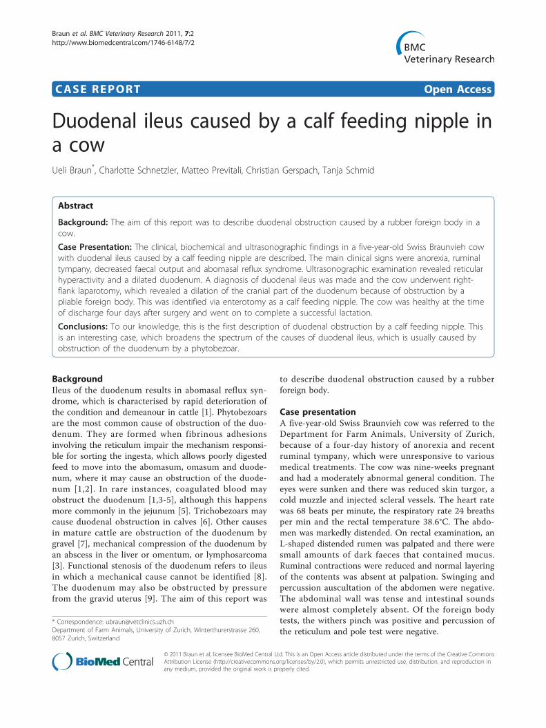
contractions per 3 minutes (normal, 3 - 4 contractions/3minutes) [10] and its contour was normal. The rumen,omasum and abomasum were severely dilated; therumen was in close proximity to the right abdominalwall and the abomasum extended to a point 10 cm cra-nial to the right stifle. The cranial part of the duodenumhad no contractions and was dilated with a diameter of7.9 cm (Figure 1). The remainder of the intestinal tractwas empty and atonic.Based on all the findings, a diagnosis of duodenal ileus
was made. A standing right-flank laparotomy was car-ried out under proximal paravertebral anaesthesia. A 25-cm incision was made 5 cm caudal and parallel to the
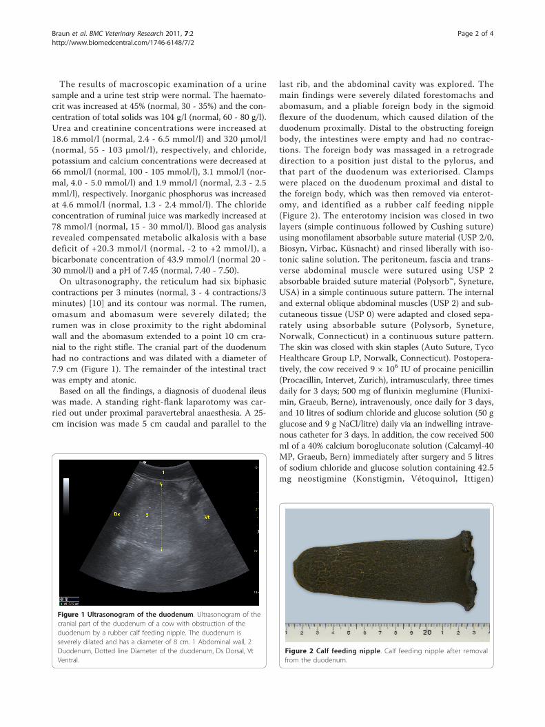
last rib, and the abdominal cavity was explored. Themain findings were severely dilated forestomachs andabomasum, and a pliable foreign body in the sigmoidflexure of the duodenum, which caused dilation of theduodenum proximally. Distal to the obstructing foreignbody, the intestines were empty and had no contrac-tions. The foreign body was massaged in a retrogradedirection to a position just distal to the pylorus, andthat part of the duodenum was exteriorised. Clampswere placed on the duodenum proximal and distal tothe foreign body, which was then removed via enterot-omy, and identified as a rubber calf feeding nipple(Figure 2). The enterotomy incision was closed in twolayers (simple continuous followed by Cushing suture)using monofilament absorbable suture material (USP 2/0,Biosyn, Virbac, Küsnacht) and rinsed liberally with iso-tonic saline solution. The peritoneum, fascia and trans-verse abdominal muscle were sutured using USP 2absorbable braided suture material (Polysorb™, Syneture,USA) in a simple continuous suture pattern. The internaland external oblique abdominal muscles (USP 2) and sub-cutaneous tissue (USP 0) were adapted and closed sepa-rately using absorbable suture (Polysorb, Syneture,Norwalk, Connecticut) in a continuous suture pattern.The skin was closed with skin staples (Auto Suture, TycoHealthcare Group LP, Norwalk, Connecticut). Postopera-tively, the cow received 9 × 10⁶ IU of procaine penicillin(Procacillin, Intervet, Zurich), intramuscularly, three timesdaily for 3 days; 500 mg of flunixin meglumine (Flunixi-min, Graeub, Berne), intravenously, once daily for 3 days,and 10 litres of sodium chloride and glucose solution (50 gglucose and 9 g NaCl/litre) daily via an indwelling intrave-nous catheter for 3 days. In addition, the cow received 500ml of a 40% calcium borogluconate solution (Calcamyl-40MP, Graeub, Bern) immediately after surgery and 5 litresof sodium chloride and glucose solution containing 42.5mg neostigmine (Konstigmin, Vétoquinol, Ittigen)
Figure 1 Ultrasonogram of the duodenum. Ultrasonogram of thecranial part of the duodenum of a cow with obstruction of theduodenum by a rubber calf feeding nipple. The duodenum isseverely dilated and has a diameter of 8 cm. 1 Abdominal wall, 2Duodenum, Dotted line Diameter of the duodenum, Ds Dorsal, VtVentral.
Figure 2 Calf feeding nipple. Calf feeding nipple after removalfrom the duodenum.
Braun et al. BMC Veterinary Research 2011, 7:2http://www.biomedcentral.com/1746-6148/7/2
Page 2 of 4
administered at a flow rate of 0.0015 mg neostigmine/kg/hour during the following 48 hours via the indwellingcatheter.One day after surgery, the demeanour of the cow had
improved, and she started eating small amounts of feedand passing olive-coloured faeces with a soft pulpy con-sistency. The general condition, appetite and faecal out-put were normal on the following day. The cow wasdischarged four days postoperatively and the skin stapleswere removed ten days after the surgery. A follow-upvia telephone seven months later revealed that the cowhad recovered completely and produced more than10,000 kg of milk during the current lactation.The clinical signs of the cow described in this report
were typical of duodenal ileus [1,2]. They included rapiddeterioration of the general condition, an enlargedL-shaped rumen and markedly diminished faecal output,but no signs of colic and an absence of dilated intestinalloops on transrectal palpation. The striking ruminal find-ings can be misinterpreted as an underlying ruminal pro-blem rather than an intestinal disorder. Laboratoryfindings indicating abomasal reflux are typically seen withimpaired passage of ingesta in the abomasum or proximalsmall intestine and have been described in cows with duo-denal ileus of varying causes [1,3,4]. Similar laboratoryfindings are also seen in cows with abomasal displacementand pyloric stenosis [11]. Haemoconcentration, azotaemia,hypochloraemia and hypokalaemia are considered to bethe main cause of rapid deterioration in the general condi-tion of cattle with duodenal ileus.Ultrasonography allowed localisation of the ileus to
the duodenum and differentiation of the problem frompyloric and jenunal stenosis. With the former, the abo-masum is dilated and the small intestines are empty,whereas with the latter multiple dilated loops of smallintestine with a diameter of at least 4 cm are typicallyseen [12]. One to three dilated intestinal loops in thecranial abdomen are typical of duodenal ileus [13,14].The normal diameter of the duodenum is 0.9 to 5.5 cm[15], whereas in cows with duodenal ileus, diametersmay range from 6.5 to 9.9 cm [13]. Of special interestwere the reticular findings because six biphasic contrac-tions per three minutes indicated reticular hypercontrac-tility. Similar findings have been described in cows withreticulo-omasal stenosis [16] and vagal indigestion [17].Treatment of duodenal ileus consists of surgical removal
of the inciting cause via right-flank laparotomy, combinedwith the administration of sodium chloride, glucose, cal-cium, potassium, non-steroidal antiinflammatory drugsand antibiotics [1,3]. In uncomplicated cases, the obstruct-ing duodenal content may be manually disintegrated andmoved distally. However, this is not usually feasible and anenterotomy is required in most cases. In the present case,a rubber foreign body, rather than a bezoar or a blood
clot, was immediately suspected after palpation of the duo-denum. It would be interesting to know whether the nip-ple was ingested during the nursing period or whether itwas accidentally eaten by the cow later.
ConclusionDuodenal ileus is usually caused by obstruction of theduodenum by a phytobezoar. In rare instances, coagu-lated blood may obstruct the duodenum or the duode-num is mechanically compressed by an abscess or aneoplasia. To our knowledge, this is the first descriptionof duodenal obstruction by a calf feeding nipple. This isan interesting case, which broadens the spectrum of thecauses of duodenal ileus.
Authors’ contributionsUB supervised the clinical and ultrasonographic examination, reviewed theliterature and prepared the manuscript. CS and MP carried out the clinicalexamination. CG performed the ultrasonographic examination. TS performedthe laparotomy. All authors read and approved the final manuscript.
Competing interestsThe authors declare that they have no competing interests.
Received: 25 October 2010 Accepted: 6 January 2011Published: 6 January 2011
References1. Braun U, Steiner A, Götz M: Clinical signs, diagnosis and treatment of
duodenal ileus in cattle. Schweiz Arch Tierheilk 1993, 135:345-355.2. Garry F, Hull BL, Rings DM, Hoffsis G: Comparison of naturally occurring
proximal duodenal obstruction and abomasal volvulus in dairy cattle.Vet Surg 1988, 17:226-233.
3. Anderson DE, Ivany Ewoldt JM: Intestinal surgery of adult cattle. Vet ClinNorth Am (Food Anim Pract) 2005, 21:133-154.
4. Lejeune B, Lorenz I: Ultrasonographic findings in 2 cows with duodenalobstruction. Can Vet J 2008, 49:386-388.
5. Braun U, Forster E, Steininger K, Irmer M, Gautschi A, Previtali M,Gerspach C, Nuss K: Ultrasonographic findings in 63 cows withhaemorrhagic bowel syndrome. Vet Rec 2010, 166:79-81.
6. Anderson DE: Surgical diseases of the small intestine. Vet Clin North Am(Food Anim Pract) 2008, 24:383-401.
7. Cebra CK, Garry FB: Gravel obstruction in the abomasum or duodenumof two cows. J Am Vet Med Assoc 209:1294-1296.
8. Van der Velden MA: Functional stenosis of the sigmoid curve of theduodenum in cattle. Vet Rec 1983, 112:452-453.
9. Koller U, Lischer C, Geyer H, Dressel C, Braun U: Strangulation of theduodenum by the uterus during late pregnancy in two cows. Vet J 2001,162:33-37.
10. Braun U, Rauch S: Ultrasonographic evaluation of reticular motility duringrest, eating, rumination and stress in 30 healthy cows. Vet Rec 2008,163:571-574.
11. Radostits OM, Gay CC, Hinchcliff KW, Constable PD: Diseases of thealimentary tract - II. Veterinary Medicine. A Textbook of the Diseases ofCattle, Horses, Sheep, Pigs, and Goats Philadelphia, Saunders Elsevier; 2007,293-382.
12. Braun U: Ultrasonography in gastrointestinal disease in cattle. Vet J 2003,166:112-124.
13. Braun U, Marmier O, Pusterla N: Ultrasonographic examination of thesmall intestine of cows with ileus of the duodenum, jejunum or ileum.Vet Rec 1995, 137:209-215.
14. Braun U: Ultrasonography of the gastrointestinal tract in cattle. Vet ClinNorth Am (Food Anim Pract) 2009, 25:567-590.
15. Braun U, Marmier O: Ultrasonographic examination of the small intestineof cows. Vet Rec 1995, 136:239-244.
Braun et al. BMC Veterinary Research 2011, 7:2http://www.biomedcentral.com/1746-6148/7/2
Page 3 of 4

16. Braun U, Schweizer G, Flückiger M: Radiographic and ultrasonogra-phicfindings in three cows with reticulo-omasal obstruction due to a foreignbody. Vet Rec 2002, 150:580-581.
17. Braun U, Rauch S, Hässig M: Ultrasonographic evaluation of reticularmotility in 144 cattle with vagal indigestion. Vet Rec 2009, 164:11-13.
doi:10.1186/1746-6148-7-2Cite this article as: Braun et al.: Duodenal ileus caused by a calf feedingnipple in a cow. BMC Veterinary Research 2011 7:2.
Submit your next manuscript to BioMed Centraland take full advantage of:
• Convenient online submission
• Thorough peer review
• No space constraints or color figure charges
• Immediate publication on acceptance
• Inclusion in PubMed, CAS, Scopus and Google Scholar
• Research which is freely available for redistribution
Submit your manuscript at www.biomedcentral.com/submit
Braun et al. BMC Veterinary Research 2011, 7:2http://www.biomedcentral.com/1746-6148/7/2
Page 4 of 4