Embed Size (px)
Citation preview
Listen to this manuscript’s
audio summary by
JACC Editor-in-Chief
Dr. Valentin Fuster.
J O U R N A L O F T H E A M E R I C A N C O L L E G E O F C A R D I O L O G Y VO L . 7 2 , N O . 2 2 , 2 0 1 8
ª 2 0 1 8 T H E A U T H O R S . P U B L I S H E D B Y E L S E V I E R O N B E H A L F O F T H E AM E R I C A N
C O L L E G E O F C A R D I O L O G Y F O U N DA T I O N . T H I S I S A N O P E N A C C E S S A R T I C L E U N D E R
T H E C C B Y - N C - N D L I C E N S E ( h t t p : / / c r e a t i v e c o mm o n s . o r g / l i c e n s e s / b y - n c - n d / 4 . 0 / ) .
JACC REVIEW TOPIC OF THE WEEK
Echocardiographic Screening forPulmonary Hypertension inCongenital Heart DiseaseJACC Review Topic of the Week
Konstantinos Dimopoulos, MD, MSC, PHD,a Robin Condliffe, MD,b Robert M.R. Tulloh, MA, DM,c Paul Clift, MD,d
Rafael Alonso-Gonzalez, MD, MSC,a Radwa Bedair, MBBCH, MD,c Natali A.Y. Chung, MD,e Gerry Coghlan, MD,f
Samantha Fitzsimmons, MBCHB, BSC,g Alessandra Frigiola, MD, MD (RES),e Luke S. Howard, MA, DPHIL,h
Petra Jenkins, MBCHB,i Damien Kenny, MD,j Wei Li, MD, PHD,a Simon T. MacDonald, BMBCH, DPHIL,k
Colm McCabe, MD,l James J. Oliver, MBCHB, PHD,m Mark S. Spence, MD, MBBCH,n Gergely V. Szantho, MD,o
Kate von Klemperer, MBBCH,p Dirk G. Wilson, BSC, MBBCH,o Stephen J. Wort, MA, MBBS, PHD,l
on behalf of the CHAMPION Steering Committee
ABSTRACT
ISS
Fro
LocBr
Ho
Lo
Co
ten
He
pit
Ho
Kin
VicpG
ma
Oli
Dim
an
na
Ph
Echocardiography is the mainstay in screening for pulmonary hypertension (PH). International guidelines suggest
echocardiographic parameters for suspecting PH, but these may not apply to many adults with congenital heart disease
(ACHD). PH is relatively common in ACHD patients and can significantly affect their exercise capacity, quality of life, and
prognosis. Identification of patients who have developed PH and who may benefit from further investigations (including
cardiac catheterization) and treatment is thus extremely important. A systematic review and survey of experts from the
United Kingdom and Ireland were performed to assess current knowledge and practice on echocardiographic screening
for PH in ACHD. This paper presents the findings of the review and expert statements on the optimal approaches when
using echocardiography to assess ACHD patients for PH, with particular focus on major subgroups: patients with right
ventricular outflow tract obstruction, patients with systemic right ventricles, patients with unrepaired univentricular
circulation, and patients with tetralogy of Fallot with pulmonary atresia. (J Am Coll Cardiol 2018;72:2778–88)
© 2018 The Authors. Published by Elsevier on behalf of the American College of Cardiology Foundation. This is an open
access article under the CC BY-NC-ND license (http://creativecommons.org/licenses/by-nc-nd/4.0/).
N 0735-1097 https://doi.org/10.1016/j.jacc.2018.08.2201
m the aAdult Congenital Heart Centre and Centre for Pulmonary Hypertension, Royal Brompton Hospital and Imperial College
ndon, London, United Kingdom; bPulmonary Vascular Disease Unit, Royal Hallamshire Hospital, Sheffield, United Kingdom;
istol Heart Institute, University Hospitals Bristol, Bristol, United Kingdom; dDepartment of Cardiology, Queen Elizabeth
spital Birmingham, Birmingham, United Kingdom; eAdult Congenital Heart Disease Service, Guy’s and St Thomas’ Hospital,
ndon, United Kingdom; fNational Pulmonary Hypertension Service, Royal Free Hospital, London, United Kingdom; gAdult
ngenital Heart Disease Unit, Southampton University Hospital, Southampton, United Kingdom; hNational Pulmonary Hyper-
sion Service, Hammersmith Hospital, Imperial College Healthcare NHS Trust, London, United Kingdom; iAdult Congenital
art Disease Unit, Manchester Royal Infirmary, Manchester, United Kingdom; jOur Lady’s Children’s Hospital and Mater Hos-
al, Dublin, Ireland; kEast Midlands Congenital Heart Centre, Leicester Cardiovascular Biomedical Research Unit, Glenfield
spital, Leicester, United Kingdom; lDepartment of Pulmonary Hypertension, Royal Brompton Hospital, London, United
gdom; mLeeds Congenital Heart Unit, Leeds Teaching Hospitals, Leeds, United Kingdom; nDepartment of Cardiology, Royal
toria Hospital, Belfast, Northern Ireland; oCardiology Department, University Hospital of Wales, Cardiff, United Kingdom; and
rown-up Congenital Heart Disease Service, Barts Heart Centre, St. Bartholomew’s Hospital, London, United Kingdom. Both the
nuscript and survey were funded by Actelion Pharmaceuticals UK Limited. Drs. Chung, Fitzsimmons, Frigiola, Kenny, McCabe,
ver, Spence, Szantho, Von Klemperer, Wilson, and Li have received nonfinancial support from Actelion Pharmaceuticals. Dr.
opoulos has received nonfinancial support from Actelion Pharmaceuticals; and has been a consultant to and received grants
d personal fees from Actelion Pharmaceuticals, Pfizer, GlaxoSmithKline, and Bayer/MSD. Dr. Condliffe has received nonfi-
ncial support from Actelion Pharmaceuticals during the conduct of the study; and has received personal fees from Actelion
armaceuticals, Bayer, and GlaxoSmithKline. Dr. Tulloh has received nonfinancial support from Actelion Pharmaceuticals;
J A C C V O L . 7 2 , N O . 2 2 , 2 0 1 8 Dimopoulos et al.D E C E M B E R 4 , 2 0 1 8 : 2 7 7 8 – 8 8 Echocardiographic Screening for PAH-CHD
2779
AB BR E V I A T I O N S
AND ACRONYM S
ACHD = adult congenital heart
disease
CHD = congenital heart disease
PA = pulmonary arterial
PAH = pulmonary arterial
hypertension
PH = pulmonary hypertension
PH-CHD = pulmonary
hypertension associated with
congenital heart disease
PR = pulmonary regurgitation
PVR = pulmonary vascular
resistance
RV = right ventricle
RVOTO = right ventricular
outflow tract obstruction
tricuspid regurgitation
P ulmonary hypertension (PH) is defined asa mean pulmonary arterial (PA) pressure$25 mm Hg (1,2) (Figure 1). PH is not uncom-
mon in adults with congenital heart disease (ACHD)and significantly affects morbidity and mortality(1,2). ACHD patients with PH have limited exercisecapacity, which results in a decreased quality of life(3–5). Moreover, PH often contributes to the develop-ment of congestive heart failure and multiorgan fail-ure, increasing the risk for hospitalization andpremature death (6,7).
Many patients with congential heart disease (CHD)associated with pulmonary arterial hypertension(PAH) benefit from the introduction of PAH-specifictherapies (8–10). Although the evidence is still fairlylimited, the use of such therapies was established forpatients with Eisenmenger syndrome after theBREATHE-5 (Effects of Tracleer [Bosentan] on Pul-monary Arterial Hypertension Related to Eisen-menger Physiology; NCT00317486) trial, whichdemonstrated a significant improvement in exercisecapacity and functional class using the endothelinreceptor antagonist bosentan (8,10). Smaller studieshave supported the use of other PAH-specific thera-pies (including phosphodiesterase-type 5 inhibitorsand prostanoids) in patients with Eisenmenger syn-drome, whereas patients after defect correction havebeen included in larger randomized trials, togetherwith patients with idiopathic PAH and connectivetissue disease (2,11). Thorough screening of all ACHDis thus crucial in identifying patients who havedeveloped PAH and who could benefit from furtherinvestigations, including cardiac catheterization andinitiation of PAH-specific therapies.
There are currently no detailed guidelines on howto use echocardiography to screen patients withACHD for the presence of PAH, especially those withunrepaired or residual defects, and those with com-plex anatomy. Although international guidelinesprovide a diagnostic algorithm and a list of supportive
and has received personal fees from Actelion Pharmaceuticals, Pfizer, Abbott
has received nonfinancial support from Actelion Pharmaceuticals; has recei
ceuticals; and has received personal fees from Bayer. Dr. Alonso-Gonzalez ha
and GlaxoSmithKline Europe; and has received education grants from Ac
received grants from Actelion. Dr. Coghlan has received nonfinancial suppo
grants and personal fees from Actelion Pharmaceuticals, Ltd.; has received
has received grants from Merck Sharp & Dohme. Dr. Howard has received n
Ltd.; has received grants, personal fees, and nonfinancial support from Bay
support from GlaxoSmithKline and Merck; and has received personal fees fro
support and conference attendance support from Actelion Pharmaceuticals. D
personal fees from Actelion Pharmaceuticals. Dr. Wort has received nonfin
received grants and personal fees from Actelion Pharmaceuticals an
GlaxoSmithKline.
Manuscript received March 14, 2018; revised manuscript received July 26, 2
echocardiographic signs for all types of PH(12), there are anatomic and physiologicalconsiderations that are specific to CHD andthus require additional expertise (Table 1)(12). For example, the estimation of theprobability of PH in symptomatic patientsbased on tricuspid regurgitation (TR) veloc-ity, as suggested in the European Society ofCardiology/European Respiratory Societyguidelines (12), does not apply to patientswith any degree of right ventricular outflowtract obstruction (RVOTO) and/or pulmonarystenosis, or patients with single-ventriclephysiology. The same may be true for otherechocardiographic signs described in theguidelines that are supportive of the presenceof PH (12). For example, flattening of theventricular septum is also present in patientswith pulmonary stenosis or regurgitation,
and a pulmonary artery larger than the aorta can befound with intracardiac shunts.We present an expert statement on the screeningfor PH in ACHD based on the findings of a survey ofexperts and a systematic review of availableevidence.
METHODS
The CHAMPION (Congenital Heart disease And pul-Monary arterial hyPertension: Improving Outcomesthrough education and research Networks) SteeringCommittee (a panel of 4 experts in PH and CHD)identified gaps in published studies, with a particularfocus on types of ACHD in which standard echocar-diographic markers of PH might not apply. A survey ofexperts in CHD and PH was then designed to deter-mine how physicians rate the importance of differentechocardiographic parameters for raising the suspi-cion of PH in various clinical scenarios (OnlineAppendix). A systematic review of all published
TR =
International, GlaxoSmithKline, and Bayer. Dr. Clift
ved grants and personal fees from Actelion Pharma-
s acted as consultant for Actelion Spain, Pfizer Spain,
telion UK and GlaxoSmithKline UK. Dr. Bedair has
rt from Actelion Pharmaceuticals, Ltd.; has received
personal fees from GlaxoSmithKline and Bayer; and
onfinancial support from Actelion Pharmaceuticals,
er PLC; has received personal fees and nonfinancial
m Endotronix. Dr. Jenkins has received nonfinancial
r. MacDonald has received nonfinancial support and
ancial support from Actelion Pharmaceuticals; has
d Bayer; and has received personal fees from
018, accepted August 14, 2018.

FIGURE 1 CHD Within the PH Clinical Classification Groups
LAP* ≤15 mm Hg LAP* >15 mm Hg
mPAP ≥25 mm Hg
• Eisenmenger syndrome• PAH-CHD with L-R shunt• PAH with small defects• PAH after CHD correction
PAH:
PRECAPILLARY POSTCAPILLARY
• Systemic ventricular dysfunction (systolic or diastolic)• Congenital cardiomyopathies• Valve disease• Congenital left heart inflow or outflow tract obstruction• Pulmonary vein stenosis
Left heart disease:
Grou
p 1
Group 2• Developmental lung diseases• Alveolar hypoventilation
Lung disease/Hypoxia:
Grou
p 3
• Congenital pulmonary artery stenosis
Pulmonary artery obstruction:
Grou
p 4
• Segmental PH• Pulmonary vasculopathy in Fontan patients**
Unclear/multifactorialmechanisms:
Grou
p 5
PH
Pulmonary hypertension (PH) is hemodynamically classified into pre- and post-capillary,
depending on whether there is an associated rise in left atrial pressure (LAP). The clinical
classification of PH defines 5 groups: patients in groups 1, 3, 4, and 5 typically have pre-
capillary hemodynamics, whereas group 2 has post-capillary PH. Examples of patients
with congenital heart disease (CHD) belonging to each of these groups are reported in
the boxes. *When a direct LAP cannot be obtained, the pulmonary arterial wedge
pressure (PAWP) or left ventricular end-diastolic pressure should be used. **Pulmonary
vascular disease may occur in patients after Fontan operation for a univentricular cir-
culation and is likely to affect cardiac output and outcome. However, in the absence of
a subpulmonary ventricle, pulmonary pressures cannot rise significantly above
20 mm Hg; hence, the standard definition of PH cannot apply to these patients. Little is
known on the pathophysiology and management of this condition; hence, we submit
that it should be included in group 5 (PH of unclear or multifactorial mechanisms).
mPAP ¼ mean pulmonary arterial pressure; PAH ¼ pulmonary arterial hypertension.
Dimopoulos et al. J A C C V O L . 7 2 , N O . 2 2 , 2 0 1 8
Echocardiographic Screening for PAH-CHD D E C E M B E R 4 , 2 0 1 8 : 2 7 7 8 – 8 8
2780
reports related to the echocardiographic assessment ofPH-CHD was performed in accordance with thePRISMA (Preferred Reporting Items for SystematicReviews and Meta-Analyses) guidelines (OnlineAppendix) (13). Preference was given to studies thatcompared echocardiographic parameters with inva-sive measures of PA pressures or pulmonary vascularresistance (PVR) and those from the modern era ofechocardiography (after 1980). Attributes related tothe risk of PH in ACHD were extrapolated from theresults of the systematic review and expert opinion ofthe CHAMPION Steering Committee. The CHAMPION
Steering Committee formulated recommendations foreach section of this review based on the results of thesystematic review and survey. The faculty memberscritically revised the recommendations. Statisticalconsiderations are presented in the Online Appendix,and the methodology for the systematic review andsurvey questions are depicted in Online Figures 1 and 2.
RESULTS
SYSTEMATIC REVIEW. Our search methodology identi-fied 512 papers, of which 411 were excluded after titleand abstract screening based on the pre-specifiedexclusion criteria provided in Online Figure 1. Theremaining 101 papers underwent full text review. Ofthese, a further 76 papers were excluded. Reasons forexclusion included no relevant information or no re-ported correlation to invasive measures of pulmonarypressure. The remaining 25 papers were thoroughlyscreened for information (Online Figure 3).
SCREENING OF CHD FOR PH. Although the recom-mended follow-up varies for different types of ACHDbased on factors such as the underlying anatomy,previous repair, and residual lesions, it is widelyaccepted that PH should be considered and investi-gated in all ACHD practices, during both baselineassessment and lifelong follow-up. However, the sys-temic review identified no data that provided guidanceon the frequency of screening of CHD patients for PAH.Expert s tatement . Regular screening of CHD pa-tients for the development of PH is recommended,the frequency of which depends on the underlyinganatomy. Screening for PH should occur at eachechocardiographic assessment, including patientswith repaired defects, although data on the specificityand sensitivity of echocardiographic parameters inACHD patients with more complex anatomy arescarce. Patients with signs of PH require further in-vestigations, including cardiac catheterization, toestablish the diagnosis.
WHICH ECHOCARDIOGRAPHIC PARAMETERS APPLY
IN SCREENING ACHD PATIENTS FOR PH? Surveyresponses on the echocardiographic parametersroutinely used in clinical practice and conditions inwhich standard parameters did not apply arepresented in the Online Appendix.
Our systematic review yielded a small number ofpapers that validated echocardiographic parametersagainst cardiac catheterization in different CHD types.The TR gradient and acceleration time of the rightventricular outflow tract (RVOT) Doppler were mostcommonly assessed in patients with no obstruction topulmonary blood flow (14–23). A good correlation wasshown between invasive PVR and pulmonary
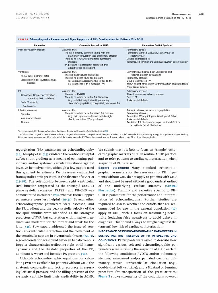
TABLE 1 Echocardiographic Parameters and Signs Suggestive of PH*: Considerations for Patients With ACHD
Parameter Comments Related to ACHD Parameters Do Not Apply In:
Peak TR velocity/gradient Assumes that:The RV is directly communicating with the
pulmonary circulation (see pulmonary atresia);There is no RVOTO or peripheral pulmonary
stenosisRA pressure is adequately estimated and
added to the TR gradient
Pulmonary atresiaPulmonary stenosis (valvular, subvalvular, or
supravalvular)Double-chambered RVTorrential TR, in which the Bernoulli equation does not apply
Ventricles Assumes that:There is biventricular circulationThere is no other cause for pressure
(or volume) overload to the RV (or to theLV in patients with a systemic RV)
Univentricular hearts, both unrepaired andrepaired (Fontan circulation)
Pulmonary stenosisDouble-chambered RVccTGA or post-atrial switch for transposition of great arteriesAtrial septal defects
RV/LV basal diameter ratio
Eccentricity index (systolic and/ordiastolic)
PA Assumes that:There is no RVOTOThere is no other cause for PA dilatation
(e.g., a left-to-right shunt), pulmonarystenosis/regurgitation, congenitally abnormal PA
Pulmonary stenosisAbsent pulmonary valve syndromeSevere PRAtrial septal defects
RV outflow Doppler accelerationtime/midsystolic notching
Early PR velocity
PA diameter
Inferior vena cava Assumes that:There is no other cause for raised RA pressure
(e.g., tricuspid valve disease, left-to-rightshunt, restrictive RV physiology)
Tricuspid stenosis or severe regurgitationPulmonary stenosisRestrictive RV physiology in tetralogy of FallotAtrial septal defectsPersistent RA dilation after repair of the defect or
arrhythmia (atrial fibrillation)
Diameter
Inspiratory collapse
RA area
*As recommended by European Society of Cardiology/European Respiratory Society Guidelines (12).
ACHD ¼ adult congenital heart disease; ccTGA ¼ congenitally corrected transposition of the great arteries; LV ¼ left ventricle; PA ¼ pulmonary artery; PH ¼ pulmonary hypertension;PR ¼ pulmonary regurgitation; RA ¼ right atrial; RV ¼ right ventricle; RVOTO ¼ right ventricular outflow tract obstruction; TR ¼ tricuspid regurgitation.
J A C C V O L . 7 2 , N O . 2 2 , 2 0 1 8 Dimopoulos et al.D E C E M B E R 4 , 2 0 1 8 : 2 7 7 8 – 8 8 Echocardiographic Screening for PAH-CHD
2781
regurgitation (PR) parameters on echocardiography(24). Murphy et al. (25) validated the ventricular septaldefect shunt gradient as a means of estimating pul-monary and/or systemic vascular resistance againstinvasive hemodynamics, although a few papers usedthis gradient to estimate PA pressures (subtractedfrom systolic aortic pressure, in the absence of RVOTO)(25–28). The relationship between right ventricular(RV) function (expressed as the tricuspid annulusplane systolic excursion [TAPSE]) and PH-CHD wasdemonstrated in children (29), whereas tissue Dopplerparameters were less helpful (29–31). Several otherechocardiographic parameters were assessed, andthe TR gradient and the peak systolic velocity of thetricuspid annulus were identified as the strongestpredictors of PVR, but correlation with invasive mea-sures was moderate for the former and weak for thelatter (31). Few papers addressed the issue of ven-tricular�ventricular interaction and the movement ofthe ventricular septum in biventricular hearts (32,33).A good correlation was found between hepatic venousDoppler characteristics (reflecting right atrial hemo-dynamics and the diastolic properties of the RV,dominant A-wave) and invasive PA pressure (34).
Although echocardiographic equations for calcu-lating PVR are available for patients without CHD, theanatomic complexity and lack of accuracy in assess-ing left atrial pressure and the filling pressure of thesystemic ventricle limit their applicability in ACHD.
We submit that it is best to focus on “simple” echo-cardiographic markers of PH in routine ACHD practiceand to refer patients to cardiac catheterization whensuspicion of PH is raised.Expert s tatement . Many standard echocardio-graphic parameters for the assessment of PH in pa-tients without CHD do not apply to patients with CHDand should not be used without a good understandingof the underlying cardiac anatomy (CentralIllustration). Training and expertise specific to PH-CHD is paramount for the performance and interpre-tation of echocardiograms. Further studies arerequired to assess whether the cutoffs that are rec-ommended for use in the general population (12)apply in CHD, with a focus on maximizing sensi-tivity (reducing false negatives) to avoid delays indiagnosis. This should always be weighed against the(current) low risk of cardiac catheterization.IMPORTANCE OF ECHOCARDIOGRAPHIC PARAMETERS IN
SUSPECTING THE PRESENCE OF PH IN SPECIFIC CHD
CONDITIONS. Participants were asked to describe howsignificant various selected echocardiographic pa-rameters were in raising the suspicion of PH in each ofthe following conditions: RVOTO and/or pulmonarystenosis; unrepaired and/or palliated complex pul-monary atresia; univentricular circulation (e.g.,double-inlet left ventricle); and a Mustard or Senningprocedure for transposition of the great arteries.Figure 2 shows schematics of the conditions covered
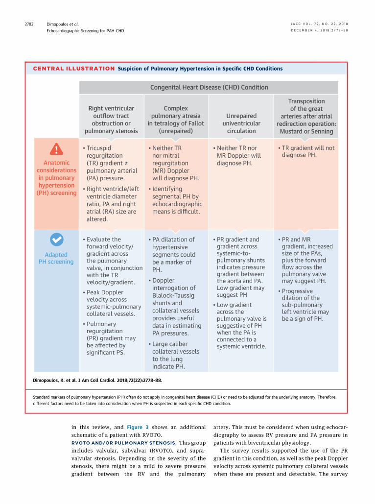
CENTRAL ILLUSTRATION Suspicion of Pulmonary Hypertension in Specific CHD Conditions
AdaptedPH screening
• Evaluate the forward velocity/gradient acrossthe pulmonary valve, in conjunctionwith the TR velocity/gradient.
• Peak Doppler velocity across systemic-pulmonary collateral vessels.
• Pulmonary regurgitation(PR) gradient may be affected by significant PS.
Anatomic considerations in pulmonary hypertension
(PH) screening
Congenital Heart Disease (CHD) Condition
• Neither TRnor mitral regurgitation(MR) Doppler will diagnose PH.
• Identifying segmental PH by echocardiographic means is difficult.
• PA dilatation of hypertensive segments couldbe a marker of PH.
• Doppler interrogation of Blalock-Taussig shunts and collateral vessels provides useful data in estimating PA pressures.
• Large caliber collateral vessels to the lung indicate PH.
• Neither TR norMR Doppler will diagnose PH.
• PR gradient and gradient across systemic-to-pulmonary shunts indicates pressure gradient between the aorta and PA. Low gradient may suggest PH
• Low gradient across the pulmonary valve is suggestive of PH when the PA is connected to a systemic ventricle.
• TR gradient will not diagnose PH.
• PR and MR gradient, increased size of the PAs, plus the forward flow across the pulmonary valve may suggest PH.
• Progressive dilation of the sub-pulmonaryleft ventricle may be a sign of PH.
Right ventricular outflow tract obstruction or
pulmonary stenosis
Complexpulmonary atresia
in tetralogy of Fallot (unrepaired)
Unrepaireduniventricular
circulation
Transpositionof the great
arteries after atrialredirection operation:
Mustard or Senning
• Tricuspid regurgitation(TR) gradient ≠pulmonary arterial (PA) pressure.
• Right ventricle/left ventricle diameter ratio, PA and right atrial (RA) size are altered.
Dimopoulos, K. et al. J Am Coll Cardiol. 2018;72(22):2778–88.
Standard markers of pulmonary hypertension (PH) often do not apply in congenital heart disease (CHD) or need to be adjusted for the underlying anatomy. Therefore,
different factors need to be taken into consideration when PH is suspected in each specific CHD condition.
Dimopoulos et al. J A C C V O L . 7 2 , N O . 2 2 , 2 0 1 8
Echocardiographic Screening for PAH-CHD D E C E M B E R 4 , 2 0 1 8 : 2 7 7 8 – 8 8
2782
in this review, and Figure 3 shows an additionalschematic of a patient with RVOTO.RVOTO AND/OR PULMONARY STENOSIS. This groupincludes valvular, subvalvar (RVOTO), and supra-valvular stenosis. Depending on the severity of thestenosis, there might be a mild to severe pressuregradient between the RV and the pulmonary
artery. This must be considered when using echocar-diography to assess RV pressure and PA pressure inpatients with biventricular physiology.
The survey results supported the use of the PRgradient in this condition, as well as the peak Dopplervelocity across systemic pulmonary collateral vesselswhen these are present and detectable. The survey
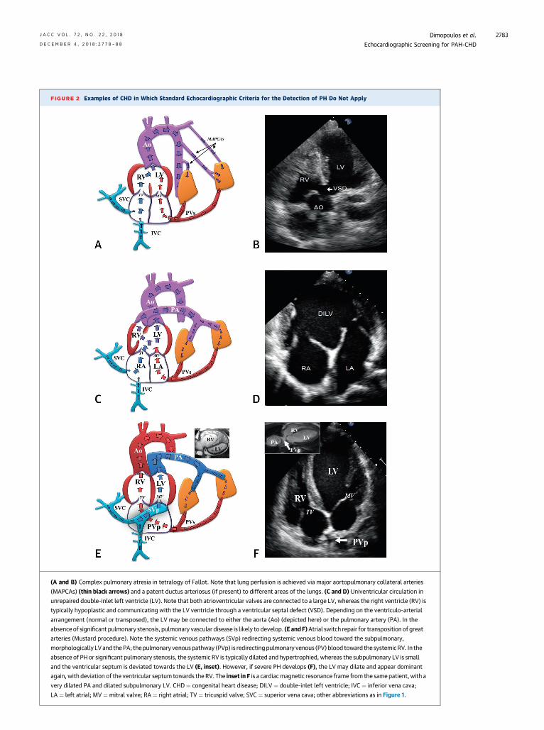
FIGURE 2 Examples of CHD in Which Standard Echocardiographic Criteria for the Detection of PH Do Not Apply
(A and B) Complex pulmonary atresia in tetralogy of Fallot. Note that lung perfusion is achieved via major aortopulmonary collateral arteries
(MAPCAs) (thin black arrows) and a patent ductus arteriosus (if present) to different areas of the lungs. (C and D) Univentricular circulation in
unrepaired double-inlet left ventricle (LV). Note that both atrioventricular valves are connected to a large LV, whereas the right ventricle (RV) is
typically hypoplastic and communicating with the LV ventricle through a ventricular septal defect (VSD). Depending on the ventriculo-arterial
arrangement (normal or transposed), the LV may be connected to either the aorta (Ao) (depicted here) or the pulmonary artery (PA). In the
absence of significant pulmonary stenosis, pulmonary vascular disease is likely to develop. (E andF)Atrial switch repair for transposition of great
arteries (Mustard procedure). Note the systemic venous pathways (SVp) redirecting systemic venous blood toward the subpulmonary,
morphologically LV and thePA; thepulmonary venous pathway (PVp) is redirectingpulmonary venous (PV) blood toward the systemicRV. In the
absence of PH or significant pulmonary stenosis, the systemic RV is typically dilated and hypertrophied, whereas the subpulmonary LV is small
and the ventricular septum is deviated towards the LV (E, inset). However, if severe PH develops (F), the LV may dilate and appear dominant
again, with deviation of the ventricular septum towards the RV. The inset in F is a cardiacmagnetic resonance frame from the same patient, with a
very dilated PA and dilated subpulmonary LV. CHD ¼ congenital heart disease; DILV ¼ double-inlet left ventricle; IVC ¼ inferior vena cava;
LA ¼ left atrial; MV ¼ mitral valve; RA ¼ right atrial; TV ¼ tricuspid valve; SVC ¼ superior vena cava; other abbreviations as in Figure 1.
J A C C V O L . 7 2 , N O . 2 2 , 2 0 1 8 Dimopoulos et al.D E C E M B E R 4 , 2 0 1 8 : 2 7 7 8 – 8 8 Echocardiographic Screening for PAH-CHD
2783
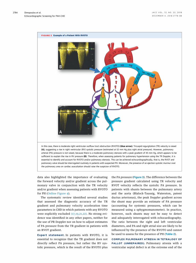
FIGURE 3 Example of a Patient With RVOTO
In this case, there is moderate right ventricular outflow tract obstruction (RVOTO) (blue arrow). Tricuspid regurgitation (TR) velocity is raised
(A), suggesting a rise in right ventricular (RV) systolic pressure (estimated at 52 mm Hg plus right atrial pressure). However, pulmonary
arterial (PA) pressure is not raised, because there is a moderate pulmonary stenosis with a peak gradient of 45 mm Hg, which appears to be
sufficient to explain the rise in RV pressure (B). Therefore, when assessing patients for pulmonary hypertension using the TR Doppler, it is
essential to identify and account for RVOTO and/or pulmonary stenosis. This can be achieved echocardiographically, that is, the RVOT and
pulmonary valve should be interrogated routinely in patients with suspected PH. Moreover, the presence of an ejection systolic murmur over
the pulmonary area on cardiac auscultation should raise the suspicion of RVOTO.
Dimopoulos et al. J A C C V O L . 7 2 , N O . 2 2 , 2 0 1 8
Echocardiographic Screening for PAH-CHD D E C E M B E R 4 , 2 0 1 8 : 2 7 7 8 – 8 8
2784
data also highlighted the importance of evaluatingthe forward velocity and/or gradient across the pul-monary valve in conjunction with the TR velocityand/or gradient when assessing patients with RVOTOfor PH (Online Figure 4).
The systematic review identified several studiesthat assessed the diagnostic accuracy of the TRgradient and pulmonary velocity acceleration timeparameters in CHD in which patients with any RVOTOwere explicitly excluded (17,19,21,22). No strong evi-dence was identified in any other papers, neither forthe use of PR Doppler nor on how to adjust estimatesof PA pressure from the TR gradient in patients withan RVOT gradient.
Expert s tatement . In patients with RVOTO, it isessential to recognize that the TR gradient does notdirectly reflect PA pressure, but rather the RV sys-tolic pressure, which is the result of the RVOTO plus
the PA pressure (Figure 3). The difference between thepressure gradient calculated using TR velocity andRVOT velocity reflects the systolic PA pressure. Inpatients with shunts between the pulmonary arteryand the aorta (Blalock-Taussig, Waterston, patentductus arteriosus), the peak Doppler gradient acrossthe shunt may provide an estimate of PA pressure(accounting for systemic pressures, which can bemeasured using a sphygmomanometer). In practice,however, such shunts may not be easy to detectand adequately interrogated with echocardiography.The ratio between the right and left ventriculardiameters, and PA and right atrial size are likely to beinfluenced by the presence of the RVOTO and cannotbe used to assess for the presence of PH (Table 2).
COMPLEX PULMONARY ATRESIA IN TETRALOGY OF
FALLOT (UNREPAIRED). Pulmonary atresia with aventricular septal defect is at the extreme end of the
TABLE 2 Summary of Expert Opinion for the Echocardiographic Diagnosis of PH in
CHD Patients
Condition/Anatomy Summary of Expert Opinion
Pulmonary stenosis/RVOTO Peak TR gradient reflects RV systolic pressure (the result ofRVOTO and PAP), not just PA pressure
Peak Doppler gradient across shunts between the PA and aortacan provide rough estimates of PA pressure, when presentand visible on echocardiography
RV/LV basal diameter ratio cannot be used to assess thepresence of PH
Unrepaired/palliated complexpulmonary atresia
Clinical suspicion is required in addition to echocardiographyThe following parameters can raise suspicion of PH:
A low peak Doppler velocity across collateral vessels orshunts
PA (or collateral vessel dilatation) or large-calibercollateral vessels
The following parameters are unrelated to PH:Peak Doppler gradients across valvesParameters relating to ventricles/atria
Univentricular circulation(e.g., DILV)
In adult patients with no pulmonary stenosis, PH diagnosis canbe made with a high degree of certainty usingechocardiography, by confirming the intracardiac anatomyand unprotected pulmonary circulation
Mustard or Senningprocedure for TGA
The following parameters should be used routinely:Systolic MR gradientPeak PR gradientIncreased size of the PAs
Eccentricity index should not be used, but progressive dilatationof the subpulmonary LV should raise the suspicion of PH
Post-capillary PH is common in patients with a systemic RV dueto RV dysfunction, TR and/or pulmonary venous pathwayobstruction; all patients should undergo cardiac catheteri-zation to differentiate between pre- and post-capillary PH
CHD ¼ congenital heart disease; DILV ¼ double inlet left ventricle; MR ¼ mitral regurgitation; PAP ¼ pulmonaryarterial pressure; TGA ¼ transposition of the great arteries; other abbreviations as in Table 1.
J A C C V O L . 7 2 , N O . 2 2 , 2 0 1 8 Dimopoulos et al.D E C E M B E R 4 , 2 0 1 8 : 2 7 7 8 – 8 8 Echocardiographic Screening for PAH-CHD
2785
spectrum of tetralogy of Fallot. Within pulmonaryatresia, there is a wide variety of conditions,depending on the anatomy of the pulmonary arteries,which range from confluent branch pulmonary ar-teries to complex forms in which there is complete ornear-complete absence of the pulmonary arteries (35).Blood supply to the lungs typically occurs through apatent ductus arteriosus and/or major aortopulmo-nary collateral vessels, which, when large, can allowexcessive flow to the segment of the lung supplied,and hence, trigger the development of pulmonaryvascular disease, which is typically segmental (i.e.,affecting certain, but not all lung segments) (Figure 1).There are limited data to suggest that PAH-specifictherapies may be beneficial in these patients; hence,the identification of PH in pulmonary atresia isessential (36,37).
The survey data demonstrated an understandingthat neither TR nor mitral regurgitation Dopplerimaging are useful in diagnosing segmental PH (mostlikely because there is no direct connection betweenthe ventricles and the pulmonary vascular tree).Doppler-derived gradients across collateral vesselsto various lung segments can be useful in identifyingpotential areas of pulmonary vascular disease, wherea lower than expected pressure gradient betweenthe aorta and lung vessels might be encountered.Moreover, PA dilation in hypertensive segmentswas believed to be a potential marker of PH (OnlineFigure 4).
The systematic review identified no studies thatassessed echocardiographic parameters for the diag-nosis of PH in complex pulmonary atresia. Dopplerinterrogation of Blalock-Taussig shunts was shown toprovide useful information in estimating PA pres-sures in patients with complex CHD (including 1 pa-tient with congenitally corrected [L-]transposition ofthe great arteries and pulmonary atresia), and thesame concept should apply to collateral vessels inpulmonary atresia (38). Schuuring et al. (39), whodescribed 7 patients with segmental PH treated suc-cessfully with PAH-specific therapies, provided noechocardiographic features.Expert s tatement . Identification of segmental PHby echocardiographic means is difficult and requiresclinical suspicion. The Doppler gradient acrosscollateral vessels identified during echocardiographycan raise the suspicion of PH in the respective lungsegment. Moreover, identification of PA (or collateralvessel) dilation or large-caliber collateral vessels tothe lung can also point toward PH. Parameters relatedto the ventricles or atria, and Doppler gradients acrossvalves are unrelated to PH in unrepaired pulmonaryatresia (Table 2).
UNREPAIRED UNIVENTRICULAR CIRCULATION.
Functional univentricular hearts occur in those inwhich there is a predominant ventricle, and typically,a small (rudimentary) ventricle that is not amenableto biventricular repair. Both the pulmonary arteryand aorta can be exposed to systemic pressures(40,41) (tricuspid or mitral atresia, double inlet left orright ventricle). PH may develop in these patientswhen the pulmonary circulation is unprotected orpartially protected (no and/or mild to moderate pul-monary stenosis).
The survey highlighted the nonapplicability of TRand mitral regurgitation Doppler gradients in thiscondition, in which the atrioventricular valve(s) pro-vide information on the pressures in the systemicventricle, but not necessarily in the pulmonary cir-culation. The PR gradient and the gradient acrosssystemic-to-pulmonary shunts (patent ductus arte-riosus or surgical shunts, such as Blalock-Taussig,Potts, or Waterston shunts) was considered signifi-cant. The latter provides an indication of the pressuregradient between the aorta and the pulmonary artery,and when the gradient is low, it may suggest PH(Online Figure 4).
Dimopoulos et al. J A C C V O L . 7 2 , N O . 2 2 , 2 0 1 8
Echocardiographic Screening for PAH-CHD D E C E M B E R 4 , 2 0 1 8 : 2 7 7 8 – 8 8
2786
The survey results were supported by 1 paperidentified in the systematic review, which assessedthe ability of echocardiography to assess PA pressuresin unrepaired or palliated univentricular hearts.Chaudhari et al. (38) demonstrated that Dopplerinterrogation of Blalock-Taussig shunts could accu-rately predict PA pressures and pulmonary blood flowin complex CHD. This concept might apply to a patentductus arteriosus or other surgical shunts, such asPotts or Waterston.Expert s tatement . Expertise is required whenassessing patients with unrepaired or palliated uni-ventricular hearts. In adult patients with no pulmo-nary stenosis (including subpulmonary stenosis dueto a small ventricular septal defect in concordantatrioventricular arrangement), and hence, no pres-sure gradient between a systemic ventricle and thepulmonary arteries, the diagnosis of severe PH can bemade with a high degree of certainty on echocardi-ography. Echocardiographic estimates of the PR peakgradients and the gradient across the pulmonaryvalve or systemic-to-pulmonary shunts are alsohelpful when screening patients with unrepaireduniventricular circulation for PH (Table 2).
TRANSPOSITION OF THE GREAT ARTERIES AFTER
ATRIAL REDIRECTION OPERATION: MUSTARD OR
SENNING. Patients with transposition of the greatarteries who have undergone atrial re-direction pro-cedures are left with a morphological RV in the sys-temic position and a morphological left ventricle inthe subpulmonary position. PH can develop in thesepatients, even after timely repair of the defect andclosure of a ventricular septal defect; these patientsmay benefit from PAH-specific therapy, although dataare limited (42,43). More commonly, post-capillaryPH develops due to RV dysfunction, significant TR,or pulmonary venous pathway obstruction.
Our survey of experts highlighted the anatomicparticularities of this condition. In particular, mitralregurgitation (rather than TR) might be used to esti-mate systolic pressure in the subpulmonary ventricleas well as PA pressures, accounting for any outflowtract obstruction. Moreover, respondents highlightedthe role of the PR gradient, pulmonary velocityDoppler, size of the pulmonary artery, and thebehavior of the ventricular septum (Online Figure 4).
The systematic review found no papers thatassessed echocardiographic parameters against inva-sive values of PA pressure in this population. Eben-roth et al. (43) described a case series of 93 patientswho underwent a Mustard procedure; of the 8 pa-tients (13%) who had a PA pressure >50% systemic, 4had anatomic explanations for this: 3 had “baffle”
(pathway) obstruction and 2 had left lung hypoplasia(43). The investigators listed the echocardiographicparameters used to suspect PH or raised sub-pulmonary ventricular pressures: shift of the ven-tricular septum toward the systemic RV; elevated PRend-diastolic velocity; or elevated mitral regurgita-tion velocity (43).Expert statement . The following parametersshould be routinely used to raise the suspicion of PHin patients who have undergone Mustard and/orSenning procedures: mitral regurgitation gradient; PRgradient; and increased size of the pulmonary ar-teries, as well as the characteristics of the forwardflow across the pulmonary valve. The eccentricityindex cannot be used as recommended by theguidelines, but a shift toward the systemic RV andprogressive dilation of the subpulmonary leftventricle (in the absence of severe pulmonary steno-sis and/or regurgitation, or significant baffle leak)may be a sign of PH. Cardiac catheterization isessential in differentiating between pre- and post-capillary PH, and anatomic causes of PH (e.g., baffleor pathway obstruction) should be excluded (Table 2).
DISCUSSION
Routine screening for PH is recommended in all pa-tients with CHD undergoing echocardiography.Echocardiography remains a fundamental part of theroutine assessment of all CHD patients, and routineechocardiographic assessment should follow a pro-tocolized approach that includes screening for PH.Standard markers of PH often do not apply in CHD orneed to be adjusted according to the underlyinganatomy. Strict guidelines for PH-CHD are difficultbecause of the lack of strong evidence; however,recommendations can be provided based on availableevidence and expert consensus.
In this review, we provided guidance based on thefindings of a systematic review of published studiesand the results of a survey of experts. Echocardiog-raphy cannot be a substitute for cardiac catheteriza-tion in most cases, with the exception, perhaps, ofsome adult cyanotic patients with large post-tricuspidshunts. It can, however, provide relevant informationthat, together with the clinical picture, can raise thesuspicion of PH and aid management (14). PH shouldbe identified promptly in CHD patients, because PHhas a significant impact on functional capacity,quality of life, and outcome. PH-CHD patients oftenbenefit from PAH-specific therapies, although evi-dence is limited mainly to patients with Eisenmengersyndrome in functional class III and to patients withrepaired defects and PAH. The consensus
J A C C V O L . 7 2 , N O . 2 2 , 2 0 1 8 Dimopoulos et al.D E C E M B E R 4 , 2 0 1 8 : 2 7 7 8 – 8 8 Echocardiographic Screening for PAH-CHD
2787
recommendations from this study should be vali-dated in large studies and registries, and updated asnew evidence and new imaging modalities emerge(32,44,45). ACHD is a vastly heterogeneous popula-tion, and providing a standardized approach thatapplies to each individual case is impossible. Thereare numerous potential pitfalls, and correct inter-pretation requires an understanding of each patient’sparticular characteristics, as well as significantexpertise in CHD and PH.
The role of noninvasive screening for PH is toidentify patients who have an increase in PA pressuresand who would therefore benefit from cardiac cathe-terization. Cardiac catheterization is the only way todistinguish between pre- and post-capillary PH and tomeasure PVR reliably, while other investigations arerequired to exclude other causes of PH (e.g., lungdisease and/or hypoxia, chronic thromboembolic dis-ease). Moreover, PH-CHDmay also be simply related tohigh pulmonary blood flow in the presence a large left-to-right shunt, which may be difficult to appreciate onechocardiography. Finally, there is limited evidenceon the use of targeted PAH therapies in patients withACHD and PHwho do not have Eisenmenger syndromewith simple defects (atrial or ventricular septal defect)or repaired CHD.
STUDY LIMITATIONS
This work has important limitations that are primarilyrelated to the extremely limited published studies (interms of number of papers and quality of evidence)to support practice and to define the sensitivityand, especially, the specificity, of echocardiographicparameters in these rare conditions. The primaryscope of the search was to inform expert opinionand statements, rather than to provide entirelyevidence-based recommendations. In the absence ofvalidated diagnostic tools, expert judgment shouldbe used to identify patients with PH by combiningechocardiographic signs with the previous probabilityof PH based on anatomy, type, and time of previous
surgery, and other clinical parameters (e.g., presenceof Down syndrome).
The survey of experts was aimed at understandingmodern practice in specialist centers, but had limitedinfluence on expert statements. Studies that compareechocardiographic parameters to the gold standard ofcardiac catheterization in various ACHD cohorts and awide range of pulmonary pressures and resistances,also incorporating previous probability based onclinical information, are urgently needed to informclinical practice. Efforts should be made to achievehomogenization of the assessment of PH in ACHD aswell as selection of proper methodologies of correla-tion with invasive measurements and other nonin-vasive cardiovascular imaging in multicenter studiesand clinical trials. This would also open the way tofuture lines of physiological and basic scienceinvestigations.
CONCLUSIONS
All patients with CHD should undergo regular assess-ment for the presence of PH during routine echocar-diography, as should patients with clinical suspicionof PH. This should occur in centers with adequateexpertise in the management of both PH and CHD. Thepaucity of evidence to guide the echocardiographicassessment urgently calls for registries and studiesthat will provide support to clinical practice.
ACKNOWLEDGMENTS The manuscript and surveywere supported by Actelion Pharmaceuticals UKLimited, who had no influence on manuscript writing.Medical writing support was provided by nspm ltd,Meggen, Switzerland. Survey responses from CHAM-PION Steering Committee members were included inanalyses.
ADDRESS FOR CORRESPONDENCE: Dr. KonstantinosDimopoulos, Adult Congenital Heart Centre, RoyalBrompton and Harefield NHS Foundation Trust,Sydney Street, SW3 6NP London, United Kingdom.E-mail: [email protected].
RE F E RENCE S
1. Diller GP, Kempny A, Inuzuka R, et al. Survivalprospects of treatment naive patients with Eisen-menger: a systematic review of the literature andreport of own experience. Heart 2014;100:1366–72.
2. Dimopoulos K, Wort SJ, Gatzoulis MA. Pulmo-nary hypertension related to congenital heartdisease: a call for action. Eur Heart J 2014;35:691–700.
3. Diller GP, Dimopoulos K, Okonko D, et al. Ex-ercise intolerance in adult congenital heart
disease: comparative severity, correlates, andprognostic implication. Circulation 2005;112:828–35.
4. Dimopoulos K, Okonko DO, Diller GP, et al.Abnormal ventilatory response to exercise inadults with congenital heart disease relates tocyanosis and predicts survival. Circulation 2006;113:2796–802.
5. Kempny A, Dimopoulos K, Uebing A, et al.Reference values for exercise limitations amongadults with congenital heart disease. Relation to
activities of daily life–single centre experience andreview of published data. Eur Heart J 2012;33:1386–96.
6. Daliento L, Somerville J, Presbitero P, et al.Eisenmenger syndrome. Factors relating to dete-rioration and death. Eur Heart J 1998;19:1845–55.
7. Diller GP, Dimopoulos K, Broberg CS, et al.Presentation, survival prospects, and predictors ofdeath in Eisenmenger syndrome: a combinedretrospective and case-control study. Eur Heart J2006;27:1737–42.

Dimopoulos et al. J A C C V O L . 7 2 , N O . 2 2 , 2 0 1 8
Echocardiographic Screening for PAH-CHD D E C E M B E R 4 , 2 0 1 8 : 2 7 7 8 – 8 8
2788
8. Diller GP, Alonso-Gonzalez R, Dimopoulos K,et al. Disease targeting therapies in patients withEisenmenger syndrome: response to treatmentand long-term efficiency. Int J Cardiol 2013;167:840–7.
9. Dimopoulos K, Inuzuka R, Goletto S, et al.Improved survival among patients with Eisen-menger syndrome receiving advanced therapy forpulmonary arterial hypertension. Circulation 2010;121:20–5.
10. Galiè N, Beghetti M, Gatzoulis MA, et al.,Bosentan Randomized Trial of Endothelin Antag-onist Therapy-5 (BREATHE-5) Investigators.Bosentan therapy in patients with Eisenmengersyndrome: a multicenter, double-blind, random-ized, placebo-controlled study. Circulation 2006;114:48–54.
11. Rosenzweig EB, Kerstein D, Barst RJ. Long-term prostacyclin for pulmonary hypertensionwith associated congenital heart defects. Circula-tion 1999;99:1858–65.
12. Galiè N, Humbert M, Vachiery JL, et al. 2015ESC/ERS Guidelines for the diagnosis and treat-ment of pulmonary hypertension: The Joint TaskForce for the Diagnosis and Treatment of Pulmo-nary Hypertension of the European Society ofCardiology (ESC) and the European RespiratorySociety (ERS): Endorsed by: Association for Euro-pean Paediatric and Congenital Cardiology (AEPC),International Society for Heart and Lung Trans-plantation (ISHLT). Eur Heart J 2016;37:67–119.
13. Liberati A, Altman DG, Tetzlaff J, et al. ThePRISMA statement for reporting systematic re-views and meta-analyses of studies that evaluatehealthcare interventions: explanation and elabo-ration. BMJ 2009;339:b2700.
14. Ajami GH, Cheriki S, Amoozgar H, Borzouee M,Soltani M. Accuracy of Doppler-derived estimationof pulmonary vascular resistance in congenitalheart disease: an index of operability. PediatrCardiol 2011;32:1168–74.
15. Bhatt DD, Manoj R, Mahajan R. Estimation ofpulmonary vascular resistance: correlation be-tween echocardiography and catheterization datain patients with congenital heart disease. Echo-cardiography 2012;29:478–83.
16. Bhyravavajhala S, Velam V, Polapragada NV,et al. Reliability of Doppler-based measurement ofpulmonary vascular resistance in congenital heartdisease with left-to-right shunt lesions. Echocar-diography 2015;32:1009–14.
17. Cevik A, Kula S, Olgunturk R, et al. Assessmentof pulmonary arterial hypertension and vascularresistance by measurements of the pulmonaryarterial flow velocity curve in the absence of ameasurable tricuspid regurgitant velocity inchildhood congenital heart disease. Pediatr Cardiol2013;34:646–55.
18. Friedberg MK, Feinstein JA, Rosenthal DN.A novel echocardiographic Doppler method forestimation of pulmonary arterial pressures. J AmSoc Echocardiogr 2006;19:559–62.
19. Kosturakis D, Goldberg SJ, Allen HD, Loeber C.Doppler echocardiographic prediction of pulmo-nary arterial hypertension in congenital heart dis-ease. Am J Cardiol 1984;53:1110–5.
20. Kouzu H, Nakatani S, Kyotani S, Kanzaki H,Nakanishi N, Kitakaze M. Noninvasive estimation ofpulmonary vascular resistance by Doppler echo-cardiography in patients with pulmonary arterialhypertension. Am J Cardiol 2009;103:872–6.
21. Pande A, Sarkar A, Ahmed I, et al. Non-invasiveestimation of pulmonary vascular resistance inpatients of pulmonary hypertension in congenitalheart disease with unobstructed pulmonary flow.Ann Pediatr Cardiol 2014;7:92–7.
22. Tabib A, Khorgami MR, Meraji M, Omidi N,Mirmesdagh Y. Accuracy of Doppler-derivedindices in predicting pulmonary vascular resis-tance in children with pulmonary hypertensionsecondary to congenital heart disease with left-to-right shunting. Pediatr Cardiol 2014;35:521–9.
23. Wang B, Feng Y, Jia LQ, et al. Accuracy ofDoppler echocardiography in the assessment ofpulmonary arterial hypertension in patients withcongenital heart disease. Eur Rev Med PharmacolSci 2013;17:923–8.
24. Atiq M, Tasneem H, Aziz K. Estimation ofpulmonary vascular resistance with Doppler dia-stolic gradients. Asian Cardiovasc Thorac Ann2008;16:221–5.
25. Murphy DJ Jr., Ludomirsky A, Huhta JC.Continuous-wave Doppler in children with ven-tricular septal defect: noninvasive estimation ofinterventricular pressure gradient. Am J Cardiol1986;57:428–32.
26. Espinola-Zavaleta N, Soto ME, Romero-Gonzalez A, et al. Prevalence of congenital heartdisease and pulmonary hypertension in Down’ssyndrome: an echocardiographic study.J Cardiovasc Ultrasound 2015;23:72–7.
27. Gabriel HM, Heger M, Innerhofer P, et al.Long-term outcome of patients with ventricularseptal defect considered not to require surgicalclosure during childhood. J Am Coll Cardiol 2002;39:1066–71.
28. Gungor H, Fatih Ayik M, Engin C, et al.Transthoracic echocardiographic and cardiopul-monary exercise testing parameters in Eisen-menger’s syndrome. Association with six-minutewalk test distance. Herz 2014;39:633–7.
29. Zakaria D, Sachdeva R, Gossett JM, Tang X,O’Connor MJ. Tricuspid annular plane systolicexcursion is reduced in infants with pulmonaryhypertension. Echocardiography 2015;32:834–8.
30. Cevik A, Kula S, Olgunturk R, et al. Dopplertissue imaging provides an estimate of pulmonaryarterial pressure in children with pulmonary hy-pertension due to congenital intracardiac shunts.Congenit Heart Dis 2013;8:527–34.
31. Roushdy AM, Ragab I, Abd El Raouf W. Nonin-vasive assessment of elevated pulmonary vascularresistance in children with pulmonary hypertensionsecondary to congenital heart disease: a compara-tive study between five different Doppler indices.J Saudi Heart Assoc 2012;24:233–41.
32. Kimura S, Nakahata Y, Honda T, et al. Nonin-vasive assessment of pulmonary vascular resis-tance and pressure in patients with congenitalheart disease: a new method using M-modeechocardiography. J Echocardiogr 2011;9:137–41.
33. Portman MA, Bhat AM, Cohen MH,Jacobstein MD. Left ventricular systolic circularindex: an echocardiographic measure of trans-septal pressure ratio. Am Heart J 1987;114:1178–82.
34. Sun DD, Hou CJ, Yuan LJ, Duan YY, Hou Y,Zhou FP. Hemodynamic changes of the middlehepatic vein in patients with pulmonary hyper-tension using echocardiography. PLoS One 2015;10:e0121408.
35. Prieto L. Management of tetralogy of fallotwith pulmonary atresia. Images Paediatr Cardiol2005;7:24–42.
36. D’Alto M, Merola A, Dimopoulos K. Pulmonaryhypertension related to congenital heart disease: acomprehensive review. Glob Cardiol Sci Pract2015;42.
37. Dimopoulos K, Diller GP, Opotowsky AR, et al.Definition and management of segmental pulmo-nary hypertension. J Am Heart Assoc 2018;7:e008587.
38. Chaudhari M, Balmer C, Heng JT, Wright J,Stümper O. Usefulness of Blalock-Taussig shuntDoppler flow velocity profiles in the assessment ofpulmonary artery pressure and flow. Eur J Echo-cardiogr 2004;5:111–7.
39. Schuuring MJ, Bouma BJ, Cordina R, et al.Treatment of segmental pulmonary artery hyper-tension in adults with congenital heart disease. IntJ Cardiol 2013;164:106–10.
40. Jacobs ML, Mayer JE Jr. Congenital HeartSurgery Nomenclature and Database Project: sin-gle ventricle. Ann Thorac Surg 2000;69:S197–204.
41. Khairy P, Poirier N, Mercier LA. Univentricularheart. Circulation 2007;115:800–12.
42. Chan E, Alejos J. Pulmonary hypertension inpatients after repair of transposition of the greatarteries. Congenit Heart Dis 2010;5:161–4.
43. Ebenroth ES, Hurwitz RA, Cordes TM. Lateonset of pulmonary hypertension after success-ful Mustard surgery for d-transposition of thegreat arteries. Am J Cardiol 2000;85:127–30.A10.
44. Jone PN, Patel SS, Cassidy C, Ivy DD. Three-dimensional echocardiography of right ventricularfunction correlates with severity of pediatric pul-monary hypertension. Congenit Heart Dis 2016;11:562–9.
45. van Riel AC, de Bruin-Bon RH, Gertsen EC,Blok IM, Mulder BJ, Bouma BJ. Simple stressechocardiography unmasks early pulmonaryvascular disease in adult congenital heart disease.Int J Cardiol 2015;197:312–4.
KEY WORDS cardiac catheterization,echocardiography, pulmonary atresia,pulmonary stenosis, transposition of greatarteries, univentricular heart
APPENDIX For expanded Methods and Re-sults sections as well as supplemental figures,please see the online version of this paper.