Embed Size (px)
Citation preview
Efficient Genome Engineering of Toxoplasma gondiiUsing CRISPR/Cas9Saima M. Sidik1, Caroline G. Hackett1, Fanny Tran2, Nicholas J. Westwood2, Sebastian Lourido1*
1 Whitehead Institute for Biomedical Research, Cambridge, Massachusetts, United States of America, 2 School of Chemistry and Biomedical Sciences Research Complex,
University of St. Andrews and EaStCHEM, North Haugh, St Andrews, Fife, Scotland, United Kingdom
Abstract
Toxoplasma gondii is a parasite of humans and animals, and a model for other apicomplexans including Plasmodium spp.,the causative agents of malaria. Despite many advances, manipulating the T. gondii genome remains labor intensive, and isoften restricted to lab-adapted strains or lines carrying mutations that enable selection. Here, we use the RNA-guided Cas9nuclease to efficiently generate knockouts without selection, and to introduce point mutations and epitope tags into the T.gondii genome. These methods will streamline the functional analysis of parasite genes and enable high-throughputengineering of their genomes.
Citation: Sidik SM, Hackett CG, Tran F, Westwood NJ, Lourido S (2014) Efficient Genome Engineering of Toxoplasma gondii Using CRISPR/Cas9. PLoS ONE 9(6):e100450. doi:10.1371/journal.pone.0100450
Editor: Ira J. Blader, University at Buffalo, United States of America
Received March 28, 2014; Accepted May 23, 2014; Published June 27, 2014
Copyright: � 2014 Sidik et al. This is an open-access article distributed under the terms of the Creative Commons Attribution License, which permitsunrestricted use, distribution, and reproduction in any medium, provided the original author and source are credited.
Data Availability: The authors confirm that all data underlying the findings are fully available without restriction. All relevant data are within the paper and itsSupporting Information files.
Funding: This work was supported in part by NIH grant 1DP5OD017892 to S.L. The funders had no role in study design, data collection and analysis, decision topublish, or preparation of the manuscript.
Competing Interests: The authors have declared that no competing interests exist.
* Email: [email protected]
Introduction
Tools for genome engineering are fundamental for the
functional dissection of genes, and have greatly advanced our
understanding of parasites for which homology to model
organisms is often limited. In the case of T. gondii, a proliferation
of tools for genetic manipulation, coupled to its relative ease of
culture, have made it a model system for the cell biology of related
apicomplexan parasites, including Plasmodium spp.: the causative
agents of malaria. The recent generation of strains deficient in
non-homologous end joining (NHEJ) through knocking out KU80
(DKU80) has facilitated genome engineering by suppressing the
high frequency of random integration naturally found in T. gondii
[1,2]. Nonetheless, these manipulations still rely on complex DNA
constructs for homologous recombination, frequently require
screening many clones for the correct rearrangement, and depend
on selectable markers to enrich for the manipulated population.
Furthermore, genome engineering remains for the most part
restricted to select, lab adapted strains or the above-mentioned
DKU80 parasite lines.
Here, we demonstrate efficient genome editing in T. gondii using
the prokaryotic CRISPR/Cas9 system, which has been shown to
facilitate RNA-guided, site-specific DNA cleavage in diverse
organisms ranging from yeast to mammalian cells [3–5]. This
prokaryotic immune system taken from Streptococcus pyogenes has
been reduced to its essential components, which include the Cas9
nuclease and a chimeric RNA (chiRNA) that directs this nuclease
to its target [4]. The chiRNA consists of a 20 nt guide (also known
as protospacer) sequence followed by an 85 nt chimeric sequence
derived from the crRNA and tracrRNAs found in the bacterial
system [4]. The 20 nt guide is perfectly complementary to the
target sequence and must be followed in the target sequence by the
nucleotides NGG, which are known as the protospacer-adjacent
motif (PAM). This short motif allows the system to distinguish
between the target and chiRNA-coding DNA sequences. Given its
size, the guide sequence can be easily programmed to target
diverse genomic loci for cleavage by Cas9. Double-stranded breaks
introduced by Cas9 are repaired by the cellular machinery
through either homologous recombination or the non-homologous
end-joining pathway (NHEJ). DNA sequences homologous to the
Cas9-targeted locus can be concurrently introduced to generate
desired mutations [3–5].
Using a single plasmid system to express both the guide RNA
and Cas9 nuclease, we can efficiently disrupt targeted loci in T.
gondii without the need for selection. We provide evidence linking
the efficiency of this process to the NHEJ pathway for DNA repair.
We also demonstrate high rates of genome editing that allow us to
introduce point mutations and epitope tags into the T. gondii
genome. Together these methods dramatically improve our ability
to manipulate the T. gondii genome and open the possibility of
genetic manipulation in diverse genetic backgrounds and with
greater throughput than previously possible.
Materials and Methods
Growth of Host Cells and Parasite StrainsAll T. gondii strains used in this work are derived from the type I
RH line. The DKU80DHXGPRT strain, referred to for clarity in
this study as DKU80, was generously provided by V. Carruthers
(University of Michigan, USA) and generated as previously
published [1,2]. Strains were maintained in Human Foreskin
Fibroblast (HFF) cells grown in Dulbecco’s Modified Eagle’s
PLOS ONE | www.plosone.org 1 June 2014 | Volume 9 | Issue 6 | e100450
Medium (DMEM) supplemented with 10% heat-inactivated fetal
bovine serum and 10 mg/mL gentamicin.
Plasmid ConstructionAll primer sequences can be found in Table S1. Plasmid pX330
encoding N-terminally FLAG tagged Cas9 with nuclear localiza-
tion sequences, and the guide RNA under mammalian promoters,
as previously published [3–5],was generously provided by Tim
Wang. The TgTUB1 promoter was cloned into the KpnI and
NcoI sites upstream of Cas9 using primers P1 and P2. Constructs
with protospacers against SAG1 (pU6-SAG1) and PKG (pU6-
PKG), as well as a universal plasmid encoding BsaI sites in place of
a protospacer, were synthesized by IDT (Figure S1). These
constructs were amplified using P3 and P4, digested with NcoI and
XbaI, and cloned into the PciI and XbaI sites of the Cas9-encoding
plasmid. The construct targeting the CDPK3 locus was generated
by annealing oligos P25 and P26, phosphorylating the duplex, and
cloning it into the BsaI-digested universal plasmid. The universal
plasmid has been deposited with Addgene (ID no. 52694).
Oligos used to facilitate homologous recombination in PKG and
CDPK3 were generated by heating complementary 90- or 119-
nucleotide oligomers ordered from IDT to 99 degrees for
2 minutes in a heat block, then removing the block from the
heating apparatus and allowing the DNA to cool to room
temperature over the course of a few hours. The sequences for
these oligomers are listed in Table S1 as P5 and P6 for the PKG
oligo and P7 and p8 for the CDPK3 oligo.
The plasmid used to generate the allelic replacements of PKG
was made by amplifying the 59 end of the locus with P17 and P18
from RH genomic DNA and the 39 end of the gene with P19 and
P20 from RH cDNA. The two fragments were spliced together
with AvrII and cloned into the PacI/AscI sites of pLIC-YFP-
HXGPRT (provided by V. Carruthers, University of Michigan,
USA), thus removing the YFP and introducing the two fragments.
The gatekeeper residue in this construct was changed to code for
T761M using site-directed mutagenesis.
Control plasmid 1 used in viability experiments was constructed
by PCR-amplifying the pyrimethamine resistance cassette from
pDHFR-TS [6] using primers P23 and P24, and cloned into the
NsiI and SbfI sites of the universal plasmid in place of Cas9.
Control plasmid 2 is the universal plasmid. Sequences for both
control plasmids are provided in File S1.
Construction of PKG allelic replacement strainsThe previously described CDPK1M strain [4,7] was transfected
with the PKG allelic replacement plasmid carrying either the wild-
type or T761M gatekeeper, and linearized with StuI to improve
homologous recombination. Parasites were selected with 25 mg/ml
mycophenolic acid and 50 mg/ml xanthine prior to subcloning.
Allelic replacements were identified using primers P21 and P22
against genomic DNA from established clonal lines.
Transfections and Plaque AssaysFiltered T. gondii were washed and resuspended in Cytomix
(10 mM KPO4, 120 mM KCl, 5 mM MgCl2, 25 mM HEPES,
2 mM EDTA) at 26108 parasites/ml. 56107 parasites were
combined with 100 mg of CRISPR/Cas9 plasmid supplemented
with 2 mM ATP, 5 mM GSH, and 150 mM CaCl2, with or
without 40 mg of double-stranded oligonucleotide, in a final
volume of 400 ml. The CaCl2 concentration used here matches the
original formulation of Cytomix [8] but was added at the time of
transfection to prevent the buildup of phosphate precipitates
during buffer storage. Parasites were electroporated in 4 mm gap
cuvettes (BTX Harvard Apparatus model no. 640) in an Electro
Square Porator (BTX Harvard Apparatus). 200 parasites were
added to confluent HFF monolayers in six-well dishes. In some
cases, as indicated in the text, 2,000 or 20,000 parasites were used
instead. If indicated, 0.2 mM Compound 2 (C2) or vehicle
(DMSO) was added to these dishes 24 hpi. All plates were
allowed to incubate for a total of eight days before staining with
crystal violet. C2 was synthesized by N. Westwood and F. Tran as
previously described [4,9,10].
Fluorescent Microscopy1.256106 T. gondii transfected with pU6-SAG1 were added to
confluent monolayers of HFF cells grown on cover slips in 24-well
dishes. Cells were fixed 24 hpi on ice with methanol for 2 minutes,
then stained using mouse-anti-SAG1 (mAb DG52), rabbit-anti-
TgAct1 [11], mouse-anti-Ty (mAb BB2 [12]), or mouse-anti-Flag
(Sigma, F1804). Alexa-594-conjugated goat-anti-rabbit (Life
Technologies, A11037) and Alexa-488-conjugated goat-anti-
mouse (Life Technologies, A11029) were used as secondary
antibodies. Antibodies against Toxoplasma were kindly provided
by L. D. Sibley (Washington University School of Medicine, USA).
Coverslips were mounted in Prolong Gold containing DAPI
(Invitrogen P-36931) and imaged using an Eclipse Ti microscope
(Nikon).
Sequence AnalysisT. gondii transfected with pU6-SAG1 or pU6-PKG plasmids
were grown in HFFs for two days, then pelleted and resuspended
in 1X PCR buffer (Sigma P2192-1VL) in PBS plus 100 mg
Proteinase K (Sigma). DNA was obtained by heating the samples
to 37uC for 1 h, 50uC for 2 h, and 95uC for 15 min. Regions of
SAG1 and PKG surrounding their protospacers were amplified
using primers P9 and P10, or P11 and P12, respectively (Table
S1). A second round of amplification was then performed using
primers P13 and P14 for SAG1 or P15 and P16 for PKG to ensure
purity of the product. The resulting fragments were cloned into
pCR-II-Blunt-Topo (Invitrogen 45-0245) per the manufacturer’s
instructions. Clones were sent to EtonBioscience Inc. for
sequencing with the primer M13F-21. Alignments were made
using SeqMan pro Version 11.0.0.
StatisticsLoss of SAG1 staining, transfection efficiency, viability, and Ty-
tagging data were arcsine transformed, then analyzed using two-
tailed Student’s t-tests. The total numbers of plaques obtained with
pU6-PKG with and without T761Q were compared using a one-
tailed t-test.
Results
CRISPR/Cas9-mediated gene disruptionTo investigate whether the CRISPR/Cas9 system could be
implemented in T. gondii, we adapted a single-plasmid system [4]
to express chimeric RNA (chiRNA) and a FLAG-tagged Cas9
protein flanked by nuclear localization signals (NLS), both using
parasite promoter sequences (Figure 1A). For Cas9 expression, we
used the previously-characterized 59UTR of the alpha-tubulin
promoter [13]. Transcription of the chiRNA needs to be driven by
an RNA polymerase III promoter to prevent polyadenylation.
However, RNA pol III promoters have not been characterized in
T. gondii. We identified the likely U6 gene through homology to the
annotated P. falciparum gene (PlasmoDB.org), and cloned 500 bp
upstream of the predicted transcription start site—corresponding
to 980,755–981,251 bp on chromosome III of the GT1 genome—
immediately upstream of the chiRNA sequence. The resulting
CRISPR/Cas9 Editing of the Toxoplasma Genome
PLOS ONE | www.plosone.org 2 June 2014 | Volume 9 | Issue 6 | e100450
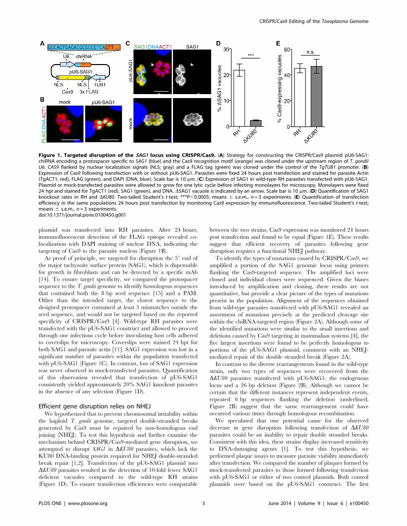
plasmid was transfected into RH parasites. After 24 hours,
immunofluorescent detection of the FLAG epitope revealed co-
localization with DAPI staining of nuclear DNA, indicating the
targeting of Cas9 to the parasite nucleus (Figure 1B).
As proof of principle, we targeted for disruption the 59 end of
the major tachyzoite surface protein (SAG1), which is dispensable
for growth in fibroblasts and can be detected by a specific mAb
[14]. To ensure target specificity, we compared the protospacer
sequence to the T. gondii genome to identify homologous sequences
that contained both the 8 bp seed sequence [15] and a PAM.
Other than the intended target, the closest sequence to the
designed protospacer contained at least 5 mismatches outside the
seed sequence, and would not be targeted based on the reported
specificity of CRISPR/Cas9 [4]. Wild-type RH parasites were
transfected with the pU6-SAG1 construct and allowed to proceed
through one infectious cycle before inoculating host cells adhered
to coverslips for microscopy. Coverslips were stained 24 hpi for
both SAG1 and parasite actin [11]. SAG1 expression was lost in a
significant number of parasites within the population transfected
with pU6-SAG1 (Figure 1C). In contrast, loss of SAG1 expression
was never observed in mock-transfected parasites. Quantification
of this observation revealed that transfection of pU6-SAG1
consistently yielded approximately 20% SAG1 knockout parasites
in the absence of any selection (Figure 1D).
Efficient gene disruption relies on NHEJWe hypothesized that to prevent chromosomal instability within
the haploid T. gondii genome, targeted double-stranded breaks
generated by Cas9 must be repaired by non-homologous end
joining (NHEJ). To test this hypothesis and further examine the
mechanism behind CRISPR/Cas9-mediated gene disruption, we
attempted to disrupt SAG1 in DKU80 parasites, which lack the
KU80 DNA-binding protein required for NHEJ double-stranded
break repair [1,2]. Transfection of the pU6-SAG1 plasmid into
DKU80 parasites resulted in the detection of 10-fold fewer SAG1
deficient vacuoles compared to the wild-type RH strains
(Figure 1D). To ensure transfection efficiencies were comparable
between the two strains, Cas9 expression was monitored 24 hours
post transfection and found to be equal (Figure 1E). These results
suggest that efficient recovery of parasites following gene
disruption requires a functional NHEJ pathway.
To identify the types of mutations caused by CRISPR/Cas9, we
amplified a portion of the SAG1 genomic locus using primers
flanking the Cas9-targeted sequence. The amplified loci were
cloned and individual clones were sequenced. Given the biases
introduced by amplification and cloning, these results are not
quantitative, but provide a clear picture of the types of mutations
present in the population. Alignment of the sequences obtained
from wild-type parasites transfected with pU6-SAG1 revealed an
assortment of mutations precisely at the predicted cleavage site
within the chiRNA-targeted region (Figure 2A). Although some of
the identified mutations were similar to the small insertions and
deletions caused by Cas9 targeting in mammalian systems [4], the
five largest insertions were found to be perfectly homologous to
portions of the pU6-SAG1 plasmid, consistent with an NHEJ-
mediated repair of the double stranded break (Figure 2A).
In contrast to the diverse rearrangements found in the wild-type
strain, only two types of sequences were recovered from the
DKU80 parasites transfected with pU6-SAG1: the endogenous
locus and a 26 bp deletion (Figure 2B). Although we cannot be
certain that the different instances represent independent events,
repeated 6 bp sequences flanking the deletion (underlined;
Figure 2B) suggest that the same rearrangement could have
occurred various times through homologous recombination.
We speculated that one potential cause for the observed
decrease in gene disruption following transfection of DKU80
parasites could be an inability to repair double stranded breaks.
Consistent with this idea, these strains display increased sensitivity
to DNA-damaging agents [1]. To test this hypothesis, we
performed plaque assays to measure parasite viability immediately
after transfection. We compared the number of plaques formed by
mock-transfected parasites to those formed following transfection
with pU6-SAG1 or either of two control plasmids. Both control
plasmids were based on the pU6-SAG1 construct. The first
Figure 1. Targeted disruption of the SAG1 locus using CRISPR/Cas9. (A) Strategy for constructing the CRISPR/Cas9 plasmid pU6-SAG1:chiRNA encoding a protospacer specific to SAG1 (blue) and the Cas9 recognition motif (orange) was cloned under the upstream region of T. gondiiU6. CAS9 flanked by nuclear localization signals (NLS; gray) and a FLAG tag (green) was cloned under the control of the TgTUB1 promoter. (B)Expression of Cas9 following transfection with or without pU6-SAG1. Parasites were fixed 24 hours post transfection and stained for parasite Actin(TgACT1; red), FLAG (green), and DAPI (DNA; blue). Scale bar is 10 mm. (C) Expression of SAG1 in wild-type RH parasites transfected with pU6-SAG1.Plasmid or mock-transfected parasites were allowed to grow for one lytic cycle before infecting monolayers for microscopy. Monolayers were fixed24 hpi and stained for TgACT1 (red), SAG1 (green), and DNA. DSAG1 vacuole is indicated by an arrow. Scale bar is 10 mm. (D) Quantification of SAG1knockout rates in RH and DKU80. Two-tailed Student’s t-test; ***P,0.0005; means 6 s.e.m., n = 3 experiments. (E) Quantification of transfectionefficiency in the same populations 24 hours post transfection by monitoring Cas9 expression by immunofluorescence. Two-tailed Student’s t-test;means 6 s.e.m., n = 3 experiments.doi:10.1371/journal.pone.0100450.g001
CRISPR/Cas9 Editing of the Toxoplasma Genome
PLOS ONE | www.plosone.org 3 June 2014 | Volume 9 | Issue 6 | e100450
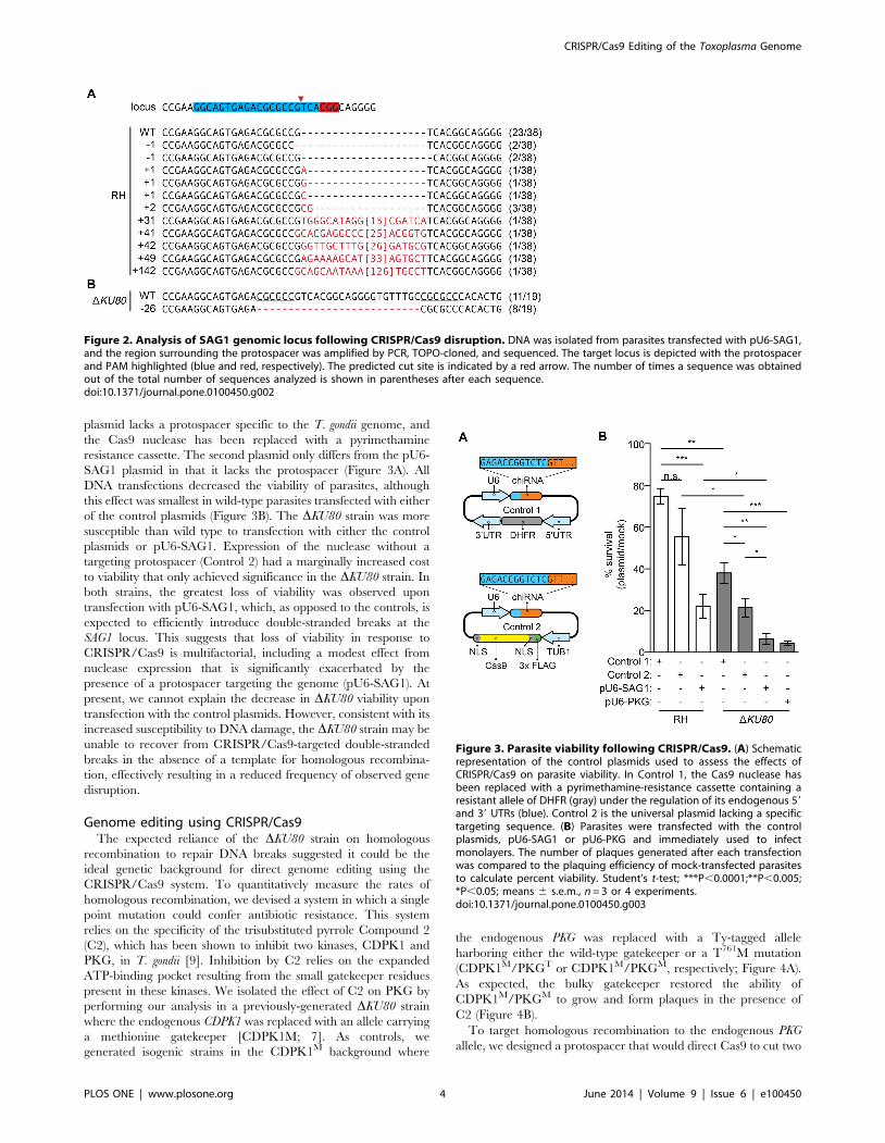
plasmid lacks a protospacer specific to the T. gondii genome, and
the Cas9 nuclease has been replaced with a pyrimethamine
resistance cassette. The second plasmid only differs from the pU6-
SAG1 plasmid in that it lacks the protospacer (Figure 3A). All
DNA transfections decreased the viability of parasites, although
this effect was smallest in wild-type parasites transfected with either
of the control plasmids (Figure 3B). The DKU80 strain was more
susceptible than wild type to transfection with either the control
plasmids or pU6-SAG1. Expression of the nuclease without a
targeting protospacer (Control 2) had a marginally increased cost
to viability that only achieved significance in the DKU80 strain. In
both strains, the greatest loss of viability was observed upon
transfection with pU6-SAG1, which, as opposed to the controls, is
expected to efficiently introduce double-stranded breaks at the
SAG1 locus. This suggests that loss of viability in response to
CRISPR/Cas9 is multifactorial, including a modest effect from
nuclease expression that is significantly exacerbated by the
presence of a protospacer targeting the genome (pU6-SAG1). At
present, we cannot explain the decrease in DKU80 viability upon
transfection with the control plasmids. However, consistent with its
increased susceptibility to DNA damage, the DKU80 strain may be
unable to recover from CRISPR/Cas9-targeted double-stranded
breaks in the absence of a template for homologous recombina-
tion, effectively resulting in a reduced frequency of observed gene
disruption.
Genome editing using CRISPR/Cas9The expected reliance of the DKU80 strain on homologous
recombination to repair DNA breaks suggested it could be the
ideal genetic background for direct genome editing using the
CRISPR/Cas9 system. To quantitatively measure the rates of
homologous recombination, we devised a system in which a single
point mutation could confer antibiotic resistance. This system
relies on the specificity of the trisubstituted pyrrole Compound 2
(C2), which has been shown to inhibit two kinases, CDPK1 and
PKG, in T. gondii [9]. Inhibition by C2 relies on the expanded
ATP-binding pocket resulting from the small gatekeeper residues
present in these kinases. We isolated the effect of C2 on PKG by
performing our analysis in a previously-generated DKU80 strain
where the endogenous CDPK1 was replaced with an allele carrying
a methionine gatekeeper [CDPK1M; 7]. As controls, we
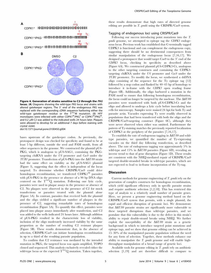
generated isogenic strains in the CDPK1M background where
the endogenous PKG was replaced with a Ty-tagged allele
harboring either the wild-type gatekeeper or a T761M mutation
(CDPK1M/PKGT or CDPK1M/PKGM, respectively; Figure 4A).
As expected, the bulky gatekeeper restored the ability of
CDPK1M/PKGM to grow and form plaques in the presence of
C2 (Figure 4B).
To target homologous recombination to the endogenous PKG
allele, we designed a protospacer that would direct Cas9 to cut two
Figure 2. Analysis of SAG1 genomic locus following CRISPR/Cas9 disruption. DNA was isolated from parasites transfected with pU6-SAG1,and the region surrounding the protospacer was amplified by PCR, TOPO-cloned, and sequenced. The target locus is depicted with the protospacerand PAM highlighted (blue and red, respectively). The predicted cut site is indicated by a red arrow. The number of times a sequence was obtainedout of the total number of sequences analyzed is shown in parentheses after each sequence.doi:10.1371/journal.pone.0100450.g002
Figure 3. Parasite viability following CRISPR/Cas9. (A) Schematicrepresentation of the control plasmids used to assess the effects ofCRISPR/Cas9 on parasite viability. In Control 1, the Cas9 nuclease hasbeen replaced with a pyrimethamine-resistance cassette containing aresistant allele of DHFR (gray) under the regulation of its endogenous 59and 39 UTRs (blue). Control 2 is the universal plasmid lacking a specifictargeting sequence. (B) Parasites were transfected with the controlplasmids, pU6-SAG1 or pU6-PKG and immediately used to infectmonolayers. The number of plaques generated after each transfectionwas compared to the plaquing efficiency of mock-transfected parasitesto calculate percent viability. Student’s t-test; ***P,0.0001;**P,0.005;*P,0.05; means 6 s.e.m., n = 3 or 4 experiments.doi:10.1371/journal.pone.0100450.g003
CRISPR/Cas9 Editing of the Toxoplasma Genome
PLOS ONE | www.plosone.org 4 June 2014 | Volume 9 | Issue 6 | e100450
bases upstream of the gatekeeper codon. As previously, the
protospacer design was checked for specificity and found to be at
least 5 bp different, outside the seed and PAM motifs, from all
other sequences in the genome. We constructed the plasmid pU6-
PKG, which is analogous to pU6-SAG1, containing the PKG-
targeting chiRNA under the U6 promoter and Cas9 under the
TUB1 promoter. Transfection of pU6-PKG into the DKU80 strain
had the same effect on viability as the pU6-SAG1 plasmid
(Figure 3), suggesting that the effect is independent of the locus
targeted. To determine whether CRISPR/Cas9 could drive
homologous recombination, we transfected CDPK1M parasites
with pU6-PKG in the presence or absence of a 90 bp DNA oligo
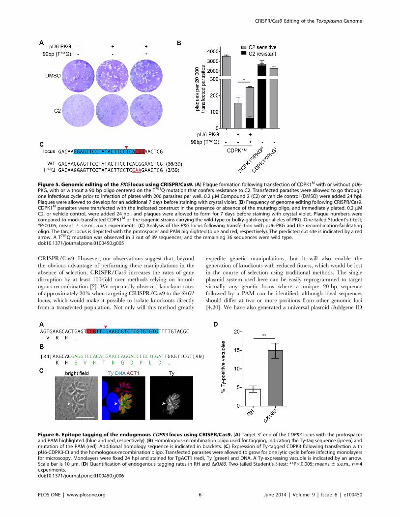
centered on the T761Q mutation. Following one lytic cycle,
parasites were used in plaque assays in the presence or absence of
C2. No plaques were observed in the presence of C2 for mock
transfections or parasites treated with the plasmid alone
(Figure 5A). In contrast, parasites that received both the plasmid
and the oligo yielded a significant number of plaques in the
presence of C2, suggesting remarkable rates of homologous
recombination (Figure 5A). To quantify this effect, parasites were
placed into plaque assays immediately after transfection, and C2
was added to the wells indicated 24 hours later. Although addition
of pU6-PKG resulted in the characteristic loss of viability,
inclusion of the oligo moderately increased viability by the same
proportion as the number of resulting C2 resistant plaques
(Figure 5B). These results demonstrate that, in the absence of
selection, CRISPR/Cas9 can initiate homologous recombination
in up to a third of the resulting parasite population.
To confirm that C2 resistance was mediated by the intended
mutation in PKG, the targeted locus was again amplified, TOPO
cloned and sequenced. This analysis exclusively revealed either the
wild-type locus or the expected T761Q mutation. Taken together,
these results demonstrate that high rates of directed genome
editing are possible in T. gondii using the CRISPR/Cas9 system.
Tagging of endogenous loci using CRISPR/Cas9Following our success introducing point mutations into the T.
gondii genome, we attempted to epitope tag the CDPK3 endoge-
nous locus. Previous work has established that C-terminally tagged
CDPK3 is functional and can complement the endogenous copy,
suggesting there should be no detrimental consequences from
similar manipulation of the endogenous locus [7,16,17]. We
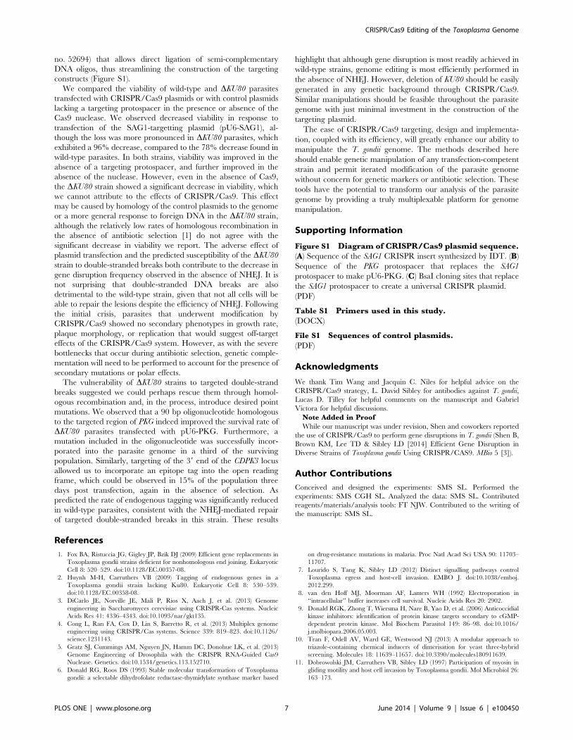
designed a protospacer that would target Cas9 to the 39 end of the
CDPK3 locus, checking its specificity as described above
(Figure 6A). We constructed plasmid pU6-CDPK3-Ct, analogous
to the other targeting constructs and containing the CDPK3-
targeting chiRNA under the U6 promoter and Cas9 under the
TUB1 promoter. To modify the locus, we synthesized a dsDNA
oligo consisting of the sequence for the Ty epitope tag [12]
followed by a stop codon and flanked by 40–43 bp of homology to
introduce it in-frame with the CDPK3 open reading frame
(Figure 6B). Additionally, the oligo harbored a mutation in the
PAM motif to ensure that following homologous recombination,
the locus could no longer be targeted by the nuclease. The DKU80
parasites were transfected with both pU6-CDPK3-Ct and the
oligo and allowed to undergo a lytic cycle before inoculating host
cells for microscopy. Samples were stained 24 hpi for both Ty and
parasite actin. Vacuoles expressing Ty were readily observed in
populations that had been transfected with both the oligo and the
CRISPR/Cas9-targeting construct (Figure 6C), although they
were never observed when either was omitted. Furthermore the
pattern of Ty-staining matches the previously reported localization
of CDPK3 at the periphery of the parasites [7,16,17].
To establish the rate of endogenous tagging in DKU80 and wild-
type parasites, we quantified the number of Ty-expressing
vacuoles on the third day following transfection, as described
above. The rate of endogenous tagging was approximately 5% in
wild-type and 15% in DKU80 parasites (Figure 6D), revealing an
inverse trend to that of gene disruption (Figure 1D). These results
are consistent with the NHEJ-mediated repair of CRISPR/Cas9
targeted double-stranded breaks in wild-type parasites, which are
not expected to lead to in-frame incorporation of the Ty-tag.
Discussion
Current methods for genome engineering of T. gondii rely on the
generation of complex constructs for homologous recombination,
which yield significant efficiency only in specific parasite strains
and require antibiotic selection [1,2,18]. This has restricted this
type of analysis to a relatively small number of parasite genes.
Here, we present a genome engineering method based on the
CRISPR/Cas9 system that permits, with a single plasmid, the
rapid and efficient disruption of genomic loci. We demonstrate
that DKU80 parasite strains are significantly more vulnerable to
these targeted disruptions than wild-type parasites, and we
postulate that this vulnerability is due to the defect in this strain’s
ability to repair double-strand breaks using NHEJ. We further
exploit the susceptibility of the DKU80 strain as a genetic
background in which to introduce targeted point mutations and
epitope tags, and we show that genome editing can be achieved in
15–30% of the manipulated parasite population without the need
for any form of selection. Together, these methods enhance our
ability to manipulate the T. gondii genome and will enable high-
throughput manipulation of a broad range of genetic loci.
Available tools for genome editing in T. gondii rely on antibiotic
selection [1,19] and are therefore difficult to compare to
Figure 4. Generation of strains sensitive to C2 through the PKGlocus. (A) Diagrams showing the wild-type PKG locus and strains withaltered gatekeeper regions. The 39 end of the endogenous PKG wasreplaced with the corresponding cDNA sequence harboring either thewild-type gatekeeper residue or the T761M mutation. (B) HFFmonolayers were infected with either CDPK1M/PKGT or CDPK1M/PKGM,and 0.2 mM C2 was added to the indicated wells 24 hours later. Plaqueswere allowed to develop for an additional 7 days before staining withcrystal violet.doi:10.1371/journal.pone.0100450.g004
CRISPR/Cas9 Editing of the Toxoplasma Genome
PLOS ONE | www.plosone.org 5 June 2014 | Volume 9 | Issue 6 | e100450
CRISPR/Cas9. However, our observations suggest that, beyond
the obvious advantage of performing these manipulations in the
absence of selection, CRISPR/Cas9 increases the rates of gene
disruption by at least 100-fold over methods relying on homol-
ogous recombination [2]. We repeatedly observed knockout rates
of approximately 20% when targeting CRISPR/Cas9 to the SAG1
locus, which would make it possible to isolate knockouts directly
from a transfected population. Not only will this method greatly
expedite genetic manipulations, but it will also enable the
generation of knockouts with reduced fitness, which would be lost
in the course of selection using traditional methods. The single
plasmid system used here can be easily reprogrammed to target
virtually any genetic locus where a unique 20 bp sequence
followed by a PAM can be identified, although ideal sequences
should differ at two or more positions from other genomic loci
[4,20]. We have also generated a universal plasmid (Addgene ID
Figure 5. Genomic editing of the PKG locus using CRISPR/Cas9. (A) Plaque formation following transfection of CDPK1M with or without pU6-PKG, with or without a 90 bp oligo centered on the T761Q mutation that confers resistance to C2. Transfected parasites were allowed to go throughone infectious cycle prior to infection of plates with 200 parasites per well. 0.2 mM Compound 2 (C2) or vehicle control (DMSO) were added 24 hpi.Plaques were allowed to develop for an additional 7 days before staining with crystal violet. (B) Frequency of genome editing following CRISPR/Cas9.CDPK1M parasites were transfected with the indicated construct in the presence or absence of the mutating oligo, and immediately plated. 0.2 mMC2, or vehicle control, were added 24 hpi, and plaques were allowed to form for 7 days before staining with crystal violet. Plaque numbers werecompared to mock-transfected CDPK1M or the isogenic strains carrying the wild-type or bulky-gatekeeper alleles of PKG. One-tailed Student’s t-test;*P,0.05; means 6 s.e.m., n = 3 experiments. (C) Analysis of the PKG locus following transfection with pU6-PKG and the recombination-facilitatingoligo. The target locus is depicted with the protospacer and PAM highlighted (blue and red, respectively). The predicted cut site is indicated by a redarrow. A T761Q mutation was observed in 3 out of 39 sequences, and the remaining 36 sequences were wild type.doi:10.1371/journal.pone.0100450.g005
Figure 6. Epitope tagging of the endogenous CDPK3 locus using CRISPR/Cas9. (A) Target 39 end of the CDPK3 locus with the protospacerand PAM highlighted (blue and red, respectively). (B) Homologous-recombination oligo used for tagging, indicating the Ty-tag sequence (green) andmutation of the PAM (red). Additional homology sequence is indicated in brackets. (C) Expression of Ty-tagged CDPK3 following transfection withpU6-CDPK3-Ct and the homologous-recombination oligo. Transfected parasites were allowed to grow for one lytic cycle before infecting monolayersfor microscopy. Monolayers were fixed 24 hpi and stained for TgACT1 (red), Ty (green) and DNA. A Ty-expressing vacuole is indicated by an arrow.Scale bar is 10 mm. (D) Quantification of endogenous tagging rates in RH and DKU80. Two-tailed Student’s t-test; **P,0.005; means 6 s.e.m., n = 4experiments.doi:10.1371/journal.pone.0100450.g006
CRISPR/Cas9 Editing of the Toxoplasma Genome
PLOS ONE | www.plosone.org 6 June 2014 | Volume 9 | Issue 6 | e100450
no. 52694) that allows direct ligation of semi-complementary
DNA oligos, thus streamlining the construction of the targeting
constructs (Figure S1).
We compared the viability of wild-type and DKU80 parasites
transfected with CRISPR/Cas9 plasmids or with control plasmids
lacking a targeting protospacer in the presence or absence of the
Cas9 nuclease. We observed decreased viability in response to
transfection of the SAG1-targetting plasmid (pU6-SAG1), al-
though the loss was more pronounced in DKU80 parasites, which
exhibited a 96% decrease, compared to the 78% decrease found in
wild-type parasites. In both strains, viability was improved in the
absence of a targeting protospacer, and further improved in the
absence of the nuclease. However, even in the absence of Cas9,
the DKU80 strain showed a significant decrease in viability, which
we cannot attribute to the effects of CRISPR/Cas9. This effect
may be caused by homology of the control plasmids to the genome
or a more general response to foreign DNA in the DKU80 strain,
although the relatively low rates of homologous recombination in
the absence of antibiotic selection [1] do not agree with the
significant decrease in viability we report. The adverse effect of
plasmid transfection and the predicted susceptibility of the DKU80
strain to double-stranded breaks both contribute to the decrease in
gene disruption frequency observed in the absence of NHEJ. It is
not surprising that double-stranded DNA breaks are also
detrimental to the wild-type strain, given that not all cells will be
able to repair the lesions despite the efficiency of NHEJ. Following
the initial crisis, parasites that underwent modification by
CRISPR/Cas9 showed no secondary phenotypes in growth rate,
plaque morphology, or replication that would suggest off-target
effects of the CRISPR/Cas9 system. However, as with the severe
bottlenecks that occur during antibiotic selection, genetic comple-
mentation will need to be performed to account for the presence of
secondary mutations or polar effects.
The vulnerability of DKU80 strains to targeted double-strand
breaks suggested we could perhaps rescue them through homol-
ogous recombination and, in the process, introduce desired point
mutations. We observed that a 90 bp oligonucleotide homologous
to the targeted region of PKG indeed improved the survival rate of
DKU80 parasites transfected with pU6-PKG. Furthermore, a
mutation included in the oligonucleotide was successfully incor-
porated into the parasite genome in a third of the surviving
population. Similarly, targeting of the 39 end of the CDPK3 locus
allowed us to incorporate an epitope tag into the open reading
frame, which could be observed in 15% of the population three
days post transfection, again in the absence of selection. As
predicted the rate of endogenous tagging was significantly reduced
in wild-type parasites, consistent with the NHEJ-mediated repair
of targeted double-stranded breaks in this strain. These results
highlight that although gene disruption is most readily achieved in
wild-type strains, genome editing is most efficiently performed in
the absence of NHEJ. However, deletion of KU80 should be easily
generated in any genetic background through CRISPR/Cas9.
Similar manipulations should be feasible throughout the parasite
genome with just minimal investment in the construction of the
targeting plasmid.
The ease of CRISPR/Cas9 targeting, design and implementa-
tion, coupled with its efficiency, will greatly enhance our ability to
manipulate the T. gondii genome. The methods described here
should enable genetic manipulation of any transfection-competent
strain and permit iterated modification of the parasite genome
without concern for genetic markers or antibiotic selection. These
tools have the potential to transform our analysis of the parasite
genome by providing a truly multiplexable platform for genome
manipulation.
Supporting Information
Figure S1 Diagram of CRISPR/Cas9 plasmid sequence.(A) Sequence of the SAG1 CRISPR insert synthesized by IDT. (B)
Sequence of the PKG protospacer that replaces the SAG1
protospacer to make pU6-PKG. (C) BsaI cloning sites that replace
the SAG1 protospacer to create a universal CRISPR plasmid.
(PDF)
Table S1 Primers used in this study.
(DOCX)
File S1 Sequences of control plasmids.
(PDF)
Acknowledgments
We thank Tim Wang and Jacquin C. Niles for helpful advice on the
CRISPR/Cas9 strategy, L. David Sibley for antibodies against T. gondii,
Lucas D. Tilley for helpful comments on the manuscript and Gabriel
Victora for helpful discussions.
Note Added in Proof
While our manuscript was under revision, Shen and coworkers reported
the use of CRISPR/Cas9 to perform gene disruptions in T. gondii (Shen B,
Brown KM, Lee TD & Sibley LD [2014] Efficient Gene Disruption in
Diverse Strains of Toxoplasma gondii Using CRISPR/CAS9. MBio 5 [3]).
Author Contributions
Conceived and designed the experiments: SMS SL. Performed the
experiments: SMS CGH SL. Analyzed the data: SMS SL. Contributed
reagents/materials/analysis tools: FT NJW. Contributed to the writing of
the manuscript: SMS SL.
References
1. Fox BA, Ristuccia JG, Gigley JP, Bzik DJ (2009) Efficient gene replacements in
Toxoplasma gondii strains deficient for nonhomologous end joining. Eukaryotic
Cell 8: 520–529. doi:10.1128/EC.00357-08.
2. Huynh M-H, Carruthers VB (2009) Tagging of endogenous genes in a
Toxoplasma gondii strain lacking Ku80. Eukaryotic Cell 8: 530–539.
doi:10.1128/EC.00358-08.
3. DiCarlo JE, Norville JE, Mali P, Rios X, Aach J, et al. (2013) Genome
engineering in Saccharomyces cerevisiae using CRISPR-Cas systems. Nucleic
Acids Res 41: 4336–4343. doi:10.1093/nar/gkt135.
4. Cong L, Ran FA, Cox D, Lin S, Barretto R, et al. (2013) Multiplex genome
engineering using CRISPR/Cas systems. Science 339: 819–823. doi:10.1126/
science.1231143.
5. Gratz SJ, Cummings AM, Nguyen JN, Hamm DC, Donohue LK, et al. (2013)
Genome Engineering of Drosophila with the CRISPR RNA-Guided Cas9
Nuclease. Genetics. doi:10.1534/genetics.113.152710.
6. Donald RG, Roos DS (1993) Stable molecular transformation of Toxoplasma
gondii: a selectable dihydrofolate reductase-thymidylate synthase marker based
on drug-resistance mutations in malaria. Proc Natl Acad Sci USA 90: 11703–
11707.
7. Lourido S, Tang K, Sibley LD (2012) Distinct signalling pathways control
Toxoplasma egress and host-cell invasion. EMBO J. doi:10.1038/emboj.
2012.299.
8. van den Hoff MJ, Moorman AF, Lamers WH (1992) Electroporation in
‘‘intracellular’’ buffer increases cell survival. Nucleic Acids Res 20: 2902.
9. Donald RGK, Zhong T, Wiersma H, Nare B, Yao D, et al. (2006) Anticoccidial
kinase inhibitors: identification of protein kinase targets secondary to cGMP-
dependent protein kinase. Mol Biochem Parasitol 149: 86–98. doi:10.1016/
j.molbiopara.2006.05.003.
10. Tran F, Odell AV, Ward GE, Westwood NJ (2013) A modular approach to
triazole-containing chemical inducers of dimerisation for yeast three-hybrid
screening. Molecules 18: 11639–11657. doi:10.3390/molecules180911639.
11. Dobrowolski JM, Carruthers VB, Sibley LD (1997) Participation of myosin in
gliding motility and host cell invasion by Toxoplasma gondii. Mol Microbiol 26:
163–173.
CRISPR/Cas9 Editing of the Toxoplasma Genome
PLOS ONE | www.plosone.org 7 June 2014 | Volume 9 | Issue 6 | e100450
12. Bastin P, Bagherzadeh Z, Matthews KR, Gull K (1996) A novel epitope tag
system to study protein targeting and organelle biogenesis in Trypanosoma
brucei. Mol Biochem Parasitol 77: 235–239.
13. Nagel SD, Nagel SD, Boothroyd JC, Boothroyd JC (1988) The alpha- and beta-
tubulins of Toxoplasma gondii are encoded by single copy genes containing
multiple introns. Mol Biochem Parasitol 29: 261–273. Available: http://eutils.ncbi.
nlm.nih.gov/entrez/eutils/elink.fcgi?dbfrom = pubmed&id = 3412377&retmode =
ref&cmd = prlinks.
14. Kim K, Boothroyd JC (1995) Toxoplasma gondii: stable complementation of
sag1 (p30) mutants using SAG1 transfection and fluorescence-activated cell
sorting. Exp Parasitol 80: 46–53. doi:10.1006/expr.1995.1006.
15. Jinek M, Chylinski K, Fonfara I, Hauer M, Doudna JA, et al. (2012) A
programmable dual-RNA-guided DNA endonuclease in adaptive bacterial
immunity. Science 337: 816–821. doi:10.1126/science.1225829.
16. McCoy JM, Whitehead L, van Dooren GG, Tonkin CJ (2012) TgCDPK3
Regulates Calcium-Dependent Egress of Toxoplasma gondii from Host Cells.PLoS Pathog 8: e1003066. doi:10.1371/journal.ppat.1003066.g006.
17. Garrison E, Treeck M, Ehret E, Butz H, Garbuz T, et al. (2012) A forward
genetic screen reveals that calcium-dependent protein kinase 3 regulates egressin Toxoplasma. PLoS Pathog 8: e1003049. doi:10.1371/journal.ppat.1003049.
t001.18. Andenmatten N, Egarter S, Jackson AJ, Jullien N, Herman J-P, et al. (2013)
Conditional genome engineering in Toxoplasma gondii uncovers alternative
invasion mechanisms. Nat Methods 10: 125–127. doi:10.1038/nmeth.2301.19. Kim K, Soldati D, Boothroyd JC (1993) Gene replacement in Toxoplasma
gondii with chloramphenicol acetyltransferase as selectable marker. Science 262:911–914.
20. Wang T, Wei JJ, Sabatini DM, Lander ES (2014) Genetic screens in human cellsusing the CRISPR-Cas9 system. Science 343: 80–84. doi:10.1126/science.
1246981.
CRISPR/Cas9 Editing of the Toxoplasma Genome
PLOS ONE | www.plosone.org 8 June 2014 | Volume 9 | Issue 6 | e100450