Embed Size (px)
Citation preview
Evaluation of Endodontic
Outcomes
Dr Vág János
Department of Conservative Dentistry
Mahmoud Torabinejad, Richard E. Walton, ENDODONTICS: PRINCIPLES AND
PRACTICE 4th edition, Chapter 21 Evaluation of Endodontic Outcomes
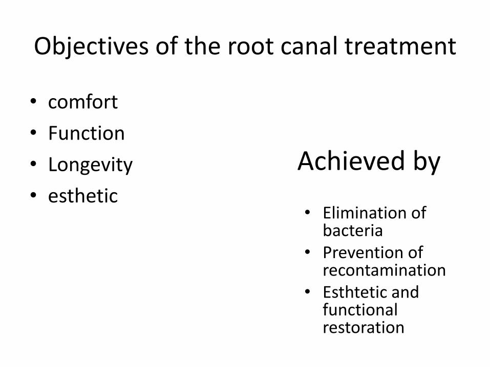
Objectives of the root canal treatment
• comfort
• Function
• Longevity
• esthetic • Elimination of
bacteria
• Prevention of recontamination
• Esthtetic and functional restoration
Achieved by
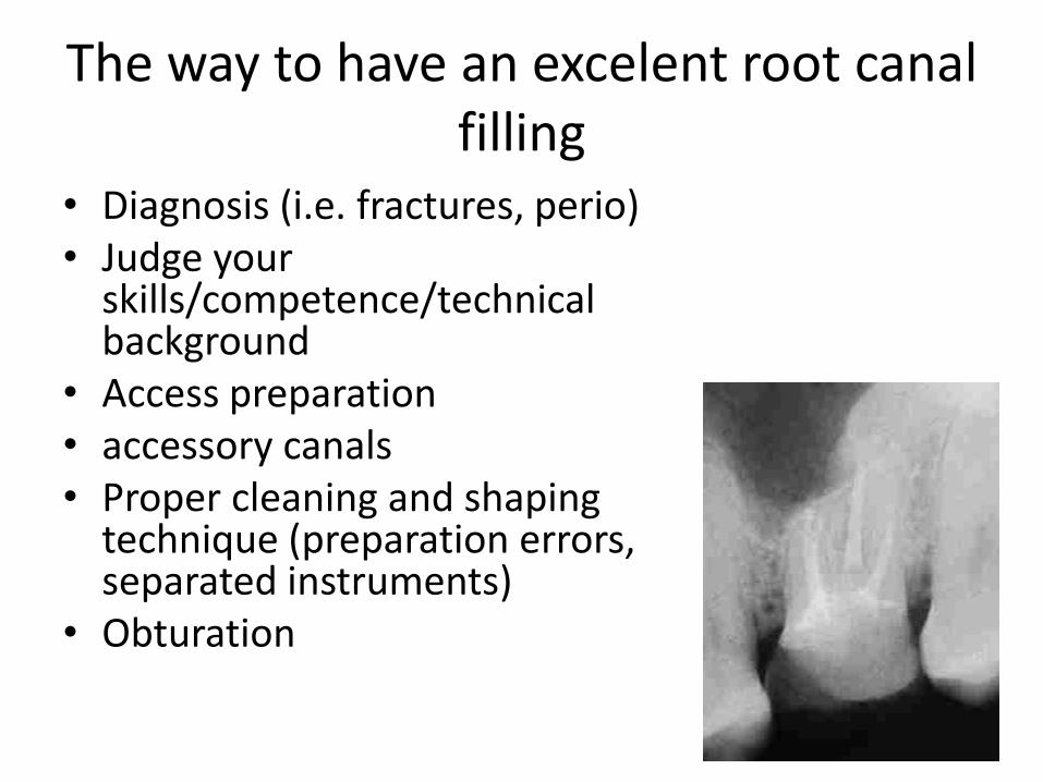
The way to have an excelent root canal
filling
• Diagnosis (i.e. fractures, perio)
• Judge your skills/competence/technical background
• Access preparation
• accessory canals
• Proper cleaning and shaping technique (preparation errors, separated instruments)
• Obturation
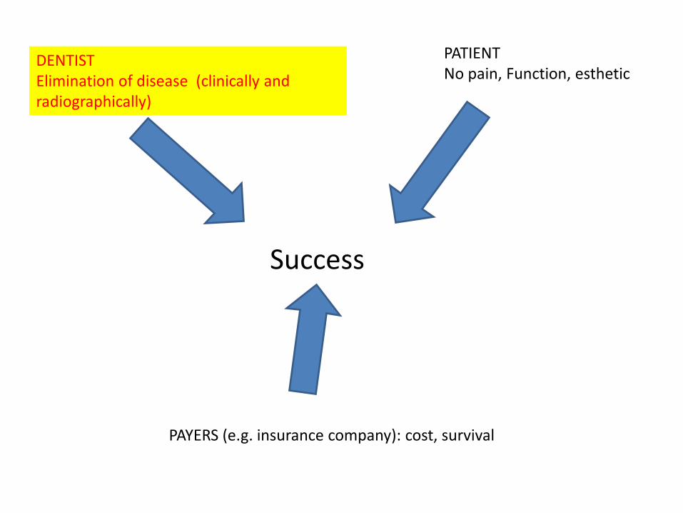
Success
PATIENT
No pain, Function, esthetic DENTIST
Elimination of disease (clinically and
radiographically)
PAYERS (e.g. insurance company): cost, survival
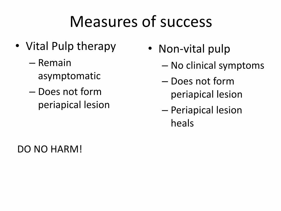
Measures of success
• Vital Pulp therapy
– Remain
asymptomatic
– Does not form
periapical lesion
• Non-vital pulp
– No clinical symptoms
– Does not form
periapical lesion
– Periapical lesion
heals
DO NO HARM!
Errors in treatment planning
• Dentist skills
• Poor prognosis
• Root fracture
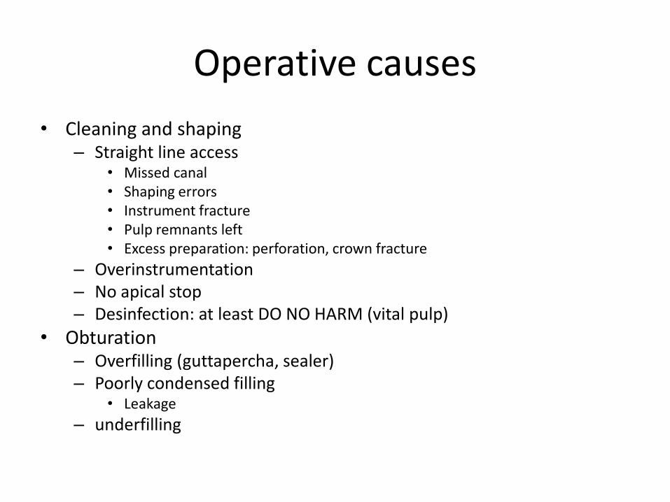
Operative causes
• Cleaning and shaping – Straight line access
• Missed canal
• Shaping errors
• Instrument fracture
• Pulp remnants left
• Excess preparation: perforation, crown fracture
– Overinstrumentation
– No apical stop
– Desinfection: at least DO NO HARM (vital pulp)
• Obturation – Overfilling (guttapercha, sealer)
– Poorly condensed filling • Leakage
– underfilling
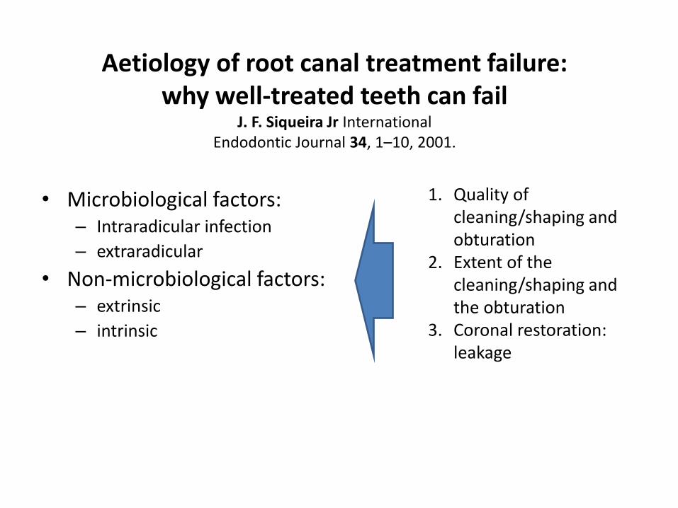
Aetiology of root canal treatment failure:
why well-treated teeth can fail J. F. Siqueira Jr International
Endodontic Journal 34, 1–10, 2001.
• Microbiological factors:
– Intraradicular infection
– extraradicular
• Non-microbiological factors:
– extrinsic
– intrinsic
1. Quality of
cleaning/shaping and
obturation
2. Extent of the
cleaning/shaping and
the obturation
3. Coronal restoration:
leakage
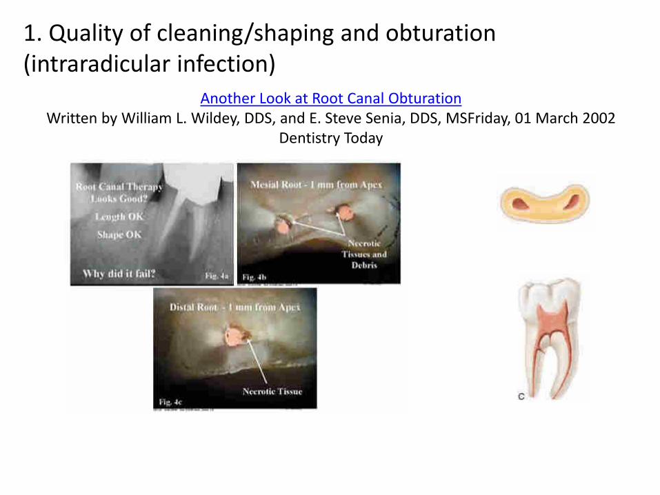
Another Look at Root Canal Obturation
Written by William L. Wildey, DDS, and E. Steve Senia, DDS, MSFriday, 01 March 2002
Dentistry Today
1. Quality of cleaning/shaping and obturation
(intraradicular infection)
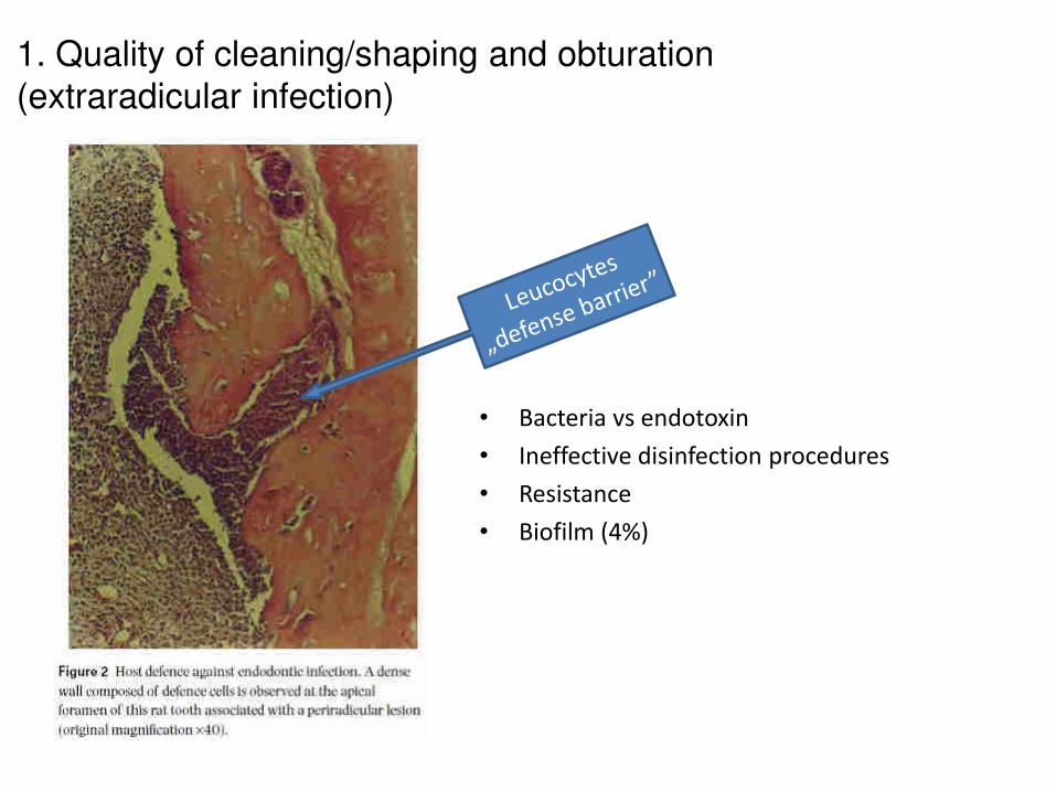
1. Quality of cleaning/shaping and obturation
(extraradicular infection)
• Bacteria vs endotoxin
• Ineffective disinfection procedures
• Resistance
• Biofilm (4%)
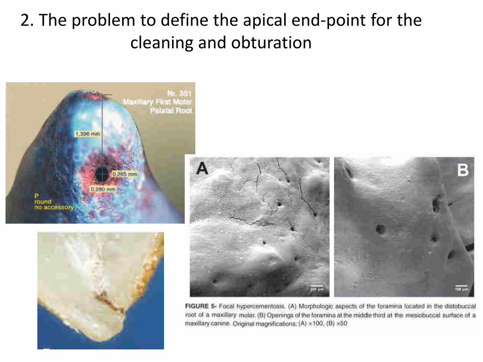
2. The problem to define the apical end-point for the
cleaning and obturation
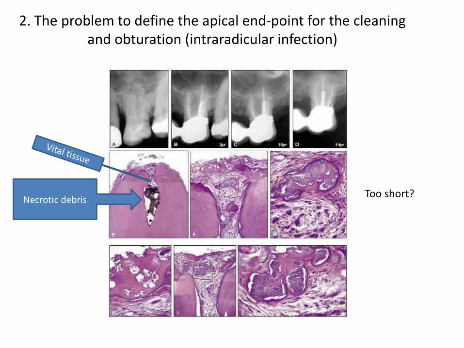
2. The problem to define the apical end-point for the cleaning
and obturation (intraradicular infection)
Necrotic debris Too short?
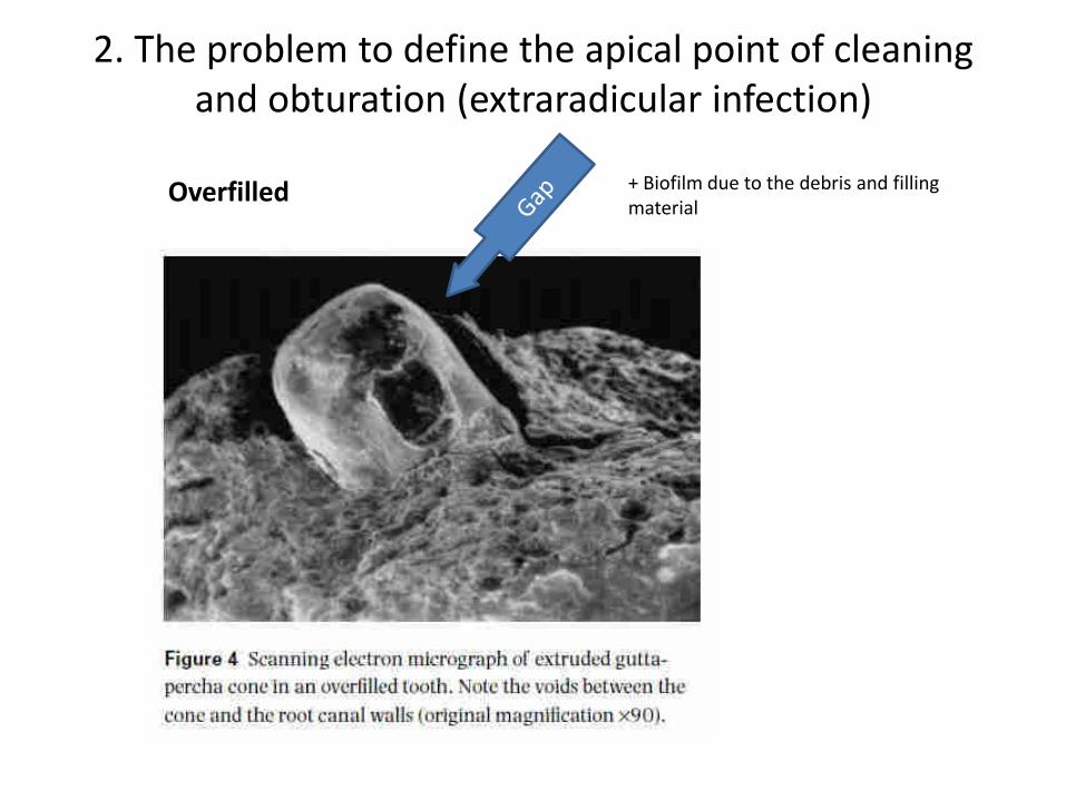
Overfilled + Biofilm due to the debris and filling
material
2. The problem to define the apical point of cleaning
and obturation (extraradicular infection)
A lekes
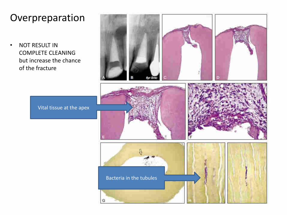
Vital tissue at the apex
• NOT RESULT IN
COMPLETE CLEANING
but increase the chance
of the fracture
Bacteria in the tubules
Overpreparation
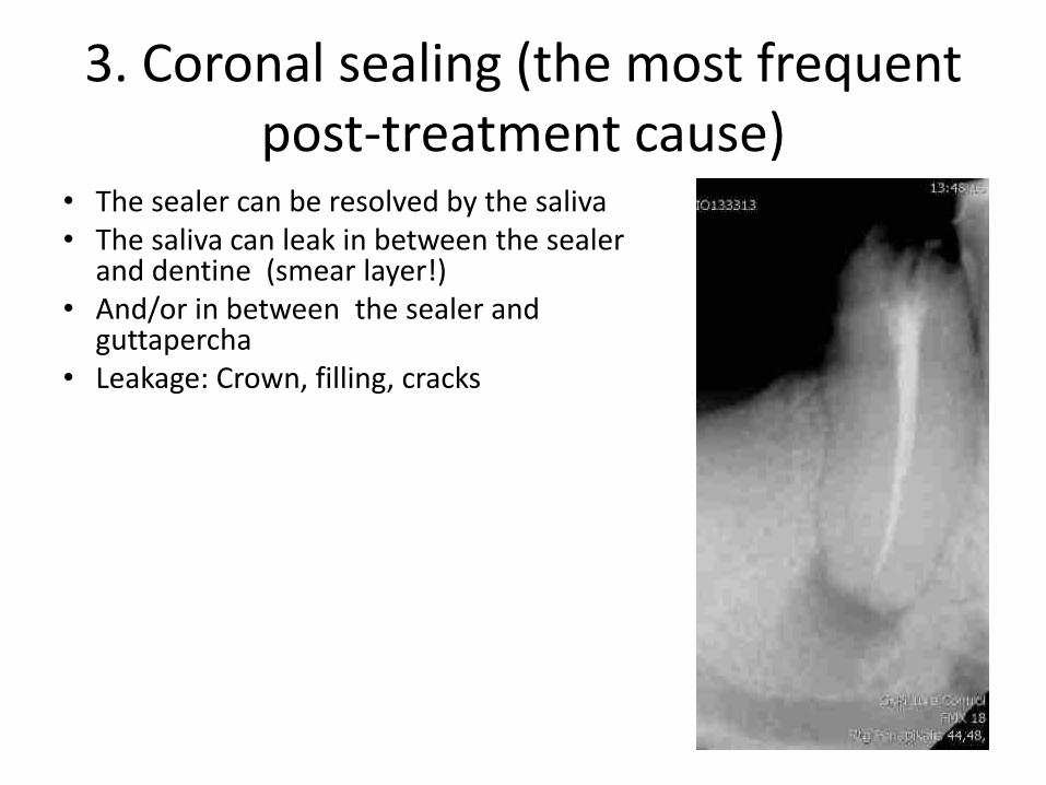
3. Coronal sealing (the most frequent
post-treatment cause) • The sealer can be resolved by the saliva
• The saliva can leak in between the sealer and dentine (smear layer!)
• And/or in between the sealer and guttapercha
• Leakage: Crown, filling, cracks
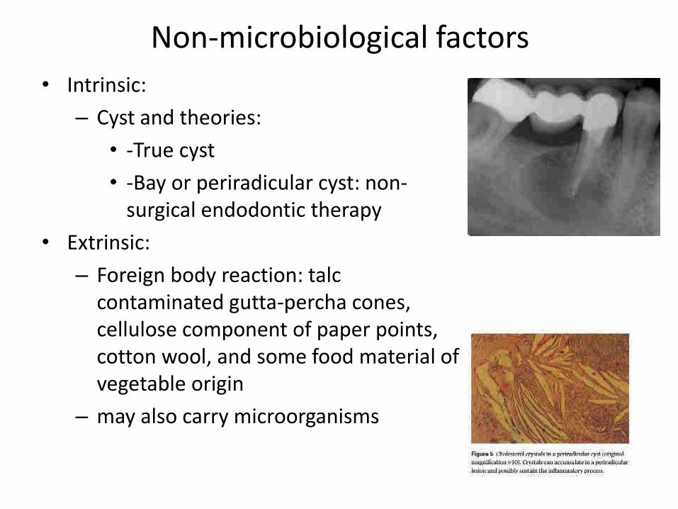
Non-microbiological factors
• Intrinsic:
– Cyst and theories:
• -True cyst
• -Bay or periradicular cyst: non-
surgical endodontic therapy
• Extrinsic:
– Foreign body reaction: talc
contaminated gutta-percha cones,
cellulose component of paper points,
cotton wool, and some food material of
vegetable origin
– may also carry microorganisms
Resolution of intraradicular infection
• Disinfection: Kill them all!
• Good sealing (obturation): enclosed the survival and
seal the gap against the nutritive tissue fluid (Bacterial
are excellent survivals)
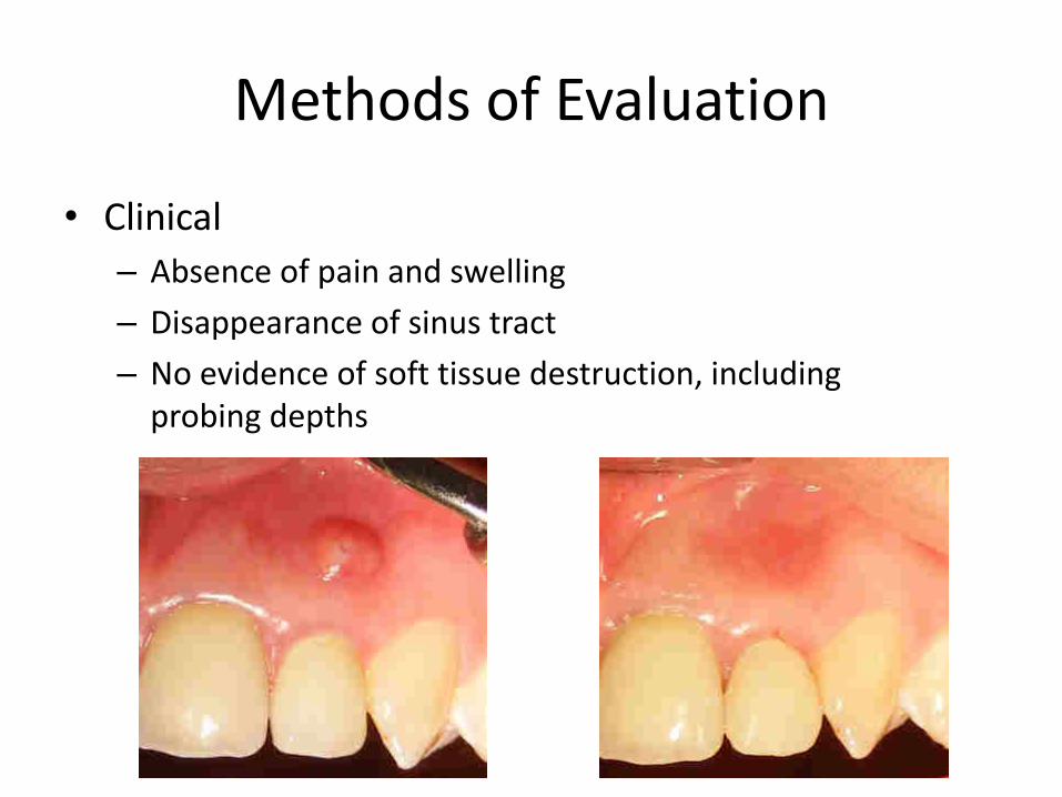
Methods of Evaluation
• Clinical
– Absence of pain and swelling
– Disappearance of sinus tract
– No evidence of soft tissue destruction, including
probing depths
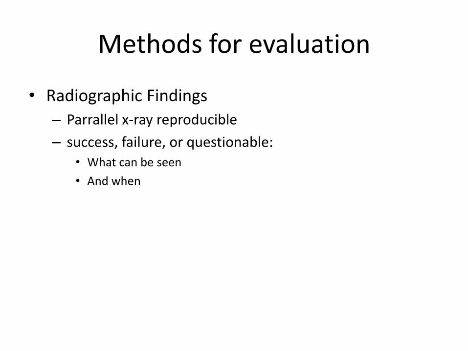
Methods for evaluation
• Radiographic Findings
– Parrallel x-ray reproducible
– success, failure, or questionable:
• What can be seen
• And when
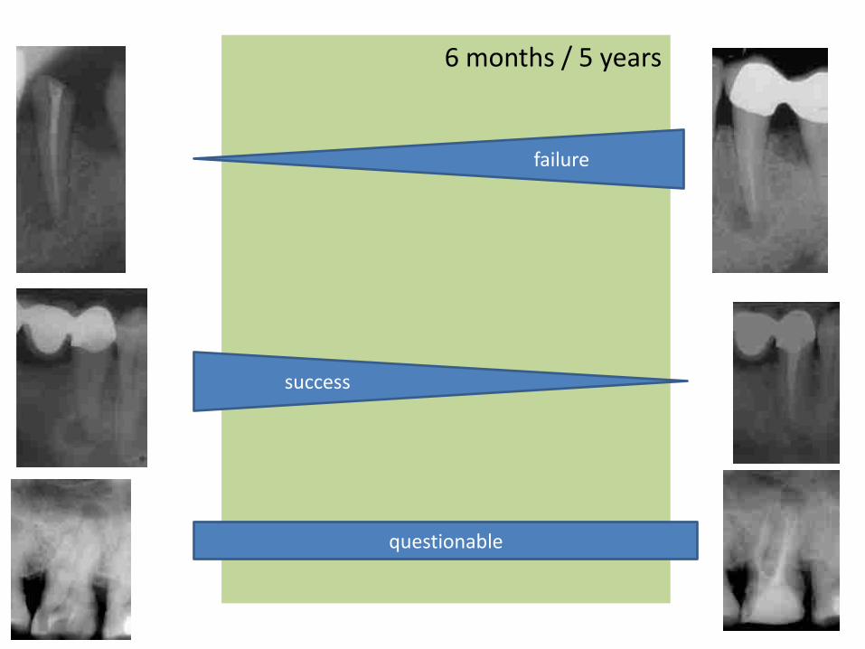
6 months / 5 years
questionable
success
failure
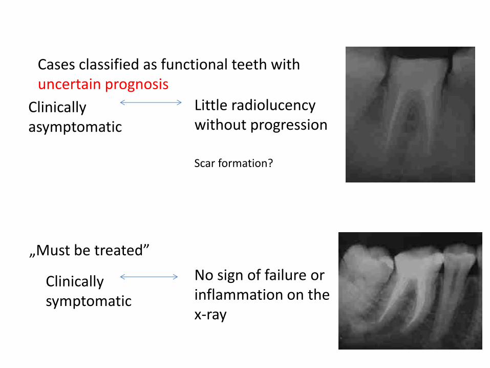
Cases classified as functional teeth with
uncertain prognosis
Little radiolucency
without progression Clinically
asymptomatic
„Must be treated”
No sign of failure or
inflammation on the
x-ray
Clinically
symptomatic
Scar formation?

Success rates
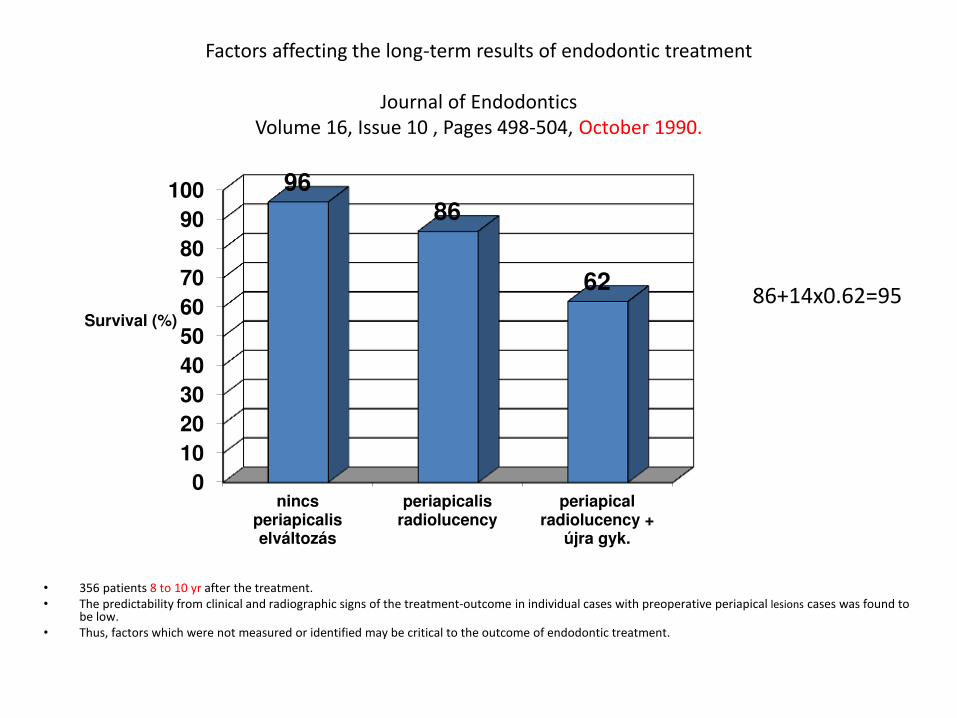
Factors affecting the long-term results of endodontic treatment
Journal of Endodontics
Volume 16, Issue 10 , Pages 498-504, October 1990.
• 356 patients 8 to 10 yr after the treatment.
• The predictability from clinical and radiographic signs of the treatment-outcome in individual cases with preoperative periapical lesions cases was found to be low.
• Thus, factors which were not measured or identified may be critical to the outcome of endodontic treatment.
0
10
20
30
40
50
60
70
80
90
100
nincsperiapicaliselváltozás
periapicalisradiolucency
periapicalradiolucency +
újra gyk.
96 86
62
Survival (%)
86+14x0.62=95
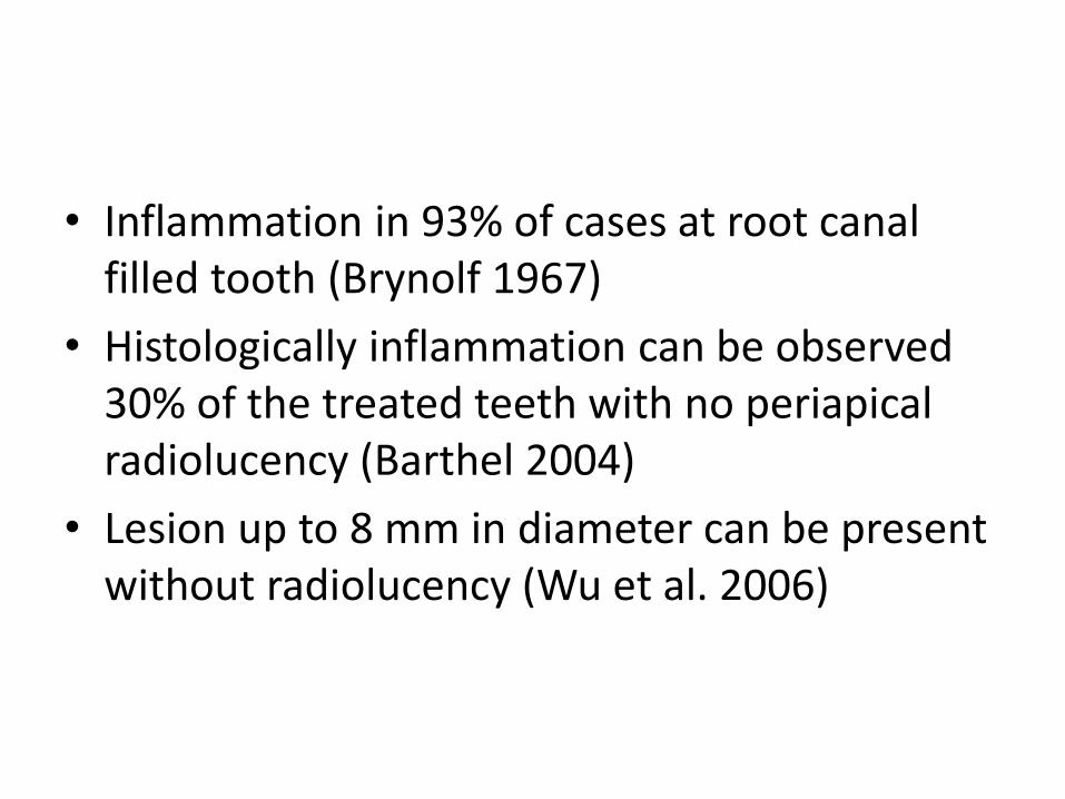
• Inflammation in 93% of cases at root canal
filled tooth (Brynolf 1967)
• Histologically inflammation can be observed
30% of the treated teeth with no periapical
radiolucency (Barthel 2004)
• Lesion up to 8 mm in diameter can be present
without radiolucency (Wu et al. 2006)
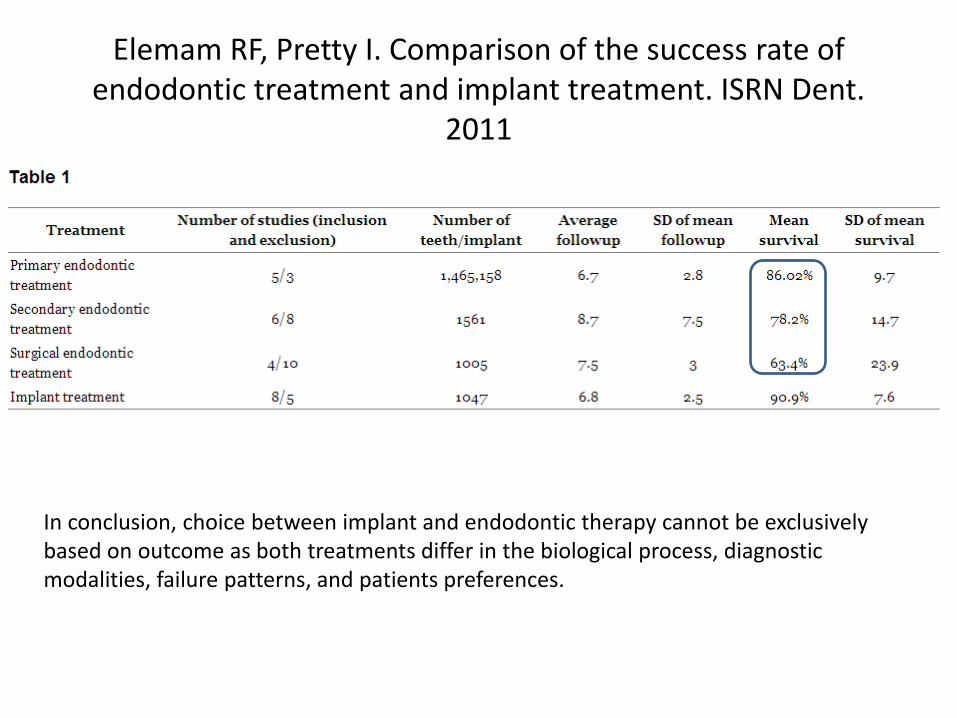
Elemam RF, Pretty I. Comparison of the success rate of
endodontic treatment and implant treatment. ISRN Dent.
2011
In conclusion, choice between implant and endodontic therapy cannot be exclusively
based on outcome as both treatments differ in the biological process, diagnostic
modalities, failure patterns, and patients preferences.