Embed Size (px)
Citation preview

Fluid and Electrolytes
Homeostasis • State of equilibrium in
body
• Naturally maintained by adaptive responses
• Body fluids and electrolytes are maintained within narrow limits
• 60% of body weight in adult
• 45% to 55% in older adult
• 70% to 80% in infants
– Varies with gender, body mass, and age


• Compartments • Intracellular fluid (ICF)
(cell membrane)
• Extracellular fluid (ECF)
– Interstitial = tissue
capillary membrane
– Intravascular (plasma)
• One third of body fluid
• 3 major components
1) Interstitial fluid
2) Intravascular
3) Trans cellular fluid • over or across the cells
Extracellular Fluid (ECF)

• Fluid between cells
– Surrounds cells
– Transport medium for nutrients, gases, waste products and other substances between blood and body cells
– Also acts as a back up fluid reservoir
Interstitial Component
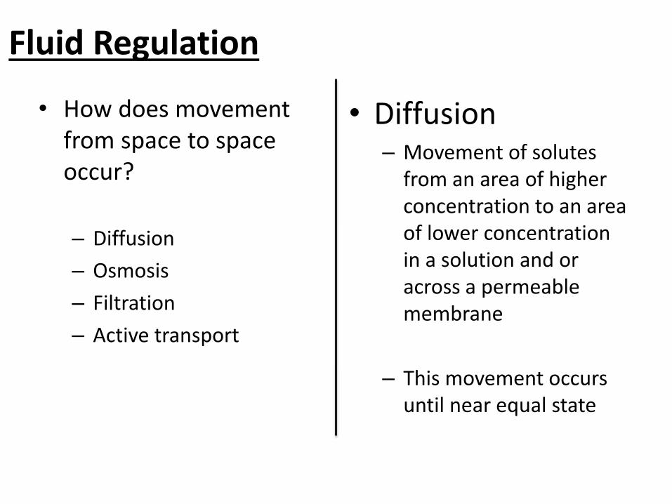
• How does movement from space to space occur?
– Diffusion
– Osmosis
– Filtration
– Active transport
• Diffusion – Movement of solutes
from an area of higher concentration to an area of lower concentration in a solution and or across a permeable membrane
– This movement occurs until near equal state
Fluid Regulation

•Osmosis - Now with water.
Fluid Regulation


Fluid Regulation Osmosis VS. Diffusion
• Filtration – Water pushing against
the confining walls of a space
• Osmosis
– Low to high
– Water potential
• Diffusion -High to low
-Movement of particles

Electrolytes Electrolyte Composition
• Substances whose molecules dissociate into ions (charged particles) when placed into water
– Cations: positively charged
– Anions: negatively charged
• ICF
– Prevalent cation is K+
– Prevalent anion is PO43-
• ECF
– Prevalent cation is Na+
– Prevalent anion is Cl-

Regulation of Electrolytes
Active transport Allows molecules to move against concentration and osmotic pressure to areas of higher concentration
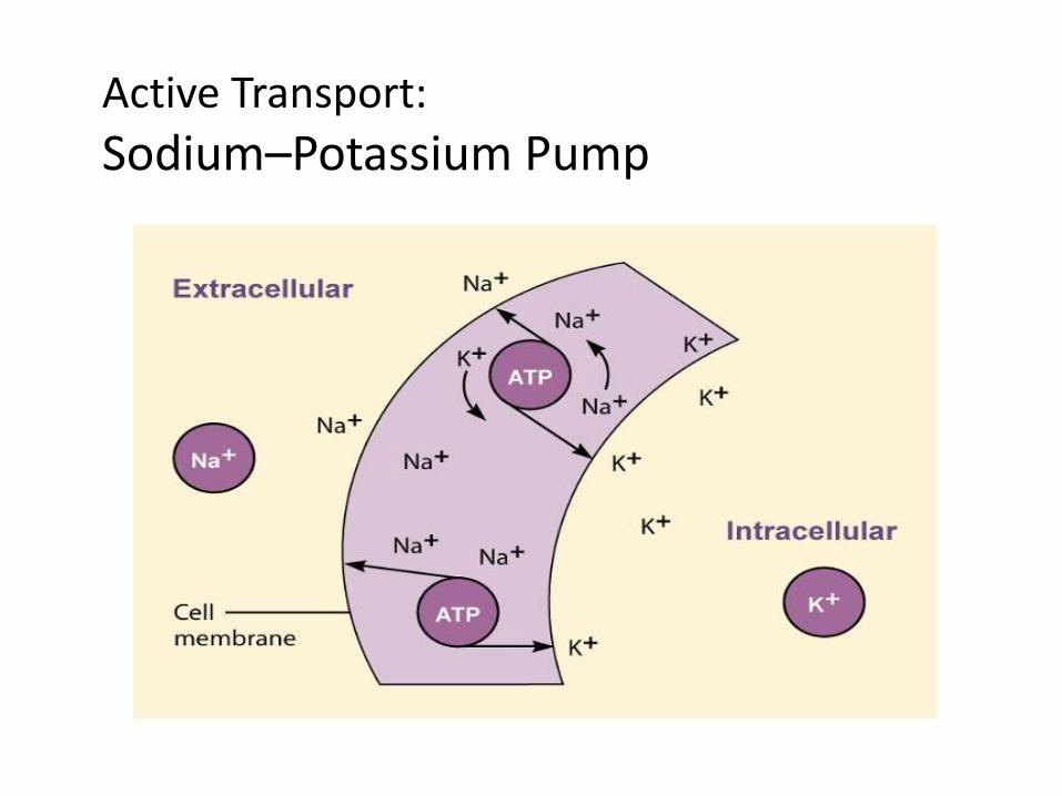
Active Transport:
Sodium–Potassium Pump

Fluid Movement in Capillaries Osmolality
• Amount and direction of movement determined by
– Capillary hydrostatic pressure
– Plasma oncotic pressure
– Interstitial hydrostatic pressure
– Interstitial oncotic pressure
• Concentration of body fluids- affects movement of fluid by osmosis.
• Reflects hydration status
• Measured by serum and urine
• Solutes measured-mainly urea, glucose, & sodium

Osmolality Fluid Volume Shifts
• Serum value 280-300 mOsm/kg
• Urine value 250-900 mOsm/kg
• Increases in serum level
– Free water loss
– Elevated Na
– Hyperglycemia
– Uremia
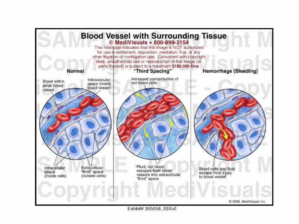
• Normally fluid shifts between intracellular and extracellular compartments to maintain equilibrium between spaces
-Fluid not lost from body, but not available for use in either compartment- considered third-space fluid shift (third-spacing)
-Enters interstitial compartment

Causes of Third-Spacing Assessment of Third-Spacing
• Burns
• Peritonitis
• Bowel obstruction
• Massive bleeding into joint or cavity
• Liver or renal failure
• Lowered plasma proteins
• Increased capillary permeability
• More difficult – fluid sequestered in deeper structures
• Signs/Symptoms
– Decreased urine output with adequate intake
– Increased HR
– Decreased BP
– Increased weight
– Pitting edema, ascites

Phases of Third-Spacing Treatment
1-Loss phase – Lasts 48-72 hours
– Symptoms of FVD
2-Reabsorption phase – Fluid gradually reabsorbed
after problem subsides
– FVO possible
– Monitor VS, I&O, Wt, and breath sounds
• Treat underlying cause if possible
• Close observation of VS • Monitor I & O more
frequently • Daily weights • Measure abdominal girth
in ascites • Measure extremities if
necessary • Monitor lab values
– albumin level important

(Fluid Volume Deficit(FVD Extracellular Fluid Volume Imbalances
• ECF volume deficit (hypovolemia)
– Abnormal loss of normal body fluids (diarrhea, fistula drainage, hemorrhage), inadequate intake, or plasma-to-interstitial fluid shift
– Treatment: replace water and electrolytes with balanced IV solutions
• Hypovolemia • Abnormally low volume of
body fluid in intravascular and/or interstitial compartments
• Causes – Vomiting – Diarrhea – Fever – Excess sweating – Burns – Diabetes insipidus – Inadequate intake – Hemorrhage – Overuse of diuretics – Third spacing

Fluid volume deficit
• What happens – Output > Intake Water
extracted from ECF • ECF hypertonic (water moves
out of cell cell dehydration) + osmotic pressure increased (stimulates thirst preceptor in hypothalamus)
• ICF hypotonic with decreased osmotic pressure posterior pituitary secretes more ADH
• Decreased ECF volume adrenal glands secrete Aldosterone

Labs Signs and Symptoms
• Acute weight loss • Decreased skin turgor • Oliguria • Concentrated urine • Weak, rapid pulse • Capillary filling time
elongated • Decreased BP • Increased pulse • Sensations of thirst,
weakness, dizziness, muscle cramps
• Increased HCT (Hematocrit)
• Increased BUN(Blood urea nitrogen)
• Increased serum osmolality
• Increased urine osmolality
• Increased specific gravity
• Decreased urine volume, dark color

Interventions Nursing Management Nursing Diagnoses
• Hypovolemia – Deficient fluid volume – Decreased cardiac
output – Potential complication:
hypovolemic shock
• Major goal prevent or correct abnormal fluid volume status before ARF occurs
• Encourage fluids
• IV fluids – Isotonic solutions (0.9%
NS or LR) until BP back to normal, then hypotonic (0.45% NS)
• Monitor I & O, urine specific gravity, DAILY WEIGHTS

Extracellular Fluid Volume Imbalances
• Monitor skin turgor
• Monitor VS and mental status
• Goal:
– Normal skin turgor, increased UOP with normal specific gravity, normal VS, clear sensorium, good oral intake of fluids, labs WNL (within normal limits)
• Fluid volume excess (hypervolemia)
• Excessive intake of fluids, abnormal retention of fluids (CHF) Congestive heart failure, or interstitial-to-plasma fluid shift
• Treatment: remove fluid without changing electrolyte composition or osmolality of ECF

Signs/Symptoms
• Excessive isotonic or hypotonic IV fluids
• Heart failure
• Renal failure- urinary
• Liver failure, cirrhosis
• Long-term use corticosteroids
• Headache, confusion, lethargy
• Edema
• Distended neck veins
• Bounding pulse,
• Polyuria
• Dyspnea, crackles, pulmonary edema
• Wt. Gain
• Seizures, coma
Causes

Nursing Management
Nursing Diagnoses Nursing implementation
• Hypervolemia
– Excess fluid volume
– Ineffective airway clearance
– Risk for impaired skin integrity
– Disturbed body image
– Potential complications: pulmonary edema, ascites
• I&O
• Monitor cardiovascular changes
• Assess respiratory status and monitor changes
• Daily weights
• Skin assessment

Nursing Implementation
• Neurologic function
• LOC (level of consciousness)
• PERLA (Pupils equal, reactive to light) and accommodation
• Voluntary movement of extremities
• Muscle strength
• Reflexes

Electrolyte Disorders Signs and Symptoms
Deficit
Excess Electrolyte
Hyponatremia
CNS deterioration
Hypernatremia
Thirst
CNS deterioration
Increased interstitial fluid
Sodium (Na)
Hypokalemia
Bradycardia
ECG changes
CNS changes
Hyperkalemia
Ventricular fibrillation
ECG changes
CNS changes
Potassium (K)

Deficit Excess Electrolyte
Hypocalcemia
Tetany
Chvostek’s,
Trousseau’s signs
Muscle twitching
CNS changes
ECG changes
Hypercalcemia
Thirst
CNS deterioration
Increased interstitial
fluid
Calcium (Ca)
Hypomagnesemia
Hyperactive DTRs
CNS changes
Hypermagnesemia
Loss of deep tendon
reflexes (DTRs)
Depression of CNS
Depression of
neuromuscular
function
Magnesium (Mg)

IV Fluid Reference