Embed Size (px)
Citation preview
Observations on the loss of catecholamine fluorescencefrom intrauterine adrenergic nerves during pregnancy
in the guinea-pigC. Bell and S. J. Malcolm
Department ofPhysiology, University ofMelbourne, Parkville, Victoria 3052, Australia
Summary. During unilateral pregnancy in the guinea-pig there is loss of formaldehyde\x=req-\induced fluorescence from the adrenergic nerves supplying the uterus and its vascula-ture. This loss occurs initially near the site of implantation at about Day 20 of gestationand spreads progressively. Implantation ofwax pellets containing progesterone into theuterine lumen or the gastrocnemius muscle of virgin guinea-pigs for 7 days producedloss of fluorescence from all local adrenergic nerves. No diminution of fluorescence was
seen when pellets containing oestradiol were substituted. Chronic denervation studiesshowed that the adrenergic axons supplying the uterus and its arteries originated fromboth the ovarian artery and the pelvic region. Our results suggest that loss of adrenergicfluorescence within the uterus during pregnancy is due to an effect of placental pro-gesterone which is localized to the uterus because the high concentration of proges-terone necessary to cause fluorescence loss is not attained in the systemic circulation.
Introduction
In some species there is, during the course of pregnancy, a progressive disappearance of the charac¬teristic formaldehyde-induced fluorescence normally associated with the adrenergic nerves of theuterus and its arterial supply. There is also a fall in the uterine content of noradrenaline. These declineshave been observed in the guinea-pig (Sjöberg, 1968), rabbit (Rosengren & Sjöberg, 1968), man
(Nakanishi, McLean, Wood & Burnstock, 1968) and dog (Ryan, Clark & Brody, 1974) and Bell ( 1972)has suggested that they constitute a protective mechanism against feto-placental ischaemia duringgeneralized maternal sympathetic activation.
During unilateral pregnancy in the guinea-pig the disappearance of fluorescence occurs in bothuterine horns, indicating that it is likely to be hormonally mediated rather than being secondary tomechanical compression of the uterine wall by the products of conception (Sjöberg, 1968). By con¬
trast, no diminution of fluorescence of adrenergic nerves in other regions of the body is seen duringpregnancy. The male internal genitalia receive their adrenergic innervation from 'short' pelvicneurones (Sjöstrand, 1965) which exhibit marked differences from the 'long' neurones of the sym¬pathetic chain in their characteristics of transmitter turnover (see Bell, 1972). It has been proposedthat the pregnancy-induced loss of fluorescence is restricted to the intrauterine axons because thesealso arise from 'short' adrenergic neurones lying in the pelvic region (Sjöberg, 1968 ; Owman, Sjöberg& Sjöstrand, 1974). Such an explanation is, however, in conflict with evidence presented by otherworkers which suggests that the adrenergic innervation of the guinea-pig uterus arises primarily from'long' adrenergic neurones and enters the uterus with the ovarian artery (Isaac, Pennefather & Silva,1969; Kulkarni, Wakade & Kirpekar, 1976). In the present study we have re-examined the origins ofthe intrauterine adrenergic nerves and made some observations on the probable mechanism of loss offluorescence during pregnancy. Ä*·-^
Materials and Methods
The animals were from our outbred colony of smooth-haired guinea-pigs in which the females are
reproductively active at 300 g body weight and the length ofgestation is between 63 and 70 days (mean65 days). The animals used were female fetuses removed by hysterotomy between Days 60 and 65 ofgestation, virgin females weighing 300-700 g, and primigravid animals in which the right oviduct hadbeen sectioned during a posterior lateral laparotomy under ether anaesthesia at least 3 weeks beforemating to ensure unilateral pregnancy. The day on which a vaginal plug was found was taken as Day 1of pregnancy and the stage ofgestation was confirmed at the time of dissection by comparing the fetalcrown-rump lengths with those recorded by Draper (1920).
HistochemistryAnimals were killed by cervical dislocation and exsanguination. Samples less than 2 mm thick
were taken from the uterine horns and body, oviducts, vagina, radial and main parametrial arteries,mesenteric arteries and atria and were placed on numbered paper tags, frozen in liquid propane andfreeze dried at -40°C and 0-13 Pa for at least 40 h. They were then incubated at 80°C for 60-90 minwithparaformaldehyde (Merck) equilibrated at 65% humidity to convert tissue noradrenaline to afluorescent product (Falck & Owman, 1965). After embedding in paraffin wax (m.p. 54°C), thesections (8-10 µ ) were examined under a Leitz Orthoplan microscope fitted with an HBO 200 high-pressure mercury burner, a BG12 excitation filter and a 510 or 530 µ barrier filter.
Denervations
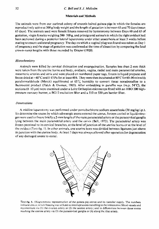
A midline laparotomy was performed under pentobarbitone sodium anaesthesia (38 mg/kg i.p.).To determine the routes by which adrenergic axons entered the uterus, forceps cooled in liquid nitro¬gen were used to freeze briefly a 2-mm length of the main parametrial artery or the paracervical ganglialying between the main parametrial artery and the cervix (Bell, 1973). The parametrial artery wasfrozen proximal to its cervical branches, at the level of junction of the uterine horns or at the level ofthe oviduct (Text-fig. 1). In other animals, one uterine horn was divided between ligatures just aboveits junction with the uterine body. At least 7 days was always allowed after operation for degenerationof any damaged axons to occur.
Text-fig. I. Diagrammatic representation of the guinea-pig uterus and its vascular supply. The numbersindicate sites at which freezing was utilized to interrupt axons travelling to the intrauterine bloodvessels andmyometrium via (11 the ovarian artery or (2) the uterine artery; and to differentiate between those axonsreaching the uterine artery via (3) the paracervical ganglia or (4) along the iliac artery.
ImplantsElongated pellets weighing 20-50 mg were formed by hand from paraffin wax (m.p. 54°C, Gurr)
containing a suspension of400 mg progesterone (Sigma)/g or 40 mgoestradiol-17ß benzoate(Sigma)/g.For intrauterine placement, a midline laparotomy was performed under pentobarbitone sodiumanaesthesia and one pellet containing steroid was inserted into the lumen of the left uterine hornthrough a transverse incision which was then closed with fine surgical silk. A ligature was placedround the horn below the pellet. A pellet of similar size containing only paraffin wax was inserted intothe right uterine horn. For intramuscular placement, a small skin incision was made, under etheranaesthesia, over the lateral portion of the right gastrocnemius muscle and a pellet containing steroidwas inserted into a tunnel made by gentle blunt dissection in the belly of the muscle. The skin incisionwas then closed with surgical silk. A pellet of similar size containing only wax was inserted into theleft gastrocnemius muscle.
PerfusionsGuinea-pigs were killed by cervical dislocation after intracardiac injection of 1000 units heparin
sodium. For investigation of intrauterine vascular connections the left uterine artery was cannulatedwith polythene tubing (PE50) through the common iliac artery. Both ovarian arteries and the contra-lateral iliac artery above the uterine artery were then ligated. The inferior vena cava was opened andthe uterine arterial cannula was connected to a reservoir of modified Krebs solution (McEwan, 1956)with a constant perfusion pressure head of 100 mmHg. When all visible blood had been washed fromthe uterus 0-5 ml India ink (Pelican) was injected into the perfusion stream and the appearance ofeachuterine horn was observed as the perfusion continued. For investigation of relationships between theuterine radial veins and radial arteries, the main uterine vein was cannulated with polythene (PE50)tubing near the oviduct and ligatures were placed on its cervical end and on the radial veins where theyentered the uterine horn. The abdominal aorta was opened and the extrinsic uterne veins were flushedfirst with 0-9% (w/v) NaCl solution, then with India ink at a perfusion pressure of 100 mmHg. Theuterus and its vasculature were then removed in toto from the animal, fixed in 10 % formalin overnightand cleared in glycerol for microscopic inspection by transmitted light.
Results
Distribution ofadrenergic axons in the uterus of the virgin femaleThe distribution of adrenergic axons within the uterus was as reported by Sjöberg (1968). The
outer longitudinal and inner circular smooth muscle layers contained scattered fluorescent fibreswhich ran parallel to the direction of the muscle fibres and did not appear to be related to bloodvessels. In addition, a dense population of fluorescent fibres was associated with the parametrialarteries and with the small arteries and arterioles running through and between the smooth musclelayers. These axons were closely applied to the outer surface of the arterial media (PI. 1, Fig. 1). Nocharacteristic differences in the pattern or density of innervation were noted along the length of theuterine horns. The appearance of the adrenergic nerves was similar in uteri from animals in oestrus ordioestrus.
Origin of the uterine adrenergic innervation
Interruption of the nerves reaching the uterus from its ovarian end in 4 animals resulted indegeneration of almost all fluorescent fibres associated with the myometrium and arteries in theovarian third of the uterine horn. There was a decreased density of fibres in the middle portion of thehorn, but the innervation of the cervical third of the horn appeared normal. By contrast, destructionof the paracervical ganglia in 3 animals produced complete or almost complete loss of fluorescence inthe cervical third of the horn and loss of some fibres from the middle portion but had no effect on
fluorescence in the ovarian third. An identical pattern of degeneration was seen in 2 other animals inwhich the main uterine artery itself was frozen at the level ofjunction of the uterine horns, but freezingthe artery at the level of the cervix had no visible effect on the density of innervation. When the ovarianor uterine artery was denervated the degree of destruction of adrenergic axons associated with par¬ticular radial arteries paralleled the degree of destruction occurring within the portion of the hornssupplied by these arteries.
Serial sections of the paracervical ganglia in one normal animal revealed that a small number (lessthan 10%) of the ganglion cells exhibited catecholamine fluorescence. However, serial cross-sectionsof the vagina and uterine body of 2 animals failed to reveal any fluorescent intramural ganglion cells.In a further 5 animals surgical separation of uterine horns from uterine body had no effect on thedensity of adrenergic axons in the horns.
Changes during pregnancy in fluorescence ofuterine adrenergic axons
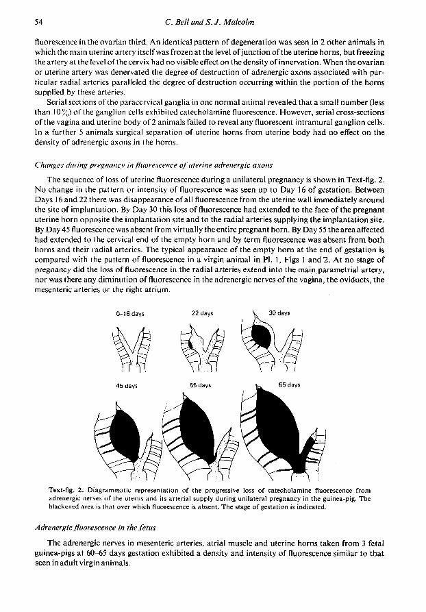
The sequence of loss of uterine fluorescence during a unilateral pregnancy is shown in Text-fig. 2.No change in the pattern or intensity of fluorescence was seen up to Day 16 of gestation. BetweenDays 16 and 22 there was disappearance of all fluorescence from the uterine wall immediately aroundthe site of implantation. By Day 30 this loss of fluorescence had extended to the face of the pregnantuterine horn opposite the implantation site and to the radial arteries supplying the implantation site.By Day 45 fluorescence was absent from virtually the entire pregnant horn. By Day 55 the area affectedhad extended to the cervical end of the empty horn and by term fluorescence was absent from bothhorns and their radial arteries. The typical appearance of the empty horn at the end of gestation iscompared with the pattern of fluorescence in a virgin animal in PI. 1, Figs 1 and "2. At no stage ofpregnancy did the loss of fluorescence in the radial arteries extend into the main parametrial artery,nor was there any diminution of fluorescence in the adrenergic nerves of the vagina, the oviducts, themesenteric arteries or the right atrium.
0-16 days 22 days 30 days
Text-fig. 2. Diagrammatic representation of the progressive loss of catecholamine fluorescence fromadrenergic nerves of the uterus and its arterial supply during unilateral pregnancy in the guinea-pig. Theblackened area is that over which fluorescence is absent. The stage of gestation is indicated.
Adrenergic fluorescence in the fetusThe adrenergic nerves in mesenteric arteries, atrial muscle and uterine horns taken from 3 fetal
guinea-pigs at 60-65 days gestation exhibited a density and intensity of fluorescence similar to thatseen in adult virgin animals.
Extrauterine connections ofuterine arteries and veins
Ink perfusion of the primary uterine vein was performed in 3 animals. Passage of ink was observedfrom the radial veins close to their junction with the main uterine vein into a plexus of small vesselswhich surrounded the radial arteries (PI. 1, Fig. 3). In transverse section these vessels were seen to besmall veins which ran in the arterial adventitia. In serial sections of several radial arteries these smallveins were seen to be in continuity with a system of capillaries lying in the arterial adventitia andconnected to small muscular vessels which arose from the lumen of the radial artery itself (PI. 1,Figs 4 and 5).
Intrauterine connections ofuterine arteries and veins
In 5 animals one uterine artery was perfused with India ink to determine whether it supplied bothuterine horns. During the first few seconds of perfusion, ink appeared only in the ipsilateral horn, butsubsequently it travelled in vessels lying superficially in the uterine body and appeared at the base ofthe contralateral horn. The ink slowly reached the middle portion of the contralateral horn, but theovarian end of the contralateral horn remained almost unmarked.
Effect of intraluminal hormones on fluorescence ofuterine adrenergic axons
In 6 animals fitted with the implants fluorescence of the intrauterine axons in the right horn wasnormal (PI. 2, Fig. 6) after 6-14 days but no fluorescence was observed in the left horn which con¬tained the progesterone-impregnated pellet (PI. 2, Fig. 7). In 2 animals, only the progesterone-containing pellet was implanted and no ligature was applied; in both fluorescence was completelyabsent from the implanted and contralateral horns by 10 days after implantation. In none of the 8animals was there loss of normal fluorescence from axons in the vagina, atria or mesenteric arteries.Direct interference ofprogesterone with the formation of the noradrenaline fluorophore was excludedby the observation that immersion of control tissues in dimethylsulphoxide containing 10 mg pro¬gesterone/ml before freeze-drying did not affect normal development of axonal fluorescence. The rateofhormone absorption during implantation, as judged by changes in pellet weight after briefdrying ina warming oven, appeared rather variable but in all instances was less than 0-5 mg/day. Implantationinto one uterine horn of pellets containing 40 mg oestradiol/g for 10-15 days had no effect on thenormal pattern of fluorescence in three animals, although marked hypertrophy of the uterus andmammary tissue indicated that absorption had occurred.
In 4 animals, subcutaneous implantation on the shoulder of 100 mg fused progesterone pellets(Organon) for 14 days (rate of absorption 0-5 mg/day) had no effect on uterine fluorescence. Dailyintramuscular injection of0-5 mg progesterone (Schering) for 7 days similarly had no effect on uterinefluorescence in another 4 animals.
Effect of implantedprogesterone on fluorescence ofadrenergic vasomotor axons in skeletal muscle
Five virgin guinea-pigs were given implants in the gastrocnemius muscles to determine whetherthe local loss of fluorescence seen during progesterone implantation in the uterine horn was specificfor that organ. The animals were killed 8-33 days later. In all cases arterioles running through thegastrocnemius in the vicinity of the pure wax pellet were accompanied by a plexus of brightlyfluorescent adrenergic axons (PI. 2, Fig. 8). By contrast, the arterioles which lay close to the proges¬terone-containing pellets were completely devoid of fluorescent axons in 4 out of the 5 animals (PI. 2,Fig. 9). In 4 other animals, pellets containing 40 mg oestradiol/g had no effect on the vasomotorfluorescence of arterioles close to the site of implantation in the gastrocnemius muscle.
Discussion
Sjöberg (1968) observed that hypogastric nerve section below the inferior mesenteric ganglion in theguinea-pig did not cause degeneration of the intrauterine adrenergic axons. He concluded that theseaxons arose from 'short' adrenergic neurones which had their cell bodies in the pelvic plexus. Otherworkers have considered that the adrenergic supply to the guinea-pig uterus is by 'long' neuroneswhich travel with the ovarian artery (Isaac et al., 1969; Kulkarni et al., 1976). The present resultsconfirm the report of Langley & Anderson (1895) that the guinea-pig uterus receives sympatheticaxons from the ovarian and pelvic ends and offer a way of reconciling the conflicting views held by laterworkers. While the ovarian end of the uterine horn is supplied almost entirely by the ovarian nerves,the cervical portion of the horn is supplied almost entirely by nerves travelling from the pelvic area.The central part of the horn receives nerves from both sources. This means that, depending on theregion of the uterus examined and probably also to some extent on variability between animals,section of the ovarian or the hypogastric supply could be interpreted as causing complete or nodenervation.
In the rabbit, Owman & Sjöberg (1966) reported the existence of adrenergic ganglion cells in theutero-vaginal wall and concluded that these were the cell bodies of the uterine adrenergic supply. Wehave been unable to find an equivalent population of ganglion cells in the wall of the uterine body orthe vagina of the guinea-pig and have also demonstrated, by complete separation of uterine horn andbody, that the adrenergic nerves to the horns do not travel from the body within the uterine wall. Bycontrast, we did observe that extirpation of the small paracervical ganglia, which lie close to theuterine artery and which supply the cell bodies of the cholinergic vasodilator fibres to this vessel (Bell,1973), produced degeneration of adrenergic axons supplying both the myometrium and the bloodvessels in the cervical part of the uterine horn. Some of these axons probably originated from the smallpopulation of paracervical ganglion cell bodies which exhibited catecholamine fluorescence. How¬ever, destruction of the paracervical ganglia would also have caused degeneration of any post-ganglionic axons of sympathetic chain origin which were travelling to the uterine artery through thisarea.
We observed that the loss of fluorescence from intrauterine adrenergic axons during pregnancywas initially restricted to that area of the uterine wall immediately adjacent to the site of implantation.With advancing gestation the area affected spread radially outwards to include both the entire preg¬nant horn and, later, the empty horn as well. This sequence of events suggests that the factor respon¬sible for loss of fluorescence originates from the fetus or placenta, and gains access to the intrauterineadrenergic axons not via the systemic circulation but by passage through the uterine tissue. Weobserved that in female fetuses near to term the fluorescence of adrenergic nerves, in the uterus andelsewhere, was similar to that observed in normal non-pregnant adult animals. As there is no reason tosuspect that fetal nerves are resistant to the hormonally-induced loss of fluorescence, this suggests thatthe placenta, rather than the fetus, is the site of origin of the factor involved.
The precise pathway by which this factor reaches those parts of the uterus remote from the im¬plantation site is not clear. We demonstrated anatomical continuity between the radial veins andradial arteries, but in the absence of information on relative pressures in the two vessels the signifi¬cance of these connections is uncertain. Despite their localization to the arterial adventitia and thesmall size of the parent vessels concerned it is possible that they represent a vasa vasorum, functionalover the period of pregnancy when hypertrophy of the radial arteries occurs. We also demonstratedanastomotic vascular connections by which material could pass across the uterine body from one hornto the other. However, as Egund & Carter (1974) have reported that no such anastomoses aredetectable in the intact animal they may not be functionally patent.
Implantation into the lumen of one uterine horn of pellets containing progesterone produceddisappearance of fluorescent nerves within the uterus similar to that seen during pregnancy. If noobstruction was placed between the two horns then both exhibited loss of fluorescence, but if theprogesterone-containing horn was ligated at its base then the effect was restricted to that horn. Nodiminution of fluorescence was seen following intraluminal implantation of pellets containingoestradiol or composed of wax alone. These results suggest that progesterone is the placental factor
responsible for intrauterine diminution of fluorescence during pregnancy; the time at which thisdiminution is first noted (about 20 days of gestation) corresponds to that at which peripheral plasmaprogesterone concentrations rise appreciably above values in non-pregnant guinea-pigs (Challis,Heap&Illingworth, 1971).
Pregnancy or progesterone implants caused loss of fluorescence from the 'long' adrenergic axons
entering the uterus with the ovarian artery as well as from the 'short' pelvic axons and progesteroneimplants in skeletal muscle also caused loss of fluorescence from adjacent vasomotor axons. Therestriction of the loss during pregnancy to the intrauterine axons may therefore not be related to anypeculiarity of these axons but to the fact that the relatively high concentration of progesterone re¬
quired to produce the effect is not attained in the systemic circulation.The mechanism by which progesterone might affect catecholamine fluorescence is unknown, but
the observation that uterine noradrenaline content is not markedly depressed during pregnancysuggests that changes in binding of the releasable amine rather than the total axonal amine may beinvolved (Bell, 1974).
This work was supported by a continuing grant from the National Heart Foundation of Australiaand by equipment grants from the Australian Research Grants Committee and the University ofMelbourne. S.J.M. received financial assistance as a B. Med. Sci. candidate from the National Healthand Medical Research Council. Organon (Aust.) generously supplied fused progesterone pellets andSchering Pty. Ltd donated injectable progesterone (Proluton). We should like to thank Dr MartheVogt for helpful discussions.
References
Bell, C. (1972) Autonomie nervous control of repro¬duction : circulatory and other factors. Pharmac. Rev.24, 657-736.
Bell, C. (1973) Selective cholinergic denervation of theuterine artery in the guinea-pig. Experientia 30, 257-258.
Bell, C. (1974) Control of uterine blood flow in preg¬nancy. Med. Biol. 52, 219-228.
Challis, J.K.G., Heap, R.B. & Illingworth, CD.(1971) Concentrations of oestrogens and proges¬terone in the plasma of non-pregnant, pregnant andlactating guinea-pigs. J. Endocr. 51, 333-345.
Draper, R.L. (1920) The prenatal growth of the guinea-pig. Anat. Ree. 18, 369-392.
Eg und, . & Carter, A.M. (1974) Uterine and placentalcirculation in the guinea-pig: an angiographie study./. Reprod. Fert. 40, 401-410.
Falck, B. & Owman, C. (1965) A detailed methodo¬logical description of the fluorescence method for thecellular demonstration of biogenic monoamines.Acta Unió. hind. (It) 7, 1-23.
Isaac, P.F., Pennefather, J.N. & Silva, O.G. (1969)The ovarian and hypogastric innervation of theguinea-pig uterus. Eur. J. Pharmac. 5, 384-390.
K.ULKARNI, P.S., Wakade, A.R. & Kirpekar, S.M.(1976) Sympathetic innervation of guinea-pig uterusand ovary. Am. J. Physio!. 230, 1400-1405.
Langley, J.N. & Anderson, H.K. (1895) The inner¬vation of the pelvic and adjoining viscera. /. Physiol.,Lond. 19,71-138.
McEwAN, L.M. (1956) Effect on the isolated rabbitheart of vagai stimulation and its modification bycocaine, hexamethonium and ouabain. /. Physiol.,Lond. 131, 678-689.
Nakanishi, H., McLean, J., Wood, C. & Burnstock,G. (1968) The role of sympathetic nerves in control ofthe nonpregnant and pregnant human uterus. /.Reprod. Med. 2, 20-33.
Owman, C. & Sjöberg, N.-O. (1966) Adrenergic nerves
in the female genital tract of the rabbit. With remarkson cholinesterase-containing structures. Z. Zell-forsch. mikrosk. Anat. 74, 182-197.
Owman, C, Sjöberg, N.-O. & Sjöstrand, N.-O. (1974)Short adrenergic neurones, a peripheral neuro¬
endocrine mechanism. In Amine Fluorescence Histo-chemistry, pp. 47-66. Eds . Fujiwara, & C. Tanaka.Igaku Shoin Ltd, Tokyo.
Rosengren, E. & Sjöberg, N.-O. (1968) Changes in theamount of adrenergic transmitter in female genitaltract of rabbit during pregnancy. Acta physiol. scand.72,412-424.
Ryan, M.J., Clark, K.E. & Brody, M.J. (1974) Neuro-genic and mechanical control of canine uterine vas¬
cular resistance. Am.J. Physiol. 227, 547-555.Sjöberg, N.-O. (1968) Consideration on the cause of
disappearance of the adrenergic transmitter in uterinenerves during pregnancy. Acta physiol. scand. 72,510-517.
Sjöstrand, N.-O. (1965) The adrenergic innervation ofthe vas deferens and the accessory male genitalglands. Acta physiol. scand. 65, Suppl. 257.
Received 18 July 1977
EXPLANATION OF PLATES
PLATE 1
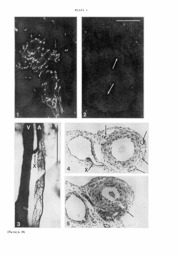
The horizontal bar in Fig. 2 represents 100 µ for Figs 1, 2, 4 and 5. The vertical bar in Fig. 3 represents1 mm for that figure.Fig. 1. Section from the middle of the uterine horn of a virgin guinea-pig, showing catecholamine fluor¬escence of the adrenergic axons associated with myometrium and with intrauterine arteries.
Fig. 2. Section from the equivalent portion of the non-pregnant horn in a unilaterally pregnant guinea-pig(gestational age 60 days). Note the absence of fluorescence around the arteries (arrows) and in themyometrium.Fig. 3. Demonstration by ink perfusion through the uterine venous supply (V) of a venous plexus whicharises from the radial vein (X) and ramifies over the accompanying radial artery (A).Figs 4 and 5. Haematoxylin-eosin-stained transverse sections from the radial artery and vein shown in Fig. 3.The plane of section in Fig. 4 is through the point of branching of the radial vein (X). The resultant venousplexus surrounding the artery can be seen as ink-containing profiles localized to the arterial adventitia(arrows). Fig. 5 is a section approximately 2 mm distal to that in Fig. 4 and shows a sidebranch of the radialartery entering the arterial adventitia (arrow). Examination of serial sections between the two points con¬
firmed that the adventitial artery and veins were in continuity.PLATE 2
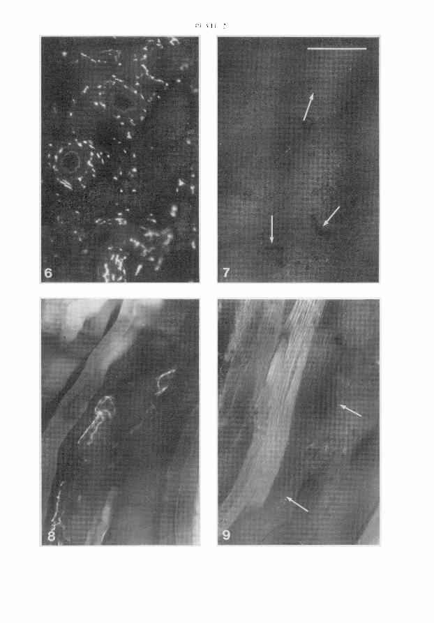
The horizontal bar in Fig. 7 represents 100 µ for all Figures.Fig. 6. Intense fluorescence of adrenergic axons associated with myometrium and intrauterine arteries froma uterine horn into which a 50 mg wax pellet had been implanted 7 days before.
Fig. 7. Absence of fluorescence from myometrium and from arteries (arrows) in the contralateral hornimplanted, 7 days before, with a 50 mg wax pellet containing 400 mg progesterone/g. Transfer of proges¬terone into the control horn was prevented by ligating the treated horn below the implant.Fig. 8. Intense fluorescence of adrenergic axons associated with arterioles in the gastrocnemius muscle,close to the site of implantation of a 20 mg wax pellet 13 days before.
Fig. 9. Absence of fluorescence from arterioles (arrows) in the contralateral gastrocnemius muscle close tothe site of implantation, 13 days before, of a 20 mg wax pellet containing 400 mg progesterone/g.


![Metabolism of [14C] methamphetamine in man, the guinea pig and](https://img.pdfslide.net/doc/110x75/58599eb01a28ab6e3290126e/metabolism-of-14c-methamphetamine-in-man-the-guinea-pig-and-.jpg)