Embed Size (px)
Citation preview
Streptozotocin Diabetes in the Mouseand Guinea Pig
G. Brosky, M.Sc, and J. Logothetopoulos, M.D., Ph.D., Toronto
SUMMARY
A single injection of streptozotocin into mice producedan extensive necrosis of the beta cells resulting in per-manent diabetes. The histological changes in the islets weresimilar to those described after the injection of alloxan.The regenerative capacity of beta cells which survived thecytotoxic injury was limited.
Hypoglycemia induced by an injection of insulin orhyperglycemia induced by glucose injections sensitizedthe beta cells to streptozotocin. In contrast, injection ofinsulin antibody effectively protected the beta cells fromthe cytotoxic injury.
Streptozotocin caused widespread necrosis of the betacells of the guinea pig and the obese hyperglycemic mouse.Both these species had proven resistant to the diabetogeniceffect of alloxan.
Diabetic guinea pigs were observed for eight months,without reversion of the diabetic syndrome. DIABETES 18:606-11, September, 1969.
Until recently alloxan was the most effective com-pound for the induction of experimental diabetes inanimals. In 1963 Rakieten1 reported that streptozotocin,a by-product of the bacterium Streptomyces achromo-genes possessing antibacterial and antitumoral prop-erties,2 caused diabetes in dogs and rats. The initialobservation attributed the diabetic syndrome to destruc-tion of the beta cells of the pancreatic islets. Laterexperimental work with rats by Arison et al.3 favoredan inhibition of insulin synthesis without cell necrosisas the diabetogenic mechanism.
We became interested in the experimental diabetesinduced by streptozotocin for the following reasons:
(a) The suggestion of a permanent biochemicallesion without necrosis of beta cells required reinvesti-gation because of its implications in the physiologyand biochemistry of insulin secretion.
(b) We wanted to test the effect of hyperglycemia
From the Banting and Best Department of Medical Re-search, University of Toronto, Toronto, Ontario, Canada.
and hypoglycemia on the specific cytotoxicity of strep-tozotocin.
(c) The diabetogenic effect of streptozotocin on theguinea pig and the obese hyperglycemic mutant mousewas worthwhile investigating because alloxan hadproved ineffective in these two species.4'5
The present paper reports the results of experimentsrelated to the above problems.
MATERIAL AND METHODS
Animals. Inbred C57 Bl/6 Jax male adult mice wereobtained from the Roscoe B. Jackson Memorial Lab-oratories, Bar Harbor. When used, the animals were*three to four-months old and weighed 22 to 28 gm.They had been fed Purina mouse breeder chow adlibitum in our animal quarters for at least a weekbefore use. Obese hyperglycemic mutant mice were alsoobtained from the same source. Guinea pigs wereobtained from a local supplier and were found to befree of pathogenic bacteria. Young adult males, weigh-ing 600 to 800 gm. were maintained on RocklandGuinea Pig Chow and water ad libitum until use.
Streptozotocin. This compound was supplied by TheUpjohn Co. after approval of the amounts requestedby the Drug Research Division of the National Insti-tutes of Health, Bethesda, Maryland. Pure streptozotocinwas used (NSC 85998). Sterile acidified saline (pH3.5) was used to dissolve the compound immediatelybefore injection into experimental animals.
Alloxan, insulin. Alloxan monohydrate was obtainedfrom the Eastman Kodak Co. Insulin-Toronto and Pro-tamine Zinc Insulin were obtained from the ConnaughtMedical Laboratories, Toronto.
Insulin antibody. The procedure to obtain insulinneutralizing guinea pig immunoglobulins was similarto that described in previous work.6 • .
Urine glucose and blood glucose. TesTape and Acetesttablets were used to evaluate glucosuria and ketonuria.Glucose in collected urine was estimated quantitativelywith Clinitest tablets (Ames Co., Canada). Blood glu-cose was determined by the ferricyanide reducing meth-
606 DIABETES, VOL. 18, NO. 9
Dow
nloaded from http://diabetesjournals.org/diabetes/article-pdf/18/9/606/344937/18-9-606.pdf by guest on 15 January 2022
G. BROSKY, M.SC, AND J . LOGOTHETOPOULOS, M.D.
od of Hoffman7 using the Autotechnikon Analyser.Injections of animals. Mice were warmed in a hot
box, restrained in a plastic tube and injected throughthe ventrolateral tail vein. Guinea pigs were injectedthrough the dorsal penis vein after application of 2 percent xylocaine solution.
Histology, autoradiography. The pancreas was evenlyspread on a paraffin-coated square of cardboard andfixed in Bouin's or Zenker-formol solutions. After astandard procedure of dehydration, clearing and paraffinembedding, sections 4 ju.. thick were stained with hema-lum-eosin, aldehyde-fuchsin, periodic acid Schiff andMasson's trichrome.
Tritiated thymidine (specific activity 6.7 c. per mM.)was injected subcutaneously at a dose of 0.5 fie. pergm. body weight in 0.2 ml. of sterile saline. Diabeticmice and their controls received eight subcutaneousinjections of tritiated thymidine at twelve-hour inter-vals. They were killed thirty-six hours following thelast injection of tritiated thymidine.
The stripping film technic of Pelc8 was applied for4 ju,. paraffin sections of the splenic part of the pancreasfixed in Bouin's solution. Sections were stained withaldehyde-fuchsin before the application of the emulsion.
EXPERIMENTAL PROCEDURES AND RESULTS
Streptozotocin diabetes in the normal mouseAdult male mice, fasted for twelve to fourteen hours,
were injected intravenously with 100 mg./kg. of strep-tozotocin in 0.2 ml. of acidified saline. This dose wasfound in preliminary experiments to produce permanentdiabetes in almost all the mice injected. Groups of twoto three mice were killed at intervals following theinjection. The following histological changes werenoticed.
Evidence of pyknotic beta cell nuclei was seen threeto four hours after the injection. By six to eight hours,most beta cells showed nuclear pyknosis while theircytoplasm became less hematoxynophilic. Some pyknoticnuclei began breaking down into smaller fragments.After twelve to sixteen hours, when the disintegrationof the pyknofck nuclei was well advanced, the cyto-plasm of the beta cells coalesced in homogeneouseosinophilic masses. Very little nuclei material could bestained after twenty-four to thirty-six hours. The shrink-ing, necrotic eosinophilic masses were surrounded byrims of intact alpha cells and a few surviving beta cells.Infiltration by leucocytes was not evident, apart from afew scattered macrophages. Endothelial cells, lining sinu-soids, were frequently encountered, and hemorrhageswithin the necrotic areas were extremely rare. After
thirty-six to forty-eight hours, the islets appeared tohave collapsed as the discrete areas of necrotic debriswere being absorbed within them. The eosinophilicnecrotic masses remained stainable with aldehyde-fuchsinuntil completely absorbed. Distorted pancreatic isletsconsisting of alpha cells and a small number of rela-tively degranulated beta cells were consistently foundafter forty-eight hours with no trace of necrotic debris.The diabetes induced in mice with streptozotocin waspermanent. Significant changes in pancreatic histologywere not seen after the initial four to five days fol-lowing the injection of streptozotocin. Most of the re-maining islets were smaller structures consisting ofalpha cells and a small number of hypertrophic betacells with variable degrees of degranulation. Occasion-ally, islets were observed which contained a larger num-ber of beta cells.
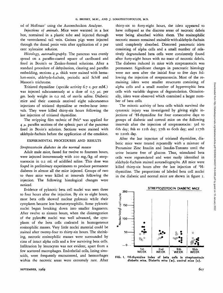
The mitotic activity of beta cells which survived thecytotoxic injury was investigated by giving eight in-jections of 3H-thymidine for four consecutive days togroups of diabetic and control mice on the followingintervals after the injection of streptozotocin: 3rd to6th day; 8th to n t h day; 57th to 60th day; and 117thto 120th day.
After the last injection of tritiated thymidine, dia-betic mice were treated repeatedly with a mixture ofProtamine Zinc Insulin and Insulin-Toronto until theurine became free of glucose. Thus, stimulated betacells were regranulated and were easily identified inaldehyde-fuchsin stained autoradiographs. All mice werekilled thirty-six hours after the last injection of 3H-thymidine. The proportions of labeled beta cell nucleiin the diabetic and normal mice are shown in figure 1.
200
150
1> 100
50
STREPTOZOTOCIN DIABETIC MICE
MM
M
M
MM
—
•• * •
o ~* °
1st.WEEK
• « #
2nd.WEEK
9th.WEEK
o
o ^*
18th.WEEK
FIG. I. 3H-thymidine index of beta cells in streptozotocindiabetic mice. Diabetic mice ( • ) , control mice (o).
SEPTEMBER, 1969 607
Dow
nloaded from http://diabetesjournals.org/diabetes/article-pdf/18/9/606/344937/18-9-606.pdf by guest on 15 January 2022
STREPTOZOTOCIN DIABETES IN THE MOUSE AND GUINEA PIG
A wave of mitotic activity was observed during thefirst two weeks. By the end of two months, the pro-portions of beta cells duplicating their DNA weresimilar to those found in controls.
No evidence was found that new formation of betacells from duct or acinar cells occurred. The density ofislets in the diabetic mice was much lower than thatin controls. Scattered small agglomerations of beta cellssurrounding ducts or lying between acini were as un-common in the diabetic mice as in the normal mice.Effects of hypoglycemia or hyperglycemia on the dia-betogenic effect of streptozotocin in the mouse
Hypoglycemia. Two units of Insulin-Toronto weregiven to fasted mice. Controls received a saline injec-tion. When symptoms of weakness developed, alternatehypoglycemic and control mice were injected intra-venously with 60 mg. streptozotocin/kg. body weight.The seventy of diabetes was evaluated by the degree ofglucosuria and the presence of ketosis. The mice werekept for fifteen days before being killed. Table 1clearly shows that hypoglycemia enhanced the cytotoxiceffect of streptozotocin on the beta cells.
TABLE 1
Severity of diabetes in control and in hypoglycemic miceinjected with 60 mg./kg. body weight of streptozotocin
Ketonuria and glucosuriaGlucosuriaNondiabetic
Insulininjected
(17)*2
105
Salineinjected
(23)*04
19
*Number of mice in parentheses.
Hyperglycemia. In the initial experiment, an acutediabetic state was induced by the intraperitoneal injec-tion of anti-insulin guinea pig globulins. Controls re-ceived saline injections. When glucosuria developed,between two to three hours after the antibody injection,experimental and control mice were alternately in-jected with 100 mg. of streptozotocin per kg. bodyweight. As shown in table 2, animals injected with in-sulin antibody were protected from the diabetogeniceffect of the compound. With the expectation of con-firming these results with hyperglycemia induced byglucose injections, a group of ten mice were injectedwith 0.15 gm. of glucose intraperitoneally ten minutesbefore the injection of 60 mg. streptozotocin per kg.body weight. A second group received two glucose in-jections at ten and sixty minutes before injection. Thethird control group was injected with streptozotocin
TABLE 2Severity of diabetes in mice injected with streptozotocin(100 mg./kg. body weight) during hyperglycemia induced
with insulin antibody
Ketonuria and glucosuriaGlucosuriaNondiabetic
Insulinantibody
(10)*00
10
Salineinjected(12)*
462
^Number of mice in parentheses.
after a previous saline injection. As table 3 shows,hyperglycemia induced by glucose injections enhancedrather than inhibited the diabetogenic effect of strepto-zotocin.
TABLE 3Severity of diabetes in mice injected with streptozotocin
(60 mg./kg.) during hyperglycemia induced by glucose
Ketonuriaand glucosuria
GlucosuriaNondiabetic
Glucose*injected
(10)$
352
Glucosetinjected
Salineinjected
(10)$
0.64
* Glucose injection ten minutes before streptozotocin.tGlucose injection ten and sixty minutes before strep-
tozotocin.JNumber of mice in parentheses.
In a third experiment, a direct comparison of theeffect of the two types of hyperglycemia was made.An acute transient diabetes induced by insulin-antibodyprotected the beta cells from the cytotoxic effect ofstreptozotocin in contrast to the effect of hyperglycemiainduced by glucose (table 4 ) .
The protective effect of hyperglycemia induced by glu-cose against the diabetogenic effect of alloxan has beenshown in the rat.9 Table 5 shows that in the mouse glu-cose-induced hyperglycemia also inhibits the diabeto-genic effect of alloxan.
TABLE 4Severity of diabetes in mice injected with streptozotocin(100 mg./kg. body weight) following injection of anti-
insulin globulins or of glucose + control globulins
Ketonuria and glucosuriaGlucosuriaNondiabetic
Insulinantibody
(9)*009
Glucose +control
globulins(10)*
262
*Number of mice in parentheses.
608 DIABETES, VOL. 18, NO. 9
Dow
nloaded from http://diabetesjournals.org/diabetes/article-pdf/18/9/606/344937/18-9-606.pdf by guest on 15 January 2022
G. BROSKY, M.SC, AND J . LOGOTHETOPOULOS, M.D.
TABLE 5Effect of glucose hyperglycemia on the diabetogenic effect
of alloxan
Ketonuria and glucosuriaGlucosuriaNondiabetic
Salineinjected
(10)*370
Glucoseinjected
(7)*025
*Number of mice in parentheses.
Streptozotocin diabetes in the obese hyperglycemicmutant mouse
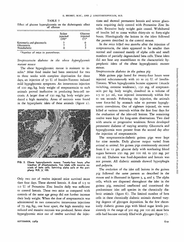
The obese hyperglycemic mouse is resistant to in-sulin.9 After food intake had been restricted for twoto three weeks with complete deprivation for threedays, an injection of 50 U. of Insulin-Toronto inducedmild hypoglycemic symptoms. An intravenous injectionof 100 mg./kg. body weight of streptozotocin to suchanimals proved ineffective in producing beta-cell ne-crosis. A larger dose of 150 mg./kg. body weight pro-duced a high mortality. Areas of necrosis were foundin the hyperplastic islets of these animals (figure 2).
FIG. 2. Obese hyperglycemic mouse. Twenty-four hours afterinjection of streptozotocin. Two islets with massive ne-crosis of beta cells. Surviving alpha cells at the peri-phery. H.E. X 100.
Only two out of twelve injected mice survived morethan four days. These showed ketosis. A dose of 0.5 to1.0 U. of Protamine Zinc Insulin daily was sufficientto control ketosis. These two mice as compared withcontrols of the same age group did not further increasetheir body weight. When the dose of streptozotocin wasadministered in two consecutive intravenous injectionsof 75 mg./kg., one hour apart, the high mortality wasreduced and massive necrosis was produced. Seven obesehyperglycemic mice out of twelve survived the injec-
tions and showed permanent ketosis and severe gluco-suria requiring daily control with Protamine Zinc In-sulin. Excessive body weight gain ceased. Withdrawalof insulin led to coma within thirty-six to forty-eighthours. Histologically the lesions in the islets followedthe pattern described in the control mouse.
In the mice killed two months after the injection ofstreptozotocin, the islets appeared to be smaller thannormal and consisted mainly of alpha cells and smallnumbers of partially degranulated beta cells. These isletsdid not bear any resemblance to the characteristic hy-perplastic islets of the obese hyperglycemic mutantmouse.Streptozotocin diabetes in the guinea pig
Male guinea pigs fasted for twenty-four hours wereinjected subcutaneously with 20 to 25 U. of Insulin-Toronto. When hypoglycemia became apparent (muscletwitching, extreme weakness), 150 mg. of streptozot-ocin per kg. body weight, dissolved in a volume of2.5 to 3.0 ml., was injected intravenously within fiveto ten seconds. Following the injection, the animalswere force-fed by stomach tube to prevent hypogly-cemic convulsions. Out of eighteen injected, six werekilled at various intervals within the first four days forthe evaluation of the islet-cell lesions. The remainingtwelve were kept for long-term observations. Two diedwith anuria or progressive weakness. Seven developedpermanent diabetes of varying severity. Glucosuria andhyperglycemia were present from the second day afterthe injection of streptozotocin.
The streptozotocin-diabetic guinea pigs were keptfor nine months. Daily glucose output varied fromanimal to animal. Six guinea pigs continuously excretedfrom 6 to 12 gm. glucose daily with nonfasting bloodsugars between 250 mg. per 100 ml. to 350 mg. per100 ml. Diabetes was food-dependent and ketosis wasnot present. All diabetic animals showed hyperphagiaand polyuria.
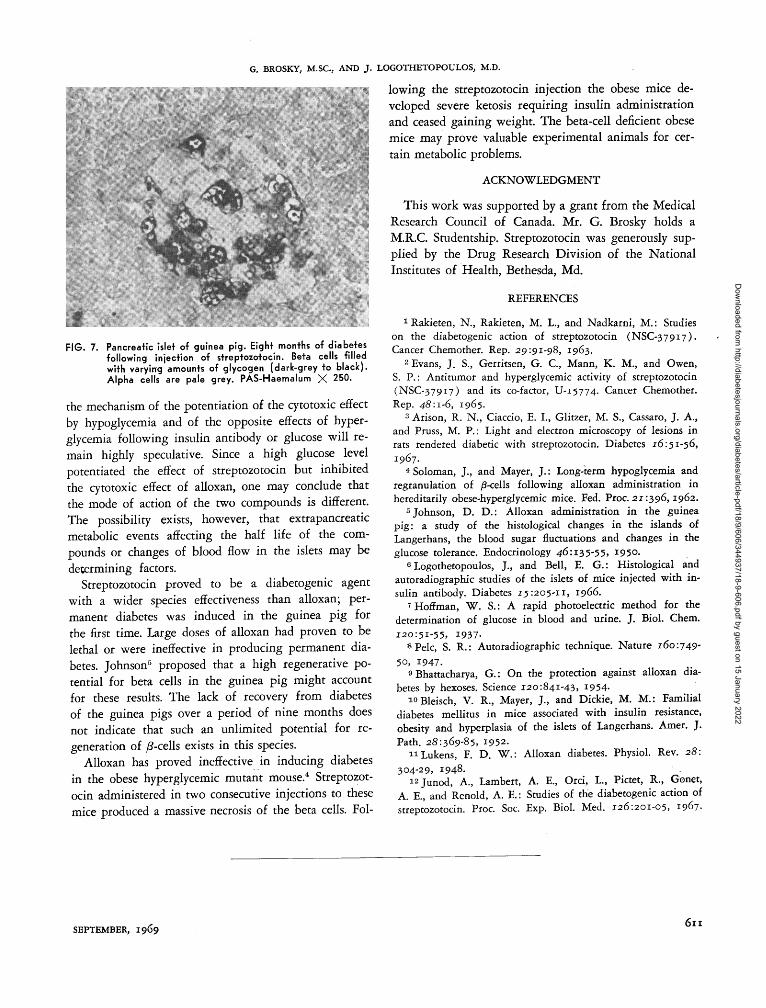
The evolution of the islet cell lesions in the guineapig followed the same pattern as described in themouse and is illustrated in figures 3, 4, and 5. The alphacells, which are dispersed throughout the islet in theguinea pig, remained unaffected and constituted thepredominant islet cell species in the chronically dia-betic animals (figure 6) . The limited number of betacells in these chronically diabetic animals showed vary-ing degrees of glycogen deposition. In the few chron-ically diabetic guinea pigs with blood sugar levels per-sistently in the range of 300 mg. per 100 ml. most betacells had become entirely filled with glycogen (figure 7).
SEPTEMBER, 1969 609
Dow
nloaded from http://diabetesjournals.org/diabetes/article-pdf/18/9/606/344937/18-9-606.pdf by guest on 15 January 2022
STREPTOZOTOCIN DIABETES IN THE MOUSE AND GUINEA PIG
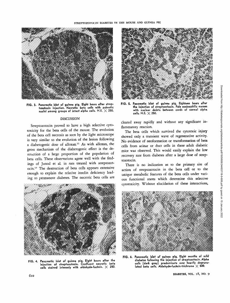
FIG. 3. Pancreatic islet of guinea pig. Eight hours after strep-tozotocin injection. Necrotic beta cells with pylcnoticnuclei among groups of intact alpha cells. H.E. X 250.
DISCUSSION
Streptozotocin proved to have a high selective cyto-toxicity for the beta cells of the mouse. The evolutionof the beta cell necrosis as seen by the light microscopeis very similar to the evolution of the lesion followinga diabetogenic dose of alloxan.11 As with alloxan, thegross mechanism of the diabetogenic effect is the de-struction of a large proportion of the population ofbeta cells. These observations agree well with the find-ings of Junod et al. in rats treated with streptozot-ocin.12 The destruction of beta cells appears extensiveenough to explain the relative insulin deficiency lead-ing to permanent diabetes. The necrotic beta cells are
FIG. 5. Pancreatic islet of guinea pig. Eighteen hours afterthe injection of streptozotocin. Pale eosinophilic masse*with nuclear debris between cords of normal alphacells. H.E. X 250.
cleared away rapidly and without any significant in-flammatory reaction.
The beta cells which survived the cytotoxic injuryshowed only a transient wave of regenerative activity.No evidence of neoformation or transformation of betacells from acinar or duct cells in these adult diabeticmice was observed. This would easily explain the lowrecovery rate from diabetes after a large dose of strep-tozotocin.
There is no indication as to the primary site ofaction of streptozotocin in the beta cell or to theunique metabolic features of the beta cells under vari-ous functional states which determine this selectivecytotoxicity. Without elucidation of these interactions,
FIG. 4. Pancreatic islet of guinea pig. Eight hours after theinjection of streptozotocin. Confluent necrotic betacells stained intensely with aldehyde-fuchsin. X 250.
(Sio
FIG. 6. Pancreatic islet of guinea pig. Eight months of milddiabetes following the injection of streptozotocin. Alphacells (dark grey) predominate over heavily degranu-lated beta cells. Aldehyde-fuchsin-trichrome X 500.
DIABETES, VOL. 18, NO. 9
Dow
nloaded from http://diabetesjournals.org/diabetes/article-pdf/18/9/606/344937/18-9-606.pdf by guest on 15 January 2022
G. BROSKY, M.SC, AND J . LOGOTHETOPOULOS, M.D.
FIG. 7. Pancreatic islet of guinea pig. Eight months of diabetesfollowing injection of streptozotocin. Beta cells filledwith varying amounts of glycogen (dark-grey to black).Alpha cells are pale grey. PAS-Haemalum X 250.
the mechanism of the potentiation of the cytotoxic effectby hypoglycemia and of the opposite effects of hyper-glycemia following insulin antibody or glucose will re-main highly speculative. Since a high glucose levelpotentiated the effect of streptozotocin but inhibitedthe cytotoxic effect of alloxan, one may conclude thatthe mode of action of the two compounds is different.The possibility exists, however, that extrapancreaticmetabolic events affecting the half life of the com-pounds or changes of blood flow in the islets may bedetermining factors.
Streptozotocin proved to be a diabetogenic agentwith a wider species effectiveness than alloxan; per-manent diabetes was induced in the guinea pig forthe first time. Large doses of alloxan had proven to belethal or were ineffective in producing permanent dia-betes. Johnson5 proposed that a high regenerative po-tential for beta cells in the guinea pig might accountfor these results. The lack of recovery from diabetesof the guinea pigs over a period of nine months doesnot indicate that such an unlimited potential for re-generation of /?-cells exists in this species.
Alloxan has proved ineffective in inducing diabetesin the obese hyperglycemic mutant mouse.4 Streptozot-ocin administered in two consecutive injections to thesemice produced a massive necrosis of the beta cells. Fol-
lowing the streptozotocin injection the obese mice de-veloped severe ketosis requiring insulin administrationand ceased gaining weight. The beta-cell deficient obesemice may prove valuable experimental animals for cer-tain metabolic problems.
ACKNOWLEDGMENT
This work was supported by a grant from the MedicalResearch Council of Canada. Mr. G. Brosky holds aM.R.C. Studentship. Streptozotocin was generously sup-plied by the Drug Research Division of the NationalInstitutes of Health, Bethesda, Md.
REFERENCES
1 Rakieten, N., Rakieten, M. L., and Nadkarni, M.: Studieson the diabetogenic action of streptozotocin (NSC-37917).Cancer Chemother. Rep. 29:91-98, 1963.
2 Evans, J. S., Gerrksen, G. C, Mann, K. M., and Owen,S. P.: Antitumor and hyperglycemic activity of streptozotocin(NSC-37917) and its co-factor, U-15774. Cancer Chemother.Rep. 48:1-6, 1965.
3 Arison, R. N., Ciaccio, E. I., Glitzer, M. S., Cassaro, J. A.,and Pruss, M. P.: Light and electron microscopy of lesions inrats rendered diabetic with streptozotocin. Diabetes 16:51-56,1967.
4 Soloman, J., and Mayer, J.: Long-term hypoglycemia andregranulation of /3-cells following alloxan administration inhereditarily obese-hyperglycemic mice. Fed. Proc. 21:396,1962.
5 Johnson, D. D.: Alloxan administration in the guineapig: a study of the histological changes in the islands ofLangerhans, the blood sugar fluctuations and changes in theglucose tolerance. Endocrinology 46:135-55. 1950.
6 Logothetopoulos, J., and Bell, E. G.: Histological andautoradiographic studies of the islets of mice injected with in-sulin antibody. Diabetes 15:205-11, 1966.
7 Hoffman, W. S.: A rapid photoelectric method for thedetermination of glucose in blood and urine. J. Biol. Chem.120:51-55, 1937-
8Pelc, S. R.: Autoradiographic technique. Nature 160:749-50, 1947.
9 Bhattacharya, G.: On the protection against alloxan dia-betes by hexoses. Science 120:841-43, 1954-
i°Bleisch, V. R., Mayer, J., and Dickie, M. M.: Familialdiabetes mellitus in mice associated with insulin resistance,obesity and hyperplasia of the islets of Langerhans. Amer. J.Path. 28:369-85, 1952.
uLukens, F. D. W.: Alloxan diabetes. Physiol. Rev. 28:304-29, 1948.
i2junod, A., Lambert, A. E., Orci, L., Pictet, R., Genet,A. E., and Renold, A. E.: Studies of the diabetogenic action ofstreptozotocin. Proc. Soc. Exp. Biol. Med. 126:201-05, 1967.
SEPTEMBER, 1969
Dow
nloaded from http://diabetesjournals.org/diabetes/article-pdf/18/9/606/344937/18-9-606.pdf by guest on 15 January 2022